Perforated Meckel's Diverticulum with Acute Appendicitis in an Infant
←
→
Page content transcription
If your browser does not render page correctly, please read the page content below
CASE REPORT

Perforated Meckel’s Diverticulum              with Acute
                          history of abdominal pain, fever and vomiting. The
                          abdominal pain was colicky and intermittent; the vomiting

Appendicitis in an Infant
Tim Jumbi, Shahbal Swaleh, Francis Osawa
School of Medicine, University of Nairobi

Correspondence to: Dr. Tim Jumbi, P.O Box 1059–00618 Ruaraka, Nairobi, Kenya. Email: mwaitim@gmail.com

Summary
                                                                 Key words: Meckel's diverticulum, Acute appendicitis
Meckel's diverticulum occurs in approximately 2% of the
                                                                 Ann Afr Surg. 2019; 16(2):***
population and is symptomatic in 16% of cases. In children,
                                                                 DOI: http://dx.doi.org/10.4314/aas.v16i2.*
the most common presentation is intestinal obstruction due to
                                                                 Conflicts of Interest: None
intussusception. The simultaneous occurrence of acute
                                                                 Funding: None
appendicitis and symptomatic Meckel’s diverticulum is rare
                                                                 © 2019 Author. This work is licensed under the Creative
and when it occurs, it may be associated with non-specific
                                                                 Commons Attribution 4.0 International License.
symptoms that present a diagnostic challenge.

Introduction
                                                                 was non-bilious and postprandial. There was no history of
Meckel's diverticulum (MD) was first reported by Meckel in
                                                                 constipation and the child was passing stool and flatus. He
1809, by describing its embryological origin as remnant of
                                                                 was clinically febrile with a temperature of 40ºC. He had a
omphalomesenteric duct (1). It is the most common
                                                                 mildly distended abdomen which was mildly tender, no
congenital malformation of the gastrointestinal tract with an
                                                                 abdominal masses were palpated and bowel sounds were
incidence of between 2% and 4% (2). The occurrence of MD
                                                                 present. Mucoid soft faeces was noted on digital rectal
is mostly asymptomatic, but is reported to be symptomatic in
                                                                 examination with no rectal masses palpated. The WBC count
approximately 16% of cases (3). Symptomatic MD is defined
                                                                 was 11.2 x 10⁹/L with a neutrophil count of 5.42 x 10³/µL.
by the presence of gastrointestinal bleeding, intestinal
                                                                 An initial abdominal x-ray showed dilated bowel loops,
obstruction and peritonitis. The most common presentation in
                                                                 predominantly small bowel with increased bowel wall
children is intestinal obstruction due to intussusception with
                                                                 attenuation. (Figure 1). An abdominal ultrasound was
the diverticulum acting as a lead point (4). The simultaneous
                                                                 reported with no features of intussusception or free
occurrence of appendicitis and symptomatic MD is rarely
                                                                 intraperitoneal fluid but was inconclusive of a diagnosis.
seen in the adult and pediatric populations. This occurrence
                                                                 We inserted a nasogastric tube for decompression and put the
may present with non-specific symptoms, which can delay
                                                                 child on nil by mouth. Additionally, we instituted
diagnosis and management in these patients. We report a
                                                                 intravenous fluids, antibiotics and antipyretics. After 6 hours
case of a 7-month old admitted with a rare occurrence of a
                                                                 of conservative management, the abdominal distension had
perforated Meckel’s diverticulum and acute appendicitis.
                                                                 subsided, the child had passed stool twice but still had fever
This case highlights the rarity and non-specific
                                                                 and abdominal tenderness that had since worsened, being
symptomatology of this occurrence with its diagnostic
                                                                 associated with guarding and rigidity. We performed an
enigma that can delay intervention in the pediatric
                                                                 explorative laparotomy, as the child had developed clear
population.
                                                                 signs of peritonitis. Intraoperatively, we found matted ileal
                                                                 and ceacal segments of bowel with fibrinous exudates that
Case Report
                                                                 had concealed a perforated Meckel’s diverticulum and an
We report a case of a 7-month old with a perforated MD and
                                                                 inflamed appendix. The MD was 2cm long with a small neck
acute appendicitis with the aim of highlighting the
                                                                 located 60cm proximal to the ileocecal valve (Figure 2).
symptomatology of this occurrence and its diagnostic
enigma, which can delay intervention in the pediatric
population. A 7-month old male was admitted to the pediatric
surgical unit at Kenyatta National Hospital with a 1-day

The ANNALS of AFRICAN SURGERY, April 2019 Volume 16 Issue 2
1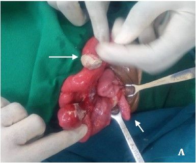
The ANNALS of AFRICAN SURGERYIwww.annalsofafricansurgery.com            Jumbi et al. Meckel’s Diverticulum with Appendicitis

                                                                        sign is gastrointestinal bleeding, reported in 25–50% of
                                                                        symptomatic patients with MD (6). In children, the most
                                                                        common presentation associated with MD is intestinal
                                                                        obstruction usually caused by intussusception, fibrous band
                                                                        volvulus, torsion of the diverticulum and Littre’s internal
                                                                        hernia (3, 7–10). Peritonitis is also a common clinical
                                                                        presentation in children, which results from a perforated
                                                                        diverticulum or diverticulitis (11, 12). Our case presented
                                                                        with non-specific symptoms suggestive of partial intestinal
Figure 1: Pre-operative plain radiographs: A. Supine view showing
dilated bowel loops with increased bowel wall attenuation B. Dorsal
                                                                        obstruction, which later progressed to peritonitis that was
decubitus view showing dilated bowel loops with multiple air fluid      caused by the perforated MD.
levels                                                                  The association between MD and appendicitis is commonly
                                                                        established in the context of an incidentally found MD
                                                                        during appendectomy (13). However, the case described
                                                                        presents a rare simultaneous occurrence of a symptomatic
                                                                        MD in the presence of acute appendicitis. The clinical
                                                                        presentation of MD and that of appendicitis can be
                                                                        indistinguishable; first, both pathologies may present acutely
                                                                        with peri-umbilical pain, migratory pain and right iliac fossa
                                                                        tenderness. Second, both may have a progressive course with
                                                                        fever and leukocytosis. Finally, both may result in
                                                                        perforation, diffuse peritoneal contamination and septic
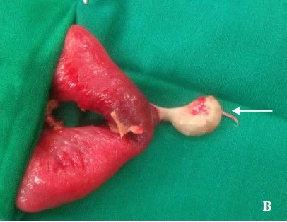
Figure 2: Intraoperative findings A. The Meckel diverticulum (white     shock (14, 15). Therefore, when a normal appearing
long arrow) is seen covered with fibrinous exudate and a perforated
                                                                        appendix is found during a laparotomy for suspected
tip. The diverticulum is in close proximity to the appendix (small
white arrow) which is enlarged and inflamed. B. Meckel’s                appendicitis, an inflamed Meckel’s diverticulum must be
diverticulum (long white arrow) is seen with a narrow neck and          sought. The severity of appendicitis may determine the
perforated tip. The adjacent bowel was resected with the diverticulum
and end to end primary anastomosis done.                                decision to resect an asymptomatic MD discovered during an
                                                                        appendectomy. This follows the recommendation that an
                                                                        incidentally detected MD should be left intact in the presence
We first removed the appendix then proceeded to do a                    of complicated appendicitis with abscess, perforation or
segmental resection of the intestine with the diverticulum and          gangrene to reduce risk of postoperative complications (16).
end-to-end anastomosis of the adjacent bowel. The patient               The decision to resect an incidentally detected MD also
made full recovery and was discharged from the clinic. The              depends on other factors and is still a subject of controversy.
resected appendix and diverticulum were sent for histology,             A systematic review of this subject found that most authors
which confirmed features of appendicitis with luminal                   favored removal of an incidentally detected asymptomatic
suppuration and infiltration of polymorphs. The diverticulum            diverticulum only if it occurred in males younger than 50
had colonic mucosa, exhibiting features of diverticulitis with          years, is longer than 2cm and contains ectopic mucosa (2).
inflammation, ulceration and perforation.                               This approach is linked to evidence that the lifetime risk of
                                                                        developing complications related to MD is low at 4–6% and
Discussion                                                              consequently, approximately 800 diverticular resections
The diagnosis of MD presents a known challenge to the                   would need to be performed to prevent 1 death (17). It
paediatric surgeon. Charles Mayo correctly stated that MD is            appears that resecting an incidentally detected MD is
frequently suspected, often looked for and seldom found (5).            unnecessary as this may increase the perioperative morbidity
The preoperative diagnosis of symptomatic MD is difficult               risk in a patient whose lifetime risk of MD-associated
and is missed in most cases especially in those that don’t              complications is rather low. In patients with symptomatic
present with bleeding. The clinical presentations are diverse           MD, treatment is surgical resection by either simple
and may include abdominal pain, intestinal obstruction,                 diverticulectomy or segmental bowel resection and
diarrhea, lower gastrointestinal bleeding with jelly stool, and         anastomosis, which are usually indicated when the
sometimes an abdominal palpable mass. The main clinical                 diverticulum has a wide base or when it is associated with
                                                            The ANNALS of AFRICAN SURGERY, April 2019 Volume 16 Issue 2
1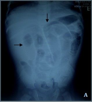
The ANNALS of AFRICAN SURGERYIwww.annalsofafricansurgery.com            Jumbi et al. Meckel’s Diverticulum with Appendicitis

palpable ectopic tissue, intestinal ischemia and perforation            10. Gerdes C, Akkermann O, Krüger V, et al. Incarceration of
(2). Recent literature suggests that the macroscopic features               Meckel's diverticulum in a left paraduodenal Treitz' hernia.
of the diverticulum do not predict the presence of ectopic                  World J Clin Cases. 2015; 3:732–5.
                                                                        11. Blevrakis E, PartalisN, Seremeti C, et al. Meckel’s
tissue and therefore a formal bowel resection is favored to
                                                                            diverticulum in paediatric practice in Crete (Greece): A 10-
simple diverticulectomy, which carries the risk of leaving
                                                                            year review. Afr J Paediatr Surg. 2011; 8:279–82.
behind hostile ectopic tissue usually at the base (18, 19). The
                                                                        12. Rattan KN, Singh J, Dalal P, et al. Meckel's diverticulum in
most common ectopic mucosa are gastric and pancreatic with                  children: Our 12-year experience. Afr J Paediatr Surg. 2016;
colonic, duodenal and biliary mucosa occurring less                         13(4):170–4.
frequently (20). We opted for a formal bowel resection and              13. Smith EB. Acute appendicitis associated with Meckel’s
anastomosis in our patient with a perforated MD, which was                  diverticulum. J Natl Med Assoc. 1960; 52(1):51–73.
an indication for such management coupled with the fear of              14. Modi S, Kanapathy PS, DeClercq S. Perforated Meckel's
leaving ectopic tissue, which was later confirmed by                        diverticulum in an adult due to faecolith: A case report and
histopathology as colonic mucosa with diverticulitis.                       review of literature. Int J Surg Case Rep. 2015; 15:143–5.
                                                                        15. Senocak R, Ince M, Kaymak S, et al. Coexistence of acute
                                                                            appendicitis and perforated Meckel’s diverticulitis: A rare
Conclusion                                                                  presentation in older adults. J Clin Gastroenterol Treat. 2016;
Children with symptomatic MD usually present with                           2:17
intestinal obstruction and peritonitis but rarely present with          16. Ueberrueck T, Meyer L, Koch A, et al. The significance of
bleeding, as seen in adults. The association between MD and                 Meckel’s diverticulum in appendicitis—A retrospective
appendicitis is commonly established in the context of an                   analysis of 233 cases. World J. Surg. 2005; 29:455–8.
incidentally found MD during appendectomy. However, a                   17. Zani A, Eaton S, Rees CM, et al. Incidentally detected Meckel
symptomatic MD in the presence of acute appendicitis is a                   diverticulum: To resect or not to resect? Ann Surg. 2008;
                                                                            247:276–81.
rare occurrence which may be associated with non-specific
                                                                        18. Varcoe RL, Wong SW, Taylor CF, et al. Diverticulectomy is
symptomatology that presents a diagnostic challenge, which
                                                                            inadequate treatment for short Meckel's diverticulum with
can delay management especially in the pediatric population.
                                                                            heterotopic mucosa. ANZ J Surg. 2004; 74:869-72.
                                                                        19. Gezer HÖ, Temiz A, İnce E, et al. Meckel diverticulum in
References                                                                  children: Evaluation of macroscopic appearance for guidance
1.   Meckel JF. Ueber die Divertikel am Darmkanal. Arch Physiol.            in subsequent surgery. J Pediatr Surg. 2016; 51(7):1177–80.
     1809; 9:421–453.                                                   20. Lin XK, Huang XZ, Bao XZ, et al. Clinical characteristics of
2.   Sagar J, Kumar V, Shah DK. Meckel's diverticulum: A                    Meckel diverticulum in children: A retrospective review of a
     systematic review. J R Soc Med. 2006; 99:501–5.                        15-year single-center experience. Medicine (Baltimore). 2017;
3.   Park JJ, Wolff BG, Tollefson MK, et al. Meckel diverticulum:           96(32):e7760
     The Mayo Clinic experience with 1476 patients (1950–2002).
     Ann Surg. 2005; 241:529–33.
4.   St-Vil D, Brandt ML, Panic S, et al. Meckel's diverticulum in
     children: A 20-year review. J Pediatr Surg. 1991; 26:1289–92.
5.   Mayo CW. Meckel’s diverticulum. Pro Mayo Clin.1933;
     8:230.
6.   Pantongrag-Brown L, Levine MS, Buetow PC, et al. Meckel’s
     enteroliths: Clinical, radiologic, and pathologic findings. Am J
     Roentgenol. 1996; 167:1447–50.
7.   Ruscher KA, Fisher JN, Hughes CD, et al. National trends in
     the surgical management of Meckel's diverticulum. J Pediatr
     Surg. 2011; 46:893–6.
8.   Ren B, Jia X, Meng X, et al. Intestinal obstruction due to axial
     torsion of a giant Meckel's diverticulum: A case report. Int J
     Colorectal Dis. 2015; 30:1133–4.
9.   DeshmukhSN, Jadhav SP, Asole AG. Axial torsion and
     gangrene of a giant Meckel's diverticulum causing small bowel
     obstruction. Sri Lanka J Surg. 2015; 33:35–6

                                                           The ANNALS of AFRICAN SURGERY, April 2019 Volume 16 Issue 2
1
You can also read


