Squamous Cell Carcinoma of the Lateral Border of the Tongue in a Patient with Toxic Multinodular Goitre (Hyperthyrodism): A Case Report
←
→
Page content transcription
If your browser does not render page correctly, please read the page content below
IBBJ
                                                                                                                                                                                  Case report
                                                                         Summer 2018, Vol 4, No 3

                                                                             Squamous Cell Carcinoma of the Lateral Border of the
                                                                              Tongue in a Patient with Toxic Multinodular Goitre
Downloaded from ibbj.org at 23:48 +0330 on Wednesday October 16th 2019

                                                                                      (Hyperthyrodism): A Case Report
                                                                                                    Sarah Ibiyemi, Ibrahim Mogaji, Oluwaremilekun Dada

                                                                         Department of Dental Services, Federal Medical Centre, Abeokuta, Ogun state, Nigeria.

                                                                                                       Submitted 6 Aug 2018; Accepted 30 Sep 2018; Published 3 Oct 2018
                                                                         Squamous cell carcinoma (SCC) of the oral cavity can mimic a myriad of ulcerative lesions in the mouth. This is
                                                                         particularly true for medically complex patients who due to their underlying systemic condition and multiple use
                                                                         of drugs, are prone to oral lesions. With increasing dental awareness, more patients are presenting early to the
                                                                         oral surgeons. The era of patients presenting first to the clinician with characteristic fungating solitary ulcer in the
                                                                         mouth with rocky hard submandibular lymph node involvement may be fast disappearing. The aim of the present
                                                                         report is to present a female patient with multiple focal area of erythema on the right lateral border of the tongue
                                                                         which though appeared innocuous but was discovered to be SCC after punch biopsy. High index of suspicion with
                                                                         proper attention to clinical details will be necessary for early diagnosis of SCC in the oral cavity, and therefore
                                                                         improve the overall prognosis of the patient.

                                                                         Keywords: Goitre, hyperthyroidism, squamous cell carcinoma, tongue

                                                                         S    quamous cell carcinoma (SCC) is considered as
                                                                              one of the most common oral cancers (1). The
                                                                         tongue is one of the oral cavity sub sites associated
                                                                                                                                                 right lateral side of her tongue for 3 months duration.
                                                                                                                                                 She has been diagnosed for toxic multinodular goiter
                                                                                                                                                 about a year before at the endocrinology unit of this
                                                                         most commonly with SCC. Previous studies have                           centre. She was on carbimazole (an antithyroid
                                                                         demonstrated that cancers of the tongue are distinct                    drug) 10 mg three times daily at the time of
                                                                         biologically and epidemiologically from other                           presentation.
                                                                         tumors of the oral cavity. Tongue cancers are                              She was conscious and alert, not pale, anicteric.
                                                                         associated with an increased proportion of patients                     She did not present any obvious respiratory or
                                                                         who are women, aged under 40 years, and non-                            painful distress. A diffuse midline neck swelling
                                                                         smokers (2). Most patients in this part of the world                    (about 6 cm in its widest diameter) consistent with
                                                                         present in the late stage, and treatment is therefore                   toxic multinodular goiter was observed. The vital
                                                                         usually palliative (3), but with increasing awareness                   signs were all within normal limits. She had no
                                                                         about oral health and early presentation of patients,                   peripheral lymphadenopathy and no pedal edema.
                                                                         high index of suspicion and good clinical acumen is                     Extra-oral      examination       revealed   no   facial
                                                                         expected so that missed diagnosis from clinicians                       asymmetry, and submandibular lymph nodes were
                                                                         will not be an increasing cause for late intervention.                  bilaterally palpable, freely mobile and not tender.
                                                                         Case presentation                                                       Temporomandibular            joints   were   bilaterally
                                                                           A 52 years old woman presented to the oral                            palpable, moved synchronously and were not tender.
                                                                         diagnosis clinic upon redness and discomfort on the                     However, intraoral examination revealed an area of

                                                                         *Correspondence: Department of Dental Services, Federal Medical Centre, Abeokuta, Ogun state, Nigeria.
                                                                         E-mail: ibrahimtasnan@gmail.com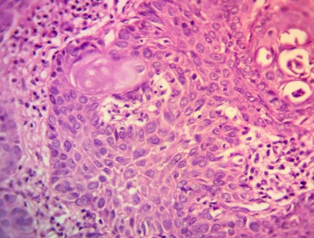
Squamous Cell Carcinoma and Multinodular Goitre
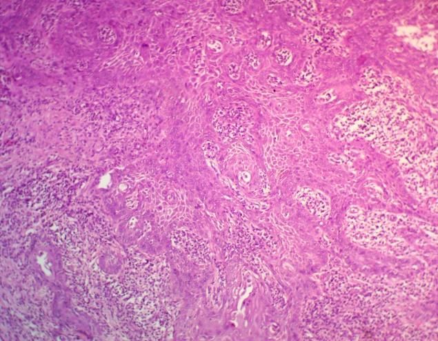
erythema located approximately around the middle Histologic section revealed a densely collagenized
third on the right lateral border of the tongue. There connective tissue stroma infiltrated by invading
were 5 different focal spots that appeared more cords, sheets and islands of neoplastic squamous
erythematous than the surrounding area. The widest epithelium (Figure 1). The neoplastic squamous
of these spots was about 4 mm in diameter. The epithelium exhibited cellular and nuclear
region was however indurate with mild tenderness. hyperchromatism and increased nucleo-
Downloaded from ibbj.org at 23:48 +0330 on Wednesday October 16th 2019
She also had an overdenture replacing lower right cytoplasmic ratio and also cellular and nuclear
central incisor. She has been using denture for two atypia (Figure 2). Some cells also exhibited
years. The dentures were done by a general vesicular nuclei. Also, chronic inflammatory cells
practitioner and they appeared well fabricated and predominantly lymphocytes, plasma cells,
fitted with no rocking movements. endothelial lined vascular channels and muscle
A full blood count and differentials, also tissue were observed. Histological diagnosis of
electrolytes and urea were ordered. A punch biopsy moderately differentiated squamous cell carcinoma
was preferred to minimize trauma and regarding the of the right lateral border of the tongue was made.
medical condition of the patient. Patient was advised
to sparingly use the denture within the week as she
insisted she could not do without it. Punch biopsy
was planned a week after presentation while results
for heamatological investigations were also
expected.
The patient presented to the clinic a week after
with vital signs being within normal limits. The
lateral border of the tongue also did not appear
differently from the day of first presentation though
patient claimed to have complied with use of denture
sparingly. Full blood counts and differentials were
within normal range thereby ruling out Figure 1. Photomicrograph showing sheets of neoplastic
epthelium within the stroma. Some squamous cells exhibit
agranulocytosis and neutropenia. Electrolyte and nuclear and cellular pleomorphism, nuclear hypercromatism
and elaborate keratin (×100).
urea were also within normal limits.
After achieving anaesthesia by blocking the
lingual nerve on the right side, a 4 mm metal punch
was used for the biopsy. Two of the focal points of
the erythema were chosen. One was taken within
apparently normal tissue, the other was taken deep
into area suspected to be the focal point of the lesion
with induration. Heamostasis was achieved with
digital pressure. One each 3.0 vicryl sutures were
placed on the biopsy site. She was placed on 2%
lignocaine gel to be applied using a pur-wrap
directly on the site. 8 hourly 500 mg paracetamol
tablet was also prescribed for 3 days. Biopsy tissue
Figure 2. Photomicrograph of neoplastic epithelium.
was preserved in 10% formalin and sent to the oral Cellular atypia and chronic inflammatory cells are apparent
(×200).
pathologist.
167 Int. Biol. Biomed. J. Summer 2018; Vol 4, No 3Ibiyemi S et al.
Discussion malignant ulcerations were misdiagnosed as
The patient with a previous history of nonneoplastic lesions up to several months before
hyperthyroidism and on medications posed a major the definite diagnosis was established (11-13).
diagnostic challenge. In addition, the presence of Valente et al. reported a case of SSC misdiagnosed
flanges of the denture around the area of the as a denture-related traumatic ulcer (11). De Sant’
erythema made this look like chronic irritation from Ana dos Santos et al. also reported misdiagnosis of
Downloaded from ibbj.org at 23:48 +0330 on Wednesday October 16th 2019
the denture. Studies have shown that chronic lip SCC as actinic cheilitis (12). A case of gingival
irritation from denture wearers have resulted in oral SCC appearing as an aphthous ulcer was also
squamous carcinoma (4). The present patient had the reported by Kumari et al. (13). This time elapsed
denture changed three months before presentation between misdiagnosis and actual diagnosis might
when she noticed the erythematous area, on the jeopardize patients’ overall prognosis; therefore,
suspicion that it was caused by the denture. attempts should be made to come to timely diagnosis
Another diagnostic challenge is the possibility of via evidenced based approach and high index of
a drug induced erythema. Carbimazole which is an suspicion. The golden rule to biopsy a lesion beyond
antithyroid drug, is a well-known medication used 2 weeks in a site even after removal of irritating
in the treatment of hyperthyroidism (5). factor, etiology or proves refractory to local therapy
Agranulocytosis is a recognized but rare and life- cannot be over emphasized (14). The patient was
threatening side effect of carbimazole therapy, therefore planned for partial glossectomy with neck
which usually occurs within the first 3 months of dissection.The present report also emphasizes the
treatment (6) but manifestation can be delayed even importance of not always attributing oral lesions in
to well over a year (7). The incidence of this medically complex patients as being due to the
particular side effect has been reported to range from underlying medical condition(s) or drug use.
0.3% to 0.6% (6) and is associated with a mortality Conflict of interest
rate of 21.5% (8). The authors declared no conflict of interest.
Clinically, patients usually present with fever
(92%), sore throat (85%), painful mouth ulcer References
(15%), anal ulcer (8%), reduced immune response, 1. Taghavi N and Yazdi I. Prognostic factors of survival rate in
and are prone to bacterial infections (9). However, oral squamous cell carcinoma: clinical, histologic, genetic and
non of these clinical features apart from the painful molecular concepts. Arch Iran Med. 2015;18:314-9.
area of erythema were observed in our patient. 2. Rusthoven K, Ballonoff A, Raben D, et al. Poor prognosis in
Moreover, results of the full blood count showed no patients with stage I and II oral tongue squamous cell carcinoma.
features of agranulocytosis or neutropeania. Cancer. 2008;112:345-51.
Because of the induration of the lesion, SSC was 3. Da Lilly-Tariah O B, Somefun A O, Adeyemo W L. Current
suspected which was confirmed by the punch biopsy evidence on the burden of head and neck cancers in Nigeria. Head
result. SSCs are usually solitary ulcers though could & Neck Oncology. 2009;1:14.
rarely be multifocal (10). The observation of 4. Panat S, Aggarwal A, Chakarvarty A. Denture induced
multifocal area of erythema on the lateral border of squamous cell carcinoma: A rare case report. J Dent Sci Rehabil.
the tongue in this patient may represent one of the 2012;3:42-4.
rare cases reported in the literature. 5. Cooper D S. Antithyroid drugs. N Engl J Med. 2005;352:905-
This present case underscores the importance of 17.s
methodical approach in acheiving diagnosis based 6. Tajiri J, Noguchi S, Murakami T, et al. Antithyroid drug-
on a sound knowledge of the differentials. induced agranulocytosis. The usefulness of routine white blood
According to the literature, many cases of oral cell count monitoring. Arch Intern Med. 1990;150:621-4.
Int. Biol. Biomed. J. Summer 2018; Vol 4, No 3 168Squamous Cell Carcinoma and Multinodular Goitre
7. Mohan A, Joseph S, Sidharthan N, et al. Carbimazole-induced -ous cell carcinoma misdiagnosed as a denture-related traumatic
agranulocytosis. J Pharmacol Pharmacother. 2015;6:228-30. ulcer: A clinical report. J Prosthet Dent. 2016;115:259-62.
8. Cooper D S, Goldminz D, Levin A A, et al. Agranulocytosis 12. De Sant'ana Dos Santos F, Isper M A, Pereira Novo-Neto J,
associated with antithyroid drugs. Effects of patient age and drug et al. Misdiagnosis of lip squamous cell carcinoma. RSBO.
dose. Ann Intern Med. 1983;98:26-9. 2012;9:114-8.
9. Mahant S, Shobhane U, Mahant P. Carbimazole induced 13. Kumari P S, Kumar G P, Bai Y D, et al. Gingival squamous
Downloaded from ibbj.org at 23:48 +0330 on Wednesday October 16th 2019
agranulocytosis with life-threatening complications, tremendous cell carcinoma masquerading as an aphthous ulcer. J Indian Soc
response with granulocyte-colony stimulating factor. Medical Periodontol. 2013;17:523-6.
Journal of Dr. DY Patil University. 2016;9:79-81. 14. Hershita S S, Nancy C, Monika P, et al. Biopsy- A vision of
10. Wood N K, Goaz P W, Alling C C, Differential diagnosis of life. International Journal of Contemporary Medical Research.
oral lesions. Mosby St. Louis:1991. 2016;3:1734-7.
11. Valente V B, Takamiya A S, Ferreira L L, et al. Oral squam
169 Int. Biol. Biomed. J. Summer 2018; Vol 4, No 3You can also read