Clinical, Dermoscopic and Histhopatological Findings in Diagnosis of Nevus Spilus
←
→
Page content transcription
If your browser does not render page correctly, please read the page content below
60

    Clinical, Dermoscopic and Histhopatological
       Findings in Diagnosis of Nevus Spilus
                                   Risa, A1, Asri, E 1, Izrul I,2 Tofrizal A3

1
  Dermato-Venereology Department of Andalas University/ Dr.M.Djamil Hospital,Padang, Indonesia
 email: amillia.risa@yahoo.com
2
  Dermato-Venereology Department of Rasidin Hospital Padang Indonesia
3
  Department of Anatomical Pathology Faculty of Medicine, Andalas University

Abstract

Introduction: Nevus spilus (NS) are seen in 0.2% to 2.3% of the population and have 0,13% to 0,2% risk for
malignant transformation. Clinical, dermoscopic, and histhopatological features were describe in this case report
in order to be easily recognize NS. Although NS is a benign cutaneous anomaly it has potential malignant
transformation and requires regular follow up. Case Report: A case of nevus spilus in 23 years-old female was
reported. There were multiple asymptomatic brownish pigmented spots over brownish patch on left cheek which
gradually increased in number and size since 1 year ago. Dermatologic state: brown macules and dark brown
papules in a speckled, overlying background café au lait macule. Dermoscopy revealed reticular pattern in
background light brown and dark reticuloglobular pattern in dark speckled. Histopathology showed elongation of
rete ridges with grouping of melanocyte cells at the tip, and proliferation of nevus cells. Conclution: Patient was
planned to treat with Nd-Yag laser.

Keywords: dermoskopi, nevus on nevus, speckled lentigenous nevus, spotty nevus

Abstrak

Pendahuluan: Nevus spilus (NS) ditemukan sekitar di 0,2% hingga 2,3% dan memiliki resiko menjadi keganasan
sekitar 0,13% to 0,2%. Gambaran klinis, dermoscopic, histhopatologis dijelaskan dalam laporan kasus ini agar
NS mudah dikenali. Walaupun NS adalah suatu lesi jinak namun memiliki postensi untuk menjadi keganasan
sehingga dianjurkan untuk di follow up secara teratur. Laporan Kasus: Satu kasus nevus spilus telah dilaporkan
pada pasien perempuan berusia 23 tahun. Terdapat beberapa bintik-bintik kecoklatan yang asimtomatik diatas
bercak kecoklatan di pipi kiri yang secara bertahap meningkat dalam jumlah dan ukuran sejak 1 tahun lalu.
Status dermatologis: makula kecoklatan dan papul kecoklatan diatas café au lait. Dermoscopy menunjukan pola
retikular dengan latar belakang coklat terang dan pola retikuloglobular gelap. Histopatologi menunjukkan
perpanjangan rete ridges dengan pengelompokan sel melanosit di ujungnya, dan proliferasi sel nevus.
Kesimpulan: Pasien direncanakan diterapi dengan laser Nd-Yag..

Kata kunci: dermoscopy, nevus on nevus, speckled lentigenous nevus, spotty nevus.

Email :heme@unbrah.ac.id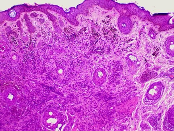
61     Heme, Vol III No 1
       January 2021

I.     INTRODUCTION                                No standard guidelines exist for the
                                                   management of patients with nevus spilus.
Nevus spilus (NS), also known as speckled          Clinical appearance (typical or atypical),
lentiginous nevus (SLN) occurs in                  history of stability or instability of pigmented
approximately 1% to 2% of the population.          elements, congenital or noncongenital onset,
They are present either at birth or in the first   perceived risk of developing melanoma, and
years of life and have therefore been regarded     cosmetic concerns are considerations when
as a variant of congenital nevus. There does       deter- mining whether to excise or
not appear to be gender or ethnic predilection.    recommend periodic clinical evaluation for
Darkly pigmented flat macules or papules are       life.3
usually present within the nevus spilus on
presentation. New pigmented elements may           II.    CASE REPORT
evolve within the lesion over time. The
background pigmentation of a nevus spilus is       A 23 year-old female, was reffered to
circumscribed and similar in appearance to a       Dermato-Venereology                  out-patient
café-au-lait macule in hue, with even light       Departement of Dr. M. Djamil Hospital, with
pigmentation.       There     are     scattered    chief complaint multiple brownish pigmented
superimposed more darkly pigmented                 spots on left cheek that not felt itchy or pain
macules or papules. The tan macular                that gradually increased in number and size
background pigmentation can range in size          since 1 year ago. Initially there was brownish
from less than 1 cm to larger than 10 cm in        patch over the left cheeck since birth that not
diameter. Lesions are most commonly found          felt itchy or pain. About 12 years ago there
on the trunk and extremities, although any         were multiple and small, brownish dots
cutaneous site may be affected. Multiple           appeared over the patch which gradually
lesions may be present and have a segmental        increased in size and number, and become
distribution.                                      darker since 2 year ago. Patients never treated
                                                   these complaints before. There is no history of
Dermoscopy reveals dark speckled foci with         yellow brownish verrocous patch on scalp,
a reticuloglobular pattern in a background         head or neck or another part of the body,
light brown and reticular pattern. Mixed           increased sweating of the left half of her body.
patterns may occur that include combinations       There is no history of muscular weakness,
of homogeneous, reticular, globular, granular,     numbness, pain or discomfort within the
and spitzoid patterns.1,2                          involved skin and multiple brown macules on
                                                   the another part of the body. There is no
Histologic sections reveal that the tan            history of easy bleeding on brownish dots.
background corresponds to a diffuse area
where there is slight epidermal hyperplasia        Physical examination we found within normal
with basilar hyperpigmentation, from no            limit. Dermatologic state we found brown
perceptible increase in melanocytes to a slight    macules and dark brown papules in a
increase in singular melanocytes along the         speckled, overlying background café au lait
basal layer. The dark spots correspond to          macule (light brown macule), Fitzpatrick skin
small lentiginous junctional and compound          type IV. Routine blood examination were
nevi featuring increased numbers of single         within normal.
melanocytes and small nests along the DEJ
and small, round melanocytes in the                Working diagnosed in this patient were nevus
superficial dermis, in cases of compound           spilus, differential diagnosis were agiminated
nevi. The diagnosis of nevus spilus is usually     acquired melanocytic nevi. We did
straightforward.1,2                                dermoscopy examination and biopsy to
                                                   establish the diagnosis.

Email :heme@unbrah.ac.id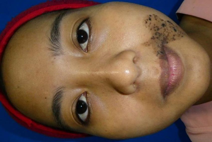
Heme, Vol III No 1     62
                                                                            January 2021

                                               Dermoscopy examination result.

                                                 A

   A

                                                 B

                                               FIGURE 2. A&B DERMOSCOPY SHOWED DARKER
  B
                                               BROWN AREA WITH A RETICULOGLOBULAR
                                               PATTERN IN DARK SPECKLED. THE BACKGROUND IS
FIGURE 1. A. SHOW THE LOCATION OF THE LESION   A LIGHT BROWN AND RETICULAR
IN LEFT CHEEK, UNILATERAL DISTRIBUTION.
B. SHOW BROWN MACULES AND DARK BROWN
PAPULES     IN   A   SPECKLED,    OVERLYING    Histopathological examination result were
BACKGROUND CAFE AU LAIT MACULE.                irregular achantosis with elongation of rete
                                               ridges, a grouping of melanocyte cells at the
                                               tip of the rete ridges at several places, as well
                                               as increased pigmentation in the basal
                                               epidermis cells in epidermis. In Dermis, we
                                               can found proliferation of nevus, cells
                                               forming nests, especially in the upper
                                               epidermis and around the adnexa, with
                                               melanophag cells containing melanin
                                               pigments.

                                                                        Health & Medical Journal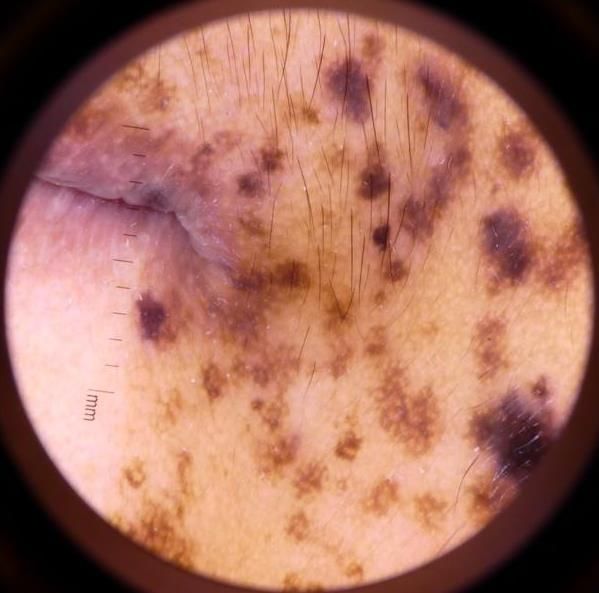
63 Heme, Vol III No 1
January 2021
genetic factors.3 in this case patient were 23
years old female.
Kamińska (Poland, 2003) reported 9 case of
nevus spilus, female were predominance, with
an average age 37 years old.7 Ghosh et al
(India, 2018) reported 7 case of nevus spilus
for 1 year in KPC medical hospital in Kolkata,
female were preponderance in this study. The
most age of presentation was at birth and
between 1-10 years old.8 Kamińska (Poland,
2003) reported 9 case of nevus spilus, female
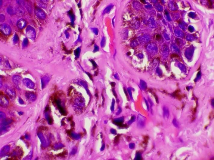
FIGURE 3. HISTOPATHOLOGICAL EXAMINATION were predominance, with an average age 37
SHOWED ELONGATION OF RETE RIDGES, years old.8 Handidwiono (Indonesia, 2015)
PROLIFERATION OF NEVUS CELLS. (HE STAIN, reported 1 case of nevus spilus in male
MAGNIFICATION 10X) patient, in mental retardation elementary
school/ among student with disabilities in
Yogyakarta.9 Danarti et al (Indonesia,2019)
reported 2 case of nevus spilus as a part of
phacomatosis pigmentokeratotica10 In out
patient of dermatology venereology
department in Dr. M. Djamil hospital, this is
the first case had been reported in the last ten
years.
Nevus Spilus be congenital or acquired and
usually appears during infancy but may also
appear later in life, typically in the intensive
growth stages.4,5 Aqil et al (Morocco, 2019)
70% of patients had these lesions from birth
FIGURE 4. GROUPING OF MELANOCYTE CELLS AT
THE TIP OF THE RETE RIDGES (HE STAIN, and 30% in adulthood.4 Kamińska (Poland
MAGNIFICATION 100X) 2003) reported, all patients presents nevus at
births.7 In this case patient had lesion since
III. DISCUSSION birth.
Nevus spilus also know as speckled Lesions are most commonly found on the
lentiginous nevus, spots on a spot, and trunk and extremities, although any cutaneous
zosteriform lentiginous nevus. It is a rare site may be affected. Multiple lesions may be
dermatologic entity, occurring in less than present and have a segmental distribution.3
0.2% of newborn infants. The reported Kamińska (Poland 2003) reported lesions are
prevalence in the pediatric age group ranges commonly located on the extremities.7 Ghosh
from 1.3% to 2.1%, and that in the adult et al (India, 2018) reported majority of lesion
population is approximately 2.3%.4,5,6 The sex were on the head and neck, and affected left
incidence is approximately equal. All ethnic side of the body, theres is no bilateral lesions
and racial groups are at risk, but there is a and associations with cutaneous or systemic
slight predilection for white individuals. findings.8 Aqil et al (Morocco, 2019) reported
Presumably, nevus spilus results from a cases of nevus spilus with the most location
localized defect in neural crest melanoblasts were on thrunk.4 In this case patient had
under the influence of environmental and unilateral lesion on the the face.
Email :heme@unbrah.ac.id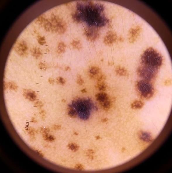
Heme, Vol III No 1     64
                                                                                January 2021

                                                   color, ranging from light brownish to brown
Typically, nevus spilus presents as an             and blackish.7
asymptomatic, tan to brown, sharply
demarcated,                well-circumscribed      Single lesion was excised for histological
pigmentation that is studded with multiple,        evaluation. The histopathological findings
smaller, darker punctate macules or papules.       revealed irregular achantosis with elongation
The lesion often starts as a light- brown oval     of rete ridges. There is a grouping of
macule/patch with few or no speckles. Over         melanocyte cells at the tip of the rete ridges at
time, the speckles slowly increase in size and     several places, as well as increased
number. These speckles can be macular or           pigmentation in the basal epidermis cells in
papular and represent lentigines or                epidermis. In dermis revealed showed
melanocytic nevi. The number of speckles           proliferation of nevus cells, cells forming
usually ranges from 8 to 10, and the size of       nests, especially in the upper epidermis and
speckles ranges from 1 to 3 mm. The size of        around the adnexa, with melanophag cells
the background pigmented patch generally           containing melanin pigments. Vaidya (USA,
ranges from 2 to 10 cm. NS could develop           2007) reported, the light brown macule or
more spot elements over the time. Nevus            patch usually shows mild melanocytic
spilus, similar to melanocytic nevus, is           hyperplasia. Fur thermore, it may show
classified as small (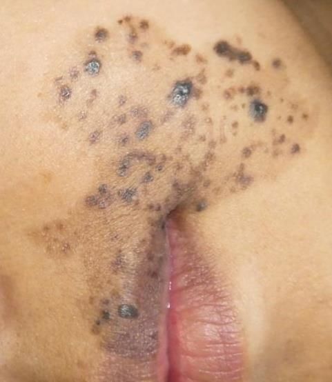
65     Heme, Vol III No 1
       January 2021

Clinical appearance (typical or atypical),         We reported a case of nevus spilus on facial,
history of stability or instability of pigmented   diagnosed was based on anamnesis, physical,
elements, congenital or noncongenital onset,       dermoscopy         and      histopathological
perceived risk of developing melanoma, and         examination. Regular clinical-dermatoscopic
cosmetic concerns are considerations when          observation is advised because of the risk of
determining whether to excise or recommend         transformation into melanoma. Any changes
periodic clinical evaluation for life.             lesion suspicious to malignant transformation
Documentation with high quality photographs        should biopsied for histopathological
can be used to aid follow up by parents and        evaluation.
physicians.3 European Society of Laser in
Dermatology recommended Q-switched laser           V.       REFERENCES
for treated Nevus spilus.15 The Q-switched         [1] Corradin MT, Cacitti V, Giulioni E, Patriarca
                                                        MM and Vettorello A. Nevus Spilus: A Review
Nd:YAG laser emits a longer, near- infrared             of the Literature. SM Dermatolog J. 2015; 1:1-7
ray of 1,064 nm that is capable of penetrating     [2] Vaidya DC, Schwartz RA, Janniger CK. Nevus
into the deeper regions of the skin. Therefore,         spilus. Pediatric dermatology. 2007;80:465-468
it is able to destroy deep-seated dermal           [3] Cuda JD, Moore RF, Busam KJ. Melanocytic
melanocytes by selective photothermolysis.16            Nevi. In : Kang S, Amagai M, Bruckner AL, Enk
                                                        AH, Margolis DJ, McMichael AJ, editors.
Patient were planned to treat with Q switched           Fitzpatrick Dermatology. Ed 9th. New York:
Nd-YAG laser.                                           McGraw Hill;2019:1944-1981
                                                   [4] Aqil N, Elloudi S, Nassiri A, Gallouj S, Baybay
Melanoma has been reported as a very rare               H, Mernissi FZ. 2019. Dermoscopy of naevus
complication in NS. The melanoma most                   spilus. J Case Rept Img. 2019. 1: 37-39
                                                   [5] EL Jouari O, Senhaji G, El Mahi H, Gallouj S,
often found on NS is superficial spreading              Zahra MF. Dermoscopy of Nevus Spilus.
melanoma, followed by nodular melanoma.                 Madridge J Dermatol Res. 2018; 3(2): 79-80
Manganoni et al (Italy, 2011) evaluated 2134       [6] Leung AK, Barankin B. Circumscribed
patients with melanoma, 27 of them presented            Pigmentation Stippled with Punctate Macules and
NS in a different body region. In the median            Papules in a 9-year-old Boy. Pediatrics in review.
                                                        2018;39;e50-53
50 months of follow-up, patients were              [7] Kamińska W G. Dermoscopy of Nevus Spilus.
monitored with clinical and dermatoscopic               Dermatologic Surgery. 2013; 39(10): 1550-1554.
observation. The risk of malignant melanoma        [8] Ghosh S, Ghosal L, Biswas SK, Bhunia D. A
in NS is higher in patients with the giant              clinicoepidemiological study of different types of
and/or zosteriform variants. Although in this           nevi in patients attending at a tertiary care hospital
                                                        in Eastern India. Journal of Pakistan Associations
case patient had a medium sized, we planing             of dermatologists. 2018; 28 (1); 51-58.
patient to follow up once per year, clinical       [9] Handidwiono R, Budiyanto A, Radiono S,
and dermatoscopy examination is required to             Rianawati T, Danarti R. Prevalensi kelainan kulit
follow up the lesion.       Patient also be             pada siswa sekolah dasar luar biasa (SDLB)
instructed how to perform self examination to           pembina Yogyakarta. MDVI. 2015;42(3):119-
                                                        127
help detect early malignant changes.               [10] Danarti R, Chusniyati N, Sulistiyowati Y.
Dermatoscopy has been shown to improve the              Phacomatosis pigmenkeratotica : two case series
diagnostic accuracy for early melanoma                  of a neurocutaneous rarity from Indonesia. J Med
detection. If there is any doubt, biopsy for            Sci. 2019;51(4): 309-316
histological evaluation should be considered.      [11] Abrusci V, Benzecry V. Medium-sized nevus
                                                        spilus of the neck treated with pulsed dye laser.
If a melanoma arises in an NS, its                      Dermatologic Therapy. 2017;00:e12497.
management and prognosis are similar to            [12] Vaidya DC, Schwartz RA, Janniger CK. Nevus
those for any other melanomas17-20                      spilus. Cutis. 2007(80); 465-467
                                                   [13] Shin J, Kim YC. Multiple agminated acquired
                                                        melanocytic nevi. Ann dermatol. 2013;25(2);
                                                        251-252.
IV.    CONCLUSION                                  [14] Shimasaki Y, Fukuta Y, Yoshida Y, Mori HH,
                                                        Yamamoto O. Acquired agminated melanocytic

Email :heme@unbrah.ac.id
Heme, Vol III No 1 66
January 2021
naevi; report of two case and review of the
literature. Acta derm venereol. 2012;92:585-663
[15] Passeron T, Genedy R, Salah L, Fusade T,
Kositratna G, Laubach J et al. Laser treatment of
hyperpigmented lesions: position statement of the
european society of laser in dermatology. Journal
of european academy of dermatology and
venereology. 2019;1-19
[16] Kim YJ, Whang KU, Choi WB, Kim HJ, Hwang
JY, Lee JH et al. Efficacy and Safety of 1,064 nm
Q-switched Nd:YAG Laser Treatment for
Removing Melanocytic Nevi. Annals of
Dermatology.2012; 24(2), 162-167
[17] Manganoni AM, Pavoni L, Farisoglio C, Sereni
E, Calzavara-Pinton P. Report of 27 cases of
naevus spilus in 2134 patients with melanoma: is
naevus spilus a risk marker of cutaneous
melanoma? Journal of the European Academy of
Dermatology and Venereology. 2011; 26(1):
129–130.
,
[18] Corradin MT Giulioni E, Fiorentino R,
.
Santeufemia DA, Lo Re G, Vettorello A In situ
malignant melanoma on nevus spilus in an elderly
patient. Acta Dermatovenerologica Alpina,
Pannonica, et Adriatica, 2014, 23(1):17-19
[19] Frings VG, Goebeler M, Kneitz H, Dermpath &
Clinic: In situ melanoma arising within a speckled
lentiginous nevus. EJD. 2018; 28.
[20] Corradin MT, Zattra E, Fiorentino R, Alaibac M,
Fortina AB. Nevus spilus and melanoma; case
report and review the literature. Journal of
cutaneous medicine and surgery. 2010 (14)2; 85-
89
Health & Medical JournalYou can also read