PROCEEDINGS OF SPIE Mobile colposcopy in urban and underserved suburban areas in Baja California - MobileODT
←
→
Page content transcription
If your browser does not render page correctly, please read the page content below
PROCEEDINGS OF SPIE
       SPIEDigitalLibrary.org/conference-proceedings-of-spie

                   Mobile colposcopy in urban and
                   underserved suburban areas in Baja
                   California

                   Marta Madiedo, Sonia Contreras, Octavio Villalobos,
                   Bruce S. Kahn, Amit Safir, et al.

                                   Marta Madiedo, Sonia Contreras, Octavio Villalobos, Bruce S. Kahn, Amit
                                   Safir, David Levitz, "Mobile colposcopy in urban and underserved suburban
                                   areas in Baja California," Proc. SPIE 9699, Optics and Biophotonics in Low-
                                   Resource Settings II, 96990I (11 March 2016); doi: 10.1117/12.2218697

                                   Event: SPIE BiOS, 2016, San Francisco, California, United States

Downloaded From: https://www.spiedigitallibrary.org/conference-proceedings-of-spie on 2/11/2018 Terms of Use: https://www.spiedigitallibrary.org/terms-of-use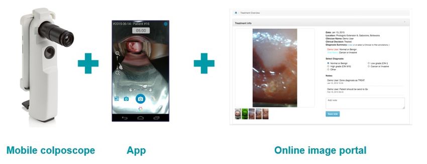
Mobile Colposcopy in Urban and Underserved Suburban Areas in
                                              Baja California

                    Marta Madiedo1, Sonia Contreras2, Octavio Villalobos1, Bruce S. Kahn3, Amit Safir4, and David
                                                              Levitz*4
                                              1
                                       Fronteras Unidas Pro-Salud, Tijuana, Baja California, Mexico
                                          2
                                            International Community Foundation, San Diego, CA
                       3
                         Department of Obstetrics & Gynecology, Scripps Clinic Medical Group, 3811 Valley Centre
                                                      Dr., San Diego, CA 92130, USA
                                       4
                                         MobileODT Ltd., Gershon Shatz 41, Tel Aviv 67017 Israel

                                                                                     ABSTRACT

                   Cervical cancer is the leading cause of cancer death for women in low resource settings, often affecting the most
                   economically disenfranchised segment of the population. The key challenge with cervical cancer is the lack of an
                   effective screening program for many of the at-risk, difficult-to-reach women. Outreach programs that utilize mobile
                   clinics to increase access to screening and care in Baja California have been developed. However, many barriers such
                   as quality assurance, efficient referral remained a challenge in this region. Visualization-based co-tests together with
                   cytology (Pap smears) as a primary screen have been proposed. Here, the mobile colposcope of the enhanced visual
                   assessment (EVA) is used to capture an image immediately following a Pap smear. EVA images were reviewed by
                   expert colposcopists. Initial or preliminary data from pilot services showed that Pap false positives and Pap false
                   negatives maybe reduced by expert review of EVA images. This suggests that reviewing of EVA images may be
                   instrumental in catching inaccurate Pap results, thereby improving care. Thus, there is a need to further explore the
                   benefits of using EVA as additional information when conducting Pap smear screenings.

                   Keywords: Low resource settings, colposcopy, translational research, imaging

                                                                                1. INTRODUCTION

                   Cervical cancer is the leading cause of cancer death for women in low-resource settings1. The most common type of
                   cervical cancer takes approximately 20 years to develop from an initial infection by the human papilloma virus (HPV),
                   through cervical dysplasia (precursor lesions), to mature cancers that put the patient’s life at risk2. Over the last 50
                   years, Organization for Economic Cooperation and Development (OECD) nations have implemented national
                   screening programs based on the Pap smear and more recently screening for human papilloma virus (HPV) infection,
                   followed by colposcopy and colposcopically-guided biopsy, and have greatly reduced the risk posed by cervical
                   cancer3. In this pathway of care, patients with a positive biopsy results are then referred to treatment with either an
                   ablative or excisional therapy.

                   In most low-income countries, this pathway of care is non-existent given the few resources available to the health
                   system. However, many middle-income countries have attempted to emulate the pathway of care offered by OECD
                   nations, albeit with mixed success. Pap screening is offered in several clinics around the Tijuana region, as well as in
                   mobile clinics that are part of outreach programs. Patients with an abnormal Pap screen come to a Tijuana clinic,
                   where colposcopy and biopsy services are available. Because there are limited colposcopy services for the
                   underserved, uninsured populations in Tijuana, is only one colposcopy clinic in Tijuana, and the prevalence of cervical
                   *
                    Address all correspondence to Bruce Kahn, kahn.bruce@scrippshealth.org. Phone: (858) 764-3225; Fax: (858)
                   764-9097

                               Optics and Biophotonics in Low-Resource Settings II, edited by David Levitz, Aydogan Ozcan, David Erickson,
                                 Proc. of SPIE Vol. 9699, 96990I · © 2016 SPIE · CCC code: 1605-7422/16/$18 · doi: 10.1117/12.2218697

                                                                           Proc. of SPIE Vol. 9699 96990I-1

Downloaded From: https://www.spiedigitallibrary.org/conference-proceedings-of-spie on 2/11/2018 Terms of Use: https://www.spiedigitallibrary.org/terms-of-use
dysplasia in this region are some of the highest in the world4, there is a tremendous need to make screening more
efficient. Also, it is essential to address the barriers patients and health systems faced to receive and provide timely
and effective screening and treatment for cervical cancer. Efficient screening can reduce the number of patients
referred to the clinic and reduce the patient backlog.
To address the need to promote efficient and quick referral and to reduce the patient backlog in Tijuana and many
other many middle-income health systems, the enhanced visual assessment (EVA) system was developed5. The EVA
system (Fig. 1) consists of a mobile colposcope built around a smartphone, a smartphone app, and an online image
portal that stores de-identified patient images for annotation and documentation. To date, the EVA system has been
used in 17 countries. However, for many health systems, some questions remain regarding how it should be integrated
into the pathway of care.
This paper presents preliminary results of a novel implementation of the EVA system in the pathway of care in Baja
California, when it is used as a screening co-test along with Pap smears. Examples of how images recorded using
EVA can be used for quality assurance / quality improvement (QA/QI) of the images are discussed, focusing on
catching Pap false positives and Pap false negatives. Finally, an overview is given of new features that are being
i-
developed into the app, and how they can be used to adapt the current pathway of care.
.:
1. ..._..`.._.
oa A O .1
Mobile colposcope App Online image portal
Fig. 1: The EVA system, consisting of a mobile colposcope built around a smartphone, an internet-connected app
controlling image acquisition, and an online image portal storing the images for documentation and analysis.
2. METHODS
2.A. EVA system specifications
The EVA system (MobileODT, Israel) consists of a mobile colposcope built around a smartphone, together with a
smartphone app, and an online image portal. In this study, mobile colposcope v2.3 was used, which included a 2nd
generation Moto G (Motorola) smartphone, a case made of medical grade plastic, a telescopic lens, and a cool white
LED for illuminating the cervix. The app controlling image capture and upload was the CervDx app (MobileODT)
that ran on Android v5.1. Images captured with the mobile colposcope were uploaded to the online image portal for
further review through a wireless connection
2.B. Outreach program
EVA systems were deployed through a cervical cancer screening outreach program by Fronteras Unidas Pro-Salud.
In the deployment, EVA was used as a primary screen co-test, together with cytology, augmenting the standard of
care (Fig. 2). The mobile clinics were part of a cervical cancer screening outreach program to underserved areas in
suburban Tijuana. Altogether, two EVA systems were deployed in the mobile clinics. The clinicians in the mobile
clinics included both medical students and nurses. Colposcopy experts trained them initially.
Proc. of SPIE Vol. 9699 96990I-2
Downloaded From: https://www.spiedigitallibrary.org/conference-proceedings-of-spie on 2/11/2018 Terms of Use: https://www.spiedigitallibrary.org/terms-of-use2.C. Screening methodology
Patients were screened for cervical cancer both by cytology (Pap smears) and visualization (EVA). During screening,
data was collected as follows: First, a speculum was inserted into the vagina. Cytology samples were acquired
routinely using spatulas, following the standard of care. Next, acetic acid was applied to the cervix using a cervical
swab. After two minutes, the acetic acid highlights cervical dysplasia by the acetowhitening process, in which suspect
areas turn white. At that point, the EVA system was used to capture cervical images.
Primary screen Secondary test Tertiary test (gold standard) Therapy
Pap Colposcopy Biopsy LEEP
EVA
Primary screen co -test
Fig 2: Standard of care for cervical cancer detection and treatment in Baja California (black) with experimental EVA
primary screen co-test (red).
Following the screening tests, cytology specimen were processed according to the standard of care. Expert
colposcopists reviewed EVA images to determine if the cervix is abnormal or not. Patients who had abnormal
screening test results with either modality were called for a follow up visit at a Level 4 colposcopy clinic in Tijuana,
Baja California, which offered colposcopy and biopsy services, as well as therapy (loop electrical excision procedure,
LEEP) when necessary (Fig. 2).
3. RESULTS AND DISCUSSION
To date, the screening program implementing cytology-EVA co-test is still ongoing, and hence results presented in
this section are only preliminary. Overall, the biggest impact of EVA images made an impact on QA/QI of the Pap
results. Specifically, review of EVA images allowed for early referral of suspicious patients that were not necessarily
detected. Because the EVA image is so similar to what clinicians see during the follow on colposcopy in the Tijuana
clinic, expert reviewers hope that EVA can be used as an additional triage to reduce a fraction of the patients that
come in for the follow up colposcopy. This suggests that there are benefits to every party. First, the clinical workload
in the colposcopy clinic, which is the bottleneck in care, has been reduced. Second, the costs associated with an
unnecessary procedure are eliminated. And third, Pap-positive, EVA-negative patients will get more appropriate care.
An additional benefit came when expert reviewers evaluated a sample of the Pap negative results. Here, EVA images
from 59 patients were evaluated, and of those 59, three EVA images were recalled back for review. While the Pap
results are processed, an abnormal or suspicious image detected can be reviewed remotely by an expert colposcopist
to determine if an expedited referral to an experienced colposcopist is necessary. Even if the Pap result is negative, a
suspicious EVA image could provide additional valuable information to evaluate and if appropriate treat a patient with
a lesion. Much like the Pap positive analysis, here again the EVA image and follow up colposcopy image were similar.
However, in contrast to catching false positive Pap results, catching false negative Pap results is much more beneficial
for the health system, as it will need to devote less resources, time, and money to a missed cancer diagnosis. And for
the patient, catching a false negative early can mean effective and timely treatment that could literally save her life.
In terms of QA/QI, the training level of the clinician who captured the EVA image greatly influenced the ability of
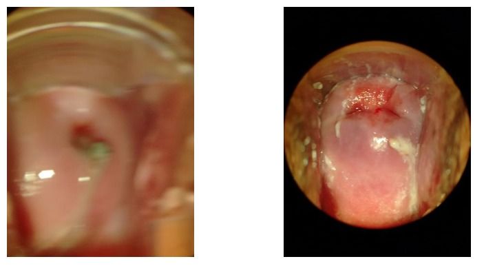
the expert reviewer to adequately conduct the triage. An example of such images is shown in Fig. 3. Fig. 3A shows
a poor quality image that suffers from what appears to be defocus and possible motion artifacts and/or blur. The image
in Fig. 3B, captured by an expert colposcopist, shows a much sharper view of the cervix. The implications on the
image reviewer’s ability to triage are significant, and this suggests that better EVA training is needed.
Future work will involve synchronous review and consultation by the multiple clinicians. An example of the proposed
clinical workflow is shown in Fig. 4. Features enabling synchronous review and consultation are currently being
developed and integrated into the CervDx app, and should begin clinical testing in the next year.
Proc. of SPIE Vol. 9699 96990I-3
Downloaded From: https://www.spiedigitallibrary.org/conference-proceedings-of-spie on 2/11/2018 Terms of Use: https://www.spiedigitallibrary.org/terms-of-use(A) (B)
Fig. 3: Comparison of colposcopy images acquired using EVA by a medical student (A) and expert colposcopist (B).
Pap Colposcopy Biopsy LEEP
EVA
Consultation
(A)
Doctor
Doctor Doctor
Doctor Doctor
Pap, HPV DNA test, EVA
Patient Nurse
Upload
Doctor
Doctor
Aggregate recommendation: Normal or abnormal?
Doctor
Where to treat /biopsy?
(B)
Fig. 4: (A) Proposed implementation of consultation feature at the primary screen. (B) Proposed new pathway of care
integrating synchronous consultation features on the CervDx app.
4. CONCLUSION
In conclusion, initial testing of the EVA system at the point of primary screen yielded promising results. EVA images
captured as a co-test together with Pap are beginning to provide valuable information to promote timely referrals and
potentially catch false positives and false negative Pap results. This has the potential to yield savings to the clinician,
patient, and health system as whole. Future developments will enable EVA review in real time at the point of care.
Proc. of SPIE Vol. 9699 96990I-4
Downloaded From: https://www.spiedigitallibrary.org/conference-proceedings-of-spie on 2/11/2018 Terms of Use: https://www.spiedigitallibrary.org/terms-of-useACKNOWLEDGEMENTS
Funding for this research is supported by a grant from the Vodafone Americas Foundation Wireless Innovation Project
(2014) http://vodafone-us.com/wireless-innovation-project/past-competitions/2014/2014-winners/mobileoct/.
REFERENCES
[1] A Jemal, F Bray, MM Center, J Ferlay, E Ward, D Forman, “Global Cancer Statistics”, Ca Cancer J Clin
61, 69–90 (2011).
[2] P Holowaty, AB Miller, T Rohan, T To, “Natural History of Dysplasia of the Uterine Cervix”, J Natl
Cancer Inst 91, 252-58 (1999).
[3] A Singer, JM Monaghan, and SC Quek. Lower Genital Tract Precancer: Colposcopy, Pathology, and
Treatment, 2nd Ed. Blackwell, Oxford (2000).
[4] LS Palacio-Mejia, G Rangel-Gomez, M Hernandez-Avila, E Lazcano-Ponce. “Cervical cancer, a disease of
poverty: mortality differences between urban and rural areas in Mexico” Salud Publica Mex 45, S315-325
(2003).
[5] C Peterson, D Rose, J Mink, and D Levitz, “Real-Time Monitoring and Evaluation of a Visual-Based
Cervical Cancer Screening Program Using a Clinical Decision Tree Algorithm”. Submitted to Diagnostics
(2016).
Proc. of SPIE Vol. 9699 96990I-5
Downloaded From: https://www.spiedigitallibrary.org/conference-proceedings-of-spie on 2/11/2018 Terms of Use: https://www.spiedigitallibrary.org/terms-of-useYou can also read