Assessment of Mandibular Condylar Morphology Using Digital Orthopantomogram in Chennai Population
←
→
Page content transcription
If your browser does not render page correctly, please read the page content below
Indian Journal of Forensic Medicine & Toxicology, July-September 2020, Vol. 14, No. 3   699

Assessment of Mandibular Condylar Morphology Using Digital
        Orthopantomogram in Chennai Population

                                           Sridhar M1, Sreedevi Dharman2
  1
   Graduate Student, 2Reader, Department of Oral Medicine and Radiology, Saveetha Dental College, Saveetha
                Institute of Medical and Technical Science, Saveetha University, Chennai, India

                                                        Abstract
      Objective: The present study was performed to evaluate the variation in shapes of mandibular condyle,
      determine the predominant shape and to assess any peculiarities in either gender in an Orthopantamogram(OPG).

      Materials and Method: Radiographic evaluation of 3200 condylar heads after analyzing 1600 digitalized
      OPG’s were done. On analyzing, there were four different morphology of condyles observed. Variations
      occurring in the shapes were assessed, and combinations of the condylar shapes present in the population
      were established.

      Result: Of 1600 pairs of condylar heads evaluated, 78% were oval in shape, followed by diamond (12%),
      crooked finger (6%) and least being bird beak (3%). Oval-oval was commonly occurring combination
      (60.5%) whereas crooked finger-bird beak combination was found to be very rare.

      Conclusion: Dental professionals must have thorough knowledge in differentiating between normal and
      abnormal condyle morphology in an OPG as it possess a diagnostic challenge for them. Asymmetries that
      occur in condyle morphologies radiographically in absence of clinical signs and symptoms of TMDs are
      considered to be normal.

      Keywords: Orthopantomographs, Bird beak, Crooked Finger, Oval, Diamond, Condyle, TMJ.

                      Introduction                               convex morphology, was considered as pathology but
                                                                 normal variation in shape of condyle does occur.
     Mandibular condyle is seen roughly as ovoid in
outline. Its dimensions are 15-20 mm mediolaterally and              Morphological alterations in the condyle can be due
8-10 mm postero-anteriorly1                                      to simple developmental variability or as remodeling
                                                                 of condyle to cope with developmental variations,
    Many assumptions were made towards the shape of
                                                                 malocclusion, trauma, endocrine disturbances and
a condyle. Most commonly reported shape of condyle
                                                                 radiation therapy2,3. Hence, a thorough understanding
was convexity throughout and it should be symmetrical
                                                                 of the morphology of mandibular condyle is essential
on both the sides i.e. right and left sides of the same
                                                                 to distinguish between normal variant from abnormal
individual. Hence, anything, which deviates from this
                                                                 conditions.

                                                                      The basic morphology of mandibular condyle is
Corresponding Author
                                                                 thought to be established early, and modified throughout
Dr. Sreedevi Dharman,
                                                                 life according to functional load4. Condyle morphology
Reader, Depatment of Oral Medicine and Radiology,
                                                                 variations occur with age, gender, facial type, Occlusal
Saveetha Dental College, Saveetha Institute of Medical
                                                                 force and also even between condyles on either sides2.
and Technical Science, Saveetha University, 162,
Poonamallee High Road, Chennai -600077                                Orthopantomographs is of diagnostic importance
Tamil Nadu, India, Email: sanjamrut@gmail.com                    as it is both cost efficient and it relatively reduces the
Telephone Number:9841009003                                      dosage of radiation received by the patients4,5. Panoramic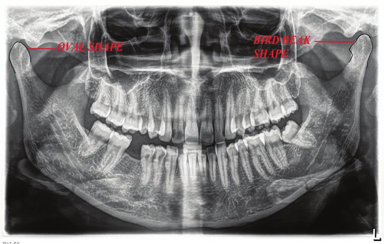
700    Indian Journal of Forensic Medicine & Toxicology, July-September 2020, Vol. 14, No. 3

radiography remains the main screening modality for                                               Results
TMJ abnormalities if clinical examination suggests
                                                                          A total of 3200 condyles were analyzed from 1600
any joint pathology. Hence, OPG’s are valuable for
                                                                      subjects with age ranging from 18 to 65 years, out of
determining the presence of osseous changes.
                                                                      which 807 were male and 793 were female.
    Our study aims to evaluate and document the
                                                                          A. Type of shape commonly seen: The shapes
variations in the shape of condyle on an OPG that aids in
                                                                      suggested by Chaudry et al were seen namely i) Oval, ii)
diagnostics i.e. distinguishing varying normal condylar
                                                                      Diamond, iii) Bird beak and iv) Crooked finger.
shapes from abnormalities. The objective of this study
is,                                                                      1. The most common shape was found to be Oval
                                                                      (78%), followed by Diamond (12%), Crooked finger
    1. To evaluate the variations in shape of condyle
                                                                      (6%) and least being Bird beak (3%).
seen in Chennai Population.
                                                                          2. The most common shape observed among both
   2. To determine the shape predominant in that
                                                                      males and females is Oval shape which accounted for
population
                                                                      about 74% in males and 72% in females respectively.
    3. To assess whether there is any peculiarity in
                                                                          3. The combination of commonly seen shape
either gender
                                                                      among both male and female is Oval-oval which
    4. To determine the occurrence of symmetry in                     accounted for about 63% and 57% respectively as shown
shape of condyle on either side.                                      in
                                                                      Figure 1-6.
                Materials and Method
                                                                           B: The gender wise distribution of shapes was
     Digital Panoramic Radiographs(OPG) (Planmeca-                    evaluated. In males, Oval shape (74%) was predominant
exposure parameters: 10 mA, 70 Kvp) which showed a                    followed by Diamond shape (18%), Crooked finger
full view of mandibular condyle on right and left side                (6%) and Bird beak shape being least common (2%). In
with optimum density and contrast were selected from                  females, Oval shape accounted for about (72%) followed
the Oral Medicine and Radiology Department, Saveetha                  by Diamond shape (18%), Crooked finger (6%) and Bird
dental college. This is a retrospective study.                        beak (4%).
     The present study comprised of radiographic                           C: To evaluate the combination of shapes occurring
evaluation of 3200 condylar heads after viewing 1600                  radiographically, revealed Oval-oval being the
digitalized OPG’s taken for routine radiographic                      commonest shape which accounted for about 60.5% and
investigation among Chennai Population. In our study,                 Crooked finger-Diamond and Crooked finger-Bird beak
radiographs of 807 males and 793 females ranging from                 is the least common combination which accounted for
the age 18-65 years were included. OPG’s in which                     about 0.02% and 0.03% respectively.
condyle heads can be visualized clearly were included
in this study. OPG’s taken for patients who came for                             Figure 1: OPG shows Crooked Finger
treatment of multiple dental caries or generalized                             appearance of mandible on either sides.
periodontal disease were selected. OPGs of patients
with history of TMD’s, Trauma, occlusal discrepancy,
developmental abnormality, were excluded from this
study. Condylar morphology of four types by Chaudry
et al were identified which are

      1) Type I - Oval shape.

      2) Type II - Bird Beak Shape

      3) Type III - Diamond shape.

      3) Type IV - Crooked finger shape.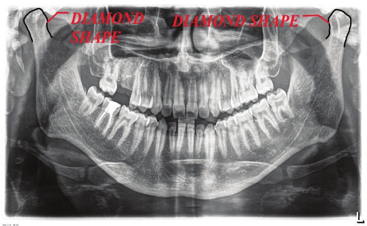
Indian Journal of Forensic Medicine & Toxicology, July-September 2020, Vol. 14, No. 3   701

Figure 2: OPG reveals Diamond Shape of condyles on both the sides of mandible

   Figure 3: OPG reveals Bird beak and Crooked finger shape Combination
702 Indian Journal of Forensic Medicine & Toxicology, July-September 2020, Vol. 14, No. 3
Figure 4: This OPG shows Oval and bird beak shape combinations of condyle
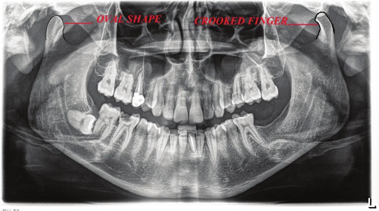
Figure 5: This OPG reveals Oval and Crooked finger shape combination of condyle
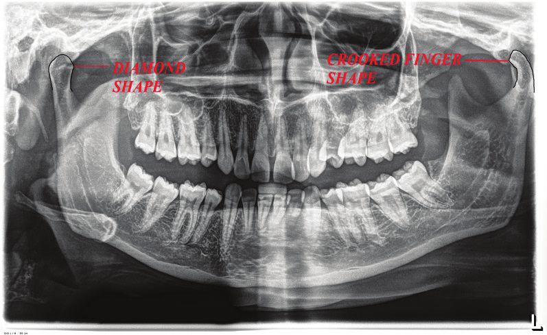
Figure 6: This OPG shows Diamond and Crooked Finger combination of condyle on either sides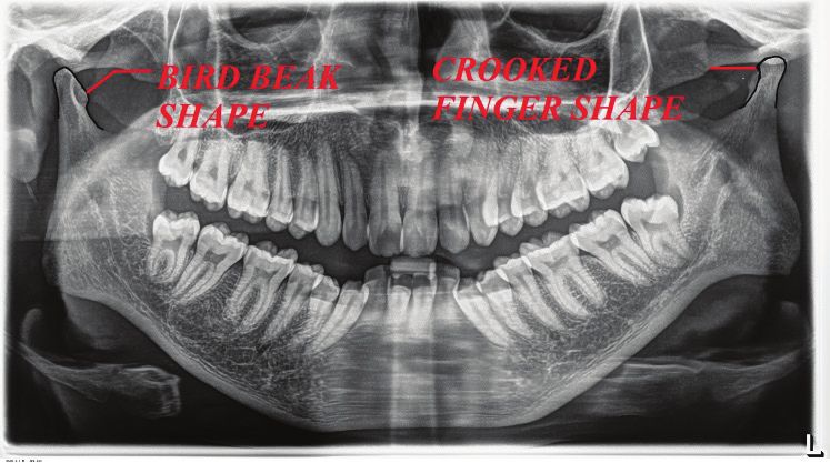
Indian Journal of Forensic Medicine & Toxicology, July-September 2020, Vol. 14, No. 3   703

                       Discussion                                 were seen more frequently in patients with clinical signs
                                                                  and symptoms of TMD.
    Condyle responds to continuous stimuli throughout
the remodeling process, and thus plays an important                   Small asymmetries between left and right condyles
role in the final morphological dimensions of the adult           were common. Small asymmetries are expected to
mandible. The condyle has a special importance in                 develop during normal condylar growth, but the manner
growth of mandible 2.                                             in which this asymmetry occurs has to be differentiated.
                                                                  Asymmetries in size differs from shape, volume or
Figure 1: Shapes of Condyle on a Radiograph
                                                                  position asymmetries 3.
      The appearance of mandibular condyle varies in
different shape and size among different age groups and                Asymmetries in condyle morphology without
individuals. In 1961, Yale et al. was the first one to report     clinical signs and symptoms of TMD’s, careful
about the different shapes of mandibular condyle6,7,8.            radiological examination of condyle are required to rule
Initially Yale classified condylar head based on superior         out TMJ pathologies.
view into three categories namely concave, convex and
flat, however later on he simplified it into five categories          Our study aimed to detect the most common shapes
namely convex, flattened, angled, rounded and concave.            on the head of condyle seen in a radiograph among
                                                                  Chennai Population. Of 3200 heads 78% were oval in
    Evaluation of the shape of condyles upon surgical             shape, followed by diamond (12%), crooked finger (6%)
exposure of TMJ revealed that most of the condyles had            and least being bird beak (3%). Our results showed a
a normal size and shape. Other varieties like excavated           variation to previous study done by Sonal V et al12,
form, oblique shape, small round condyles and flattened           where the most common shape was found to be oval
condyles were noted 9.                                            (60%), followed by bird beak (29%), diamond (9%), and
                                                                  crooked finger (2%).
    Using different radiographic techniques many
studies were done to detect the condylar morphology, to               In the same study, the oval was the most common
compare the accuracy of detecting condylar changes in             shape in both males and females which accounted for
temporomandibular disorders.                                      about 61% and 46% 12. Similar results were obtained
                                                                  from our study in which oval being the commonest
     The most prevalent morphologic changes are
                                                                  shape showed a high prevalence of about 74% in males
detected in the TMJ of elderly persons due to the onset
                                                                  and 72% in females as shown in the figure 3.
of joint degeneration 4.
                                                                       Combination of condyle shapes in OPG revealed
     The normal morphological variations like diamond,
                                                                  that Oval-oval combination, was 60.5%, followed
bird beak, crooked finger, oval should not be mistaken
                                                                  by Oval-diamond combination (12.25%), Diamond-
with TMJ pathologies like flattening of articular surface,
                                                                  diamond (7.6%), Bird beak-bird beak (5.43%), Oval-
erosions, pencil shaped condyles, osteophytes, anterior
                                                                  crooked finger(5.31%), Crooked finger-crooked finger
lipping of condyle and ely’s cyst.
                                                                  (4.93%) and Crooked finger-Diamond and Crooked
    Flattening is loss of an even convexity of condyle            finger-Bird beak is the least common combination which
surfaces, Osteophyte is local outgrowth of bone arising           accounted for about 0.02% and 0.03% respectively. In
from a mineralized joint surface, Erosion is local area           the similar study conducted by Sonal V et al, Oval-oval
of rarefaction in the cortical plate of a joint surface,          combination (67%) was most prevalent followed by
Sclerosis is thickening of the cortical bone on a joint           Oval-bird beak (25%), Oval-diamond (5%), Bird beak-
surface, Ely’s cyst is sub cortical cyst is rounded               bird beak (3%) and Crooked finger- crooked finger is
radiolucent area that may be just below the cortical plate        least common combination which accounted for about
or deep in trabecular bone.                                       1% only 12.

     Radiological variations of condyle should be always                In the study Sonal et al, the combination of shapes
correlated with clinical signs and symptoms to arrive at          commonly seen in male and females were evaluated
the diagnosis of TMDs. Anuna Laila Mathew et al, in               where oval-oval combination was seen most prevalent in
their study revealed that, radiographic abnormalities in          58% of female population and 42% of male population
                                                                  12
the condylar morphology increased with age11. They                   . In our study, Oval-oval combination was seen in 63%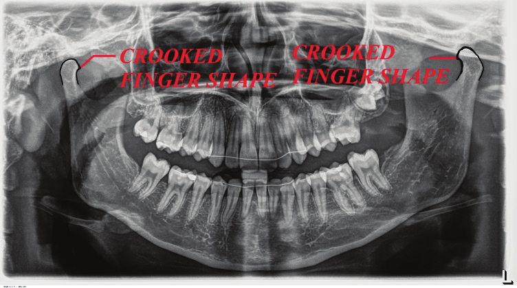
704    Indian Journal of Forensic Medicine & Toxicology, July-September 2020, Vol. 14, No. 3

of male population and 37% of female population.                      the ethical standards of the responsible committee on
                                                                      human experimentation (institutional and national) and
    Gender wise distribution of shapes was recorded. In               with the Helsinki Declaration of 1975, as revised in 2008
males, Oval shape (74%) was predominant followed by                   (5). Informed consent was obtained from all patients for
Diamond shape (18%), Crooked finger (6%) and Bird                     being included in the study.’
beak shape being least common (2%). In females, Oval
shape accounted for about (72%) followed by Diamond                                            References
shape (18%), Crooked finger (6%) and Bird beak (4%).
But in previous study done by Sonal V et al, only the                 1.    Standring S. Gray’s Anatomy: Thethe
                                                                                                                Anatomical
prevalence of commonest shape was recorded which                            Basis of Clinical Practice. 39 ed. London:
showed Oval being most common in both males (62%)                           Elsevier Ltd.; 2005. p. 519-30.
and females (46%).                                                    2.    Alomar X, Medrano J, Cabratosa J, Clavero
                                                                            JA, Lorente M, Serra I, et al. Anatomy of the
     Panoramic radiography is a screening tool for                          Temperomandibular joint. Semin Ultrasound CT
diagnosing TMDs. Various advanced imaging techniques                        MR 2007; 28:170-83.
are needed to confirm any pathology in addition to
                                                                      3.    Mongini F. The importance of radiography in the
diagnosis made by OPG such as Radionuclide bone
                                                                            diagnosis of TMJ dysfunctions. A comparative
scanning which is a useful technique for showing early
                                                                            evaluation of Trans cranial radiographs and serial
functional and biochemical bone changes, CT images
                                                                            tomography. J Prosthet Dent 1981; 45:186-98.
which are highly accurate for osseous abnormality
13and Cone-beam computed tomography images which                      4.    Ross BR, Johnston MC. Developmental anomalies
are superior over others for the bony morphology of                         and dysfunction. In: Zarb GA, Carlsson GE, Sessle
mandibular condyles and detection of condylar cortical                      BJ, Mohl ND (eds). Temporomandibular joint and
erosion14. CBCT is a useful tool to measure and evaluate                    masticatory muscle disorders. Mosby 1994; 221-
the condylar dimensions.                                                    222.
                                                                      5.    Habets LL, Bezuur JN, Jimenez Lopez V, Hansson
                         Conclusion                                         TL. The OPG: An aid in TMJ diagnostics. III.
                                                                            A comparison between lateral tomography
    Dentists should have thorough knowledge on the
                                                                            and dental rotational panoramic radiography
characteristic normal mandibular condyle variations
                                                                            (Orthopantomography).     J    Oral    Rehabil
to give a diagnosis of TMD’s. Amount of condyle
                                                                            1989;16:401-6.
morphology asymmetry is considered as ‘normal
asymmetry’ in the absence of signs and symptoms                       6.    Yale SH, Rosenberg HM, Ceballos M, Haupt-
of TMD’s. Genetic, acquired, functional factors, age                        Fuehrer JD. Laminagraphic cephalometry in the
groups, individuals have a role in morphologic changes                      analysis of mandibular condyle morphology. A
of condyle. Thus variability in the shapes and sizes of                     preliminary report. Oral Surg Oral Med Oral Path
condyles should be an important factor in diagnosing                        1961;14: 793-805.
the disorders of temperomandibular joint. OPG’s are                   7.    Yale SH, Ceballos M, Kresnoff CS, Hauptfuehrer
preferred imaging technique as it is easily prescribed                      JD. Some observation on the classification of
by most of the dental surgeons and is a screening tool                      mandibular condyle types. Oral Surg Oral Med
for TMD’s. Hence, Dental professionals must have                            Oral Pathol 1963;16: 572-577.
thorough knowledge in differentiating between normal                  8. Yale SH, Allison BD, Hauptfuehrer JD. An
and abnormal condyle morphology in an OPG, as it                          epidermiological assessment of mandibular
possess a diagnostic challenge for them.                                  condyle morphology. Oral Surg Oral Med Oral
                                                                          Path 1966;21(2): 169-177.
    Declaration on Conflict of Interest: we have no
conflict of interest                                                  9.    Juniper RP . The shape of the condyle and position
                                                                            of the meniscus in temporomandibular joint
      Financial support and sponsorship: Nil                                dysfunction. Br J Oral Maxillofac Surg 1994;32:
                                                                            71-76.
      Source of Funding: Self
                                                                      11. Anuna Laila Mathew, Amar A, Sholapurkar ,
      ‘All procedures followed were in accordance with
Indian Journal of Forensic Medicine & Toxicology, July-September 2020, Vol. 14, No. 3 705
Keerthilatha M, Pai. Clinical Study. Condylar 13. Raustia AM, Pyhtinen J. Computed Tomography
Changes and Its Association with Age, TMD, of the Temporomandibular joint. In: Delbalso AR
and Dentition Status: A Cross-Sectional (Ed.), Maxillofacial Imaging. Saunders 1990;650-
Study. International Journal of Dentistry. 672.
Volume 2011 |Article ID 413639 | 7 pages | 14. Ludlow JB, Laster WS, See M, Bailey LJ, Hershey
12. Sonal V, Sandeep P, Kapil G, Christine R. HG. Accuracy of measurements of mandibular
Evaluation of condylar morphology using anatomy in cone beam computed tomography
panoramic radiography. J Ad Clin Res Insights images. Oral Surg Oral Med Oral Pathol Oral
2016; 3:5-8. Radiol Endod 2007;103: 534-542.You can also read