A retrospective study of demographic profile of keloid over the pinna in central Karnataka
←
→
Page content transcription
If your browser does not render page correctly, please read the page content below
International Journal of Otorhinolaryngology and Head and Neck Surgery
Chaitanya V et al. Int J Otorhinolaryngol Head Neck Surg. 2018 May;4(3):726-729
http://www.ijorl.com                                                                       pISSN 2454-5929 | eISSN 2454-5937

                                                          DOI: http://dx.doi.org/10.18203/issn.2454-5929.ijohns20181860
Original Research Article

 A retrospective study of demographic profile of keloid over the pinna in
                            central Karnataka
                 Chaitanya V.1, Kavitha Y.1*, Basavaraju K. P.1, Upendra Kumar Joish2
  1
   Department of Otorhinolaryngology, 2Department of Radiodiagnosis, JJM Medical College, Davangere, Karnataka,
  India

  Received: 29 January 2018
  Revised: 02 March 2018
  Accepted: 05 March 2018

  *Correspondence:
  Dr. Kavitha Y.,
  E-mail: kavithajoish@gmail.com

  Copyright: © the author(s), publisher and licensee Medip Academy. This is an open-access article distributed under
  the terms of the Creative Commons Attribution Non-Commercial License, which permits unrestricted non-commercial
  use, distribution, and reproduction in any medium, provided the original work is properly cited.

   ABSTRACT

   Background: Unusually higher number of patients was observed to seek medical attention for keloids over the pinna
   in a geographical area in Central Karnataka. This study was conducted to find the demographic profile of such
   patients.
   Methods: A retrospective observational study was conducted in two tertiary care centres, in which medical case files
   of all patients with documented diagnosis of keloid over the pinna between January 2013 to October 2017, were
   reviewed for their demographic profile and clinical presentation.
   Results: A total of 482 patients had presented with keloids of pinna in the duration studied. Of these 474 were
   females and 8 were males, with a mean age of 29 years. The most common age group of patients (37.3%) was 21 to
   30 years followed by 31 to 40 years (25.7%). The most common antecedent event to keloid formation was piercing of
   the helix of the pinna. The commonest location of keloid formation in the pinna was found to be helix of the pinna
   (92.7%) The mean time interval between the antecedent event and keloid formation was 14 months.
   Conclusions: Higher number of patients seeks medical attention for keloid over the pinna in geographical region of
   central Karnataka. Most of them had undergone ear piercing and had presented in their early adulthood. Ear piercing
   over the helix of pinna was more commonly associated with keloid formation. Further studies are intended to be done
   on the etiological factors for higher incidence of keloids and feasible preventive measures.

   Keywords: Keloid over the pinna, Ear piercing, Demographic profile, Helix of the pinna

INTRODUCTION                                                         young ladies having a psychological impact on them.
                                                                     Keloids are prone for recurrence with need for prolonged
Ear piercing is a widely prevalent custom among ladies in            treatment. Various treatment options have been tried
the Indian Subcontinent. Minor complications like                    including surgical excision, steroid injections,
allergy, infections following the procedure has been often           radiotherapy etc. with no significant permanent results.3-5
seen. Most agonising is the formation of keloid with                 Studies have claimed genetic predisposition for
studies claiming a 2.5% risk of development of keloid                development of keloids.6 In our Institution, which is
post ear piercing.1 Keloids over pinna are also a known              located in the interior parts of Southern India, we have
complication following minor incisions, drainage of                  observed an unusually high number of patients seeking
auricular collections, trauma etc.2 Patients seek medical            medical attention for keloids. This prompted us to further
assistance mostly for cosmetic reasons. Keloids over the             probe into our records and quantify and analyse our
pinna can be cosmetically disfiguring especially among               observations.

                 International Journal of Otorhinolaryngology and Head and Neck Surgery | May-June 2018 | Vol 4 | Issue 3   Page 726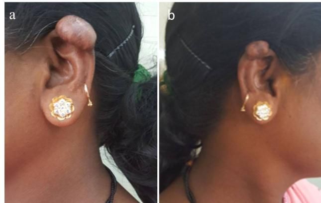
Chaitanya V et al. Int J Otorhinolaryngol Head Neck Surg. 2018 May;4(3):726-729
METHODS history of trauma to the affected ear and 3 had underwent
piercing of the lobule of pinna. Of the females, 442 had
This retrospective study was carried out in the undergone ear piercing in the past and the remainder 32
departments of otorhinolaryngology of two tertiary care had a history of trauma to the ear. Of the females who
centres located in same town in Central Karnataka in had undergone ear piercing, 75% of them had undergone
December 2017. Prior Institutional ethical committee piercing of the helix of pinna in 2nd decade of life or later
approval for the study was obtained. The study included (>10 years of age). The mean time interval between the
all cases found to be diagnosed as keloid over the pinna antecedent event (ear piercing or trauma) and keloid
in the period from January 2013 to October 2017 based formation was 14 months with a minimum of 3 months to
on a departmental patient data register search. After a maximum of 3 years. Though all these cases had
noting the patient identification numbers and diagnoses, presented with a lump over the pinna, 25.8% of them had
the medical records section was requested to provide the complained of intractable itching and pain. The
detailed medical records/case files of those cases. commonest location of keloid formation in the pinna was
found to be helix of the pinna (92.7%) (Figure 1 and 2),
The case files were scrutinised for demographic data, both in males and females. Keloids of the lobule of pinna
clinical presentation, past antecedent event leading to accounted for 7.3% of cases. Among our cases, right ear
keloid formation and treatment received. Wherever was found to be more frequently affected than left ear by
available, clinical photographs in the case files were also keloid formation. Bilateral involvement was seen in 102
examined. Such patients were contacted and their consent cases (21.2%) (Figure 3).
was taken to use the clinical photographs of their ear
lesions with concealed identities, for publication as part
of this study. The data obtained was tabulated in terms of
gender, age, antecedent event with time interval for
keloid formation.
Patients had undergone various combinations of
treatment. Some patients had received intralesional
injection of Triamcinolone only, at weekly intervals for 4
weeks, while others underwent excision of keloid under
local anaesthesia followed by weekly steroid injection to
the excision site after wound healing for 4 weeks.
Patients, who had failed to respond to initial weekly
intralesional steroid injections, were subjected to excision
followed by weekly steroid injections for 4 weeks. The
same strength of triamcinolone acetonide (40 mg/ml) had
been used in all patients.
RESULTS
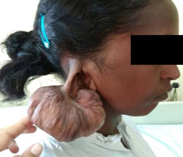
Figure 1: A large mass over the helix of right pinna in
A total of 482 number of patients had presented with a 21 year old female. Patient sought medical attention
keloids of pinna in the duration studied. Of these cases, due to rapid increase in size of this mass over three
474 were females and 8 were males. Patients ranged from months during third trimester of pregnancy. Post
6 years to 68 years of age with a mean age of 29 years. excision, the mass was proved to be a keloid on
Clinical characteristics of cases are depicted in Table 1. A histopathology. This patient had undergone high
major chunk of the patients (37.3%) were of the age helical ear piercing six months prior to her initial
group 21 to 30 years followed by age group 31 to 40 presentation.
years of age (25.7%). Among 8 male patients, 5 had prior
Table 1: Clinical characteristics of cases.
Number of cases
Age group History Location of keloid
Bilaterality
Ear Piercing Trauma Lobule Helix
Below 10 years 2 6 1 7 0
11 to 20 years 76 4 8 72 11
21 to 30years 158 18 10 166 67
31 to 40 years 117 6 9 114 23
41 to 50 years 62 2 7 57 1
Above 50 years 30 1 0 31 0
445 37 35 447 102
International Journal of Otorhinolaryngology and Head and Neck Surgery | May-June 2018 | Vol 4 | Issue 3 Page 727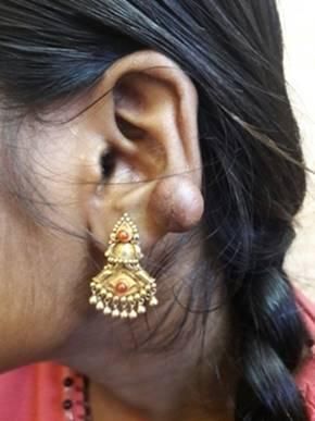
Chaitanya V et al. Int J Otorhinolaryngol Head Neck Surg. 2018 May;4(3):726-729

                                                                    common among adolescents and ladies of the location.
                                                                    Moreover, lobule of pinna is free from cartilage, whereas
                                                                    helix is cartilaginous. As per Staley et al and Bashir et al,
                                                                    cartilaginous areas are more prone for complications
                                                                    compared to soft tissue piercing.8,9 However, Simplot et
                                                                    al mentioned similar incidence in both.1 Again bilateral
                                                                    involvement can be explained by bilateral ear piercing
                                                                    custom. Cases with keloids over the lobule had
                                                                    documented history of trauma to the lobule of the pinna.

                                                                    The mean time interval between antecedent event, ear
                                                                    piercing or trauma, to noticing a lump in the ear, was 14
                                                                    months. A minimum of 3 months and a maximum time
                                                                    interval of 3 years was observed. A study by Brissett et al
                                                                    had documented a period between 3 months to 1 year for
                                                                    keloid formation following skin trauma.10 In the present
                                                                    study, the delayed noticing of the lump by the patients
                                                                    was mainly documented among individuals seeking
                                                                    treatment in their 4th decade or later. As they had
 Figure 2: A 24 year old female with history of helical             presented to the Department much later than they had
  ear piercing one year prior, presented with a lesion              intitially noticed the lumps, the time interval between
   over her left pinna in the low helical region, with              antecedent event and first noticing the ear lump could
severe itching. Post excision this lesion was confirmed             have been subject to recall bias among such people. Most
                     to be a keloid.                                of these patients had complained of ear lumps persisting
                                                                    for a decade or more and had sought medical attention
                                                                    only when the lumps were too ‘heavy’ or ‘large’ or
                                                                    associated with ‘unbearable itching’ as documented in
                                                                    their case files (25.8%). In a study by Lee et al, 86% had
                                                                    presented with itching and 46% had presented with
                                                                    pain.11

                                                                    Incidence of keloid formation could not be calculated as
                                                                    no baseline data on total number of ear piercing events
                                                                    was available. There are hardly few studies on incidence
                                                                    and prevalence of keloids post ear piercing. Simplot et al,
                                                                    compared the incidence of complications of cartilage
                                                                    piercing versus soft tissue piercing and found a 2.5%
                                                                    incidence of keloids.1 Keloid occurrence has also been
                                                                    documented to vary with skin pigmentation. Darker
  Figure 3: Keloid in the high helical region of the (a)
                                                                    complexion has increased inherent risk as has been seen
left pinna, in a 31 year old female, who had undergone
                                                                    in Africans as well as dark skinned people in the far west
bilateral helical ear piercing 9 months prior. Also note
                                                                    with an incidence as high as 16%.12-14 Increased risk is
a smaller lesion over the helix of right pinna (b) in the
                                                                    also seen in puberty and pregnancy among genetically
                      same patient.
                                                                    predisposed.15
DISCUSSION
                                                                    With this baseline data, further studies will be undertaken
                                                                    to understand the etiology of higher occurrence of keloids
In the present study, majority of the cases who had                 in the population specified in the location of this study
presented with keloids were females in 2nd and 3rd decade           with regards to genetic predisposition, ear piercing
of their life. This is similar to a study by Ramakrishnan et        methodology etc.
al, who found higher incidence of keloid formation in the
age group 11 to 30 years.7 This can be attributed to the
                                                                    CONCLUSION
apprehension and anxiety over the facial disfigurement
caused by keloids that they seek medical attention.
                                                                    Higher number of patients seeks medical attention for
Moreover, ear piercing is not common among males and
                                                                    keloid over the pinna in this geographical region of
primarily involves lobule piercing. Though as a ritual, ear
                                                                    central Karnataka. Most of them had an antecedent event
piercing is done in the lobule of the ear, this was a less
                                                                    of ear piercing and had presented in their early adulthood.
frequent location of keloid formation in this study. The
                                                                    Ear piercing over the helix of pinna was more commonly
commonest location of keloid over the pinna was helix.
                                                                    associated with keloid formation. Further studies are
Piercing of helix of pinna as a fashionable trend is
                                                                    intended to be carried out on the possible etiological

                International Journal of Otorhinolaryngology and Head and Neck Surgery | May-June 2018 | Vol 4 | Issue 3   Page 728
Chaitanya V et al. Int J Otorhinolaryngol Head Neck Surg. 2018 May;4(3):726-729
factors for higher incidence of keloids in this region and 9. Bashir MM, Afzal S, Khan FA, Abbas M. Factors
feasible preventive measures. Associated with Postpiercing Auricular Cartilage
Keloids. Journal of the College of Physicians and
Funding: No funding sources Surgeons Pakistan. 2011;21(10):606-10.
Conflict of interest: None declared 10. Brissett AE, Sherris DA. Scar contractures,
Ethical approval: The study was approved by the hypertrophic scars, and keloids. Facial Plast Surg.
Institutional Ethics Committee 2001;17(4):263-72.
11. Lee SS, Yosipovitch G, Chan YH, Goh CL.
REFERENCES Pruritus, pain, and small nerve fiber function in
keloids: a controlled study. J Am Acad Dermatol.
1. Simplot TC, Hoffman HT: Comparison between 2004;51(6):1002-6.
cartilage and soft tissue ear piercing complications. 12. Gauglitz GG, Korting HC, Pavicic T, Ruzicka T,
Am J Otolaryngol. 1998;19:305-10. Jeschke MG. Hypertrophic scarring and keloids:
2. Sand M, Sand D, Brors D, Altmeyer P, Mann B, pathomechanisms and current and emerging
Bechara FG. Cutaneous lesions of the external ear. treatment strategies. Mol Med. 2011;17(1-2):113-
Head Face Med. 2008;4(1):2. 25.
3. Chowdri NA, Mattoo MMA, Darzi MA. Keloids 13. Child FJ, Fuller LC, Higgins EM, Du Vivier AWP.
and hypertrophic scars: results with intra-operative A study of the spectrum of skin disease occurring in
and serial postoperative corticosteroid injection a black population in southeast London. Br J
therapy. Aust N Z J Surg. 1999;69:655-9. Dermatol. 1999;141:512-7.
4. Botwood N, Lewanski C, Lowdell C. The risks of 14. Moshref SS, Mufti ST. Keloid and Hypertrophic
treating keloids with radiotherapy. Br J Radiol. Scars: Comparative Histopathological and
1999;72:1222-4. Immunohistochemical Study. Med Sci. 2010;17:3–
5. Venkatramani H, Yadav P. Complete excision with 22.
staged reconstruction in the treatment of earlobe 15. Aköz T, Gideroğlu K, Akan. Combination of
keloid after ear piercing. Plast Reconstr Surg. different techniques for the treatment of earlobe
1999;104:1574-5. keloids. M. Aesthetic Plast Surg. 2002;26(3):184-8.
6. Marneros AG, Norris JE, Olsen BR, Reichenberger
E. Clinical genetics of familial keloids. Arch
Dermatol. 2001;137(11):1429-34.
7. Ramakrishnan KM, Thomas KP, Sundararajan Cite this article as: Chaitanya V, Kavitha Y,
CR.Study of 1,000 patients with keloids in South Basavaraju KP, Joish UK. A retrospective study of
India. Plast Reconstr Surg. 1974;53(3):276-80. demographic profile of keloid over the pinna in
8. Staley R, Fitzgibbon JJ, Anderson C. Auricular central Karnataka. Int J Otorhinolaryngol Head Neck
infections caused by high ear piercing in Surg 2018;4:726-9.
adolescents. Pediatrics. 1997;99:610-1.
International Journal of Otorhinolaryngology and Head and Neck Surgery | May-June 2018 | Vol 4 | Issue 3 Page 729You can also read