Innovative Left Ventricular Assist Device in High-fidelity Patient Simulator
←
→
Page content transcription
If your browser does not render page correctly, please read the page content below
Open Access Technical
                                         Report                                                       DOI: 10.7759/cureus.7763

                                         Innovative Left Ventricular Assist Device in
                                         High-fidelity Patient Simulator
                                         Wayne Lindsay 1 , Tiffany Nelms 2 , Sean O'Hara 1 , Zachary Sletten 3

                                         1. Emergency Medicine, Brooke Army Medical Center, San Antonio, USA 2. Simulation, Brooke Army
                                         Medical Center, San Antonio, USA 3. Emergency Medicine, San Antonio Military Medical Center, San
                                         Antonio, USA

                                         Corresponding author: Wayne Lindsay, wjlindsay0602@email.campbell.edu

                                         Abstract
                                         Left ventricular assist devices (LVADs) are implantable mechanical devices that pump blood from
                                         the apex of the left ventricle to the aorta in order to assist the forward flow of blood; they are
                                         most commonly used as a bridge to transplant for patients with heart failure. As of February
                                         2019, a total of 25,145 patients with ventricular assist devices have been reported in the
                                         Interagency Registry for Mechanically Assisted Circulatory Support (Intermacs). As this number
                                         continues to grow, more and more of these patients will inevitably be seen in the acute care
                                         setting outside of their defined LVAD center. Currently, however, LVAD emergencies represent a
                                         high-acuity low-occurrence event with limited opportunities for exposure and mastery for most
                                         physicians. Therefore, a growing need exists for emergency care providers to familiarize
                                         themselves with these devices and the management of LVAD emergencies. We present a novel
                                         model for the simulation of LVAD emergencies created through simple modifications of a Laerdal
                                         3G Manikin.

                                         Categories: Emergency Medicine, Medical Education, Medical Simulation
                                         Keywords: lvad, left ventricular assist device, simulator, simulation, emergency medicine, lvad
                                         emergency, emergency room

                                         Introduction
                                         The left ventricular assist device (LVAD) was initially developed in the 1960s as a bridge to
                                         cardiac transplant. Since the number of patients awaiting cardiac transplant has continued to
                                         rise, LVADs have also commonly become destination therapy. This device may also be used for
                                         temporary support in cardiomyopathies and patients diagnosed with New York Association’s
                                         class IV heart failure [1]. As of 2019, there were an estimated 25,145 patients with LVAD devices
                                         and the number continues to grow [1]. These patients are primarily managed at centers with both
                                         LVAD-trained cardiologists and support staff. Yet, with the increasing number of patients relying
Received 02/27/2020
                                         on these devices, it is inevitable that acute care will need to be provided outside of dedicated
Review began 03/17/2020
Review ended 04/13/2020                  LVAD centers. A recent study suggests that the average patient with an LVAD will be seen in the
Published 04/21/2020                     emergency department (ED) seven times a year, with a 64% admission rate [2]. Patients with an
                                         LVAD are at high risk of experiencing a complication, and, unfortunately, there are a variety of
© Copyright 2020
Lindsay et al. This is an open access    emergent complications associated with having an LVAD, including bleeding, thrombosis, and
article distributed under the terms of   infection [3]. Additionally, there are many components of the physical examination unique to
the Creative Commons Attribution         patients with an LVAD, including blood pressure measurement, cardiac auscultation, and
License CC-BY 4.0., which permits
                                         assessment of the device and its components [3]. As such, it is imperative that emergency
unrestricted use, distribution, and
                                         providers gain familiarity with the device and the notable physiological changes in these
reproduction in any medium, provided
the original author and source are       patients. For an emergency physician not routinely caring for patients with an LVAD, LVAD
credited.                                emergencies represent a high-acuity low-occurrence (HALO) event with limited opportunities
                                         for training and familiarization.

                                         How to cite this article
                                         Lindsay W, Nelms T, O'hara S, et al. (April 21, 2020) Innovative Left Ventricular Assist Device in High-
                                         fidelity Patient Simulator. Cureus 12(4): e7763. DOI 10.7759/cureus.7763
The LVAD is composed of several key components that this simulator aims to mimic: the pump,
                                   which sits internally and pumps blood from the left ventricle into the thoracic aorta, the
                                   driveline, which is the percutaneous cable that exits the abdominal wall to connect the pump to
                                   the external components, an external computer controller, which both monitors and controls
                                   pump performance with a display screen, and a power supply, which typically rests along the
                                   patient’s hip with a backup battery located in an additional pouch [4].

                                   The LVAD pump provides continuous forward flow from the left ventricle to the aorta; as such
                                   there are notable physiological changes in these patients. There is no longer a definable systolic
                                   and diastolic component to the cardiac cycle. These patients will lack a palpable pulse, and
                                   healthcare providers will not be able to record a traditional blood pressure [5]. This low-cost
                                   simulator may be used to introduce these physiological changes and provide training on
                                   common LVAD pathologies such as driveline site infections, bleeding events, and power source
                                   failure.

                                   Technical Report
                                   This simulation was built with a Laerdal 3G Manikin (Laerdal, Stavanger, Norway); however,
                                   other manikin models may be used in its place. The following steps and figures serve to outline
                                   the simple process of configuring an LVAD simulator.

                                   A small square of manikin-colored material should be cut approximate to the size of your
                                   intended window dressing; for this model, a Sorbaview 2000 window dressing (Centurion,
                                   Williamston, MI, USA) was used with a window of approximately 4 x 4 cm. Red grease paint and
                                   modeling clay were used for creating a moulage, representing erythema with mild bleeding as
                                   seen in Figure 1. The moulage can differ depending on the intended simulation case.

                                      FIGURE 1: The Sorbaview 2000 window dressing

2020 Lindsay et al. Cureus 12(4): e7763. DOI 10.7759/cureus.7763                                                                  2 of 8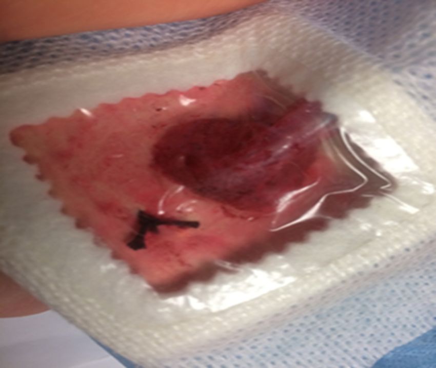
A Bardex 20-French silicone Foley catheter (Bard Medical, Covington, GA, USA) was used to
                                   represent the driveline coming from the patient’s abdomen and connecting to the controller. The
                                   Foley catheter (Figure 2) with the tip removed was then inserted through a small cut in the
                                   dressing and sutured in place with black Ethicon braided suture on the manikin material along
                                   the edge of the window (Figure 1).

                                      FIGURE 2: Foley catheter attached to the manikin-like material
                                      with suture and hemostat

                                   A discarded PositionPro hospital bed remote (Stryker, Kalamazoo, MI, USA) was then used as the
                                   patient controller with a printed image modeled after a Heartmate III controller (Thoratec
                                   Corporation, Pleasanton, CA, USA) (Figure 3). The Foley catheter was then taped to the bed
                                   remote representing the driveline attaching to the controller, while the remote’s cord was run to
                                   a simulated battery pack.

2020 Lindsay et al. Cureus 12(4): e7763. DOI 10.7759/cureus.7763                                                                3 of 8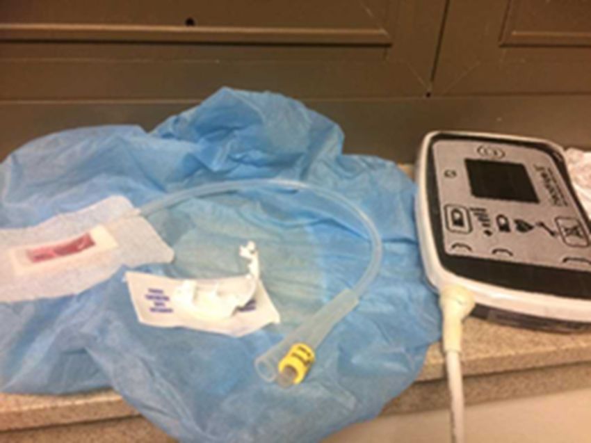
FIGURE 3: The printed controller panel attached to the hospital
bed remote
Two large squared batteries, taken from an Audio-Technica wireless microphone
receiver (Audio-Technica Corp., Tokyo, Japan), were then placed in the waist packs from a hiking
backpack (Figure 4) to represent the primary battery and the backup most patients will carry at
all times. A fanny pack with dual pockets may also serve as the battery containers. The LVAD
controller was then connected to the front of the waist packs between the batteries (Figure 5).
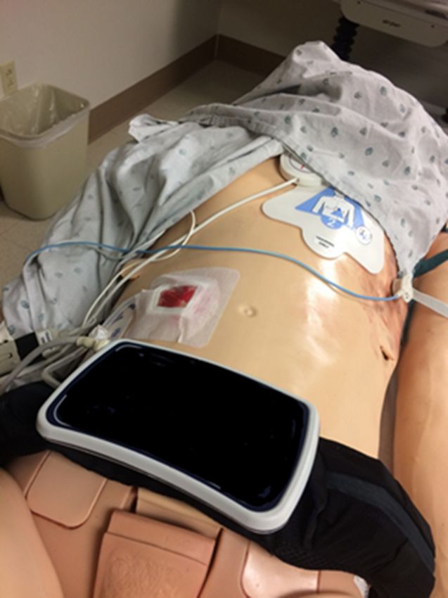
2020 Lindsay et al. Cureus 12(4): e7763. DOI 10.7759/cureus.7763 4 of 8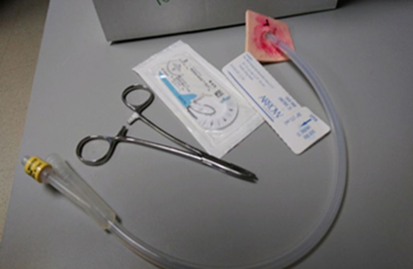
FIGURE 4: The simulated battery pack with a spare pack

2020 Lindsay et al. Cureus 12(4): e7763. DOI 10.7759/cureus.7763                               5 of 8
FIGURE 5: The controller resting in the middle of the battery
                                      packs

                                   The Sorbaview 2000 window dressing piece was then adhered to the right mid-abdomen with the
                                   Foley catheter portion feeding out laterally connecting to the simulated controller (Figure 6).
                                   The driveline may exit a patient from either their right or left abdomen to connect to the
                                   controller [4].

2020 Lindsay et al. Cureus 12(4): e7763. DOI 10.7759/cureus.7763                                                              6 of 8
FIGURE 6: The cloth tape dressing in place on the right
abdomen
Finally, an electric toothbrush was placed in the manikin’s chest cavity and turned on to
represent the LVAD pump functioning within the heart, creating the continuous hum that would
be heard when auscultating these patients (Figure 7).
FIGURE 7: The mechanical toothbrush used to represent the
pump
2020 Lindsay et al. Cureus 12(4): e7763. DOI 10.7759/cureus.7763 7 of 8Discussion
The number of patients with LVADs will continue to grow and will inevitably come under the care
of healthcare providers unfamiliar with LVADs. Patients with LVADs have frequent ED visits and
subsequent admissions with a wide array of emergent complications [2]. These encounters
represent HALO events for the majority of emergency physicians. There are many aspects of
managing LVAD patients that are unique and potentially unfamiliar to include techniques for
measuring blood pressure and troubleshooting the device components [3]. LVAD simulation
training provides life-saving education and guidance on how to care for these patients.
With only minor modifications, simulation centers can create a functioning LVAD simulator that
can be incorporated into their simulation curriculum. The Brooke Army Medical Center (BAMC)
Simulation Department has successfully incorporated this simulator into training for the BAMC
Emergency Medicine Residency with cases including ventricular fibrillation, driveline bleeding,
and surgical site infection.
Conclusions
The basic modifications outlined in this article allow for the creation of a high-fidelity low-
cost LVAD simulator that can be used to provide familiarity with LVAD physiology and education
on the complex pathology unique to these patients. The model is both reusable and can be
reverted back to the manikin’s original state with relative ease.
Additional Information
Disclosures
Human subjects: All authors have confirmed that this study did not involve human participants
or tissue. Animal subjects: All authors have confirmed that this study did not involve animal
subjects or tissue. Conflicts of interest: In compliance with the ICMJE uniform disclosure form,
all authors declare the following: Payment/services info: All authors have declared that no
financial support was received from any organization for the submitted work. Financial
relationships: All authors have declared that they have no financial relationships at present or
within the previous three years with any organizations that might have an interest in the
submitted work. Other relationships: All authors have declared that there are no other
relationships or activities that could appear to have influenced the submitted work.
References
1. Kormos RL, Cowger J, Pagani FD, et al.: The Society of Thoracic Surgeons Intermacs database
annual report: evolving indications, outcomes, and scientific partnerships. Ann Thorac Surg.
2019, 107:341-353. 10.1016/j.healun.2018.11.013
2. Tainter CR, Braun Ö, Teran F, et al.: Emergency department visits among patients with left
ventricular assist devices. Intern Emerg Med. 2018, 13:907-913. 10.1007/s11739-017-1776-8
3. Long B, Robertson J, Koyfman A, et al.: Left ventricular assist devices and their complication: a
review for emergency clinicians. Am J Emerg Med. 2019, 37:1562-1570.
10.1016/j.ajem.2019.04.050
4. Left Ventricular Assist Device. (2016). Accessed: June 13, 2019:
https://anmedhealth.org/portals/0/PDFS/Piedmont%20Heart%20Upstate%20Heart%20Forum.pdf
5. Intermediate- and long-term mechanical circulatory support. (2019). Accessed: June 10, 2019:
https://www.uptodate.com/contents/intermediate-and-long-term-mechanical-circulatory-
support.
2020 Lindsay et al. Cureus 12(4): e7763. DOI 10.7759/cureus.7763 8 of 8You can also read