Frequency and prognostic value of NPM1 mutations in Sudanese acute myeloid leukemia patients
←
→
Page content transcription
If your browser does not render page correctly, please read the page content below

Frequency and prognostic value of NPM1 mutations in Sudanese acute myeloid leukemia patients Eman Ali Elzain 1, *and Hiba BadrEldin Khalil 2 1Department of Haematology, Faculty of Medical laboratory Sciences, International University of Africa, Khartoum, Sudan. ORCID ID: 0000-0002-7797-0369. 2 Hematology and Stem Cell Technology, Al Neelain Stem Cell center, Al Neelain University, Khartoum, Sudan. ORCID ID: 0000-0003-0305-7726. GSC Biological and Pharmaceutical Sciences, 2021, 14(02), 181–184 Publication history: Received on 18 January 2021; revised on 19 February 2021; accepted on 21 February 2021 Article DOI: https://doi.org/10.30574/gscbps.2021.14.2.0055 Abstract Acute myeloid leukemia (AML) is a malignancy of proliferative, clonal, abnormally differentiated cells of the hematopoietic system, described byevolution and genetic heterogeneity. Molecular genetics of AML regarding the prognosis of patients primarily representing by NPM1. NPM1 is a nucleolar protein that placed on chromosome 5q35.1 which transported between the nucleus and cytoplasm. It is concerned in multiple functions, including ribosomal protein assembly and transport, control of centrosome duplication, and regulation of the tumor suppressor ARF & p53. NPM1 mutations that relocalize NPM1 from the nucleus into the cytoplasm are associated with development of acute myeloid leukemia. The aim of this study was to determine the frequency and shed light on the prognostic value of NPM1 mutations among Sudanese patients with acute myeloid leukemia. A cross sectional study recruited 100 patients in this study clinically diagnosed firstly (not transformed from any other malignancy) as AML patients based on the diagnostic protocol concern radioisotopecentre of Khartoum (RICK) hospital; such as morphological identification, immunophenotyping analysis, and molecular genetics, Of the 100 AML patients, 54% were newly diagnosed and 46% were admitted by chemotherapy treatment and follow up. EDTA blood or bone marrow samples were collected from patients for performing CBC, hematological studies including FAB classification, PCR protocols and sequencing. Genomic DNA was extracted from all samples using guanidine method. NPM1 mutations detection, sequencing technique was done, then sequencing analysis by software (Mega 7 Software) revealed that there were no NPM1mutations in Sudanese AML patients. The Sudanese AML patients carry wild type NPM1. Keywords: Acute Myeloid Leukemia; NPM1; Sudanese 1. Introduction Acute myeloid leukemia (AML) is the most familiar hematologic malignancy, characterized by uncontrolled construction of hematopoietic stem cells follow-on in abnormal accumulation of myeloblasts. Generally, based on the cytogenetic abnormalities, the prognosis of AML patients is grouped into three risk groups: good, intermediate, and poor [2]. Acute myeloid leukemia is the result of more than one mutation; one of the most common is NPM1mutations. NPM1 mutations represent frequent genetic alterations in patients with AML associated with a favorable prognosis. Moreover, NPM1 mutations display distinct biological and clinical features that led to its inclusion as a provisional disease entity Corresponding author: Eman Ali Elzain Department of Haematology, Faculty of Medical laboratory Sciences, International University of Africa, Khartoum, Sudan. ORCID ID: 0000-0002-7797-0369. Copyright © 2021 Author(s) retain the copyright of this article. This article is published under the terms of the Creative Commons Attribution Liscense 4.0.
GSC Biological and Pharmaceutical Sciences, 2021, 14(02), 181–184
in the 2016 World Health Organization (WHO) classification of myeloid neoplasms [3]. Majority of cases NPM1
mutations result in a frame shift due to an insertion of four bases, which gather in exon 12. Different types of NPM1
mutations have been illustrated according to the inserted tetranucleotide, the most frequent being type A mutations
(TCTG) in 80%, followed by type B (CATG) and type D (CCTG) mutations in about 10%, and a spectrum of other
mutations accounting for 10% of cases. In rare cases, insertions from 2 to 9 bases can occur (e.g. types E, F). The altered
NPM1 protein (NPM1c+) contains an additional C terminal nuclear export signal (NES) motif and loses at least one
tryptophan residue, causing an aberrant cytoplasmatic localization of the protein [4]. Its roles, it relies on its ability to
shuttle between the nucleolus, nucleus and cytoplasm using subcellular localization signals. This ability is impaired in
30% of AMLs as a result of NPM1c mutations, which disrupt the nucleolar localization signal of NPM1 and generate a
nuclear export signal in its place. Mutant NPM1 is known to bind to and alter the subcellular distribution of several
proteins, including HEXIM1, p19Arf and nuclear factor-kB;8 however, the relevance of these interactions to AML is
unclear [5], The intrinsic genetic heterogeneity of AML, which has been well described, includes a sizeable minority
whose blasts have NPM1 mutations [6]. AML patients whose blasts have an NPM1 mutation but do not have a FLT3
internal tandem duplication (ITD) represent a common molecular category and have a relatively good prognosis [7]. So
this study was designed to determine the frequency and prognostic value of NPM1 mutations among Sudanese AML
patients, Moreover, according to our knowledge there was no published data about NPM1 among Sudanese population.
2. Methods
2.1. Patients
100 patients already diagnosed with AML which was diagnosed using morphology, cytochemistry, immunophenotyping
and genetic analysis. EDTA blood or Bone marrow samples were collected from the patients, 34 samples were Bone
marrow and 66 samples were peripheral blood sample Clinical and laboratory data, including CBC, French–American–
British (FAB) sub class, blast percentage, leukocytes count, Red blood cell count, and hemoglobin (Hb) level were also
collected., the patients grouped as 54 were newly diagnosed recruited at the time of diagnosis prior to chemotherapy
achievement and 46 were under treatment who attend the RICK as follow up, accordingly, All of them were screened
for NPM1 mutations.
DNA was extracted using manual guanidine technique
2.2. Analysis of the NPM1mutations
The extracted DNA were analyzed for detection of NPM1 mutation on chromosome 5q35.1 exon 12 using conventional
PCR. 2µl of genomic DNA was amplified in 10µl reaction mixture containing 2µl ready to load mastermix (containing
FIRpol DNA polymerase, 5x reaction buffer B, 12.5 Mm MgCl, 1Mm for each deoxyribonucleotidetriphosphate-dNTP)
and 1µl of each of the forward and reverse primers. Primers sequences used for NPM1 mutations were mentioned in
table No (1). PCR amplification was performed using PCR thermal cycler. Amplification process consisted of initial
denaturation at 95C0 for 2 mins, 30 cycles of 30 sec at 95C0for denaturation, 58.5C0 for 30 sec for annealing. 50 sec at
72C0 for extension and 72C0 for 5 mins as final extension. Bioinformatic analysis was done using (Mega 7 software
program) [1].
Table 1 The primers of NPM1 used in the study.
NAME PRIMER
NPM1 F 5’-GGCCATATGGGTCTCTGTTC -3’
NPM1 R 5'-AACACGGTAGGGAAAGTTCTCA-3'
F: Forward. R: Reverse
3. Results
This study included One hundred patients diagnosed as AML based on morphological, immunophenotype, and
molecular genetics with acute myeloblastic leukemia; 54 of them were males and 46 were females; the mean of age is
(39 years). (23%) of the patients were FAB type M0 as shown in Fig (1). NPM1 mutations detection, sequencing
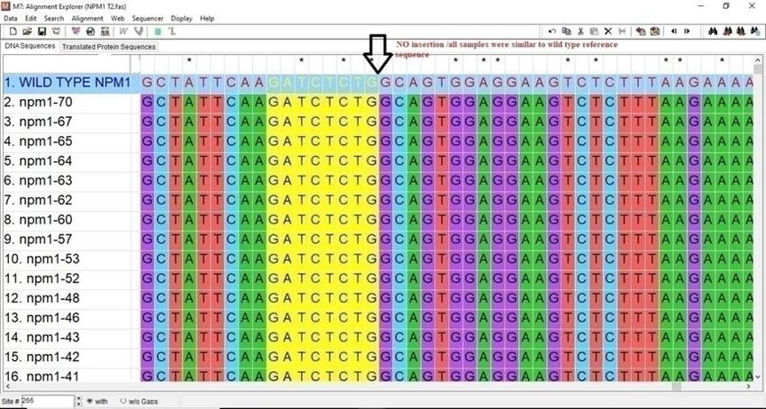
technique was done, and then sequencing analysis by software (Mega 7 Software) revealed that there were no
NPM1mutations in Sudanese AML patients shown in Fig (2).
182
GSC Biological and Pharmaceutical Sciences, 2021, 14(02), 181–184

                        Figure 1The percentage of AML subs types in the patient of this study.

                   Figure 2The alignment of NPM1 sample number (20) with wild Types reference.

4. Discussion
NPM1 gene characterize as candidate for prognosis and treatment of AML are which revealed that the Sudanese AML
patient carry wild type NPM1 which were almost resemble previous study done in Sudan showed minimum prevalence
of mutated NPM1 types (3%) [8], this may due to different technique used QRT. PCR comparing to PCR/sequencing
which may indicating of methylation effect on the gene (epigenetic effect) rather than nucleotide changes, Therefore
when comparing the phenotypes result of Sudanese NPM1 wild types with previous studies from other population
revealed were the same figure [9]. One of explanation AML samples used for NPM1 typing missing the karyotype data,
may be most of samples were abnormal karyotypes. Therefore the absence of mutant types of NPM1 among Sudanese
population may due to weak role of these mutant types on AML prognosis this low genetic variability due founder effect
on this founder effect or genetic drift. Mutations of NPM1 gene are significantly associated with increasing age and it
has been reported that they are less frequently seen in patients under the age of 35 years [10]. As more than 50% of the
Sudanese patients were 35years or less, this could possibly be the reason behind the absence of the NPM1 mutations in
this study. Another possible explanation is that different frequencies have been reported in different ethnic regions, and

                                                                                                                     183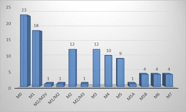
GSC Biological and Pharmaceutical Sciences, 2021, 14(02), 181–184

also globally found significantly lower frequency of NPM1 mutations in Asian populations, for example, 11% in a large
study from China [11].

5. Conclusion
NPM1 mutations are part of genetic abnormalities of acute myeloid leukemia (AML) mainly for diagnosis, treatment but
for prognosis they have no clear role in Sudanese patients.

Compliance with ethical standards

Acknowledgments
The cooperation of the medical staff at the RICK Hospital in Khartoum is appreciated.

Disclosure of conflict of interest

We have no conflicts of interest to disclose

Statement of informed consent
There is no information about any individual

References
[1]    Kumar S, Stecher G, Tamura K. MEGA7: molecular evolutionary genetics analysis version 7.0 for bigger datasets.
       Molecular biology and evolution. 2016 Mar 22;33(7):1870-4.
[2]    Rezaei N, Arandi N, Valibeigi B, Haghpanah S, Khansalar M, Ramzi M. FMS-like tyrosine kinase 3 (FLT3) and
       nucleophosmin 1 (NPM1) in Iranian adult acute myeloid leukemia patients with normal karyotypes: mutation
       status and clinical and laboratory characteristics. Turkish Journal of Hematology. 2017 Dec;34(4):300.
[3]    Arber DA, Orazi A, Hasserjian R, Thiele J, Borowitz MJ, Le Beau MM, Bloomfield CD, Cazzola M, Vardiman JW. The
       2016 revision to the World Health Organization classification of myeloid neoplasms and acute leukemia. Blood.
       2016 May 19;127(20):2391-405.
[4]    He R, Oliveira JL, Hoyer JD, Viswanatha DS. Molecular Hematopathology. InHematopathology 2018 Jan 1;712-
       760.
[5]    Wang JA. Cytoplasmic Mislocalization of CTCF by Leukemic Oncogene NPM1c in Acute Myeloid Leukemia. 2019.
[6]    Papaemmanuil E, Gerstung M, Bullinger L, Gaidzik VI, Paschka P, Roberts ND, Potter NE, Heuser M, Thol F, Bolli
       N, Gundem G. Genomic classification and prognosis in acute myeloid leukemia. New England Journal of Medicine.
       2016 Jun 9;374(23):2209-21.
[7]    Uckelmann HJ, Armstrong SA, Stone RM. Location, location, location: Mutant NPM1c cytoplasmic localization is
       required to maintain stem cell genes in AML. Cancer cell. 2018 Sep 10;34(3):355-7.
[8]    Altaybe T. Frequency of AML1: ETO, MLL: AF9, FUS: ERG, and NPM1 gene in Sudanese AML patients. [thesis].
       Bahri: Khartoum; 2014.
[9]    Dovey OM, Cooper JL, Mupo A, Grove CS, Lynn C, Conte N, Andrews RM, Pacharne S, Tzelepis K, Vijayabaskar MS,
       Green P. Molecular synergy underlies the co-occurrence patterns and phenotype of NPM1-mutant acute myeloid
       leukemia. Blood, The Journal of the American Society of Hematology. 2017 Oct 26;130(17):1911-22.
[10]   Owaidat HM. Prognostic Value of Day 14 Bone Marrow Morphology in Adult AML patients. CU Theses. 2018.
[11]   Gou H, Zhou J, Ye Y, Hu X, Shang M, Zhang J, Zhao Z, Peng W, Zhou Y, Zhou Y, Song X. The prevalence and clinical
       profiles of FLT3-ITD, FLT3-TKD, NPM1, C-KIT, DNMT3A, and CEBPA mutations in a cohort of patients with de
       novo acute myeloid leukemia from southwest China. Tumor Biology. 2016 Jun 1;37(6):7357-70.

                                                                                                                   184
You can also read