UC Davis Dermatology Online Journal - eScholarship
←
→
Page content transcription
If your browser does not render page correctly, please read the page content below
UC Davis
Dermatology Online Journal

Title
Anti-MDA-5 negative, anti-Ku positive clinically amyopathic dermatomyositis

Permalink
https://escholarship.org/uc/item/1qg7x8gf

Journal
Dermatology Online Journal, 27(4)

Authors
Carrington, Alexis E
Tartar, Danielle M
Sivamani, Raja K

Publication Date
2021

DOI
10.5070/D3274053154

Copyright Information
Copyright 2021 by the author(s).This work is made available under the terms of a Creative
Commons Attribution-NonCommercial-NoDerivatives License, available at
https://creativecommons.org/licenses/by-nc-nd/4.0/

Peer reviewed

 eScholarship.org                                 Powered by the California Digital Library
                                                                  University of California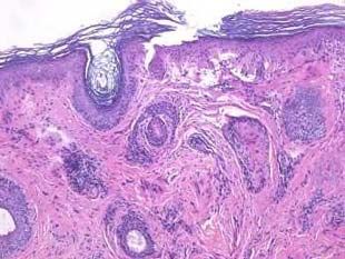
Volume 27 Number 4| April 2021
Dermatology Online Journal || Case Presentation                                                                             27(4):6

Anti-MDA-5 negative, anti-Ku positive clinically amyopathic
dermatomyositis
Alexis E Carrington1 MD, Danielle M Tartar1 MD PhD, Raja K Sivamani1-5 MD MS AP
Affiliations: 1Department of Dermatology, University of California Davis, Sacramento, California, USA, 2Department of Biological
Sciences, California State University, Sacramento, California, USA, 3College of Medicine, California Northstate University,
Sacramento, California, USA, 4Pacific Skin Institute, Sacramento, California, USA, 5Zen Dermatology, Sacramento, California, USA
Corresponding Author: Raja K Sivamani MD MS AP, Department of Dermatology, University of California-Davis, 3301 C Street, Suite
1300, Sacramento, CA 95816., Tel: 916-734-6111, Email: rksivamani@ucdavis.edu

                                                                      patient with negative melanoma differentiation-
  Abstract                                                            associated gene 5 antibody (anti-MDA5) and anti-Ku
  We present a patient with anti-MDA5 negative, anti-                 positive CADM.
  Ku positive clinically amyopathic dermatomyositis
  (CADM). A 61-year-old woman presented with a chief
  complaint of a 20-year history of a pruritic rash that
  was active on her face, chest, hands, legs, and back. A
                                                                      Case Synopsis
  mildly scaly, erythematous, photo-distributed                       A 61-year-old woman with a past medical history of
  eruption along with slightly violaceous, scaly papules              type two diabetes mellitus, longstanding history of
  accentuated on the wrist, metacarpophalangeal                       atopic dermatitis (not biopsy confirmed), major
  joints, proximal interphalangeal and distal                         depressive disorder, and hyperlipidemia presented
  interphalangeal joints. Antibody profile was                        with a chief complaint of a rash that was currently
  significant for positive ANA and anti-dsDNA, elevated               active on her face, chest, hands, legs, and back. She
  anti-TIF-1gamma (RDL)/p155, and weakly positive                     noted that the rash had been present for over 20
  anti Ku. Biopsy was consistent with dermatomyositis.                years and was pruritic. Prior treatment had included
  Melanoma differentiation-associated gene 5                          topical corticosteroids, most recently fluocinolone to
  antibody (anti-MDA-5) has been identified as the                    the scalp and hydrocortisone to the trunk and
  most commonly associated autoantibody found in                      extremities. In addition, intramuscular triamcinolone
  CADM and is associated with poor prognosis and a                    injections had been administered without
  biomarker for the diagnosis of rapidly progressive
                                                                      improvement. Interestingly, prior to presentation,
  interstitial lung disease. To our knowledge, our
                                                                      she had recently received 20 treatments of excimer
  patient is the first case of negative anti-MDA-5 and
                                                                      laser (narrow-band UVB) with some improvement,
  anti-Ku positive CADM.
                                                                      but did note persistent flaring. Medication allergies
                                                                      included fluconazole and there was no personal or
Keywords: antibodies, autoimmune disorder,
                                                                      family history of skin cancer. Review of systems was
dermatomyositis                                                       positive for arthralgia and back pain and negative for
                                                                      fever, fatigue, unintentional weight loss, weakness,
                                                                      or myalgias. The patient’s systemic medications
Introduction                                                          included fenofibrate, metformin, sertraline,
                                                                      simvastatin, and alendronate.
Clinically amyopathic dermatomyositis (CADM) is an
inflammatory autoimmune disease with cutaneous                        On examination, the patient was found to have
manifestations of dermatomyositis but no or                           erythematous, photo-distributed, slightly infiltrated
minimal muscle involvement [1]. We present a                          plaques and scattered papules along the forehead,

                                                                -1-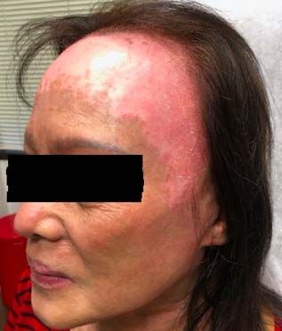
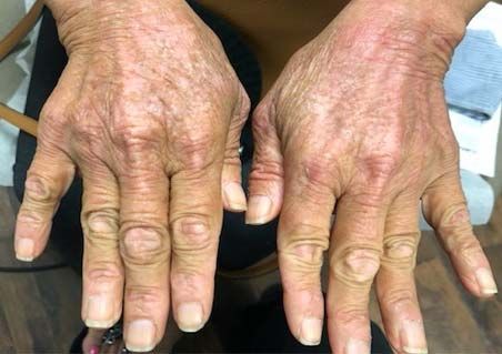
Volume 27 Number 4| April 2021
Dermatology Online Journal || Case Presentation                                                                               27(4):6

 A                                B                                                    C
Figure 1. A) Erythematous, scaly, slightly infiltrated plaques and scattered papules in a photodistributed pattern along the forehead,
temples, and cheeks. Recent excimer laser treatment likely contributed to the marked erythema of the forehead. B) Slightly violaceous,
scaly papules accentuated on the wrist, metacarpophalangeal joints, proximal interphalangeal joints, and distal interphalangeal joints
associated with ragged cuticles and sparse dilated capillaries on the bilateral dorsal hands. C) Biopsy of the right upper cutaneous lip
showed interface dermatitis with superficial and deep perivascular and periadnexal inflammation and thickened basement membranes.

temples, posterior neck, upper back, chest, and                         owing to the COVID-19 pandemic. The patient
cheeks with minimal scale (Figure 1A) and notable                       declined shortness of breath, cough, or other
sparing of the photoprotective areas. She also                          respiratory symptoms. She was started on
presented with slightly violaceous, scaly papules                       hydroxychloroquine 200mg twice daily as well as
accentuated on the wrist, metacarpophalangeal                           triamcinolone 0.1 % cream topically. However, the
joints, proximal interphalangeal joints, and distal                     patient discontinued the hydroxychloroquine
interphalangeal joints associated with ragged                           secondary to worsening of itch. She was then started
cuticles and sparse dilated capillaries on the bilateral
dorsal hands (Figure 1B). Strength assessment
revealed 5/5 strength throughout.
Creatinine kinase, aldolase, AST, and ALT were all
within normal limits. Accordingly, muscle MRI,
biopsy, or EMG were not done. Antibody profile was
significant for a positive ANA titer, 1:1280, speckled
(negative
Volume 27 Number 4| April 2021
Dermatology Online Journal || Case Presentation                                                                   27(4):6

on methotrexate 15mg weekly with improvement                   patients [7], whereas Hall noted that 9 of 11 anti-
(Figure 2). Systemic corticosteroids were avoided              MDA-5+ DM patients had a symmetric inflammatory
given the possible avascular necrosis of femoral head          polyarthropathy resembling rheumatoid arthritis [8].
seen on the pelvic CT.                                         Interestingly, whereas our anti-MDA-5-negative
                                                               patient did experience arthralgias, her rheumatoid
                                                               factor, erythrocyte sedimentation rate, and C-
Case Discussion                                                reactive protein were negative.
Clinically amyopathic dermatomyositis is further               Ku protein is a DNA-binding protein involved in DNA
classified as hypomyopathic or amyopathic                      repair. Anti-Ku antibodies have been reported in
dermatomyositis. The former lacks clinical evidence            several auto-immunity conditions, including
of muscle weakness, yet laboratory investigation,              myositis, systemic lupus erythematosus (SLE), and
EMG, or muscle biopsy confirms muscle                          Sjögren syndrome [9]. Inflammatory myopathies
involvement. The latter also has no clinical evidence          with anti-Ku antibodies are commonly seen in an
of muscle weakness but also lacks laboratory, biopsy,          overlap syndrome with different connective
or EMG evidence of underlying muscle inflammation              autoimmune diseases, such as scleroderma and SLE;
[2]. The pathognomonic skin manifestations of                  in addition, reports of anti-Ku positive inflammatory
CADM are similar to that of dermatomyositis,                   myopathies have found significant associations with
including Gottron papules (Figure 1B, C), heliotrope           ILD [10]. Interestingly, our patient presented with a
rash, and shawl sign on the posterior neck, shoulders,         weak anti-Ku antibody and absence of myopathy or
and upper back. A photodistributed exanthem,                   scleroderma symptoms, which, to our knowledge,
alopecia, and periungual telangiectasias are well-             has not been reported in the literature.
described [2,3].                                               Currently, classification criteria do not require the
Melanoma differentiation-associated gene 5                     presence of myositis-specific antibodies or myositis-
antibody (anti-MDA-5) has been identified as the               associated antibodies to diagnose CADM [3]. In
most commonly associated autoantibody found in                 addition, cutaneous lupus and CADM antibody
CADM [4,5] and is associated with poor prognosis. It           profile can overlap, as in our patient with a positive
is a biomarker for the diagnosis of rapidly                    anti-dsDNA.
progressive interstitial lung disease (ILD), [1,2]. In         A variety of malignancies have been found to be
fact, anti-MDA5+ dermatomyositis (DM) patients                 associated with CADM. A comprehensive review
who developed rapidly progressive ILD had higher               found CADM to have similar associated malignancies
anti-MDA5 antibody levels compared to those                    to classic dermatomyositis, such as genitourinary
without rapidly progressive ILD [5]. To our                    and respiratory malignancies [11]. However, this
knowledge, our patient is the first reported with              review also showed that breast cancer was the most
negative anti-MDA-5 and anti-Ku positive CADM.                 commonly reported neoplasm in CADM, whereas
Melanoma differentiation-associated gene 5 is a RIG-           lung cancer was the most commonly observed
1-like receptor dsRNA helicase enzyme encoded in               malignancy in classic dermatomyositis [11].
the IFIH1 gene and functions as a virus sensor by              Patients with dermatomyositis are at the highest risk
pattern recognition receptor for dsRNA [6]. In both            of having an underlying malignancy during the first
Japanese and North American DM patients, anti-                 five years of diagnosis. Despite conflicting studies on
MDA-5 positive DM patients have also been shown                malignancy risk in DM versus CADM, age appropriate
to have an increased frequency of rare, acral                  cancer screening is still recommended in CADM
ulcerative skin changes [6]. Arthralgias are common            patients [3]. Regarding cancer screening, there is no
in patients with positive anti-MDA-5 [5]. A cohort             consensus regarding the recommended frequency
study reported hand swelling in 40% and                        or extent of evaluation for patients with CADM or
arthritis/arthralgia in 70% of 77 patients with DM             DM. However, given the higher association of

                                                         -3-
Volume 27 Number 4| April 2021
Dermatology Online Journal || Case Presentation 27(4):6
pulmonary pathologies, including rapidly for complications and brings up the discussion of
progressive ILD, in CADM patients frequent ideal cancer screening guidelines.
evaluations could prove beneficial.
Potential conflicts of interest
Conclusion RKS serves as a scientific advisor for LearnHealth and
This is a unique case of CADM with an absence of Arbonne and as a consultant to Burt’s Bees,
anti-MDA-5 and presence of anti-Ku antibodies. This Physicians Exclusive, Nutrafol, Abbvie, Leo, Sun and
presentation reflects of the necessity of evaluating Regeneron Pharmaceuticals.
References
1. Takada T, Asakawa K, Barrios R. A Japanese-American female with diversity in dermatomyositis. Ann Transl Med. 2017;5:160. [PMID:
rapidly progressive interstitial lung disease associated with 28480196].
clinically amyopathic dermatomyositis. Clin Rheumatol. 7. Fiorentino D, Chung L, Zwerner J, Rosen A, Casciola-Rosen L. The
2020;40:1159-1165. [PMID: 32676922]. mucocutaneous and systemic phenotype of dermatomyositis
2. Tartar DM, Chung L, Fiorentino DF. Clinical significance of patients with antibodies to MDA5 (CADM-140): a retrospective
autoantibodies in dermatomyositis and systemic sclerosis. Clin study. J Am Acad Dermatol. 2011;65:25–34. [PMID: 21531040].
Dermatol. 2018;36:508–24. [PMID: 30047434]. 8. Hall JC, Casciola-Rosen L, Samedy L-A, et al. Anti-melanoma
3. Concha JSS, Tarazi M, Kushner CJ, Gaffney RG, Werth VP. The differentiation-associated protein 5-associated dermatomyositis:
diagnosis and classification of amyopathic dermatomyositis: a expanding the clinical spectrum. Arthritis Care Res . 2013;65:1307–
historical review and assessment of existing criteria. Br J Dermatol. 15. [PMID: 23436757].
2019;180:1001–8. [PMID: 30561064]. 9. Belizna C, Henrion D, Beucher A, et al. Anti-Ku antibodies: Clinical,
4. Sato S, Hirakata M, Kuwana M, et al. Autoantibodies to a 140-kd genetic and diagnostic insights. Autoimmun Rev. 2010;9:691–4.
polypeptide, CADM-140, in Japanese patients with clinically [PMID: 20621654].
amyopathic dermatomyositis. Arthritis Rheum. 2005;52:1571–6. 10. Rigolet A, Musset L, Dubourg O, et al. Inflammatory myopathies
[PMID: 15880816]. with anti-Ku antibodies: a prognosis dependent on associated
5. Moghadam-Kia S, Oddis CV, Aggarwal R. Anti-MDA5 Antibody lung disease. Medicine . 2012;91:95–102. [PMID: 22391471].
Spectrum in Western World. Curr Rheumatol Rep. 2018;20:78. 11. Udkoff J, Cohen PR. Amyopathic Dermatomyositis: A Concise
[PMID: 30382445]. Review of Clinical Manifestations and Associated Malignancies.
6. Sontheimer RD. MDA5 autoantibody-another indicator of clinical Am J Clin Dermatol. 2016;17:509–18. [PMID: 27256496].
-4-You can also read