Update on Melanoma 2019 - Bruce M. Brenner, MD, FACS Associate Professor of Surgery Associate Program Director, General Surgery
←
→
Page content transcription
If your browser does not render page correctly, please read the page content below
Update on Melanoma 2019

       Bruce M. Brenner, MD, FACS
      Associate Professor of Surgery
Associate Program Director, General Surgery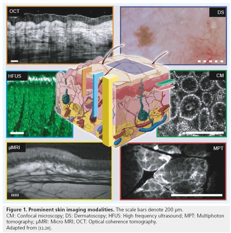
Objectives
• To identify new technologies for melanoma diagnosis
• To list recent changes in the melanoma staging
• To define adequate margins for excision of melanoma
• To describe indications for sentinel node biopsy and
  complete node dissection
• To discuss indications for adjuvant therapy in melanoma
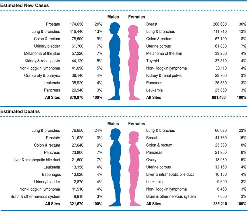
Cancer Statistics 2019
Florida
8360 Cases

               Siegel RL, Miller KD, Jemal A. Cancer statistics, 2019. CA Cancer J Clin. 2019 Jan;69(1):7-34.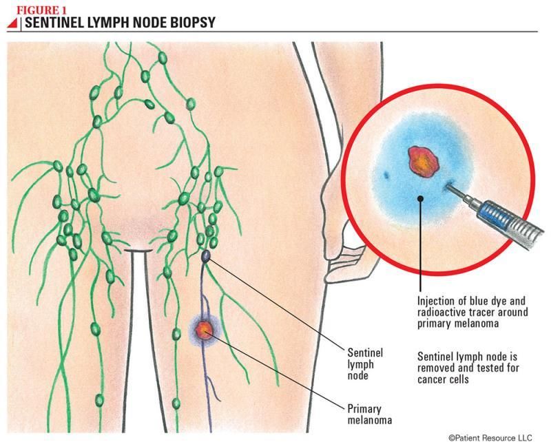
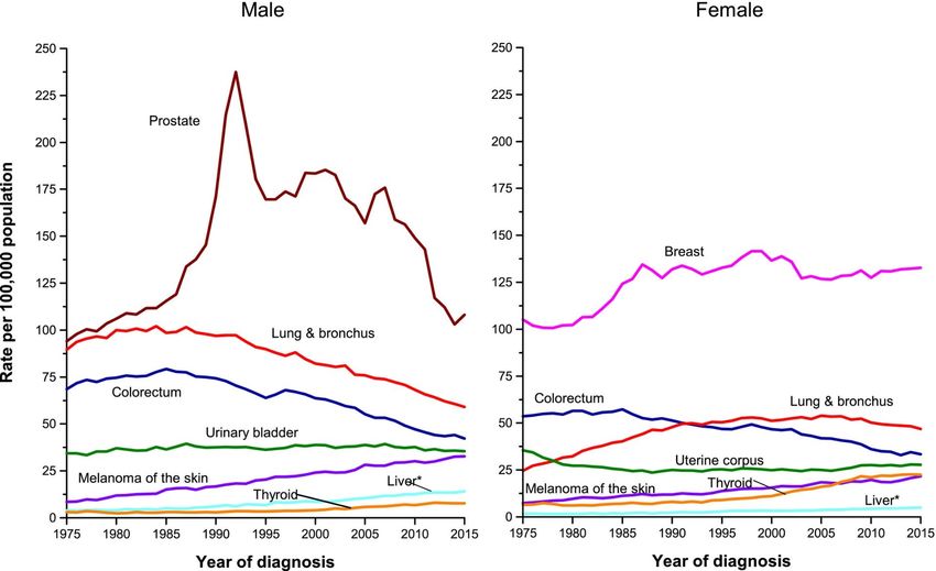
Trends in Cancer Incidence
Melanoma Survival by Stage and Race
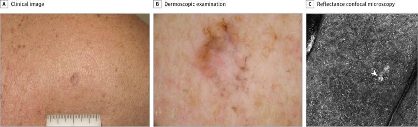
Melanoma Diagnosis

• Biopsy is the gold standard
   • Dermoscopy may help direct biopsies

• New technologies
   • Melafind
       • Optical scanning device, off market
   • Reflectance confocal microscopy
       • Real-time imaging at cellular resolution
       • Much more specific than dermoscopy for equivocal lesions*
           • Avoid unnecessary biopsies

                  *Dinnes J, et al; Cochrane Skin Cancer Diagnostic Test Accuracy Group. Reflectance confocal
                  microscopy for diagnosing cutaneous melanoma in adults. Cochrane Database Syst Rev. 2018 Dec 4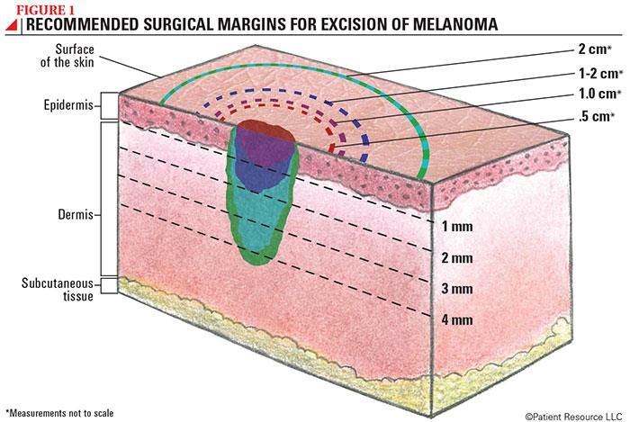
Melanoma Diagnosis

• Other New tech
   • Optical coherence tomography
        • Deeper penetration, lower
          resolution than LCM
   • High-frequency ultrasound
   • These need more data in
     cutaneous melanoma
        • Accuracy unclear
        • Lack of guidelines for
          diagnosis

• Total body photography
   • Screening high risk patients with
     multiple moles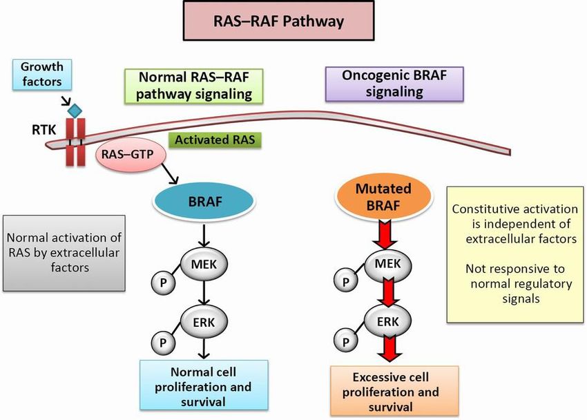
Melanoma Staging 2019
TX: Primary tumor thickness cannot be assessed
T0: No evidence of primary tumor
Tis (melanoma in situ)
T1        ≤1.0 mm                Unknown or unspecified
T1a       1.0‐2.0 mm            Without ulceration
T2b       >1.0‐2.0 mm            With ulceration
T3        >2.0‐4.0 mm            Unknown or unspecified
T3a       >2.0‐4.0 mm            Without ulceration
T3b       >2.0‐4.0 mm            With ulceration
T4        >4.0 mm                Unknown or unspecified
T4a       >4.0 mm                Without ulceration

T4b       >4.0 mm                With ulceration
* Mitoses removed from staging
                                                                       Survival by T for Stage I-II Melanoma
                                      Gershenwald JE, et alMelanoma staging: Evidence-based changes in the American Joint Committee on
                                      Cancer eighth edition cancer staging manual. CA Cancer J Clin. 2017
                                      Nov;67(6):472-492.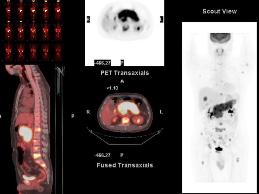
N Staging 2019
NX        Node path not assessed*        No in-transit**

N0        No regional metastases         No

N1        One node or in-transit met

N1a       One clinically occult***       No

N1b       One clinically detected        No

N1c       No lymph node disease          Yes

N2        2-3 nodes or in‐transit with 1 node

N2a       Two or 3 clinically occult     No

N2b       2-3, ≥1 clinically detected    No

N2c       One or more nodes              Yes

N3        ≥4 nodes or any in‐transit with ≥ 2 nodes, or any matted nodes

N3a       ≥4 clinically occult           No

N3b       ≥4 nodes, ≥1 clinically or matted****    No

N3c       ≥2 or matted nodes             Yes

*Pathological N category is not required for T1 melanomas
**Includes in-transit, microsatellite, and satellite
*** No micro or macroscopic ****Not pathologic extranodal extension
M Staging 2019
M0       No distant metastasis                 Tis          N0b                    M0   0
                                               T1a          N0                     M0   IA
M1       Distant metastasis
                                               T1b          N0                     M0   IA
M1a      Skin, soft tissue, nonregional node   T2a          N0                     M0   IB
                                               T2b          N0                     M0   IIA
M1a(0) LDH* not elevated
                                               T3a          N0                     M0   IIA
M1a(1)   LDH elevated                          T3b          N0                     M0   IIB
                                               T4a          N0                     M0   IIB
M1b      To lung with or without M1a
                                               T4b          N0                     M0   IIC
M1b(0)   LDH not elevated                      T0           N1b, N1c               M0   IIIB
                                               T0           N2b, N2c, N3b or N3c   M0   IIIC
M1b(1)   LDH elevated
                                               T1a/b‐T2a    N1a or N2a             M0   IIIA
M1c      Non‐CNS visceral sites                T1a/b‐T2a    N1b/c or N2b           M0   IIIB
                                               T2b/T3a      N1a‐N2b                M0   IIIB
M1c(0)   LDH not elevated                      T1a‐T3a      N2c or N3a/b/c         M0   IIIC
M1c(1)   LDH elevated                          T3b/T4a      Any N ≥N1              M0   IIIC
                                               T4b          N1a‐N2c                M0   IIIC
M1d**    CNS                                   T4b          N3a/b/c                M0   IIID
M1d(0)   LDH not elevated                      Any T, Tis   Any N                  M1   IV

M1d(1)   LDH elevated
*LDH now included for all subcategories
**CNS now separate category
Staging Workup
• All Stages
• H & P with focus on lymphatic exam
• Imaging to evaluate specific symptoms
• Stages I – II
• Routine imaging or lab tests not recommended
• Stage IIIA (Positive sentinel node)
• Consider imaging for baseline staging
• Stage III B/C/D (SLN +) or Clinical Node + or In-transit
• Needle biopsy of palpable lesions
• Imaging for baseline staging (Contrast CT Chest/Abd/Pelvis or whole body
PET/CT, + Brain MRI if ≥ IIIC)Melanoma Staging
Margins of Resection

• WHO Trial (1988)*
   • Breslow thickness ≤ 2mm
   • 1 vs 3 cm margins
   • No difference in disease-free or overall survival
   • Possible increase in local recurrence if > 1mm

• Intergroup Melanoma Trial**
   • Breslow thickness 2-4 mm
   • 2 vs 4 cm margins
   • No difference in DFS or OS

• Others – Swedish, UK, French

                   *Veronesi U, et al. Thin stage I primary cutaneous malignant melanoma. Comparison of excision with margins of 1 or 3 cm. N Engl JMed. 1988 May
                   5;318(18):1159-62.
                   **Balch CM, et al. Efficacy of 2-cm surgical margins for intermediate-thickness melanomas (1 to 4 mm). Results of a multi-institutional randomized
                   surgical trial. Ann Surg. 1993 Sep;218(3):262-7.
Current Margin Recommendations
Management of Lymphatic Basins

• Clinically positive nodes require complete lymphadenectomy
• Sentinel node biopsy indications (ASCO/SSO Guidelines) for
  patients with clinically negative nodes
   • Thin (≤ 1mm) - 5.2% nodal mets
       • T1a – Not routinely indicated
       • T1b – Consider, discussion with patient
   • Intermediate thickness (1 – 4 mm) – 16 – 20% with nodal mets
       • Indicated for all (T2 and T3)
   • Thick
       • Recommended, discuss with patient
Lymphatic Drainage Patterns
Sentinel Node Techniques
Impact of SLN Biopsy

• MSLT-I trial*
   • 2001 patients with primary cutaneous melanoma
   • Randomized to WLE with SLN vs WLE and observation
   • Significantly improved survival in SLN+ compared to SLN-
       • Both intermediate and thick melanomas
   • ~20% of intermediate thickness had lymph node mets
       • Survival improved in SLN+CLND vs observation and therapeutic
         dissection
            • 56% vs 41.5% at 10 years
   • No effective adjuvant therapy available at that time (trial closed in 2002)

                   *Morton DL, et al; MSLT Group. Final trial report of sentinel-node biopsy versus nodal observation in melanoma. N Engl J Med.
                   2014 Feb 13;370(7):599-609.
Lymphadenectomies
Management of Lymphatic Basins
• Role of completion lymphadenectomy with positive SLN
   • Not recommended unless more extensive nodal involvement
       • Trials randomized for CLND vs no CLND
       • Patients without dissection all followed with frequent ultrasound
       • Regional control improved
       • No difference in disease-specific or overall survival
       • ~ 65% of mets under 1mm
           • DeCOG-SLT (German)*
                • Some patients got adjuvant interferon
           • MSLT-II (International)**
                • Positive non-sentinel nodes strongly associated with recurrence
                • 12% of patients RT-PCR positive only
       • Role of CLND with more extensive nodal involvement is still unclear

                 *U. Leiter, R. Stadler, C. Mauch, et al. Complete lymph node dissection versus no dissection in patients with sentinel lymph
                 node biopsy positive melanoma (DeCOG-SLT): a multicentre, randomized phase 3 trial. Lancet Oncol, 17 (2016), pp. 757-767
                 **M.B. Faries, J.F. Thompson, A.J. Cochran, et al. Completion dissection or observation for sentinel-node metastasis in melanomaN Engl J Med, 376 (2017),
                 pp. 2211-2222
Adjuvant Systemic Therapy
• Interferon
• No clear benefit in overall survival
• Anti-CTLA4 (Ipilimimab/Yervoy, FDA approved 2015)
• Enhance effector T cell function
• EORTC 18071 – significantly improved RFS and OS (HR 0.76 and 0.72)
• Significant toxicity due to high dose regimen
• Trial of high vs low dose in progress
• BRAF/MEK inhibitors (dabrafenib/trametinib, FDA 2018)
• Only effective in V600E or V600K BRAF mutant melanoma (50% of cases)
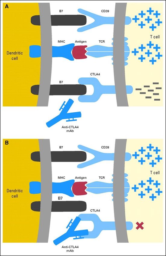
• COMBI-AD – Significant improvement in 3 year RFS and OS (0.47 and 0.57)Anti – CTLA4
Mechanism
Fig 4. Cytotoxic T lymphocyte-associated antigen 4 (CTLA4) blockade prevents downregulation of T cells. (A) CTLA4 receptors are upregulated after T-cell activation and bind
to B7, sending an inhibitory signal that downregulates T-cell activation. (B) Anti-CTLA4 antibodies have a greater affinity for CTLA4 than B7 and inhibit the interaction of B7
and CTLA4. Thus, the inhibitory signal produced by B7 binding to CTLA4 is blocked, and T-cell activation is prolonged. MHC, major histocompatibility complex; TCR, T-cell
receptor; mAb, monoclonal antibody.
Published in: John M. Kirkwood; Ahmad A. Tarhini; Monica C. Panelli; Stergios J. Moschos;
Hassane M. Zarour; Lisa H. Butterfield; Helen J. Gogas; Journal of Clinical Oncology 2008 263445-3455.BRAF-MEK Inhibitors From: Muñoz-Couselo E, et al. Recent advances in the treatment of melanoma with BRAF and MEK inhibitors. Ann Transl Med. 2015 Sep;3(15):207.
Adjuvant Therapy

• Anti-PD-1
   • Block PD-1/PD-L1 interaction, decreases blocking of immune recognition
   • Pembroluzimab (Keytruda) – FDA approved 2019
       • EORTC1325/KEYNOTE-054
           • Pembrolizumab vs placebo
           • Improved RFS (HR 0.57)
       • PDL-1 status did not matter
   • Nivolumab (Opdivo) – FDA 2017
       • CHECKMATE-238
           • Nivolumab vs Ipilumimab
           • Improved RFS (HR 0.65)
Mechanism of Anti-PD-1 From: Homet Moreno B, Parisi G, Robert L, Ribas A. Anti-PD-1 therapy in melanoma. Semin Oncol. 2015 Jun;42(3):466-73.
Management of In-transit Metastases

• Disease in the subdermal
  lymphatics
• Occur in up to 6% of patients
• May have multiple lesions and/or
  recurrences
• Surgical resection is first option
   • Patients are stage III and therefore
     candidates for adjuvant systemic therapy
Unresectable Stage III
• Limb Perfusion/infusion
• Good response rates of 75% or more
• Complete responders have improved survival
• Indications unclear in light of new systemic options
• ILI less morbid/minimally invasive
• Radiation
• Mainly used in palliation
• Systemic
• Limited data in Stage III
• Combination therapyUnresectable Stage III
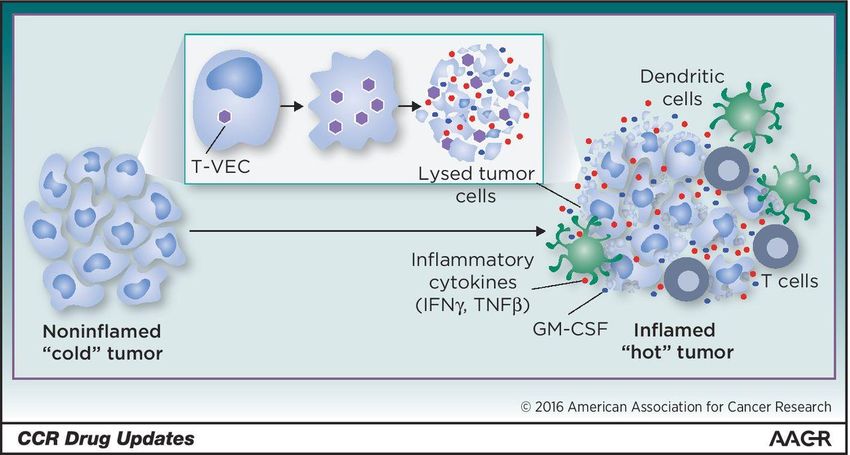
• T-VEC (talimogene laherparepvec) injection
• Modified herpes virus preferentially targets cancer cells
• Oncolytic and causes GM-CSF production
From: Ott PA, Hodi FS. Talimogene Laherparepvec for the Treatment of Advanced
Melanoma. Clin Cancer Res. 2016 Jul 1;22(13):3127-31.Role of Adjuvant Radiation in Melanoma
• Very little data is available from randomized trials
• Historical bias against XRT in melanoma
• No data in patients who received modern adjuvant systemic therapy
• Potential current indications*
• Regional nodes after dissection for clinically palpable/bulky disease
• May decrease local recurrence, but no effect on survival
• Nodal recurrence after regional dissection
• Local control of desmoplastic or other melanomas with neurotropism
• In patients withCurrent Adjuvant Studies • Safety and Efficacy of Pembrolizumab Compared to Placebo in Resected High-risk Stage II Melanoma (MK-3475- 716/KEYNOTE-716, Merck) - Recruiting • Recurrence-free survival is primary outcome • DDFS and OS secondary outcomes • Neoadjuvant PD-1 Blockade in Patients With Stage IIB/C Melanoma (University of Pennsylvania) – Not open yet • Single dose of pembrolizumab preop • Continued treatment postop up to 1 year • Evaluate effect on sentinel node positivity • Secondary outcomes include DFS and OS
Current Adjuvant Studies

• Neoadjuvant L19IL2/L19TNF(Pivotal) - Recruiting
   • Intratumoral injections of cytokines, stage III
   • Outcome is RFS

• Vaccine trials
   • 6MHP, With or Without Systemic CDX-1127 (Celldex) - Recruiting
       • Peptide vaccine (6MHP) with adjuvants and monoclonal antibody (CDX-
         1127)
       • Endpoints are adverse events and level of immune response
   • MART-1 Antigen With or Without TLR4 Agonist GLA-SE in Treating Patients
     With Stage II-IV Melanoma That Has Been Removed by Surgery - Active
       • Outcome is immune response
Future Directions • New diagnostic tools • Further refinement of staging and indications for SLN • New immunotherapy and targeted therapy agents • Adjuvant therapy for stage II • Neoadjuvant therapy for stage III • Continue defining the role of completion lymphadenectomy
You can also read