Imperforate hymen presenting with massive haematocolpos and acute urinary retention in a teenage girl: A case report
←
→
Page content transcription
If your browser does not render page correctly, please read the page content below
Tanzania Journal of Health Research                                       Doi: http://dx.doi.org/10.4314/thrb.v14i4.9
Volume 14, Number 4, October 2012

Imperforate hymen presenting with massive haematocolpos and acute urinary
retention in a teenage girl: A case report

IPYANA H. MWAMPAGATWA1* and BARAKA A. MPONDA2
1 School of Medicine, University of Dodoma, Dodoma, Tanzania

2Department of Obstetrics and Gynaecology, Dodoma Regional Hospital, Dodoma, Tanzania

_____________________________________________________________________________
Abstract: Imperforate hymen is relatively rare but it is the most frequently encountered obstructive
anomaly of the female lower genital tract. The clinical presentation vary significantly from patient to
patient depending on the age at diagnosis but in most cases the diagnosis is missed in early childhood
and therefore the diagnosis is made after puberty when the patient present with haematocolpos,
heamatometra or both. When this happens, the presentation could even be tricky because the patient may
presents with unlikely symptoms apart from cryptomenorhoea like, urinary retention or bowel
obstruction or both. Here we present a 16 years old girl with imperforate hymen and presented with
history of lower abdominal pain and distension associated with acute urinary retention. She was treated
by hymenotomy and improved dramatically and was discharge 6th day post operatively. This case
report is presented to address to clinicians the possibility of imperforate hymen with haematocolpos as a
differential diagnosis in adolescent girls particularly those who have not started having their menses in
their teens and present with acute urinary retention so that their external genitalia are carefully examined
to exclude the possibility of imperforate hymen as a cause of acute urinary retention due to the
haematocolpos.
_____________________________________________________________________________
Keywords: Imperforate hymen, urinary retention, haematocolpos, hymenotomy, Tanzania

Introduction

Normal vaginal development requires the fusion of components that are derived from
two embryologic structures, the mesodermal Müllerian ducts and the endodermal
urogenital sinus (UGS). The upper half of the vagina develops from the Müllerian ducts
while the lower half develops from the UGS. This is normally followed by canalization
to form a normal patent vagina. The hymen represents the junction of the sinovaginal
bulbs with the UGS; hence it is formed from the endoderm of the urogenital sinus
epithelium (Golan et al., 1989; Moore et al., 2003). By the fifth month of gestation, the
canalization of the vagina is complete while the hymen usually ruptures (perforates)
before or shortly after birth and remains as a thin mucous membrane (Acien, 1992). An
imperforate hymen therefore results when there is failure of the tissues of the endoderm
of the urogenital sinus to completely canalize. Imperforate hymen itself is relatively rare

*
    Correspondence: mwampagatwai@yahoo.com.

                                                     1
Tanzania Journal of Health Research Doi: http://dx.doi.org/10.4314/thrb.v14i4.9
Volume 14, Number 4, October 2012
with the incidence of about 0.1% of all newborn female babies (Edmonds, 2000) but by
far it is the commonest lower female genital tract abnormalities.
The presentation of imperforate hymen may be challenging in some cases such
that the diagnosis may initially be missed. Patients may present with a lower abdominal
mass mimicking pelvic tumour or sometimes presenting with acute urinary or bowel
obstruction due to massive accumulation of blood in the vagina and/or uterus (Posner
& Spandorfer, 2005). In this case report, we describe a presentation of imperforate
hymen in 16 years old school girl who presented with acute urinary retention and a
lower abdominal mass at the Dodoma Regional Hospital in central Tanzania.
Case History
A 16 years old primary school girl was referred to Dodoma Regional Hospital from
Kondoa District Hospital 160km away with a diagnosis of bladder tumour with urine
retention. She was seen at casualty and she further gave history of lower abdominal
pain which started few days prior to the onset of inability to pass urine. Basing on the
presented history and diagnosis in the referral letter, the doctors at casualty section
admitted her in the surgical ward. Being a young girl, two more possible diagnosis
were thought to be likely (Pregnancy or ovarian tumour) but urine for pregnancy test
was negative, and the ultrasound revealed fluid collection in the lower abdomen
though the exact location was not pin pointed. The uterus and ovaries couldn’t be seen
on ultrasound. Based on these findings she was then transferred to Gynaecological
Ward for further evaluation.
Further questioning, the girl admitted not to have yet attained menarche but to
have been experiencing rather cyclic lower abdominal pain over the past two to three
years. She further gave history she never have had penetrative vaginal sexual
intercourse. Upon general clinical examination it was noted that she was healthy, pink,
afebrile but in pain. She was also of normal stature and development of secondary
sexual characteristics. Abdominal examination revealed a supra-pubic mass equivalent
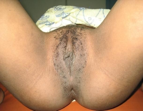
to 20-week pregnancy that was tense and tender on palpation. Pelvic inspection showed
a normal vulva but with brownish bulging membrane in the introitus (Figure 1A).
Bimanual pelvic examination through the rectum also revealed a markedly distended
vagina bulging into the anterior rectal wall (Figure 1B). Urinary catheterization was
performed and about 1300ml of clear urine was drained from the urinary bladder and
the abdominal mass diminished to about 16 weeks (gestation equivalent) but the
bulging membrane in the introitus was still there.
Based these findings in history and examination the diagnosis of imperforate
hymen was reached. This diagnosis was communicated to the patient and the mother
who escorted the girl. It was further explained to them the definitive treatment of the
2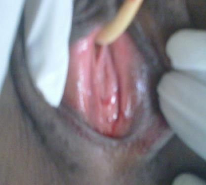
Tanzania Journal of Health Research                                 Doi: http://dx.doi.org/10.4314/thrb.v14i4.9
Volume 14, Number 4, October 2012

condition for which both the mother and the girl consented for both the procedure and
its outcomes in terms of socio-cultural implications bearing in mind that the virginity of
the girl will be lost after the procedure. Preparations for hymenoctomy were done for
the next day. On the day of operation the patient was taken to theatre for the operation.
She was placed in the dorsal lithotomy position, the bladder was drained, and a sterile
perineal preparation performed.

A                                                     B
Figure 1: Haematometra/haematocolpos presenting as a supra-pubic mass after catheterization to drain
the retained urine (A); Bulging of the hymen due to massive haematocolpos (B)

Hymenotomy was performed under local anaesthesia where a cruciate incision was
made on the hymen and about 1500mls of viscous chocolate-coloured blood was
drained. After that the hymeneal leaflets were then trimmed sharply from the hymeneal
ring and the cut edges of the leaflet bases were then over sewn with interrupted
absorbable sutures.

                                                 3
Tanzania Journal of Health Research Doi: http://dx.doi.org/10.4314/thrb.v14i4.9
Volume 14, Number 4, October 2012
Figure 2: The appearance of the external genitalia on 4th day post operation
Post operatively the girl improved remarkably and was discharged home on 6 th day.
Because she was coming from far, she was instructed to go back to the referring district
Hospital after 2 weeks for follow up. Report so obtained 4 months after the operation
during supervision visits gave a success story that the patient attended their hospital
and she was doing fine (Figure 2).
Discussion
Imperforate hymen is generally a rare occurrence in clinical practice of gynaecology.
Literature has reported incidences of around 0.1% of all newborn female babies
(Edmonds, 2000). It is this rarity that brings the difficulty in making the diagnosis easily
at birth compounded by a bizarre presentation in some cases whose diagnosis is made
at puberty. In most cases the diagnosis is reached when the girl reaches menarche and
start having accumulation of blood in the vaginal (haematocolpos) and the uterus
(haematometra). This accumulation of blood compresses the bladder and the urethra to
cause urine retention (Buick et al., 1999). At times other unusual clinical features may
show up and further complicate the picture making the diagnosis even more
challenging. These include low back pain and/or constipation due to massive
haematocolpos (Buick et al., 1999; Isenhour et al., 1999).
Our patient presented with an acute urine retention of about two days duration
and because it is very likely that a thorough clinical examination was not done at birth
nobody had a clue of imperforate hymen and again pelvic examination was not done
4Tanzania Journal of Health Research Doi: http://dx.doi.org/10.4314/thrb.v14i4.9
Volume 14, Number 4, October 2012
when she arrived at the hospital hence, the diagnosis was missed for the second time
and pregnancy and urinary bladder tumour were given a priority. From this picture it
becomes evident that an imperforate hymen should also be thought as a cause of urine
retention in young girls who present with acute urine retention. This should be
accompanied by a carefully pelvic examination to substantiate the diagnosis.
The diagnosis of imperforate hymen may be reached at birth from a thorough
clinical examination or in early childhood when the little girl presents with mucocolpos
or hydrocolpos but more frequently the diagnosis is made at puberty when the girl
presents with haematocolpos or hematometra or both (Posner & Spandorfer, 2005).
Commonly imperforate hymen is asymptomatic and therefore the diagnosis is often
missed before puberty and it is made when the girl reaches menarche. In our case the
diagnosis was only reached about a year after the onset of irregularly cyclic abdominal
pain when she had a massive haematocolpsos. This delay came because nobody had in
mind the presence of the condition. A good clinical examination at birth would have
helped to have the diagnosis at hand before this massive haematocolpos appeared
(Posner & Spandorfer, 2005).
In countries where Magnetic Resonance Imaging (MRI) can be done examination
of the pregnant woman is done, the diagnosis of imperforate hymen may be reached
prenatally where there is a protrusion of the hymen into the introitus depending on the
amount of fluid accumulation in the vagina (Adaletli et al., 2007). This can be a tool
because the diagnosis is already in the thought of an attending doctor. But it is obvious
that the use of MRI cannot be considered to be a diagnostic tool of choice in resource
constrained countries and therefore clinical examination is critically important.
The clinical presentation in patients with imperforate hymen and transverse
vaginal septum may be similar in many aspects but in cases with transverse vaginal
septum there is usually no bulging at the outlet as well as the location of the obstruction
which may be high these patients (Wall et al., 2003). In our patient, bulging of the
membranes at the introitus clearly differentiated between the imperforate hymens from
transverse vaginal septum.
Thorough physical pelvic inspection and examination in patients with
imperforate hymen with haematocolpos and/or haematometra with its obstructive
symptoms should be sufficient to make the diagnosis though imagining studies may
add value. Ultrasound is in most cases a diagnostic tool but MRI may be employed in
complicated cases where the location of the collected fluid cannot be pin pointed (Frates
et al., 2004.). When ultrasound is chosen, the rectal route is preferred for it provides a
better visualization (Kushir et al., 1997).
Conclusion
5Tanzania Journal of Health Research Doi: http://dx.doi.org/10.4314/thrb.v14i4.9
Volume 14, Number 4, October 2012
Adolescent girls with acute urinary retention particularly those who have not yet stated
menstruation in their mid-teens, imperforate hymen with haematocolpos should be
considered as a possible differential diagnosis and a critical inspection of the external
genitalia are performed so that the diagnosis is reached the earliest and prompt
treatment provided.
Acknowledgements
The consent and assent to take photographs and publish this case report was obtained
from the mother and the girl, respectively. The consent was signed by the mother on
behalf of the girl. The authors would like to thank the Medical Officer in-Charge of
Dodoma Regional hospital for permission to publish this case report.
References
Acien, P. (1992) Embryological observations on the female genital tract. Human
Reproduction 7, 437-445.
Adaletli, I., Ozer, H., Kurugoglu, S., Emir, H. & Madazli, R. (2007) Congenital
imperforate hymen with hydrocolos diagnosed using prenatal MRI. American
Journal of Roentgen 189 23-25.
Buick, R. & Chowdhary, S. (1999) Bakache: a rare diagnosis and unusual complication.
Pediatric Surgery International 15, 586-587.
Edmonds, D. (2000) Congenital malformations of the genital tract. Obstetrics and
Gynecology Clinics of North America 27, 49-62.
Frates, M., Kumar, J., Benson, C., Ward, V. & Tempany, C. (2004) Fetal anomalies:
Comparison of MRI imaging and ultrasound diagnosis. Radiology 232, 398-404.
Golan, A., Langer, R., Bukovsky, I., Caspi, E.(1989) Congenital anomalies of the
Müllerian system. Fertility and Sterility 51, 747-755.
Isenhour, J., Henry, M. & Marx, J. 1999 Hematocolpometra manifesting as constipation
in young female. Academic Emergency Medicine 6, 752-753.
Kushir, O., Garde, K. & Blankstein, J. (1997) Rectal sonography for diagnosis of
hematocolpometra : a case report. Journal of Reproductive Medicine 45, 519-520.
Posner, J. & Spandorfer, P. (2005) early detection of imperforate hymen prevents
morbidity from delays in diagnosis. Pediatrics 115, 108-112
Wall EM, Stone B, Klein BL. (2003) Imperforate hymen: a not-so hidden diagnosis.
American Journal of Emergency Medicine 21,249-250
6You can also read