False-Negative Results of Real-Time Reverse-Transcriptase Polymerase Chain Reaction for Severe Acute Respiratory Syndrome Coronavirus 2: Role of ...
←
→
Page content transcription
If your browser does not render page correctly, please read the page content below
Case Report | Thoracic Imaging
eISSN 2005-8330
https://doi.org/10.3348/kjr.2020.0146
Korean J Radiol 2020;21(4):505-508

False-Negative Results of Real-Time Reverse-
Transcriptase Polymerase Chain Reaction for Severe
Acute Respiratory Syndrome Coronavirus 2:
Role of Deep-Learning-Based CT Diagnosis and
Insights from Two Cases
Dasheng Li, MM1, Dawei Wang, PhD2, Jianping Dong, MM3, Nana Wang, MM1, He Huang, MM1,
Haiwang Xu, MB1, Chen Xia, MS2
Departments of 1Radiology and 3Infection, Beijing Haidian Section of Peking University Third Hospital (Beijing Haidian Hospital), Beijing, China;
2
 Institute of Advanced Research, Infervision, Beijing, China

The epidemic of 2019 novel coronavirus, later named as severe acute respiratory syndrome coronavirus 2 (SARS-CoV-2), is
still gradually spreading worldwide. The nucleic acid test or genetic sequencing serves as the gold standard method for
confirmation of infection, yet several recent studies have reported false-negative results of real-time reverse-transcriptase
polymerase chain reaction (rRT-PCR). Here, we report two representative false-negative cases and discuss the supplementary
role of clinical data with rRT-PCR, including laboratory examination results and computed tomography features. Coinfection
with SARS-COV-2 and other viruses has been discussed as well.
Keywords: COVID-19; SARS-COV-2; rRT-PCR; False-negative results; Laboratory examination; Computed tomography

INTRODUCTION                                                               (1, 2). Diseases caused by this novel coronavirus were
                                                                           named as coronavirus disease 2019 (COVID-19) by the World
  At the end of December 2019, pneumonia of unknown                        Health Organization. To date, the epidemic has gradually
causes broke out in Wuhan, Hubei, China. Subsequently, a                   spread to over 30 provinces of China and 26 countries
novel coronavirus was found to be the causative pathogen,                  worldwide. The nucleic acid test or genetic sequencing for
which was later named as severe acute respiratory syndrome                 SARS-CoV-2 was regarded as the gold standard method for
coronavirus 2 (SARS-CoV-2) by the Coronavirus Study Group                  confirmation of infection. Here, we report two false negative
of the International Committee on Taxonomy of Viruses                      results of real-time reverse-transcriptase polymerase chain
based on its phylogeny, taxonomy, and established practices                reaction (rRT-PCR) and discuss complementary approaches,
                                                                           such as computed tomography (CT) in combination with rRT-
Received February 19, 2020; accepted after revision February 25,           PCR to achieve a more reliable diagnosis in clinical practice.
2020.
                                                                           This study was approved by the Institutional Review Board
Corresponding author: Dasheng Li, MM, Department of Radiology,
Beijing Haidian Section of Peking University Third Hospital (Beijing       of the Beijing Haidian Hospital, and the requirement of
Haidian Hospital), 29 Zhongguancun Rd, Haidian District, Beijing           informed consent was waived since patient information was
100080, China.                                                             anonymized to ensure privacy.
• Tel: (86) 13621219887 • Fax: (86) 10-8579 5622
• E-mail: 724501143@qq.com
This is an Open Access article distributed under the terms of              CASE REPORT
the Creative Commons Attribution Non-Commercial License
(https://creativecommons.org/licenses/by-nc/4.0) which permits
                                                                           Case 1
unrestricted non-commercial use, distribution, and reproduction in
any medium, provided the original work is properly cited.                    A 10-month-old boy presented with fever for 3 hours

Copyright © 2020 The Korean Society of Radiology                                                                                             505
Li et al.
and was admitted to the Fever Clinic of the Beijing Haidian people from the Hubei province, but a recent travel history
Hospital. His parents and sister were confirmed with to Chongqing was reported. Physical examination showed
COVID-19 2 days before. They contracted it after having fever with a body temperature of 38.5°C. Respiratory
dinner with a family friend who had recently returned from symptoms at admission included dry throat and difficulty
Wuhan. Physical examination showed fever with a peak breathing; no cough, sputum, or stuffy/runny nose was
body temperature of 38°C that returned to normal by itself. observed. Other symptoms included nausea, vomiting,
Laboratory examination showed normal leukocyte (9.32 and diarrhea. Laboratory examination revealed increased
x 109/L) and neutrophil (1.93 x 10⁹/L) counts, increased leukocyte (13.69 x 10⁹/L) and neutrophil (10.42 x 10⁹/L)
differential count of lymphocytes (68.8%), and an elevated counts, decreased differential count of lymphocytes (12.6%),
C-reactive protein level (11 mg/L). and an elevated C-reactive protein level (155 mg/mL).
The patient had been admitted to the Fever Clinic 2 weeks Chest CT showed emphysema in both upper lungs and
before because of influenza A infection as evidenced by a diffuse ground-glass opacities in the right lower lobe,
weakly positive nucleic acid test result. Subsequently, the highly suggestive of viral pneumonia. In addition, the DL-
patient underwent isolated medical observation before his based computer-aided diagnostic system also indicated a
family was diagnosed with COVID-19. During the medical high risk of pneumonia with the infected area accounting
observation, the nucleic acid test presented weakly positive for 8.9% of the whole lungs (Fig. 2). Subsequently, throat
for influenza A again, and CT showed diffuse ground- swab specimens were promptly collected for SARS-CoV-2
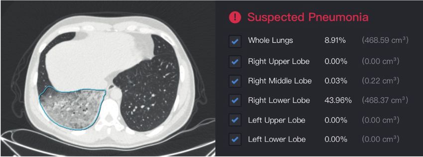
glass opacities in both lungs. A deep learning (DL)-based rRT-PCR. A negative result for SARS-CoV-2 was observed in
computer-aided diagnostic system for pneumonia, which was the first rRT-PCR test. A second consecutive SARS-CoV-2
trained with CT scans of patients with COVID-19, suggested rRT-PCR test was conducted immediately thereafter, and
this patient to have pneumonia, with the lesion volume a positive result was obtained. The patient was further
accounting for 13.3% of the whole lungs (Fig. 1). Later, confirmed with COVID-19 with additional positive rRT-PCR
throat swab specimens from the patient were tested with tests.
rRT-PCR for SARS-CoV-2. After two consecutive negative
results, a third SARS-CoV-2 rRT-PCR test confirmed the DISCUSSION
infection.
Since rRT-PCR tests serve as the gold standard method
Case 2 to confirm the infection of SARS-CoV-2, false-negative
A 36-year-old man presented with fever for 5 days (peak results could hinder the prevention and control of
body temperature: 40°C) and was admitted to the Fever the epidemic, particularly when this test plays a key
Clinic of the Beijing Haidian Hospital. The patient had reference role in deciding the necessity for continued
no direct contact history with patients with COVID-19 or isolated medical observation or discharge. Regarding the
A B
Fig. 1. Chest CT scans for 10-month-old patient in Case 1.
A. Thin-slice CT scan that shows glimpse of lesions (breathing-induced motion artifacts are heavy for patient). CT shows diffuse ill-defined
ground-glass opacities in both upper lung lobes. B. Representative of DL-based segmentation of lesions in left lung that shows overview of
automatically calculated abnormality proportions. Artificial intelligence alarms suspected pneumonia based on relatively large proportion of
abnormalities in lung. Detailed abnormality proportions in whole lungs, right upper lobe, right middle lobe, right lower lobe, left upper lobe, and
left lower lobe were calculated and listed. CT = computed tomography, DL = deep learning
506 https://doi.org/10.3348/kjr.2020.0146 kjronline.orgCase Report: False Negative Results of rRT-PCR for SARS-CoV-2 A B Fig. 2. Chest CT scans for patient in Case 2. A. Thin-slice CT scan that shows glimpse of lesions. CT shows diffuse ground-glass opacities in dependent area of right lower lobe. B. Representative of DL-based segmentation of lesions in lower lobe of right lung that shows overview of automatically calculated ratios. Artificial intelligence alarms suspected pneumonia based on relatively large proportion of abnormalities in lung. Detailed abnormality proportions in whole lungs, right upper lobe, right middle lobe, right lower lobe, left upper lobe, and left lower lobe were calculated and listed. underlying reasons for false-negative rRT-PCR results, a results. To some extent, CT features and rRT-PCR results previous published study suggested that insufficient viral were complimentary in the diagnosis of COVID-19. From a specimens and laboratory error might be responsible (3). clinical perspective, CT features could be utilized as the We speculated from these two cases that infection routes, first and immediate reference for doctors to screen the disease progression status (specimen collection timing highly suspected cases and to take necessary actions while and methods), and coinfection with other viruses might rRT-PCR serves as a confirmation tool, the results of which influence the rRT-PCR test accuracy, which should be further could be utilized later to decide the subsequent action of studied with more cases. continuing isolated treatment or discharge. Notably, our False-negative rRT-PCR results were seen in many hospital was facilitated with a DL-based computer-aided hospitals. By monitoring data collected at our hospital diagnostic system (InferRead CT Pneumonia, Infervision, from January 21 to 31, 2020, two out of ten negative Beijing, China) for pneumonia, which greatly improved cases shown by the rRT-PCR test were finally confirmed to the detection efficiency for patients highly suspected with be positive for COVID-19, yielding an approximately 20% COVID-19 by alarming the technician within 2 minutes false-negative rate of rRT-PCR. Although the false-negative when any suspected cases was found after CT examination. estimate would not be accurate until we expand the The automatic lesion segmentation on CT was also helpful observational time span and number of monitored cases, the to evaluate the progression of COVID-19 quantitatively. drawback of rRT-PCR was revealed. Clinical manifestations, With an integrated approach of DL, CT features, and rRT-PCR laboratory examination results, and chest CT features of results, the screening and treatment of COVID-19 would be patients with COVID-19 were also of great value in helping more effective. the detection and diagnosis. Thus, an integrated criterion Furthermore, we observed conflicting laboratory should be established for the diagnosis of SARS-CoV-2 examination results in these two patients. Patient in Case 2 infection. In addition to the epidemiological information, was infected only by SARS-CoV-2 and presented decreased we focused on two aspects of information: chest CT features lymphocytes and elevated C-reactive protein, consistent and laboratory examination results. with the typical tendency found in the COVID-19 cohort. Of note, approximately 96% of patients with COVID-19 In contrast, the patient in Case 1 was coinfected with presented with chest CT abnormalities, such as multiple influenza A and presented with increased lymphocytes and bilateral and peripheral ground-glass opacities and elevated C-reactive protein. The difference in laboratory consolidation (3, 4), making chest CT features essential in examination results could be a potential indicator of a recognizing COVID-19. The National Health Commission of different infection status, including SARS-CoV-2 infection China revised the diagnostic criteria in the Hubei province, alone or coinfection with other viruses, which, however, where a severe epidemic occurred (5). A new diagnostic should be further validated with more cases. type called “Clinical diagnosis” was set according to the In conclusion, we reported two false-negative results presence of pneumonia on chest CT, regardless of rRT-PCR of rRT-PCR for SARS-CoV-2 infection and mentioned the kjronline.org https://doi.org/10.3348/kjr.2020.0146 507
Li et al.

possible tandem approaches for clinical practices to ensure          Gulyaeva AA, et al. Severe acute respiratory syndrome-related
an early and accurate diagnosis of COVID-19. In addition,            coronavirus: the species and its viruses – a statement of the
                                                                     Coronavirus Study Group. bioRxiv, 2020. Available at: https://
the potential role of laboratory examination results in
                                                                     doi.org/10.1101/2020.02.07.937862. Accessed February 11,
differentiating the infection status was revealed as well.
                                                                     2020
                                                                2.   Zhu N, Zhang D, Wang W, Li X, Yang B, Song J, et al.; China
Conflicts of Interest                                                Novel Coronavirus Investigating and Research Team. A novel
The authors have no potential conflicts of interest to               Coronavirus from patients with pneumonia in China, 2019. N
disclose.                                                            Engl J Med 2020;382:727-733
                                                                3.   Xie X, Zhong Z, Zhao W, Zheng C, Wang F, Liu J. Chest CT for
                                                                     typical 2019-nCoV pneumonia: relationship to negative RT-
ORCID iDs
                                                                     PCR testing. Radiology 2020 Feb 12 [Epub]. https://doi.
Dasheng Li                                                           org/10.1148/radiol.2020200343
  https://orcid.org/0000-0002-5071-5739                         4.   Chung M, Bernheim A, Mei X, Zhang N, Huang M, Zeng
Dawei Wang                                                           X, et al. CT imaging features of 2019 novel Coronavirus
  https://orcid.org/0000-0002-8670-1961                              (2019-nCoV). Radiology 2020 Feb 4 [Epub]. https://doi.
                                                                     org/10.1148/radiol.2020200230
Chen Xia
                                                                5.   National Health Commission of the People’s Republic of China.
  https://orcid.org/0000-0002-8839-0220
                                                                     Diagnosis and treatment protocols of pneumonia caused by a
                                                                     novel coronavirus (trial version 5). Beijing: National Health
REFERENCES                                                           Commission of the People's Republic of China, 2020

1. Gorbalenya AE, Baker SC, Baric RS, de Groot RJ, Drosten C,

508                                                                          https://doi.org/10.3348/kjr.2020.0146   kjronline.org
You can also read