A rare combination of Dermoid cyst and Cystadenoma: Are collision tumors in the ovary a real entity? - SciELO
←
→
Page content transcription
If your browser does not render page correctly, please read the page content below
Clinical Case Report
A rare combination of Dermoid cyst and Cystadenoma: Are collision
tumors in the ovary a real entity?
Tushar Kalonia1 , Neha Kumari1 , Akanksha Malik1 , Arvind Kumar1 ,
Anupama Bahadur2 , Sanjeev Kishore1
How to cite: Kalonia T, Kumari N, Malik A, Kumar A, Bahadur A, Kishore S. A rare combination of Dermoid
cyst and Cystadenoma: Are collision tumors in the ovary a real entity?. Autops Case Rep [Internet]. 2021;11:e2021249.
https://doi.org/10.4322/acr.2021.249
ABSTRACT
Collision tumors have been reported in various organs like the gastrointestinal tract, lung, skin, adrenals, central nervous
system, lymph nodes, uterus, but are rarely seen in the ovary. Collision tumors are two histologically distinct neoplasms
in the same organ without any intermixture between them. Here we present a case of a collision tumor of the ovary
represented by a mucinous cystadenoma and teratoma. It is imperative for a surgical pathologist to correctly diagnose
the collision tumor components and differentiate them from mixed tumors as it will dictate the appropriate treatment
based on the individual biological aggressiveness of each component.
Keywords
Cystadenoma, Mucinous; Ovary; Teratoma
INTRODUCTION
Collision tumor refers to the simultaneous Mature cystic teratoma co-existing with a mucinous
coexistence of two distinct tumors in the same tissue cystadenocarcinoma is infrequently encountered, with
without any transition zone or mixing interface. only a handful of cases reported to date.
Collision tumors have been described in various Here we report a collision tumor of the ovary
body organs, e.g., liver, bone, kidney, brain, lung. with the simultaneous association of dermoid cyst and
However, collision tumors in the ovary are rarely seen.1 benign mucinous cystadenoma.
Case reports of collision tumors in the ovary were
reported depicting the admixture of granulosa and
CASE REPORT
cystadenocarcinoma, granulosa and teratoma, serous
adenocarcinoma, and steroid cell tumor.2-4 A 36-year-old female was referred to the
The cystadenomas account for 30% of the ovarian Gynecology Department complaining of dysmenorrhea
tumors, and the mature cystic teratomas comprise 10-20% for the past 3 months associated with intermenstrual
of the ovarian tumors.5 However, together only 2-10% of spotting. The abdominal examination revealed a freely
teratomas are associated with mucinous cystadenomas.6 mobile mass in the right lower abdominal quadrant.
1
All India Institute of Medical Sciences, Rishikesh,Tushar Kalonia, Deapartment of Pathology and Laboratory Medicine. Rishikesh,
Deharadun, Uttarakhand, India
2
All India Institute of Medical Sciences, Rishikesh, Tushar Kalonia, Department of Gynecology and Obstetrics. Rishikesh, Deharadun,
Uttarakhand, India
Copyright: © 2021 The Authors. This is an Open Access article distributed under the terms of the Creative
Commons Attribution License, which permits unrestricted use, distribution, and reproduction in any medium,
provided the original work is properly cited.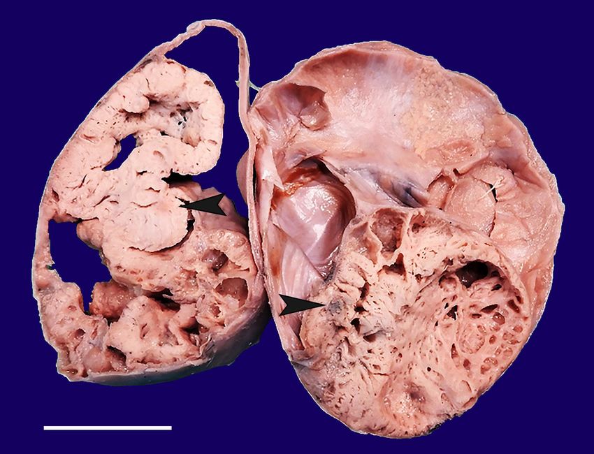
A rare combination of Dermoid cyst and Cystadenoma: Are collision tumors in the ovary a real entity?

The pelvic CT scan revealed cystic lesions on bilateral                  when normal tissue intervenes between both tumors,
adnexa, the left side with 7.4x6.2x6.1 cm, and the                       and there is no histological admixture at the interface.
right side with 14.5x17.4x18.5 cm.Thus, the bilateral                    The occurrence of a transition zone between the two
adnexal masses were excised along with hysterectomy.                     tumors makes it difficult to differentiate between two
The cut surface of bilateral ovaries shows solid and                     colliding tumors or a true mixed tumor.3,7
cystic areas with focal papillaroid areas along with an                       The histogenesis of the ovarian mucinous
occasional greyish brown, firm area, which revealed a                    cystadenoma has not yet been fully clarified. There has
teratomatous component (Figure 1).                                       been the suggestion of a surface epithelial metaplasia
     The Paraffin-embedded samples from left adnexa                      origin or a teratomatous origin. The ultrastructure
stained with H&E stain showed solid cystic ovarian                       studies and mucin histochemical studies have supported
parenchyma partially lined by stratified squamous                        the surface epithelial metaplasia theory.8,9 The frequent
epithelium along with the presence of pilosebaceous                      co-existence of mature cystic teratoma and mucinous
units, cystic spaces filled with keratinous debris with                  cystadenoma is the basis for the teratoma theory.
pigment-laden macrophages in its wall. A diagnosis                       Germ cell tumors comprise approximately 30% of the
of mature cystic teratoma was rendered on the left                       primary ovarian tumors, and of these, 95% are mature
adnexa. Right adnexa showed ovarian stroma and                           cystic teratomas. The most common histological
many cysts like spaces lined by simple non-stratified                    combination of collision tumors in the ovary is the
columnar cells with abundant intracellular mucin and                     coexistence of teratoma with mucinous tumors.10
distinct areas of cartilaginous differentiation, pilo                    The main differentiating feature between collision
sebaceous units, and keratinous debris separated                         tumor and teratoma with cystadenoma component
by ovarian parenchyma. (Figure 2 A to D). Hence a                        is that the former presents a distinct collar of normal
diagnosis of a mature cystic teratoma with mucinous                      ovarian parenchyma separating the teratoma and
cystadenoma was given However ,intraoperative frozen                     cystadenoma component, while the latter entity shows
sections examination was reported as suggestive of                       an intermixture of both the histological components.
mucinous cystadenoma on both sides. (Figure 2C)                          Various hypothesis has been proposed for the existence
                                                                         of collision tumors in the ovary. The first hypothesis
                                                                         is that the first tumor’s presence causes changes in
DISCUSSION                                                               the tissue microenvironment, making it conducive for
                                                                         the seeding of a metastatic tumor or development of
    The origin of teratomas is still widely disputed. The                the second tumor. The second theory proposes that
most accepted theory is that they arise from primordial                  the existence of two primary tumors is just a chance
germ cells.6 The diagnosis of a collision tumor is made                  occurrence. This theory was supported by Vang et al.,11
                                                                         who showed the ovarian mucinous tumors associated
                                                                         with mature cystic teratomas exhibited morphological
                                                                         and immunophenotypic diversity. The third theory
                                                                         proposes that both the primary coexisting tumors have
                                                                         a common stem cell origin.12
                                                                              The evidence of a clonal relationship could be
                                                                         provided by additional molecular analyses demonstrating
                                                                         shared genetic changes in the mucinous tumor and
                                                                         adjacent conventional teratomatous elements. Thus,
                                                                         supporting the theory of a teratomatous origin for
                                                                         these mucinous tumors.
                                                                              In a study by Fuji et al. 13 the homozygous
                                                                         genetic patterns similar to those of the teratomatous
                                                                         components were shown in mucinous tumors arising
Figure 1. Gross view of the cut surface of the tumor                     together with mature cystic teratomas, and it was
with cystic and greyish-white solid area(arrowheads)                     suggested that these mucinous tumors developed from
that represent teratomatous component in microscopy.                     pre-existing mature cystic teratomas.

2-5                                                                                          Autops Case Rep (São Paulo). 2021;11:e2021249
Kalonia T, Kumari N, Malik A, Kumar A, Bahadur A, Kishore S
Figure 2. Photomicrographs of the tumor. A – Section is lined by keratinous stratified squamous epithelium. The
sub epithelium shows lobules of glands (H&E,100X); B – The section shows a cartilage formation (H&E,200X);
C – Part of the cyst cavity is lined by benign columnar epithelium with moderate amount of mild eosinophilic
cytoplasm. Tangential cut of the glands are also noted (Frozen Section,100X); D – The section is lined by columnar
glandular epithelium having basally located nucleus with bland nuclear feature and moderate amount of eosinophilic
cytoplasm(H&E,100X).
Although not all mucinous tumors are of germ- CONCLUSION
cell origin, mucinous elements in dermoid cysts may
be of teratomatous origin as they are more likely to Collision tumors involving ovaries are rare.
be intestinal rather than Mullerian in differentiation.14 The surgeon should establish good perioperative
Another study published stated that molecular communication with the pathologists and whenever
findings which are significant allelic imbalance for possible, a specialized gynecology pathologist is
microsatellite markers, indicating homozygosity rather advisable since a thorough grossing and recognition of
than heterozygosity for chromosomal polymorphisms close differential diagnosis lead the correct diagnosis.
in some ovarian mucinous cystadenomas associated
with mature cystic teratomas, were consistent with
a germ cell origin. 15 Okada et al. 16 demonstrated REFERENCES
a possible association between dermoid cyst and
1. Bostanci MS, Yildirim OK, Yildirim G, et al. Collision
multiseptated cyst. Multiseptated cyst contains fatty
tumor: dermoid cysts and mucinous cystadenoma in
tissue foci. It was also stated that recognizing of the the same ovary and a review of the literature. Obstet
potential for the coexistence of these two neoplasms in Gynecol Cases Rev. 2015;2(2):1-3. http://dx.doi.
the same ovary is essential to make a correct diagnosis. org/10.23937/2377-9004/1410031.
Autops Case Rep (São Paulo). 2021;11:e2021249 3-5A rare combination of Dermoid cyst and Cystadenoma: Are collision tumors in the ovary a real entity?
2. Ozbey C, Erdogan G, Pestereli HE, Simsek T, dx.doi.org/10.1111/j.1440-1827.1995.tb03480.x.
Karaveli S. Serous papillary adenocarcinoma and PMid:7581934.
adult granulosa cell tumor in the same ovary. An
unusual case. APMIS. 2005;113(10):713-5. http:// 10. Tang P, Soukkary S, Kahn E. Mature cystic teratoma of the
dx.doi.org/10.1111/j.1600-0463.2005.apm_255.x. ovary associated with complete colonic wall and mucinous
PMid:16309432. cystadenoma. Ann Clin Lab Sci. 2003;33(4):465-70.
PMid:14584762.
3. Dgani R, Rozenman D, Lifschitz-Mercer B. Granulosa cell
tumor arising in an ovary with mature cystic teratoma. 11. Vang R, Gown AM, Zhao C, et al. Ovarian mucinous
Int J Gynaecol Obstet. 1993;41(3):287-9. http://dx.doi. tumors associated with mature cystic teratomas.
org/10.1016/0020-7292(93)90562-B. PMid:8102994. Am J Surg Pathol. 2007;31(6):854-69. http://dx.doi.
org/10.1097/PAS.0b013e31802efb45. PMid:17527072.
4. Nirenberg A, Ostor AG, Quinn MA. Collision tumor:
serous adenocarcinoma and steroid cell tumor of 12. Brandwein-Gensler M, Urken M, Wang B. Collision
the ovary. Pathology. 1992;24(2):60-2. http://dx.doi. tumor of the thyroid: a case report of metastatic
org/10.3109/00313029209063624. PMid:1322520. liposarcoma plus papillary thyroid carcinoma. Head
5. Kumar V, Abbas A, Fausto N, Aster J. Robbins and Neck. 2004;26(7):637-41. http://dx.doi.org/10.1002/
Cotran pathologic basis of disease. Philadelphia, Pa: Els hed.20024. PMid:15229907.
Saunders; 2010.
13. Fujii K, Yamashita Y, Yamamoto T, et al. Ovarian
6. Talerman A, Vang R. Germ cell tumor of the ovary. mucinous tumors arising from mature cystic teratomas: A
In: Kurman RJ, Ellenson LH, Ronnett BM. Blaustein’s molecular genetic approach for understanding the cellular
pathology of the female genital tract. 6th ed. Springer; origin. Hum Pathol. 2014;45(4):717-24. http://dx.doi.
2011. p. 847-907. org/10.1016/j.humpath.2013.10.031. PMid:24485845.
7. Matias-Guiu X, Caixas A, Costa I, Cabezas R, Prat J. Compound 14. Russel P, Farnsworth A, Russel P, Farnsworth A. Surgical
medullary-papillary carcinoma of the thyroid: true mixed pathology of the ovaries 2nd ed. New York: Churchill
versus collision tumor. Histopathology. 1994;25(2):183-5. Livingstone; 1987. 273-8.
http://dx.doi.org/10.1111/j.1365-2559.1994.tb01578.x.
PMid:7982684. 15. Magi-Galluzi C, O’Connell JT, Neffen F. Are mucinous
cystadenomas of the ovary derived from germ cells. A
8. Fenoglio CM, Ferenczy A, Richart RM. Mucinous
genetic analysis. Mod Pathol. 2001;14:140A.
tumors of the ovary. Ultrastructural studies of mucinous
cystadenomas with histogenetic considerations. 16. Okada S, Ohaki Y, Ogura J, Ishihara M, Kawamura T,
Cancer. 1975;36(5):1709-22. http://dx.doi. Kumazaki T. Computed tomography and magnetic
org/10.1002/1097-0142(197511)36:53.0.CO;2-7. PMid:1192360.
coexisting with surface epithelial tumors in the same
9. N o m u r a K . M u c i n h i s t o c h e m i s t r y o f o v a r i a n ovary. J Comput Assist Tomogr. 2004;28(2):169-73.
mucinous cystadenomas expressing gastrointestinal http://dx.doi.org/10.1097/00004728-200403000-00003.
characteristics. Pathol Int. 1995;45(6):430-5. http:// PMid:15091118.
This study was carried out at AIIMS, Rishikesh, Uttarakhand, India
Authors’ contributions: Tushar Kalonia was responsible for the manuscript preparation. Neha Kumari for its
conceptualization, Akanksha Malik for the literature research. Arvind Kumar for the final editing, Anupama
Bahadur for the critical revision, and Sanjeev Kishore for the design of the study.
Ethics statement: The authors retain informed consent and authorization to publish the data, and the manuscript
Is by the institutional policy.
Conflict of interest: The authors have no conflict of interest to declare.
Financial support: The authors declare that no financial support was received.
Submitted on: October 9th, 2020
Accepted on: December 2nd, 2020
4-5 Autops Case Rep (São Paulo). 2021;11:e2021249Kalonia T, Kumari N, Malik A, Kumar A, Bahadur A, Kishore S Correspondence Dr Arvind Kumar Department of Pathology and Laboratory Medicine, All India Institute of Medical Sciences, Rishikesh 249203, Uttarakhand, India Phone: +919410994467 drarvindkumar10@gmail.com Autops Case Rep (São Paulo). 2021;11:e2021249 5-5
You can also read