Minimal Change Disease and Phenylketonuria in an Adult Patient: The Two Sides of Protein Homeostasis
←
→
Page content transcription
If your browser does not render page correctly, please read the page content below
European Journal
                                                                      of Case Reports in
                                                                  Internal Medicine

          Minimal Change Disease and Phenylketonuria in an Adult Patient:
                                       The Two Sides of Protein Homeostasis
                         Cristina Pires Correia, Inês Neves, Paulo Chaves, Teresa Cardoso, Roberto Silva, Jorge Almeida
                                        Internal Medicine Department, Centro Hospitalar São João, Porto, Portugal

                                 Doi: 10.12890/2020_001821 - European Journal of Case Reports in Internal Medicine - © EFIM 2020

Received: 17/06/2020
Accepted: 30/06/2020
Published: 31/07/2020

How to cite this article: Pires Correia C, Neves I, Chaves P, Cardoso T, Silva R, Almeida J. Minimal change disease and phenylketonuria in an adult patient:
two sides of protein homeostasis. EJCRIM 2020;7: doi:10.12890/2020_001821.

Conflicts of Interests: The Authors declare that there are no competing interests.
This article is licensed under a Commons Attribution Non-Commercial 4.0 License

ABSTRACT
Phenylketonuria (PKU), the most common inborn error of amino acid metabolism, has been associated with an increased risk of renal
impairment, the reason for which is not fully understood. We report the case of a 33-year-old female patient diagnosed with PKU in
adulthood after suspicion of the disease in her daughter. The diagnostic approach revealed proteinuria, and minimal change disease (MCD)
was identified. Therapeutic measures and follow-up are discussed. The possible link between these two disorders is interesting. Attenuated
developmental delay of untreated PKU could be explained by the decreased accumulation of phenylalanine secondary to proteinuria. On
the other hand, MCD may be a possible, although as yet unreported, pathophysiological mechanism explaining the renal repercussions that
have been found in adult PKU patients, who should be screened for proteinuria.

LEARNING POINTS
• Phenylketonuria (PKU) is associated with renal impairment so patients with PKU should be periodically screened for proteinuria
   because podocytopathies, such as minimal change disease (MCD), may be an underlying misdiagnosed condition.
• MCD in adults demands tight surveillance in case of severe complications including nephrotic syndrome, haematuria and acute kidney
   injury, and although remission with glucocorticoid therapy is typical, 50–75% of patients will relapse.
• MCD may be a previously unreported cause of proteinuria and renal involvement in adult patients with PKU, where control of
   hypertension and moderation of protein overload are essential.

KEYWORDS
Minimal change disease (MCD), proteinuria, nephrotic syndrome, phenylketonuria (PKU), phenylalanine

CASE DESCRIPTION
We describe a 33-year-old female patient evaluated by an internal medicine consultant due to the possibility of hereditary and/or
metabolic disease after global developmental delay was detected in her 6-month-old daughter. Maternal PKU was suspected because of
the association between global psychomotor developmental delay, microcephaly and a family history of cognitive impairment. A serum
aminogram uncovered a remarkably elevated phenylalanine (Phe) level of 1,663 nmol/ml (normal range 50–90 nmol/ml) with reduced
tyrosine, confirming the diagnosis of classic PKU. The patient’s medical history included non-medicated arterial hypertension and learning
difficulties in school.

DOI: 10.12890/2020_001821                                                                European Journal of Case Reports in Internal Medicine © EFIM 2020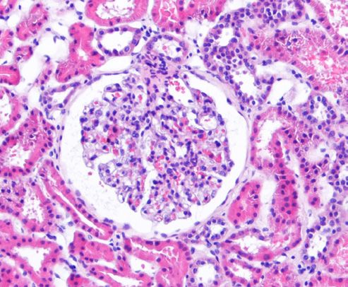
European Journal
of Case Reports in
Internal Medicine
On physical examination, the patient presented with peripheral oedema and a BMI of 28 kg/m2, without any other significant findings.
Neuropsychological evaluation revealed alterations in executive functions as well as visual-perception and constructive impairment. Brain
T2-weighted magnetic resonance imaging (MRI) showed widespread high-intensity areas in the deep white matter, especially the bilateral
occipital lobes (Fig. 1). Laboratory findings including a complete blood count, renal and hepatic function were unremarkable.
Figure 1. Axial T2-weighted MR images showing
the symmetrical increase in the T2-weighted
signal in the periventricular white matter,
especially in bilateral occipital lobes, common in
phenylketonuria
The patient started antihypertensive medication and dietary restriction of Phe, but maintained poor therapeutic compliance, with Phe
concentrations above the recommended level of 120–600 µmol/l. She also maintained uncontrolled hypertension, requiring three
antihypertensive drugs (including an ACE inhibitor), while 24-hour urine documented proteinuria of 1.4–1.6 g/day, with normal levels of
urea (10 mg/dl; normal 6–23 mg/dl), creatinine (1.01 mg/dl; normal 0.50–1.20 mg/dl) and albumin (3.7 g/dl; normal 3.5–5.2 g/dl). A complete
infectious and autoimmune study was requested because of the proteinuria.
Further serological studies including a viral hepatitis panel, HIV, anti-nuclear antibody, rheumatoid factor, anti-Ds-DNA, anti-ANCA and
anti-citrullinated peptide antibody and urinary eosinophils were negative. C3/C4 were normal. Serum protein electrophoresis revealed
hypogammaglobulinemia. Phospholipase A2 receptor antibodies were negative. Renal ultrasound did not show any relevant changes.
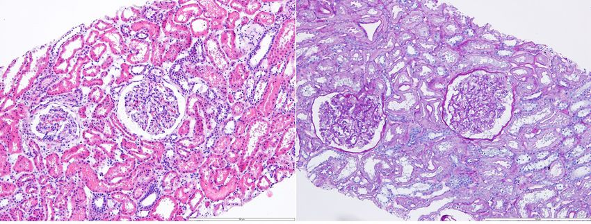
Therefore, in light of the still unexplained proteinuria, a renal biopsy was performed which exhibited 20 glomeruli without mesangial
expansion or increased cellularity by light microscopy and the absence of complement or immunoglobulin deposits by immunofluorescence
microscopy (Figs. 2 and 3). Electron microscopy revealed diffuse effacement of the podocyte foot processes, supporting the diagnosis of
minimal change disease (MCD) (Fig. 4).
The treatment approach included optimising hypertension control, reinforcing the dietary restriction of Phe, and stopping consumption of
synthetic amino acid (AA) formulas. In light of the normal renal function and mild level of proteinuria, our patient did not immediately start
glucocorticoid therapy but was kept under tight surveillance for 18 months. Although further studies revealed proteinuria of less than 1 g/
day, a glucocorticoid regimen is now being planned.
DISCUSSION
Phenylalanine hydroxylase (PAH) deficiency, otherwise known as phenylketonuria (PKU), is the most frequent inborn error of amino acid
metabolism. It is an autosomal recessive disorder which if left untreated results in global developmental delay and severe intellectual
impairment [1].
We report the unexpected diagnosis of MCD in an adult patient with PKU, after proteinuria was investigation in a complex diagnostic
approach. The association of these two rare disorders is unique and although it may be a coincidence, patients with PKU are known to have
an increased risk of renal impairment, the reason for which is not fully understood [2, 3]. Considering the difficulty in studying longitudinal
outcomes in rare diseases, the causes of renal disease cannot always be thoroughly investigated. Moreover, renal disease in these patients
could easily be missed, especially in the early stages.
DOI: 10.12890/2020_001821 European Journal of Case Reports in Internal Medicine © EFIM 2020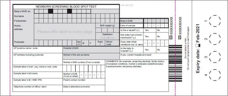
European Journal
                                                                                   of Case Reports in
                                                                              Internal Medicine

   Figure 2. Renal biopsy revealing renal cortex with slight lesions indicating acute tubular damage and without other significant changes, including the absence of complement or
   immunoglobulin deposits by immunofluorescence microscopy (IgA, IgG, IgM, C3c, C1q and fibrinogen) (left: HE 100×, right: PAS 100×)

                                                                                            Figure 3. Light microscopy showing glomeruli with open capillary loops and no evidence of cellular
                                                                                            crescents, fibrinoid necrosis or endocapillary hypercellularity

    Figure 4. Electron microscopy disclosing diffuse effacement of the podocyte foot processes, the typical histological lesion of minimal change disease

DOI: 10.12890/2020_001821                                                                                 European Journal of Case Reports in Internal Medicine © EFIM 2020
European Journal
of Case Reports in
Internal Medicine
We highlight the fact that although proteinuria is a common condition associated with several disorders, in our case a specific disease was
diagnosed with significant implications for treatment and follow-up. Furthermore, it is easy to test for proteinuria and these patients should
be periodically screened because podocytopathies like MCD may be an associated misdiagnosed condition.
A few published reports have described somatic comorbidities in PKU patients, such as renal impairment, proteinuria, arterial hypertension
and obesity [2, 3]. According to the literature, PKU is a potential cause of tubulointerstitial disease associated with increased oxidative stress,
local damage due to high Phe excretion and the highly concentrated AA used as a complement in Phe-restricted diets, which lead to greater
renal workload, thereby affecting kidney function. Indeed, after MCD diagnosis, the suspension of synthetic AA consumption resulted in
reduced proteinuria in our patient [2, 3].
This case is remarkable both because of the rarity of a delayed PKU diagnosis after the implementation of the newborn blood spot screening
programme (Fig. 5), and also because of the few signs of intellectual impairment presented by this adult patient despite identification of
the classic form of PKU. The attenuated developmental delay due to untreated PKU in this patient could be explained by the decreased
accumulation of Phe secondary to proteinuria associated with MCD. Plasma and urine amino/organic acid analyses should be carried out in
adult patients with learning difficulties and unexplained neuropsychological manifestations.
Figure 5. Newborn blood spot screening card, including phenylketonuria (since 1966) congenital hypothyroidism (since 1977), cystic fibrosis (since 1988) and various inborn errors
of metabolism (since 2002)
MCD may be secondary to drug intake, neoplasms, infections and allergies, or else primary, which is probably the case in this patient[4].
The pathogenesis underlying MCD is unclear. MCD is a major cause of nephrotic syndrome (approximately 90%) in children and in a
minority of adults (approximately 10%) [4]. We underline the early identification of MCD in this patient, explaining the mild proteinuria levels
documented so far. Moreover, we highlight the importance of surveillance for serious manifestations of MCD, such as nephrotic syndrome,
haematuria and acute kidney injury [4, 5].
We emphasise the importance of blood pressure control and stopping consumption of synthetic AA formulas. The mainstay of therapy for
MCD is prednisolone, with >70% of adults, particularly younger patients, achieving complete remission within 18 months [4, 5]. Although
5–10% of untreated patients can spontaneously remit within 3 months, according to the literature, when persistent nephrotic syndrome is
evident, glucocorticoid therapy should not be withheld due to possible thrombotic and infectious complications [4, 5].
PKU presents with unclear multisystemic manifestations which are currently under study, especially in adult patients. Investigation of the
link between PKU and MCD could provide better understanding of the pathophysiology of both disorders as protein homeostasis is affected
in each condition. Moreover, there are therapeutic and follow-up implications for both disorders and affected patients should be screened
for proteinuria.
DOI: 10.12890/2020_001821 European Journal of Case Reports in Internal Medicine © EFIM 2020European Journal
of Case Reports in
Internal Medicine
REFERENCES
1. van Wegberg AMJ, MacDonald A, Ahring K, Bélanger-Quintana A, Blau N, Bosch AM, et al. The complete European guidelines on phenylketonuria: diagnosis and treatment.
Orphanet J Rare Dis 2017;12:162.
2. Hennermann JB, Roloff S, Gellermann J, Vollmer I, Windt E, Vetter B, et al. Chronic kidney disease in adolescent and adult patients with phenylketonuria. J Inherit Metab Dis
2013;36:747–756.
3. Burton BK, Bradford Jones K, Cederbaum S, Rohr F. Prevalence of comorbid conditions among adult patients diagnosed with phenylketonuria. Mol Genet Metab 2018:125;228–
234.
4. Vivarelli M, Massella L, Ruggiero B, Emma F. Minimal change disease. Clin J Am Soc Nephrol 2017;12:332.
5. Lee H, Yoo KD, Oh YK, Kim DK, Oh KH, Joo KW, et al. Predictors of relapse in adult-onset nephrotic minimal change disease. Medicine (Baltimore) 2016;95(12):e3179.
DOI: 10.12890/2020_001821 European Journal of Case Reports in Internal Medicine © EFIM 2020You can also read