Primary Hepatic Liposarcoma Diagnosed by Dynamic Computed Tomography
←
→
Page content transcription
If your browser does not render page correctly, please read the page content below
J Radiol Sci 2011; 36: 113-117

Primary Hepatic Liposarcoma Diagnosed by Dynamic
Computed Tomography
Shin-Chun Fang1 Cheng -Kuang Chang1 Yu-Chun Lin2 Chih-Yung Yu1 Chang -Hsien Liu1
Guo -Shu Huang1

Department of Radiology1, Department of Pathology2, Tri-Service General Hospital, Taipei, Taiwan

ABSTRACT
        Primary hepatic liposarcoma is extremely rare and only a few cases have been reported in the English litera-
   ture. Here, we report a case of myxoid-type primary hepatic liposarcoma in a 63-year-old man presenting with low
   back pain. Before surgery, dynamic abdominal computed tomography (CT) revealed a well-marginated mass in the
   left lobe of the liver with a fat component. There was no enhancement of the fat component and only mild delayed
   enhancement of the soft-tissue component after the administration of contrast. The pathological diagnosis was a
   myxoid liposarcoma of liver. The patient was doing well at least five years after a left segmentectomy of the liver.
   A CT study was used preoperatively in the diagnosis of a hepatic liposarcoma and was helpful in differentiating the
   liposarcoma from other fat-containing tumors.

      Fat-containing tumors of the liver are uncommon enti-              L4/5 disk with compression of the right nerve root. However,
ties. Among these, hepatic liposarcoma is an exceedingly                 a fat-containing tumor was incidentally found in the left
rare tumor. Metastatic spread of soft tissue liposarcoma is              lobe of the liver. His past history included hypertension and
relatively common, but the liver is involved in only 10%                 diabetes mellitus, both controlled with medication for 10
case [1-3]. Only a few cases of primary liposarcoma have                 years. He had no other risk factors for liver disease, such
been reported (Table 1). Imaging modalities are used to                  as viral hepatitis or cirrhosis. A physical examination of the
analyze the characteristics of the tumor. We report a case               abdomen showed a soft and flat abdomen, with no palpable
of primary hepatic liposarcoma followed for more than                    mass or tenderness. Laboratory tests showed blood urea
five years after surgery and discuss the possible preop-                 nitrogen = 43 mg/dL (normal: 6-20 mg/dL) and creatinine
erative diagnosis of this type of hepatic tumor on dynamic               = 1.7 mg/dL (normal: 0.7-1.2 mg/dL). Other laboratory data,
computed tomography.                                                     including liver function test, α-fetoprotein, and other tumor
                                                                         markers, were within the reference ranges. Viral markers
                                                                         for hepatitis B and C were negative. Dynamic computed
Case report                                                              tomography (CT) of the abdomen revealed a well-defined
                                                                         encapsulated fat-containing mass of about 8 cm × 8.5 cm
     A 63-year-old man presented with low back pain                      × 9 cm in the lateral segment of the left lobe of the liver
radiating to the thigh for two months. Magnetic resonance                (Fig. 1). The mass showed a predominant fat component and
imaging (MRI) of the lumbar spine showed bulging of the                  some soft-tissue component. During dynamic study, the fat

 Correspondence Author to: Chang-Hsien Liu
 Department of Radiology, Tri-Service General Hospital, Taipei, Taiwan
 No. 325, Sec. 2, Cheng-Kung Road, Taipei, 114, Taiwan

J Radiol Sci June 2011 Vol.36 No.2                                                                                                   113
Dynamic Ct in diagnosing hepatic liposarcoma
component within the lesion showed no significant enhance- network (Fig. 2). Serial follow-ups with ultrasound and CT
ment from the arterial phase to the delayed phase. The soft- over five years revealed no evidence of tumor recurrence or
tissue component showed mild delayed enhancement. These metastasis.
characteristics are suggestive of a hepatic liposarcoma. An
exploratory laparotomy with lateral segmentectomy of the
left lobe of the liver was subsequently performed. The Discussion
gross specimen showed a yellow–brown–gray tumor with
a soft, gelatinous consistency. Microscopically, sections of Lipomatous tumors of the liver are not frequently
tumor indicated a myxoid liposarcoma, characterized by encountered in the clinical context. Hepatic liposarcoma is
a hypocellular myxoid lesion mixed with mature adipose one of the exceedingly rare lipomatous tumors. Most liposa-
tissue, lipoblasts, and a rich chicken-wire-type capillary rcomas in the liver are metastases, which usually originate
Table 1. Location, imaging findings, type, and prognosis of previously reported cases of primary liposarcoma in
adults
Clinical signs
Author/year Age/sex Location Diameter (cm) CT findings Type Prognosis
and symptoms
Wolloch/1973 [4] 22/F right lobe NAa NA NA myxoid survival 46 days
Kim/1985 86/M liver NA RUQb pain, NA NA NA
capsule massive ascites,
and pleural
effusion
Kim/1987 [12] 30/F left lobe 14 × 10 × 6 abdominal pain CTc: exophytic dedifferentiated tumor-free for at
least 10 months
Aribal/1993 48/F hilum NA intermittent NA NA NA
right-sided
abdominal pain
Khan/2000 [9] 50/M right lobe 23.9 × 19 × 14 right CT: fat density, NA NA
hypochondrium exophytic
pain for 6 months
Nelson/2001 [1] 54/F right and 16 cm diameter abdominal CT: solid mass, myxoid postoperative
left lobes (left), pain, nausea extensive bleeding and
3 cm diameter and vomiting, hemorrhage in death
(right) abdominal recurrent tumor
distention,
weight loss
Kuo/2006 [5] 61/F right lobe 11 × 11 × 13 fever, nausea, CT: cystic lesion myxoid two recurrences
vomiting, and bilateral intra- and cervical
jaundice, body- hepatic ducts were metastases;
weight loss compressed survival 27
months
Kim/2007 [6] 63/F left lobe 7 × 6 × 4.3 asymptomatic, CT: lobulated mass; well differenti- tumor-free for
abnormal liver fatty; small area of ated at least 8 months
function test nodular enhance-
ment
Fang/current 63/M left lobe 8 × 8.5 × 9 asymptomatic, CT: well-defined myxoid tumor-free for
incidental finding encapsulated, at least 5 years
fatty tumor; mild
delayed enhance-
ment in soft-tissue
component within
the lesion
aNA: not available
bRUQ: right upper quadrant of the abdomen
cCT: computed tomography
114 J Radiol Sci June 2011 Vol.36 No.2Dynamic Ct in diagnosing hepatic liposarcoma
Figure 1
1a 1b
1c 1d
Figure 1. a-d. Dynamic contrast CT of liver without contrast, in the arterial phase, portal venous phase, and the three-
minute delayed phase. a. A well-defined encapsulated low-attenuation mass (arrowhead) with a negative Hounsfield-unit
component (white arrow) in the left lateral lobe of the liver. b-d. On images from the arterial phase to the delayed phase,
the fat component within the lesion shows no significant contrast enhancement, and the soft-tissue component (black arrow)
shows mild delayed enhancement.
from the extremities or retroperitoneum [1-3]. The first symptoms include fever, nausea and vomiting, jaundice,
published case of primary hepatic liposarcoma we found and body weight loss [7, 8]. In the current study, the patient
in the English literature was reported by Wolloch et al. in had no clinical abdominal symptoms. Early diagnosis is
a 22-year-old man in 1973 [4]. According to the literature, difficult without an imaging study because the liver is a
primary hepatic liposarcoma occurs most often in adults, in silent organ in the abdomen; thus, a tumor may not induce
the fifth and sixth decades of life (Table 1) [1, 5, 6]. It has symptoms until it becomes large. Furthermore, there are
also been described in children, who had a liposarcoma in few tumor-specific characteristics by which liposarcoma
the hepatic hilum, presenting as obstructive jaundice and can be recognized. In previous cases, patients have had
abdominal pain [1, 7, 8]. almost normal liver function tests and tumor markers were
The clinical presentation of primary hepatic liposar- within physiologically normal ranges.
coma varies, and patients may be free of symptoms. Right- Fat tissue appears with unique characteristics on every
sided abdominal pain is the most common symptom. Other imaging modality, and these are used in the diagnosis and
J Radiol Sci June 2011 Vol.36 No.2 115Dynamic Ct in diagnosing hepatic liposarcoma
Figure 2
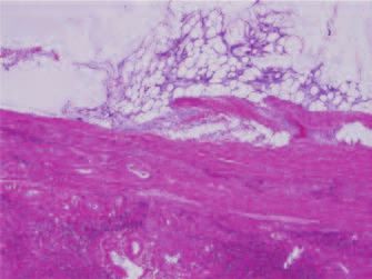
2a 2b
Figure 2. a. The hepatic tumor appears as a fatty tumor with a myxoid background (hematoxylin/eosin stain, × 40). b.
High-power magnification of the fatty tumor, with lipoblasts in the myxoid background indicating a myxoid liposarcoma
(hematoxylin/eosin stain, × 1000)
differentiation of tumors. Ultrasound is the most conven- Additionally, the mass shows mild enhancement of the soft
ient tool for screening the solid organs in the abdomen. The tissue component in the delayed phase image. It strongly
fat component generally appears hyperechogenicity on a suggests the diagnosis of hepatic liposarcoma. The image
hepatic screen. On CT, fat component shows low attenua- appearance is unlike hepatic angiomyolipoma and hepa-
tion, in a range of –10 to –100 HU [3]. Tumors with large tocellular carcinoma, showing strong enhancement in the
amounts of fat, such as lipomas or liposarcomas, usually arterial phase, then becoming isodense or hypodense in the
demonstrate typical negative HU characteristics. In our portal phase or delayed phase [3, 6].
patient, the fat component within the lesion showed nega- Five major histological categories of liposarcoma have
tive HU values, from –20 to –70. On MRI, fat displays a been reported: (1) myxoid, (2) round cell, (3) well differenti-
hyperintense signal on T1-weighted images [2]. On in-phase ated, (4) dedifferentiated, and (5) pleomorphic [11]. Myxoid,
images, the signal from fat and water are additive, while on well-differentiated, and dedifferentiated liposarcomas have
out-of-phase images the fat signal is subtracted from the been recorded in the English literature [1, 4-6]. Myxoid
water signal. Lesions containing fat and water therefore liposarcoma is more common than the other two types.
show a loss of signal intensity on out-of-phase images when Curative surgery is still the most effective way to manage
compared with in-phase images. the disease [5]. In our case, presence of the adipose tissue in
Fat-containing tumors of the liver are not common. histology is compatible with low attenuated area in tumor
They can contain macroscopic fat or intracellular lipid [2, 3]. on CT image. The mild enhanced soft tissue component
Primary liposarcoma belongs to the “macroscopic fat” cate- after contrast administration could be correlated with
gory, although it also contains a soft-tissue component. the picture of lipoblasts and capillary network in myxoid
Other lesions containing macroscopic fat include angi- matrix. The prognosis of hepatic liposarcoma varies with
omyolipoma, lipoma, hepatocellular carcinoma with fatty the histological type. The well-differentiated type is consid-
change…etc [3, 10]. Correct detection and differentiation ered to be a low-grade malignancy. The myxoid type has an
of macroscopic fat-containing tumors is possible with CT. intermediate prognosis. Some patients are free of disease
Hepatic lipoma, angiomyolipoma, and hepatocellular carci- for months and years, but a few patients die of the complica-
noma should be included in the differential diagnosis of tions caused by recurrent tumor [5-6, 11]. A sonographic
liposarcoma. examination is recommended to detect recurrance.
In our patient, the mass shows fat and soft tissue In conclusion, primary liposarcoma of the liver is
components. The finding of a hepatic mass with two extremely rare. It usually has a heterogeneous hypodense
components narrows the differential diagnosis to liposa- appearance on CT, and can be preoperatively differentiated
rcoma, angiomyolipoma and hepatocellular carcinoma. from other fat-containing tumors with dynamic CT.
116 J Radiol Sci June 2011 Vol.36 No.2Dynamic Ct in diagnosing hepatic liposarcoma
Reference 7. Soares FA, Magnani Landell GA, Peres LC, Olivera
MA, Vincente YAMVA, Tone LG. Liposarcoma of
1. Nelson V, Fernandes NF, Woolf GM, Geller SA, Petrovic hepatic hilum in childhood: report of a case and review
LM. Primary liposarcoma of the liver: a case report and of literature. Med Pediatr Oncol 1989; 17: 239-243
review of the literature. Arch Pathol Lab Med 2001; 125: 8. Wright NB, Skinner R, Lee RE, Craft AW. Myxoid
410-412 liposarcoma of the portal hepatis in childhood. Pediatr
2. Basaran C, Karcaaltincaba M, Akata D, et al. Fat- Radiol 1993; 23: 620-621
containing lesions of the liver: cross-sectional imaging 9. Khan A, Sherlock DJ, Wilson G, Butterworth D. Sono-
findings with emphasis on MRI. Am J Roentgenol 2005; graphic appearance of primary liver liposarcoma. J Clin
184: 1103-1110 Ultrasound 2001; 29: 44-47
3. Prasad SR, Wang H, Rosas H, et al. Fat-containing 10. Kutami R, Nakashima Y, Nakashima O, Shiota K,
lesions of the liver: radiologic-pathologic correlation. Kojiro M. Pathomorphologic study on the mechanism
Radiographics 2005; 25: 321-331 of fatty change in small hepatocellular carcinoma of
4. Wolloch Y, Dintsman M, Garti I. Primary malignant humans. J Hepatol 2000; 33: 282-289
tumors of the liver. Isr J Med Sci 1973; 9: 6-11 11. Enzinger FM, Weiss SW. Soft Tissue Tumors. 3rd ed. St
5. Kuo LM, Chou HS, Chan KM, Yu MC, Lee WC. A Louis, Mo: Mosby; 1995: 431-465
case of huge primary liposarcoma in the liver. World J 12. Kim YI, Yu ES, Lee KW, Park EU, Song HG. Dedif-
Gastroenterol 2006; 12: 1157-1159 ferentiated liposarcoma of the liver. Cancer 1987; 60:
6. Kim J, Woo J, Lee M, et al. Imaging findings of primary 2785-2790
well-differentiated liposarcoma of the liver: a case
report. Acta Radiol 2007; 48: 1061-1065
J Radiol Sci June 2011 Vol.36 No.2 117You can also read