Imaging Hepatocellular Liver Injury using NIR-labeled Annexin V
←
→
Page content transcription
If your browser does not render page correctly, please read the page content below
A P P L I C AT I O N N O T E

                                                                        Pre-clinical in vivo Imaging

                                                                        Authors:
                                                                        Kristine O. Vasquez
                                                                        Jeffrey D. Peterson, Ph.D.
                                                                        PerkinElmer, Inc.
                                                                        Hopkinton, MA

Imaging Hepatocellular
Liver Injury using                                    Abstract

NIR-labeled Annexin V                                   Drug induced liver injury (DILI) is a major reason
                                                        for late stage termination of drug discovery
                                                        research projects, highlighting the importance
     of early integration of liver safety assessment in the drug development process. A technical
     approach for in vivo toxicology determination was developed using Acetaminophen (APAP),
     a commonly used over-the-counter analgesic and antipyretic drug, to induce acute
     hepatocellular liver injury. PerkinElmer imaging technology and near infrared (NIR) labeled
     Annexin V (Annexin-VivoTM 750) were used to detect and quantify and necrosis associated
     with this type of liver toxicity. Both fluorescent tomographic imaging (FMT® 4000 and IVIS®
     SpectrumCT), and higher throughput epifluorescence imaging (IVIS SpectrumCT), provided
     excellent detection of Annexin-Vivo fluorescence in the liver. Histology and plasma alanine
     transaminase (ALT) confirmed the kinetics of tissue necrosis, and more extensive liver damage
     was seen but with an apparent decrease in tissue PS and plasma ALT by 48 hours, suggesting
     a decline in the induction of tissue destruction. Compared to conventional plasma/serum
     assays, in vivo imaging can offer fast, quantitative imaging results directly assessing the tissue
     of interest.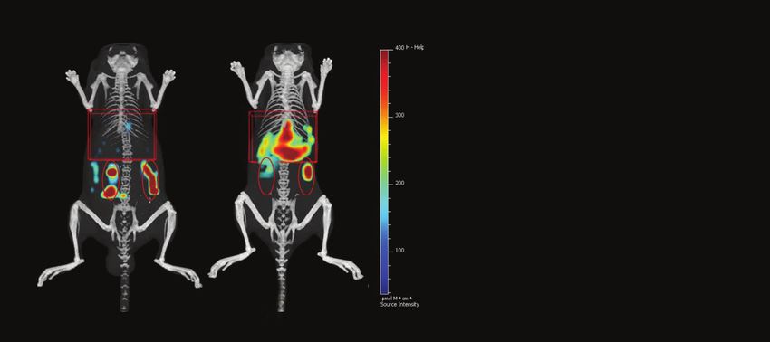
Materials
    Fluorescent Agent                                                                  Table 1. Basic Properties of Annexin-Vivo 750 Fluorescent Tumor Imaging Agent.
    The Annexin-Vivo 750 (AV750) fluorescent agent is specific for                        		                                Annexin-Vivo 750
    phosphatidyl serine exposed on the surface of cells undergoing                        Agent Type                        Phosphatidyl Serine Targeted Agent
    either apoptotic or necrotic cell death. This agent was designed to                   Molecular Weight                  34,000 g mol-1
    non-invasive detect and image cellular death occurring due to
                                                                                          Ex/Em		                           755/772 nm
    tumor chemotherapy, adverse drug effects on normal tissue, or
                                                                                          Blood Half-life                   Distribution t1/2 ~30 min
    spontaneous cell death associated with disease progression in
    mice. The imaging dose for this agent was as recommended in                           Tissue Half-life                  ~14 h
    the product insert (2 nmol/25 g mouse).                                            Agent Summary. Characteristics of the agent (MW/size, excitation/emission [Ex/Em],
                                                                                       and blood/tissue pharmacokinetics) were determined in multiple independent studies.
    Mice for APAP – Induced Liver Injury Model
    Seven to eight week-old male C57BL/6 mice were purchased from
    Charles River Laboratories (Wilmington, MA) and maintained in a
    pathogen-free animal facility with water and low-fluorescence
    mouse chow (Harlan Tekland, Madison, WI). Handling of mice and
    experimental procedures were in accordance with PerkinElmer
    IACUC guidelines and approved veterinarian requirements for
    animal care and use. The animals are fasted overnight
    (approximately 18 h) prior to drug administration.

                                                                                                                                Serum ALT,
                   APAP susceptible male
                                                                                                           Depilated
                      C57BL/6 mice                                                                          region
                                                                                Annexin-Vivo
                      (FASTED O/N)                                              750 Injection

                                                                                            2h

                                                                            0     12          24
                                                                                                                                Liver Injury
                                      APAP, IP                           Time post-APAP (h)
                                     500 mg/kg

                                                                                                                                        Imaging
    Figure 1. Optimal Imaging Protocol for Hepatocellular Liver Injury

2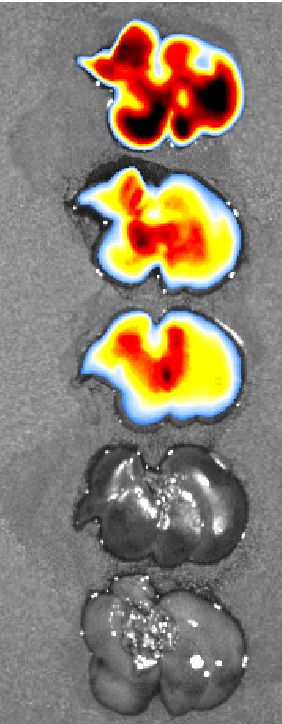
Hepatocellular Injury Imaging Protocol
Preparing Mice for Imaging                                                    IVIS Spectrum Imaging
• Use 7-8 week old C57BL/6 male mice for APAP-induced liver                  • If using the epifluorescence (2D) feature of an IVIS imaging
  injury, as this particular drug response is very mouse-strain and             system, place the anesthetized animal in the supine position (i.e.
  gender dependent. Other drugs that induce similar liver injury                belly-up) to facilitate optimal liver signal detection. The systems
  may differ in mouse strain/gender dependence or be                            provide gas anesthesia to the imaging chamber.
  independent of these variables.
                                                                              • F or the IVIS Spectrum/SpectrumCT, use the 745 ex/800 em filter
• Two
     weeks before the imaging study, switch to low fluorescence               set. For the IVIS Lumina use either 745 ex/ICG em (standard
  mouse chow. Regular mouse chow contains chlorophyll that                      Lumina filter set) or 745 ex/800 em.
  auto-fluoresces around 700 nm and can interfere with signal
                                                                              • For
                                                                                 rapid animal screening, IVIS systems permit imaging of five
  collected from this agent.
                                                                                mice at the same time using Field of View D (FOV D). It is
• Animal
          hair is highly effective at blocking, absorbing, and scattering     essential to use guards between animals to prevent signal
  light during optical imaging. Even light within the NIR spectrum,             contamination from neighboring animals.
  which typically shows minimal scattering and absorbance in tissue,
                                                                              • F or the IVIS Spectrum and IVIS SpectrumCT, optical tomography
  is significantly absorbed and scattered by hair.
                                                                                (FLIT) can also be used, with imaging of one mouse at a time.
• Always
        remove the fur on and around the areas of the animal                  Transillumination scan fields should be established with 15-20
  that are to be imaged (Nair depilatory lotion; Church & Dwight                scan points covering the liver and some area above and below
  Co, Princeton NJ), whether performing 2D or 3D optical imaging,               the liver. This will take 12-15 minutes for acquisition.
  within 1-24 h prior to imaging. Take care to minimize the time of
                                                                              •R
                                                                                econstruction of 3D datasets should be performed using
  Nair treatment and rinse mice well with warm water. IP injection
                                                                               appropriate thresholding and masking out of regions far outside
  of ketamine/xylazine (100 mg/kg and 20 mg/kg, respectively) may
                                                                               the scan field prior to reconstruction. Images should be
  be needed to keep mice anesthetized for the procedure.
                                                                               represented with 0.62 mm voxel size and with optimal color
•M
  ice should be fasted overnight (~18h) for most consistent                   scales for appropriate 3D fluorescence representation.
 induction of liver injury. Water is provided.
                                                                              • L iver quantification in APAP and control mice is performed by
Preparing Annexin-Vivo 750                                                      placement of 2D or 3D regions of interest (ROI) to capture the
• Each vial contains 10 mouse doses of Annexin-Vivo 750 in                    fluorescent signal within the liver region.
  solution (Total volume 1 mL). This material provides sufficient
                                                                              • F or further information on 2D (epifluorescence) or 3D (FLIT) IVIS
  reagent for imaging approximately 10 mice (weighing ~25
                                                                                imaging, please refer to tech notes under the help tab of the
  grams each) when using the recommended dose of 100 μL of
                                                                                Living Image® (LI) software.
  Annexin-Vivo 750 per mouse.
                                                                              FMT 4000 Imaging
• Annexin-Vivo
             750 is stable for up to six months when stored at
                                                                              • The FMT 4000 imaging system is a dedicated fluorescence
  2-8 ºC and protected from light.
                                                                                tomography system, but epifluorescence images are also
Introducing Hepatocellular Liver Injury with APAP                               acquired at the same time. As with IVIS imaging, it is important
• APAP must be freshly prepared from stock powder as a 300-500                 to position mice in the supine position, in this case within an
  mg/kg solution (20 mg/ml in PBS) for effective induction of liver             animal imaging cassette.
  injury. Warming the mixture to 55 °C for 10 minutes may be
                                                                              •U
                                                                                se the height adjustment knobs on the cassette to make sure
  required for generation of a homogeneous solution.
                                                                               that the anesthetized animal is gently but securely compressed
• Fasted
        mice are injected IP with a single, bolus dose of either             between the top and bottom plates.
  375-625 ul APAP or PBS (control group) in a 20 g mouse.
                                                                              • S lide the cassette into the docking station with the animal in the
  Optimal peak induction of liver cellular death is around 24 h
                                                                                prone position. The system provides gas anesthesia to the
  for this particular treatment.
                                                                                cassette within the imaging chamber. Set up the scan field with
Animal Imaging Considerations                                                   30 to 50 scan points with at least 1-2 rows of scan points
At the desired time (recommended 22 h post-APAP), inject 100 ul                 surrounding (on all sides) the liver region. Acquisitions typically
of AV750 intravenously into APAP and control mouse groups. Other                take 5-7 minutes.
drugs that induce hepatocellular injury may require establishing the
                                                                              • S elect AV750 as the imaging agent, which will select the optimal
optimal timecourse post-drug or even several days of dosing prior
                                                                                laser and filter combination for 750 nm imaging.
to AV750 injection.
                                                                              •3
                                                                                D reconstructions are performed automatically by TrueQuant
• The
   optimal imaging time for AV750 is two hours post injection.
                                                                               software. Images should be represented with optimal
  The optimal re-injection time is every 1-2 days, which allows for
                                                                               thresholding and optimal color scales.
  the clearance of the agent from the liver. It is important to note
  that kidney signal can be retained somewhat longer but with                 • L iver quantification in APAP and control mice is performed by
  minimal impact on liver imaging.                                              placement of 3D ROIs to capture the fluorescent signal within
                                                                                the liver region.
                                                                                                                                                       3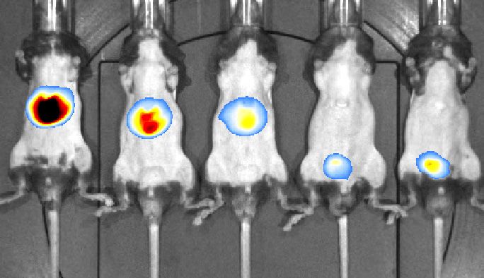
Introduction and Results
    Drug induced liver injury (DILI) has been the most frequent single                                  Pairing either the IVIS SpectrumCT or the FMT 4000 with the
    cause of safety-related drug withdrawals from the marketplace for                                   imaging agent Annexin-Vivo 750 (AV750) allows the imaging and
    the past 50 years and continues to be an important consideration                                    detection of cellular death within the liver. Acetaminophen (APAP), a
    in drug discovery research today. Liver safety issues further limit                                 commonly used over-the-counter analgesic and antipyretic drug, is
    use of many approved drugs or prevent consideration for                                             known to cause centrilobular hepatic necrosis when used at high
    approval. The majority of drugs showing evidence of DILI show                                       doses. When male C57BL/6 mice were fasted overnight and injected
    hepatocellular toxicity, a form of liver injury with cellular death                                 with a single dose (200-500 mg/kg) of APAP, the resulting liver
    and little or no evidence of hepatobiliary obstruction, cholestasis,                                necrosis peaked at 24 hours as detected by imaging with AV750 in
    or steatosis. The most overtly toxic of such drugs will cause DILI                                  living animals (Fig 1). Dose ranging of APAP (100, 200, 300, and 500
    in anyone receiving a high enough dose, however many drugs                                          mg/kg) showed maximal tissue destruction at 300 and 500 mg/kg
    (at recommended doses) show adverse liver finding in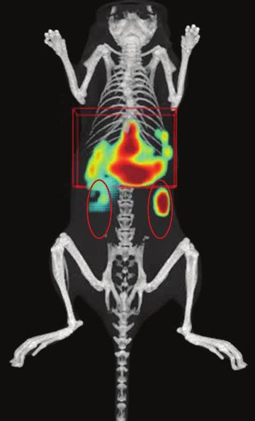
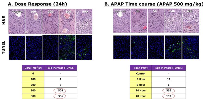
A. Dose Response (24 h) B. APAP Time Course (APAP 500 mg/kg)
Images are shown from representative tissue regions/sections for APAP dose response (A) and time course (B) studies. Formalin-fixed, paraffin-embedded tissue
sections from APAP treated mice were assessed by H&E (upper panels) and fluorescent TUNEL staining (lower panels). Representative images were acquired on the
Vectra® Imaging system (Perkinelmer, and spectral unmixing was used to specifically eliminate background autofluorescence by spectral signature. Lower panels
show the corresponding fold increases of apoptosis in liver sections using InForm® advanced image analysis software (PerkinElmer).
Figure 4. Histology Corroboration
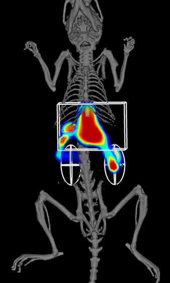
B. 3D FMT 4000 Fluorescence Imaging
A. 3D IVIS FLIT/CT Imaging with Quantum FX µCT Co-registration
L iver
Liver
K idneys
Kidneys
PBS APAP
APAP-treated (500 mg/kg) and control mice were also assessed using 3D tomographic imaging on the IVIS Spectrum (A) and FMT 4000 (B) imaging systems.
The IVIS SpectrumCT data was acquired with 15 minute scans of the liver region, including the built in CT functionality. In a separate study, FMT 4000 acquisitions
of the liver region were acquired with 5 minute scans and separate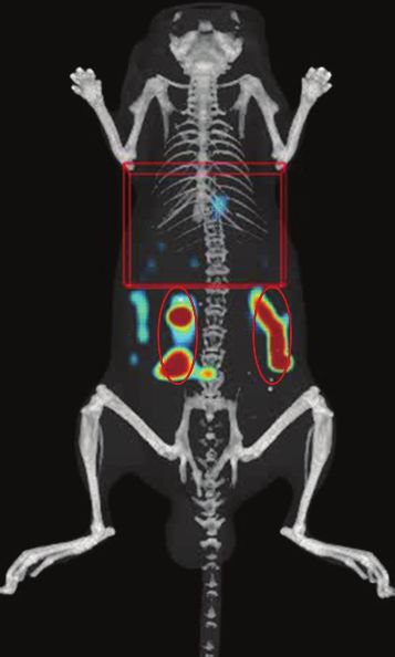
Conclusions
The present studies provides evidence that APAP induced liver                                                                   Liver Imaging
injury can be quantified non invasively in vivo with Annexin-Vivo                                                               Vasquez KO, Casavant C, Peterson JD. Quantitative whole body
750 used in conjunction with PerkinElmer's imaging platform.                                                                    biodistribution of fluorescent-labeled agents by non-invasive
Rapid 2D epifluorescence imaging of 5 mice at a time provides a                                                                 tomographic imaging. PLoS One 6(6):e20594 (2011).
robust and quantitative method for assessing hepatocellular toxicity                                                            Shuhendler AJ, Pu K, Cui L, Uetrecht JP, Rao J. Real-time imaging
in the liver in living mice, and the results correlated well with                                                               of oxidative and nitrosative stress in the liver of live animals for
direct imaging of excised liver fluorescence. The time course and                                                               drug-toxicity testing. Nat Biotechnol. 32(4):373-80 (2014).
dose-response of APAP - induced liver injury were quantified by
imaging, and the results correlated well with serum ALT levels,                                                                 Filonov GS, Piatkevich KD, Ting LM, Zhang J, Kim K, Verkhusha VV.
direct imaging of excised livers, qualitative assessment of liver                                                               Bright and stable near-infrared fluorescent protein for in vivo
damage by H&E staining of tissues, and the increase in TUNEL                                                                    imaging. Nat Biotechnol 29(8):757-61 (2011).
staining. Tomographic (3D) imaging provided additional depth
                                                                                                                                APAP-induced Liver Injury
detection and detail of signal within the liver, as well as detection
                                                                                                                                Hinson JA, Roberts DW, James LP. Mechanisms of acetaminophen-
of the expected kidney clearance of Annexin V.
                                                                                                                                induced liver necrosis. Handb Exp Pharmacol 196:369-405 (2010).
References                                                                                                                      Liu HH, Lu P, Guo Y, Farrell E, Zhang X, Zheng M, Bosano B, Zhang
Imaging Technology                                                                                                              Z, Allard J, Liao G, Fu S, Chen J, Dolim K, Kuroda A, Usuka J,
B.W. Rice, M.D. Cable, and M.B. Nelson. In vivo imaging                                                                         Cheng J, Tao W, Welch K, Liu Y, Pease J, de Keczer SA,
of light-emitting probes. Journal of Biomedical Optics 6(4):                                                                    Masjedizadeh M, Hu JS, Weller P, Garrow T, Peltz G. An integrative
432-440 (2001).                                                                                                                 genomic analysis identifies Bhmt2 as a diet-dependent genetic
                                                                                                                                factor protecting against acetaminophen-induced liver toxicity.
H. Xu and B.W. Rice. In vivo fluorescence imaging with multivariate
                                                                                                                                Genome Res 20(1):28-35 (2010).
curve resolution spectral unmixing technique. Journal of Biomedical
Optics 14(6):064011 (2009).                                                                                                     Ramachandran R, Kakar S. Histological patterns in drug-induced
                                                                                                                                liver disease. J Clin Pathol 62(6):481-92 (2009).
P. Mohajerani, A. Adibi, J. Kempner and W. Yared. Compensation
of optical heterogeneity-induced artifacts in fluorescence molecular
tomography: theory and in vivo validation. Journal of Biomedical
Optics 14:034021 (2009).
R. Weissleder. A clearer vision for in vivo imaging. Nature
Biotechnology 19:316-317 (2001).

PerkinElmer, Inc.
940 Winter Street
Waltham, MA 02451 USA
P: (800) 762-4000 or
(+1) 203-925-4602
www.perkinelmer.com

For a complete listing of our global offices, visit www.perkinelmer.com/ContactUs

Copyright ©2014, PerkinElmer, Inc. All rights reserved. PerkinElmer® is a registered trademark of PerkinElmer, Inc. All other trademarks are the property of their respective owners.

011715_01
You can also read