Ultrasonographic identification and characterization of congenital portosystemic shunts and portal hypertensive disorders in dogs and cats ...
←
→
Page content transcription
If your browser does not render page correctly, please read the page content below
Chapter 3

Ultrasonographic identification and
characterization of congenital
portosystemic shunts and portal
hypertensive disorders in dogs
and cats
  Viktor Szatmári, Jan Rothuizen

  ABSTRACT

   In the first part of this chapter the hemodynamic, anatomic and pathophysiologic features
of canine portal vein disorders are described. Understanding these principles is necessary for
correct interpretation of the ultrasonographic images. In the second part of the chapter an
ultrasonographic scanning protocol is described, which is recommended to be used for
thorough evaluation of the portal venous system. A short section at the very end will discuss
the specific features of feline portal vein disorders.

INTRODUCTION

   In the first part of this chapter the hemodynamic, anatomic and pathophysiologic features
of canine portal vein disorders are described. Understanding these principles is necessary for
correct interpretation of the ultrasonographic images. In the second part of the chapter an
ultrasonographic scanning protocol is described, which is recommended to be used for
thorough evaluation of the portal venous system. A short section at the very end will discuss
the specific features of feline portal vein disorders.

Normal abdominal vascular anatomy in dogs

   The aorta, the caudal vena cava (CVC) and the portal vein (PV) are the three great
abdominal vessels, which all course parallel to the vertebral column. The aorta is the most
dorsal one and its major branches from cranial to caudal are: the celiac, the cranial mesenteric,
the right and left renal arteries, and before the final trifurcation: the right and left external iliac
arteries. (1) The celiac artery branches further into three arteries, of which the largest is the
common hepatic artery, which runs cranially between the portal vein and the CVC (Fig. 1).
All the above described vessels can be visualized with ultrasound. The smallest intrahepatic
branches of the hepatic artery terminate in the hepatic sinusoids.
WSAVA Standards for Clinical and Histological Diagnosis of Canine and Feline Liver Diseases
                                 Society of Comparative Hepathology

  A

   B
   Figure .1. Normal canine abdominal blood vessels.
   A. Corrosion cast of the portal vein (blue) and the cranial part of the abdominal aorta
(red) in an adult normal beagle. R right portal branch, L left portal branch, CA celiac artery,
CMA cranial mesenteric artery, HA hepatic artery; CrMV cranial mesenteric vein, CaMV
caudal mesenteric vein. Before preparation the spleen, jejunum, ileum and colon were
removed and the descending duodenum was retracted.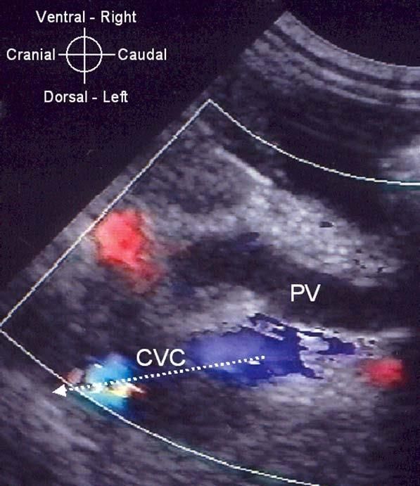
Ultrasonographic identification and characterization of portosystemic shunts and portal hypertensive disorders in
                                                  dogs and cats

   B. Color Doppler ultrasound image of a normal portal vein (PV) of a Yorkshire terrier.
   The diameter of the portal vein is uniform along its whole length. The image was made via
the right flank with the dog in left lateral recumbency (plane-4). Dotted arrows indicate the
direction of blood flow. Gastroduodenal vein (GDV), Splenic vein (SPLV), Hepatic artery
(HA) (From Szatmári V et al. Standard planes for ultrasonographic examination of the portal
system in dogs. J Am Vet Med Assoc 2004;224:713-716, with permission)

   The CVC is formed by the confluence of the right and left common iliac veins at the level
of the aortic trifurcation. The CVC courses ventral to the aorta and after entering the thoracic
cavity it terminates in the right atrium. The abdominal CVC collects the blood of the left and
the right renal veins, and as it passes through the liver collects the blood of the hepatic veins.
The hepatic veins drain the blood of the hepatic sinusoids and are straight vessels running in
the liver lobes to a craniomedial direction. The left gonadal vein (ovarian vein in females and
testicular vein in males) enters the left renal vein, whereas the right gonadal vein is a direct
tributary of the caudal vena cava. (2) The renal and hepatic veins can be visualized with
ultrasound, but the gonadal veins because of their small diameter cannot.
   The PV is formed by the confluence of the cranial and caudal mesenteric veins. (2) The PV
collects the blood of the splenic vein at the level where the celiac artery originates from the
aorta, as well as the blood of the gastroduodenal vein immediately caudal to the portal
bifurcation. The left gastric vein is a tributary of the splenic vein. The right gastric vein is
either a tributary of the gastroduodenal vein or of the PV. In the latter case it enters the PV
directly cranial to the gastroduodenal vein after a course along the lesser curvature of the
stomach. (3) At the hilus of the liver the trunk of the PV bifurcates into a larger left and a
smaller right portal branch. The right branch courses dorsally, the left one ventrally. The right
branch supplies the right lateral and the caudate hepatic lobes, whereas the left branch
supplies the left lateral, left medial, quadrate and right medial lobes. The smallest portal
branches terminate in the hepatic sinusoids where their blood mixes with the hepatic arterial
blood. The splenic vein, the gastroduodenal vein and the left and right portal branches can be
visualized with ultrasound.
   The azygos vein is a thin vessel that courses dorsal to the aorta and after passing through
the diaphragm it enters the cranial vena cava, which latter terminates in the right atrium. (2)

The CVC and the azygos vein together with their tributaries belong to the systemic venous
system, and the portal vein together with its tributaries form the portal venous system. No
macroscopic communication exists between the systemic and the portal venous systems.

  Portosystemic shunting occurs when anomalous veins allow the portal blood to enter the
systemic veins directly without first flowing through the hepatic sinusoids. (4) Portosystemic
shunting can occur via acquired portosystemic collaterals or via congenital portosystemic
shunts. (4-6)

Diagnostic approach to dogs suspected of having portosystemic
shunting

   Because portosystemic shunting can cause a great variety of clinical signs, and
ultrasonographic visualization of the anomalous veins has been a diagnostic challenge,
measuring fasting venous blood ammonia level has become a routine procedure to justify or
rule out the presence of portosystemic shunting. (7, 8)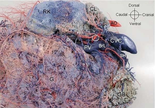
WSAVA Standards for Clinical and Histological Diagnosis of Canine and Feline Liver Diseases
                                 Society of Comparative Hepathology

   Determining the blood ammonia level before performing an abdominal ultrasound
examination can greatly increase the positive and negative predictive values of
ultrasonography in finding the anomalous vein since hyperammonemia can only be caused by
a few diseases such as congenital portosystemic shunts (CPSSs), acquired portosystemic
collaterals (APSCs), or urea cycle enzyme deficiency. (9, 10) Differentiating these conditions
non-invasively (e.g. by ultrasound) is crucial because CPSSs is the only disease that requires
surgical treatment, the other two do not. Some other diseases may also cause
hyperammonemia, but they can be differentiated from the above mentioned disorders by
clinical examination and laboratory tests. (9)
   Abdominal ultrasonography can readily diagnose CPSSs in non-sedated dogs. Moreover,
the anatomy of the shunting vessel (i.e. intra- or extrahepatic) can be precisely determined. (9,
11-13)
       This knowledge is important not only for the surgeon, but also for predicting the
prognosis. (14) The other great advantage of ultrasonography is that the other conditions
causing elevated blood ammonia and bile acids levels, such as acquired portosystemic
collaterals, may also be diagnosed. (9)

Pathophysiology of canine portal hypertension

   Acquired portosystemic collaterals (APSCs) are formed as the result of sustained hepatic or
prehepatic portal hypertension by enlargement of extrahepatic rudimentary vessels, through
which no blood normally passes. (5, 6) The term prehepatic refers to disorders that affect the
portal vein (i.e. extravascular compression or intravascular obstruction). (6) The term hepatic
refers to the diseases of the liver itself, and the term posthepatic refers to conditions that affect
the thoracic caudal vena cava (CVC) or the heart. (6) Posthepatic portal hypertension (e.g.
right-sided congestive heart failure) never results in APSC-formation because not only the
portal, but also the caval pressure increases. (6) Posthepatic portal hypertension results in an
enlarged liver and dilated hepatic veins due to congestion, whereas prehepatic portal
hypertension results in a small liver due to insufficient portal venous perfusion. In hepatic
portal hypertension the small or normal sized liver have a slightly or severely abnormal (echo-
)structure. A common, but not consistently occurring consequence of any kind of portal
hypertension is accumulation of free abdominal fluid (pure or modified transudate). (6)

Acquired portosystemic collaterals in dogs
   Collateral-formation is a compensatory mechanism to maintain normal portal pressure by
allowing the portal blood to be drained into the lower pressure systemic veins. (5, 6) Collateral
veins run simultaneously in several anatomic ways, however dogs tend to consistently
develop the so called spleno-renal collaterals. (6) As a result of the spleno-renal collateral
circulation the left gonadal vein (testicular vein in males and ovarian vein in females)
becomes dilated (15) because a portion of the portal venous blood is forced to flow via the
splenic vein through the preexisting embryonic connections to the left gonadal vein, and from
here through the left renal vein eventually to the CVC (Fig. 2).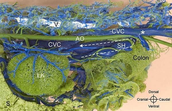
Ultrasonographic identification and characterization of portosystemic shunts and portal hypertensive disorders in
                                                  dogs and cats

  Figure 2. The left gonadal vein, which is a normal tributary of the left renal vein.
  A. Dilated left ovarian vein (LOV) in a 5-month-old female great Dane with spleno-renal
collaterals as a result of sustained portal hypertension of hepatic origin. C conglomeration of
collateral vessels, CVC caudal vena cava, LRV left renal vein.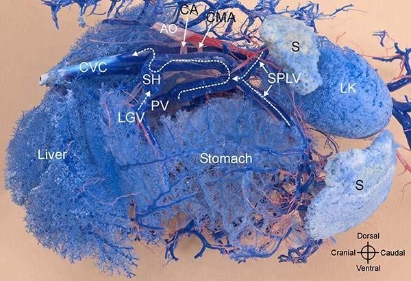
WSAVA Standards for Clinical and Histological Diagnosis of Canine and Feline Liver Diseases
                                 Society of Comparative Hepathology

   B. Normal left ovarian vein in a 9-month-old female Labrador retriever. The left ovarian
vein (LOV) enters the left renal vein from caudal It is much thinner than the left renal vein
(LRV).

   In addition to the dilated left gonadal vein, the origin of an APSC can occasionally be
found with ultrasound at the point where congenital extrahepatic spleno-caval shunts arise
from the portal vein (PV) (see later). In these cases normal flow may be seen caudal to the
APSC-origin and hepatofugal (i.e. away from the liver) flow cranial to it (Fig. 3A). In other
dogs with portal hypertension the flow in the PV may be so slow that no color signals can be
detected (Fig. 3B).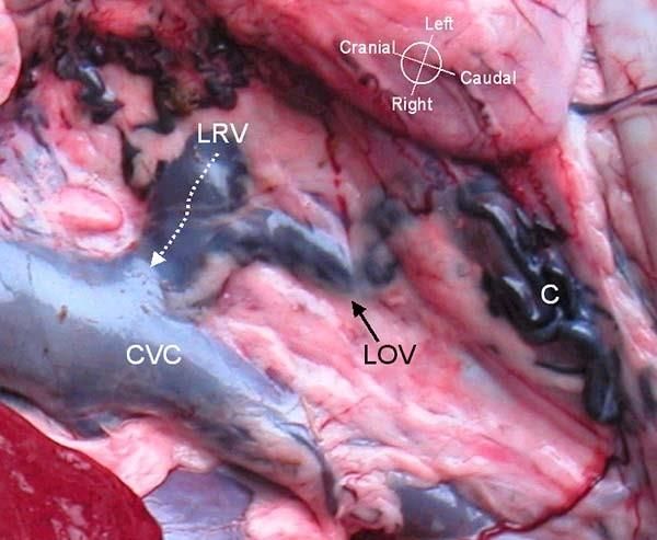
Ultrasonographic identification and characterization of portosystemic shunts and portal hypertensive disorders in
                                                  dogs and cats

  Figure 3. Portal venous flow in two dogs with hepatic portal hypertension.
  (Reproduced from Szatmári V et al. Ultrasonographic findings in dogs with
hyperammonemia: 90 cases (2000-2002). J Am Vet Med Assoc 2004;224:717-727, with
permission)
  A. Color Doppler ultrasound image of the portal vein and the origin of an acquired
portosystemic collateral (SH) in a 5-year-old West highland white terrier with sustained
portal hypertension of hepatic origin. Cranial to the collateral-origin (PVcrSH) hepatofugal
portal flow can be seen. Note that the anomalous vein (SH) runs caudally. Dotted arrows
indicate the direction of blood flow. In the portal vein caudal to the collateral origin
(PVcaudSH) normal flow can be seen.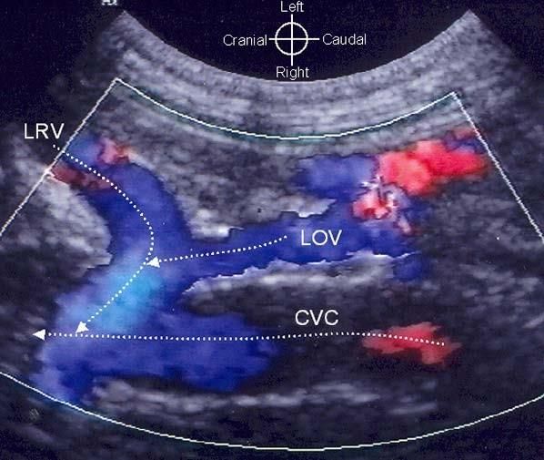
WSAVA Standards for Clinical and Histological Diagnosis of Canine and Feline Liver Diseases
                                Society of Comparative Hepathology

   B. Color Doppler ultrasound image of the portal vein in a 6.5-year-old female Jack Russell
terrier with sustained portal hypertension due to primary hypoplasia of the portal vein shows
undetectably slow flow in the portal vein. Note that no color signals are seen in the portal
vein (PV), whereas aliasing artifact is apparent in the caudal vena cava (CVC). The dotted
arrow indicates the direction of blood flow.

Etiology of canine portal hypertension
   Once the presence of portal hypertension has been established among others by visualizing
a dilated left gonadal vein, the next diagnostic step is to identify the underlying cause.
Ultrasonography is able to determine the cause of prehepatic portal hypertension as well as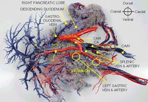
Ultrasonographic identification and characterization of portosystemic shunts and portal hypertensive disorders in
                                                  dogs and cats

diagnose arterioportal fistula. Hepatic portal hypertension may only be suspected with
ultrasound.
   Prehepatic portal hypertension can be caused by compression of the portal vein by a
neoplasia or a cyst or by an obstruction of the portal vein by a thrombus or a tumor. Both
conditions can readily be diagnosed with ultrasound.
   Congenital arterioportal fistula is a developmental anomaly characterized by a direct
connection between a portal venous and a hepatic arterial branch. (9, 16) The high arterial
pressure is responsible for the classical ultrasonographic changes:
    (a) extremely dilated and tortuous portal branch in a liver lobe
    (b) hepatofugal flow in the PV (with a variable or an arterial Doppler spectrum)
    (c) APSCs (Fig 4). (9)
   Ascites (pure transudate) is usually present.

   Figure 4. Congenital arterio-portal fistula.
   Color Doppler ultrasound image of the liver of a 6-month-old male American Staffordshire
terrier reveals dilated portal branches (PVbr) in the affected liver lobe. The flow-direction in
the portal vein (PV) is hepatofugal indicated by the dotted arrow. A ascites
   (Reproduce from Szatmári V et al. Ultrasonographic findings in dogs with
hyperammonemia: 90 cases (2000-2002). J Am Vet Med Assoc 2004;224:717-727, with
permission)
WSAVA Standards for Clinical and Histological Diagnosis of Canine and Feline Liver Diseases
                                 Society of Comparative Hepathology

   Hepatic portal hypertension must be suspected in dogs with high blood ammonia level if
ultrasonography discloses a dilated left gonadal vein and excludes arterioportal fistula and
compression or obstruction of the PV. Hepatic portal hypertension can be caused by
parenchymal liver diseases (chronic hepatitis of various etiologies) (17-19) or anomalies of the
portal branches [e.g. primary hypoplasia of the portal vein (PHPV)]. (9, 20) Diagnosing and
differentiating these conditions require histopathologic examination of liver biopsy
specimens.
   In mild cases of PHPV portal hypertension does not develop, hence blood ammonia level
remains normal, however in severe cases APSCs develop as a consequence of sustained
hepatic portal hypertension. (9, 20)
   Primary hypoplasia of the portal vein is simultaneously present with arterioportal fistula
and might accompany CPSSs. (9) When a CPSS and PHPV coincide in a dog, PHPV cannot be
diagnosed preoperatively. In these dogs portal hypertension and APSCs will not develop
because there is an already existing connection between the portal and the systemic veins (i.e.
the CPSS). (21) Currently, the earliest time point when PHPV can be suspected in a dog with
CPSS is during surgical attenuation of extrahepatic CPSSs, with intraoperative Doppler
ultrasonography. (22)

Congenital portosystemic shunts in dogs

   Portosystemic shunting is considered to be congenital when a single, usually large-bore
vein is present without a concurrent portal hypertension. (4) Congenital portosystemic shunts
are classified as intrahepatic and extrahepatic. Since a CPSS has equal or larger diameter
compared to the PV-segment caudal to the shunt, it offers a lower resistance way for the blood
to reach the systemic veins than the way through the hepatic sinusoids. As blood tends to flow
towards the lowest possible resistance, the vast majority of the portal blood flows via the
shunt because hepatic sinusoids represent much higher resistance to flow. Therefore, the liver
receives no or only a trivial fraction of the portal blood, which is insufficient for a normal
hepatic development and function. (4, 23)

Intrahepatic congenital porto-caval shunts
   Intrahepatic porto-caval shunts occur predominantly in large breed dogs, (24) particularly
often in Bernese mountain dogs and retrievers. Intrahepatic CPSSs originate either from the
left or from the right portal branch and appear as the direct continuations of the PV as the
diameters of the shunt and of the affected portal branch are the same as that of the PV. (9)
Because the majority of blood flows through the portal branch that continues as the shunt, the
contralateral portal branch remains very thin due to hypoperfusion. All intrahepatic CPSSs
terminate in the CVC either directly or via a hepatic vein. (25) The intrahepatic CPSS is usually
a single vein, but exceptionally they can have two loops. (9, 26)
   Intrahepatic CPSSs that originate from the left portal branch courses cranioventrally and to
the left (similarly to a normal left portal branch) to the diaphragm, then turns abruptly dorsally
to enter the CVC via a dilated segment of the left hepatic vein. (25) In these dogs the right
portal branch is very thin (Fig. 5). (9)
Ultrasonographic identification and characterization of portosystemic shunts and portal hypertensive disorders in
dogs and cats
Figure 5. Patent ductus venosus.
Corrosion cast of a left divisional intrahepatic portocaval shunt of a 2-month-old Irish
wolfhound. The portal vein (PV) has the same diameter as the left portal branch (BrL) and
the shunt (SH). The right portal branch (O blue) is very narrow, however the corresponding
hepatic arterial branch (O red) is relatively wide. CVC caudal vena cava, RK right kidney,
AO aorta, CL caudate liver lobe, P pancreas, G guts, GB gallbladder.
Intrahepatic CPSSs that originate from the right portal branch appear as the direct
continuation of the right portal branch (Fig. 3.6). The dilated right portal branch runs
consistently dorsolaterally and to the right from the PV, like a normal right portal branch, but
then, instead of tapering, it turns medially to enter the CVC. (9, 25) The dorsolaterally running
segment is either short or long (i.e. central and right divisional shunt, respectively). Whatever
morphology of a right-sided intrahepatic CPSS has, the left portal branch is severely
underdeveloped.
A dog with simultaneous intrahepatic CPSS and arterioportal fistula has been reported. (27)WSAVA Standards for Clinical and Histological Diagnosis of Canine and Feline Liver Diseases
Society of Comparative Hepathology
Figure 6.
Congenital intrahepatic portocaval shunt from the right portal branch.
Corrosion cast of a central divisional intrahepatic portocaval shunt of a 7-month-old mixed
breed dog 3 months after partial shunt ligation. The portal vein (PV) continues via the right
portal branch (arrow) to the caudal vena cava (CVC). Three months after shunt attenuation
the left portal branch (BrL) is still rather narrow. HV hepatic vein. There is ligature around
the shunt. The PV and CVC slightly caudal to the liver have been removed.
Extrahepatic congenital portosystemic shunts
Extrahepatic portosystemic shunts occur mostly in small breeds, particularly often in
Maltese dogs, miniature schnauzers and small terriers (Yorkshire, Jack Russell, cairn), but are
occasionally seen in large breeds. Extrahepatic CPSSs originate from the splenic vein, from
the right gastric vein, or from both as communicating loops and enter either the abdominal
CVC or the thorax: spleno-caval, right gastric-caval, spleno-azygos and right gastric-azygos
shunt. (9) Shunts with two loops (one arising from the right gastric vein and the other one from
the splenic vein) are categorized as right gastric shunts because the right gastric vein is the
main loop and this has an identical morphology with the single shunts that arise from the right
gastric vein. Though extrahepatic CPSSs are named after a portal tributary, they all divert the
blood of the PV via a short and dilated segment of the involved tributary. Extrahepatic CPSSs
with other anatomy than the above described four types are extremely rare in dogs. (28)
Extrahepatic CPSSs arising from the splenic vein
Spleno-caval shunts are the most common type of congenital extrahepatic CPSSs. They
usually form a short loop between the PV and the CVC. Although the anomalous vein may
have a long cranially extending loop, the points of origin and termination are always the same.
Since the point of shunt origin is very close to the point where the splenic vein enters the PV
and the short segment of the splenic vein that is between the PV and the shunt-origin is dilatedUltrasonographic identification and characterization of portosystemic shunts and portal hypertensive disorders in
dogs and cats
and the flow hepatofugal (i.e. away from the liver) in it, the CPSS seems to originate from the
PV itself and the splenic vein seems to enter the shunting vessel (Fig. 7A). The origin of the
spleno-caval shunts is slightly cranial to the level where the celiac artery originates from the
aorta (i.e. approximately at the level of the cranial pole of the right kidney, Fig. 7B). The
termination of the spleno-caval shunts in the CVC is always at the same point, i.e. slightly
cranial to the level of the shunt-origin (Fig 8A). (9, 13)
Figure 7. Congenital extrahepatic spleno-caval shunt.
A. Color Doppler ultrasound image of the portal vein in longitudinal section made via the
right flank with the dog in left lateral recumbency (plane-4). The shunt (SH) originates from
the splenic vein (SPLV), very close to the point where the SPLV normally enters the portal
vein. Both the intestinal and the splenic blood are diverted. In the portal vein segment
between the shunt-origin and the entering point of the gastroduodenal vein (PVcrSH) the flow
is hepatofugal. This PVcrSH is thinner than the portal vein caudal to the shunt-origin
(PVcaudSH). Cranial to the entering point of the gastroduodenal vein (GDV) the portal flow
is hepatopetal (PVcrGDV). Dotted arrows indicate the direction of blood flow. (From
Szatmári V et al. Ultrasonographic evaluation of partially attenuated congenital extrahepatic
portosystemic shunts in 14 dogs. Vet Rec 2004;155:448-456, with permission)WSAVA Standards for Clinical and Histological Diagnosis of Canine and Feline Liver Diseases
Society of Comparative Hepathology
B. Corrosion cast of the abdominal blood vessels of a 2-month-old cairn terrier. The veins
are blue and the arteries are red. The shunt (SH) terminates in the caudal vena cava (CVC)
slightly cranial to the point where the celiac artery (CA) originates from the aorta (AO). The
left gastric vein (LGV) and both the dorsal and ventral branches of the splenic vein (SPLV)
enter the SH. The portal vein (PV) is thin cranial to the SH-origin. CMA Cranial mesenteric
artery, LK left kidney, S spleen (the middle part of the spleen has been removed). Dotted
arrows indicate the direction of blood flow in a living animal.
In cases of spleno-azygos shunts, the shunting vessel approaches the CVC at the point
where the spleno-caval shunts terminate, but instead of entering it the shunting vessel courses
dorsal to the CVC and eventually enters the thorax (Fig 8B). The point of origin of these
spleno-azygos shunts is the same as that of the spleno-caval shunts (Fig 7A).
Since the diameter of the shunt is always wider than that of the PV caudal to the shunt-
origin the PV cranial to the shunt-origin and the intrahepatic portal branches remain
hypoperfused. Therefore, the PV-segment cranial to the shunt-origin is always thinner than
the PV-segment caudal to the shunt-origin. In most cases of extrahepatic CPSSs the left and
right portal branches are also very thin. When they have a relatively normal diameter, color
Doppler shows an undetectably slow or very slow hepatopetal (i.e. towards the liver) flow in
them. The flow-direction in the PV-segment cranial to the shunt-origin is hepatofugal in most
dogs, however could be slow hepatopetal (i.e. flow to the liver) or alternating (“to-and-fro”).
The blood of the gastroduodenal vein was found to be responsible for the hepatofugal flow
because the gastroduodenal blood finds lower resistance to flow towards the shunt (i.e. to
caudal) than towards the hepatic sinusoids (Fig 3.7A).Ultrasonographic identification and characterization of portosystemic shunts and portal hypertensive disorders in
dogs and cats
Figure 8. Congenital extrahepatic portosystemic shunts in two dogs.
Color Doppler ultrasound images show the termination of congenital extrahepatic
portosystemic shunts. The transducer was positioned caudal to the last right rib (flank) with
the dog in dorsal recumbency. Dotted arrows indicate the direction of blood flow. (From
Szatmári V et al. Ultrasonographic findings in dogs with hyperammonemia: 90 cases (2000-
2002). J Am Vet Med Assoc 2004;224:717-727, with permission)
A. Spleno-caval shunt in a 3-month-old female cairn terrier at the point where the shunt
(SH) enters the caudal vena cava (CVC). This point is located always slightly cranial to the
point where the celiac artery (CA) originates from the aorta. CMA cranial mesenteric arteryWSAVA Standards for Clinical and Histological Diagnosis of Canine and Feline Liver Diseases
Society of Comparative Hepathology
B. Spleno-azygos shunt in a 3.5-month-old male Jack Russell terrier. The shunting vessel
(SH) courses dorsal to the CVC, and enters the thorax.
The morphology of the APSCs that originate from the PV can be very similar to that of
congenital extrahepatic spleno-caval shunts (Figs 3A,.7A). Moreover, in both cases
hepatofugal portal flow cranial to the origin of the anomalous vein could be seen. The
differences are: an APSC runs caudally from its origin and tends to disappear among the
intestines, furthermore the diameters of the PV cranial and caudal to the APSC-origin are
roughly equal (Fig 3A). In contrast, congenital extrahepatic spleno-caval or spleno-azygos
shunts tend to run cranially from the origin and can always be followed to their terminations
(CVC or diaphragm). Thirdly, the PV-segment that is cranial to the origin of a spleno-caval or
a spleno-azygos shunt is always thinner than the PV-segment caudal to it (Fig.7). Moreover,
an APSC is thinner than the PV caudal to the anomalous vein, unlike in cases of CPSSs. The
simultaneous presence of the dilated left gonadal vein proves that an extrahepatic anomalous
vein originating from the PV with a hepatofugal flow is the origin of an APSC and not that of
a CPSS. (9)Ultrasonographic identification and characterization of portosystemic shunts and portal hypertensive disorders in
dogs and cats
Extrahepatic CPSSs arising from the right gastric vein
Right gastric-caval shunts have either one or two loops. In the former case only the cranial
loop (right gastric-caval loop) is present (Fig 9 A, B). In the latter case both the cranial loop
and the caudal loop (spleno-caval loop) are present and they anastomose before entering the
CVC (Fig 10).
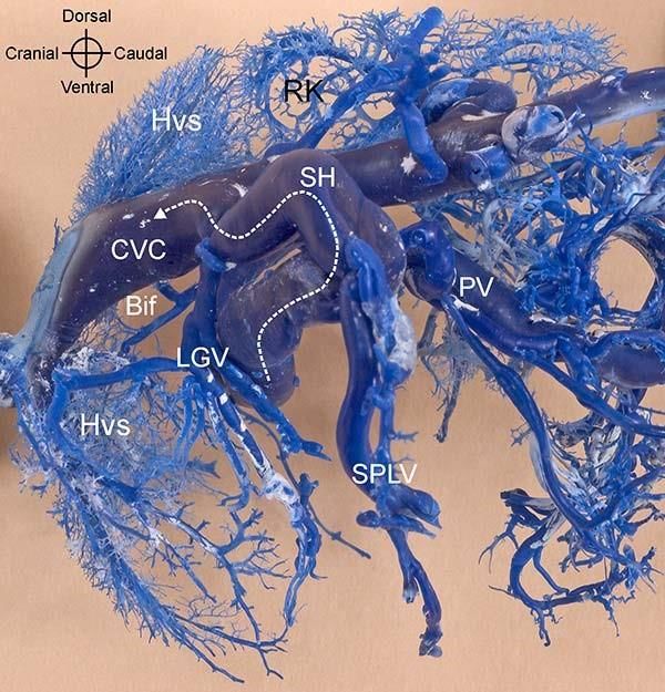
Figure 9. Right gastric-caval shunt with one shunt loop.
Corrosion cast of the abdominal veins of a Dachshund with congenital extrahepatic right
gastric-caval shunt. In this patient only the cranial shunt-loop (SH) has developed. The left
kidney has been removed. RK right kidney
A. The shunt (SH) originates at the point where the gastroduodenal vein (GDV) enters the
portal vein (PV). The right gastric vein is a tributary of the GDV. The PV caudal to the SH-
origin is thinner than the SH and becomes very thin cranial to the SH-origin. The right portal
branch runs to dorsal and the left portal branch to ventral. Bif portal bifurcation, CVC
caudal vena cava, GEV left and right gastroepiploic veins, JVs jejunal veins, SPLV splenic
veinWSAVA Standards for Clinical and Histological Diagnosis of Canine and Feline Liver Diseases
Society of Comparative Hepathology
B. The portal vein (PV) becomes very thin cranial to the origin of the SH. Compare the PV-
diameter caudal to the SH with that just caudal to the PV-bifurcation (Bif). Both the splenic
vein (SPLV) and the left gastric vein (LGV) enter the SH. The SH terminates in the caudal
vena cava (CVC). HVs hepatic veins.Ultrasonographic identification and characterization of portosystemic shunts and portal hypertensive disorders in
dogs and cats
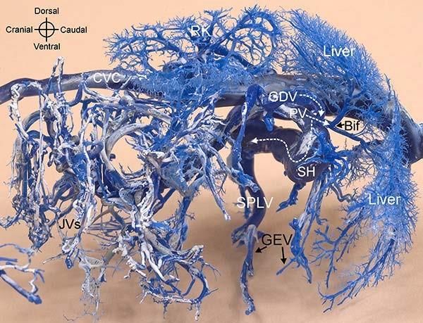
Figure 10. Right gastric-caval shunt with two shunt loops.
Corrosion cast of the abdominal veins of a Yorkshire terrier with a right gastric-caval
shunt. Both the cranial shunt-loop (Shcr) and the caudal shunt-loop (SHca) are present and
they anastomose with each other (o). The portal vein (PV) becomes narrower cranial to the
point of the Shca-origin (*), and even more narrow cranial to the point of the Shcr-origin (+).
The Shcr originates at the point where the gastroduodenal vein (GDV) enters the PV. By
surgical closure of the common trunk of the SH adjacent to the caudal vena cava (CVC) both
shunt-loops can be ceased. The arrows indicate the flow-directions in the vessels in a living
animal. Bif Portal bifurcation with the very thin right and left portal branches, HVs Hepatic
veins, GEV Right and left gastroepiploic veins, SPLVv Ventral branch of the splenic vein,
SPLVd Dorsal branch of the splenic vein. (The CVC has been removed caudal to the SH-
termination.)
The cranial loop arises from the right gastric vein and the morphology of this loop varies
slightly depending whether the right gastric vein is a tributary of the gastroduodenal vein or of
the PV itself originating between the portal bifurcation and the gastroduodenal vein. (3) In both
cases the shunt originates immediately caudal to the portal bifurcation via the dilated right
gastric vein. When the right gastric vein is a direct tributary of the PV, the blood of the PV is
drained via the dilated right gastric vein to the CVC. When the right gastric vein is a tributary
of the gastroduodenal vein, the blood of the PV is drained via a short and dilated segment of
the gastroduodenal vein through the right gastric vein into the CVC and the blood of the
gastroduodenal vein flows through the shunt (i.e. the right gastric vein) without reaching first
the PV. Regardless of the anatomic variation of the right gastric vein, the course of the shunt
(i.e. the right gastric vein) is always the same, namely it makes a long loop from the liver
hilus first laterally to the left body wall, then from here to caudomedially to eventually enter
the CVC at the point where the spleno-caval shunts terminate (i.e. slightly cranial to the celiacWSAVA Standards for Clinical and Histological Diagnosis of Canine and Feline Liver Diseases
Society of Comparative Hepathology
artery, Fig 11). The caudal shunt-loop of a right-gastric-caval shunt resembles a spleno-caval
shunt.
Exceptionally, the shunting vessel does not enter the CVC, but courses dorsal to it and
enters the thorax (right gastric-azygos shunts).
A
B
Figure 11. Right gastric-caval shunt.Ultrasonographic identification and characterization of portosystemic shunts and portal hypertensive disorders in
dogs and cats
Color Doppler ultrasound images of (the cranial loop of) a congenital extrahepatic right
gastric-caval shunt in a 6.5-month-old female Yorkshire terrier. The dog is in right lateral
recumbency and the transducer is placed caudal to the last left rib (plane-6). Dotted arrows
indicate the direction of flow.
A. The shunt (SH) originates at the liver hilus and runs towards the left body wall making a
roughly 90° angle with the portal vein (PV). The continuation of the shunt (traced caudally) is
shown in Figure B. The PV cannot be seen cranial to the shunt origin because it is extremely
thin due to hypoperfusion (see the corrosion cast).
B. The shunt (SH) terminates in the caudal vena cava (CVC) cranial to the celiac artery
(CA), similarly to a congenital extrahepatic spleno-caval shunt. Note the large-caliber
shunting vessel (SH) immediately under the left body wall. CMA cranial mesenteric artery.
(From Szatmári V et al. Ultrasonographic findings in dogs with hyperammonemia: 90 cases
(2000-2002). J Am Vet Med Assoc 2004;224:717-727, with permission)
All CPSSs that arise from the right gastric vein are very wide, with a diameter comparable
to that of the CVC. At surgical exploration the cranial loop of the right gastric-caval shunt is
found to follow the lesser curvature of the stomach, similarly to a normal right gastric vein.
The caudal shunt loop, which is not consistently present, originates at the region where
spleno-caval shunts are expected, but unlike the spleno-caval CPSSs, it courses from caudal to
cranial and not from ventral to dorsal like the spleno-caval shunts do. The caudal loop drains
the blood of the PV via the dilated segment of the splenic vein to the common trunk (Fig 10).
The PV becomes slightly thinner cranial to the origin of the caudal shunt-loop (i.e. cranial to
the splenic vein) with hepatopetal flow direction. The PV cranial to the origin of the cranial
shunt-loop is so thin that cannot be visualized by ultrasound. (9) Right gastric-caval shunts are
frequently found in Maltese dogs.
Hyperammonemia without portal vein disorder
Urea cycle enzyme deficiency is a rare congenital metabolic disease. (10) Since no
morphologic changes are present, the abdominal ultrasound examination reveals normal sized
and structured liver and kidneys and the absence of vascular abnormalities. (9)
Peritoneal absorption of ammonia containing urine may result in hyperammonemia, (29)
however this condition can easily be differentiated from portosystemic shunting by measuring
high plasma creatinine concentration.
Irish wolfhound pups have a physiologic period of hyperammonemia. (30)
Abdominal ultrasonography of portal vein disorders
Secondary changes
Before starting the examination of abdominal vessels, a detailed B-mode ultrasonographic
study of the abdominal organs has to be performed. Determining the presence and amount of
free abdominal fluid, the size and structure of the liver and kidneys is particularly important.
The size of the left and right halves of the liver should be separately evaluated. The urinary
bladder should also be examined for the presence of sediment or stone.WSAVA Standards for Clinical and Histological Diagnosis of Canine and Feline Liver Diseases
Society of Comparative Hepathology
Typical findings in dogs with CPSS is a small liver, normal or enlarged kidneys (often with
hyperechoic medulla) and no free abdominal fluid, (9, 11, 13) whereas dogs with APSCs have
small, normal sized or asymmetric liver (usually the left side is small and the right side is
enlarged), variable amount of ascites (from none to large amount) and slightly or markedly
abnormal hepatic echo structure. (9) The kidneys are of normal size of acquired diseases,
however may be enlarged with congenital portal hypertensive disorders such as PHPV.
Normal sized liver and kidneys do not exclude CPSSs.
Large amount of free abdominal fluid may hinder ultrasonographic visualization of the
abdominal vessels, but hyperammonemia in dogs with severe peritoneal effusion cannot
possibly be the result of CPSSs or an urea cycle enzyme deficiency, since a dog with CPSS
cannot have portal hypertension or so severe hypoalbuminemia that would result in formation
of a large amount of transudate in the abdominal cavity. (6) Small amount of free abdominal
fluid is normal in healthy pups, hence it may also be seen in puppies with CPSSs or with urea
cycle enzyme deficiency. (9)
The spleen has usually a normal size in dogs with both CPSSs and APSCs. (9) The reason
why splenomegaly does not develop in dogs with portal hypertension could be that acquired
spleno-renal collaterals are the most consistently developing APSCs in dogs, and these
collateral veins prevent the spleen from congestion. (9)
Diagnosis of CPSS is based on visualizing the anomalous vessel. Measuring flow velocities
has no use. Ultrasonographic evaluation of the abdominal vasculature is recommended to be
performed in 7 standard planes. (15)
Accurate recognition of CPSSs by ultrasound is only possible, if the anomalous vein is
traced from its origin to its termination, or the other way around. Finding the point where a
CPSS enters the CVC may be easier than finding the origin of a CPSS, (13) however finding a
vein that enters the CVC does not mean that it is a CPSS because several other veins enter the
CVC. In contrast, when a vein that originates from the PV displays hepatofugal flow, it is
surely an extrahepatic CPSS or an APSC, even without knowing the point of its termination
because in normal animals veins only enter and do not originate from the PV.
Diagnosing APSCs ultrasonographically requires a different approach from that of CPSSs
because recognizably large collateral veins only occasionally arise directly from the PV,
moreover they are thin and tortuous and most of the times are hidden among the intestines.
Therefore, their origins and courses can only exceptionally be ultrasonographically revealed.
However, the dilated left gonadal vein, i.e. the termination of the spleno-renal collaterals, was
found to be a highly specific and sensitive indicator of APSCs in dogs, and its
ultrasonographic visualization is simple (Fig 12 A,B). (9)Ultrasonographic identification and characterization of portosystemic shunts and portal hypertensive disorders in
dogs and cats
A
BWSAVA Standards for Clinical and Histological Diagnosis of Canine and Feline Liver Diseases
Society of Comparative Hepathology
Figure 12. Acquired portosystemic (spleno-renal) collaterals.
A. Dilation of the left ovarian vein (LOV) results from acquired spleno-renal collaterals in
a miniature schnauzer after partial attenuation of a congenital extrahepatic spleno-caval
shunt. This color Doppler ultrasound image was made via the left flank with the dog in right
lateral recumbency (plane-7). Arrows indicate the directions of blood flow. LRV left renal
vein, CVC caudal vena cava.
(From Szatmári V et al. Ultrasonographic evaluation of partially attenuated congenital
extrahepatic portosystemic shunts in 14 dogs. Vet Rec 2004;155:448-456, with permission)
B. Gray scale ultrasound image of spleno-renal acquired collaterals (C) caudal to the left
kidney (LK) in a 1-year-old female Dutch schapendoes with sustained portal hypertension of
hepatic origin.
A ascites, CVC caudal vena cava (From Szatmári V et al. Ultrasonographic findings in
dogs with hyperammonemia: 90 cases (2000-2002). J Am Vet Med Assoc 2004;224:717-727,
with permission).
“Double caudal vena cava” refers to an innocent congenital anomaly: the left and right
common iliac veins fuse to form the CVC more cranial than normal, namely between the left
and right renal veins. (31) Thus, the left renal vein enters the left common iliac vein and the
right renal vein enters the CVC. The left and right common iliac veins have the same
diameters and run symmetrically on the respective side of the aorta (Fig 13 A,B). The only
significance of this anomaly is that ultrasonographically the left common iliac vein may be
mistaken with a dilated left gonadal vein. (9) However, careful examination can overcome this
mistake. Of course, a “double caudal vena cava” does not cause high blood ammonia level,
but it can be simultaneously present with a CPSS or with APSCs.
AUltrasonographic identification and characterization of portosystemic shunts and portal hypertensive disorders in
dogs and cats
B
Figure 13. “Double caudal vena cava” (From Szatmári V et al. Ultrasonographic findings
in dogs with hyperammonemia: 90 cases (2000-2002). J Am Vet Med Assoc 2004;224:717-
727, with permission)
A. Color Doppler ultrasound image of the left renal vein (LRV) as it enters the left common
iliac vein (LCIV) in a 3-month-old female cairn terrier. Compare to Figure 9A! Dotted
arrows indicate the direction of blood flow. The LCIV and RCIV fuse to form the CVC cranial
to the LRV.
B. Gray scale ultrasound image of the aortic trifurcation of the dog shown on Figure 10A.
The left and right common iliac veins (LCIV, RCIV) run on the corresponding side of the
aorta (AO). Plane-7, LEIA left external iliac artery.
Machine settings
Color Doppler parameters should be adjusted with care so that a vessel would be uniformly
colored. Namely, the color gain must be set so that color signals would be seen in the entire
lumen of a given vessel, but not outside the vessel. Traditionally, flow towards the transducer
is coded with red, and flow away from the transducer with blue. Higher flow-velocities are
coded with lighter hues of the appropriate color. The pulse repetition frequency (PRF) should
also be appropriately adjusted, since a given PRF-setting is able to detect only a limited range
of velocities. (32) If the PRF is set too high, slow flow may be missed. When the flow-velocity
is higher than the upper limit of the velocity-range set that belongs to that particular PRF
setting, then aliasing artifact occurs. Aliasing artifact means that if a flow velocity is higher
than the upper limit of the velocity range belonging to a particular PRF, then no more lighter
shades are available of the appropriate color, therefore these velocities will be coded with the
opposite color, i.e. the color that indicates flow to the opposite direction. (32)WSAVA Standards for Clinical and Histological Diagnosis of Canine and Feline Liver Diseases
Society of Comparative Hepathology
Standard scanning planes for evaluating portal vein disorders
When a portal vein disorder is suspected, a routine abdominal ultrasound examination
should be done and the abdominal vessels in all the following seven scanning planes should
be examined.
Plane-1: left lateral recumbency, transverse intercostal section — the
starting point
The transducer is placed in one of the last right intercostal spaces. One should find the
intercostal space through which only the liver is seen without the right kidney, and the cross-
sections of the aorta, the CVC and the PV are visualized. When the right kidney appears, the
transducer should be angled cranially or slided to a more cranial intercostal space, whereas,
when air-containing lungs appear, the transducer should be angled caudally or slided to a
more caudal intercostal space. When the PV cannot be imaged because of the duodenal gas,
the transducer should be shifted dorsally within the same intercostal space and directed
ventromedially.
Normal anatomy. From dorsal to ventral the cross-sections of the aorta, CVC and PV are
seen; their cross-sectional areas are roughly equal (Fig 14).
Figure 14. Normal great abdominal vessels.Ultrasonographic identification and characterization of portosystemic shunts and portal hypertensive disorders in
dogs and cats
Gray scale ultrasound image of the liver of a healthy male adult beagle in plane-1 (i.e.
transverse section via one of the last right intercostal spaces with the dog in left lateral
recumbency). This is the starting point of the systematic ultrasound examination of the portal
system. From dorsal to ventral the cross-sections of the aorta (AO), caudal vena cava (CVC)
and portal vein (PV) are seen. The cross-sectional areas of the three vessels are
approximately equal. (From Szatmári V et al. Standard planes for ultrasonographic
examination of the portal system in dogs. J Am Vet Med Assoc 2004;224:713-716, with
permission).
Congenital intrahepatic porto-caval shunts. The images do not differ from normal, except
for the presence of a prominent hepatic artery between the CVC and PV. The hepatic artery is
a pulsating vessel with a smaller diameter compared to that of the PV.
Congenital extrahepatic spleno-caval, spleno-azygos, right gastric-caval and right gastric-
azygos shunts. The PV is thinner than the aorta, sometimes to such an extent that cannot even
be recognized. The shunt might directly appear in this section.
Portal hypertension with APSCs. Visualization of the PV may often be hindered by ascitic
fluid. If the PV is visible, the diameter is either smaller or larger compared to the aorta.
Plane-2: left lateral recumbency, transverse intercostal section to image
the right portal branch
Starting from plane-1, the PV is traced by angling or sliding the transducer cranially to the
point where the longitudinal image of the right portal branch appears.
Normal anatomy. The right portal branch can consistently be found as a well-defined vein
originating from the PV and running dorsolaterally to the right while becoming gradually
thinner due to ramification (Fig 15A).WSAVA Standards for Clinical and Histological Diagnosis of Canine and Feline Liver Diseases
Society of Comparative Hepathology
Figure 15. Right portal branch in 6 dogs.
Plane-2, transverse section via a right intercostal space with the dog in left lateral
recumbency to image the right portal branch.
A. Normal right portal branch (PVbrR) at the point of its origin from the portal vein (PV) in a
healthy adult male beagle. The right portal branch is thinner than the PV and becomes
gradually thinner to the periphery due to ramification. The right portal branch runs
dorsolaterally and to the right. CVC caudal vena cava. (From Szatmári V et al. Standard
planes for ultrasonographic examination of the portal system in dogs. J Am Vet Med Assoc
2004;224:713-716, with permission).Ultrasonographic identification and characterization of portosystemic shunts and portal hypertensive disorders in
dogs and cats
B. Color Doppler ultrasound image shows the right portal branch in a 1.5-year-old cairn
terrier with a congenital extrahepatic spleno-caval shunt. The caudal vena cava (CVC) is well
recognizable, however at the place of the portal vein (PV) and right portal branch (PVbrR),
only the walls of these collapsed vessels can be seen as hyperechoic structures. The color
signs originate from the adjacent hepatic artery branch (HA hepatic artery branch of the
right lateral liver lobe). AO aorta.
Congenital intrahepatic porto-caval shunts. Each right-sided intrahepatic CPSS originates
from the right portal branch as its direct continuation. The right portal branch is wide and does
not taper to the periphery. The first segment of the shunt consistently runs dorsolaterally to
the right, like a normal right portal branch, but then instead of ramification, it turns medially
to enter the CVC (Figs 15C, D). With little transducer-manipulation the entire course of the
shunt can be traced to its caval termination.WSAVA Standards for Clinical and Histological Diagnosis of Canine and Feline Liver Diseases
Society of Comparative Hepathology
Figure 15.C. Central divisional congenital porto-caval shunt. Gray scale ultrasound image
of a 4-month-old male large mixed breed dog with a short intrahepatic porto-caval shunt
(SH) that originates from the right portal branch (PVbrR). Compare the length of the shunt
with the one shown in Figure D! CVC caudal vena cava, PV portal vein.
Three months after the surgical attenuation of this shunt the dog was euthanized and a
corrosion cast of the abdominal veins was made (see Fig 3.5).Ultrasonographic identification and characterization of portosystemic shunts and portal hypertensive disorders in
dogs and cats
Figure 15.D. Right divisional congenital intrahepatic portocaval shunt. Gray scale
ultrasound image of an intrahepatic porto-caval shunt that originates from the right portal
branch (PVbrR) in a 5.5-month-old male Labrador retriever. In this single image the direct
connection between the right portal branch and the caudal vena cava (CVC) can be
appreciated. The right portal branch is as wide as the portal vein (PV) and remains wide
towards the periphery. (From Szatmári V et al. Ultrasonographic findings in dogs with
hyperammonemia: 90 cases (2000-2002). J Am Vet Med Assoc 2004;224:717-727, with
permission).
Left-sided intrahepatic CPSS must be suspected, when the findings described in plane-1 are
compatible with an intrahepatic CPSS, and the right portal branch is absent or very thin in
plane-2 (Fig 15E). Often a hepatic artery branch is found at the place where the right portal
branch is expected (Fig 15F). On B-mode images this artery looks like a very thin right portal
branch, but color Doppler mode reveals fast flow and pulsed-wave Doppler mode shows
arterial spectrum in it confirming that it is a hepatic artery branch that courses adjacent to the
hypoperfused right portal branch.WSAVA Standards for Clinical and Histological Diagnosis of Canine and Feline Liver Diseases
Society of Comparative Hepathology
Figure 15.E. Gray scale ultrasound image of the right portal branch (PVbrR) in a 5.5-
month-old male Bernese mountain dog with an intrahepatic porto-caval shunt that originates
from the left portal branch. The portal vein (PV) has similar diameter to that of the caudal
vena cava (CVC), however the right portal branch is very thin.Ultrasonographic identification and characterization of portosystemic shunts and portal hypertensive disorders in
dogs and cats
Figure 15.F. Undetectable right portal branch of a dog with a left divisional congenital
intrahepatic portocaval shunt. An artery is seen at the place where the right portal branch is
expected. The localization and course of this vessel is compatible with a right portal branch,
but the flow velocity is much higher in it (color aliasing). The hepatic artery and portal
branches run adjacent to each other, but in this case the portal branch is undetectably thin.
Dotted arrows indicate the direction of blood flow, CVC caudal vena cava, HA hepatic artery
branch of the right lateral liver lobe, PV portal vein.
Congenital extrahepatic spleno-caval, spleno-azygos, right gastric-caval and right gastric-
azygos shunts. The right portal branch as well as the PV itself are usually so thin that they
cannot be visualized either on B-mode or on color Doppler images (Fig 15B).
Portal hypertension with APSCs. The right portal branch could only be exceptionally
visualized because of the ascites and small liver size. It could be thinner or wider than normal
or might have a normal diameter corresponding to the diameter of the PV, and shows normal
arborization, but undetectably slow flow (i.e. no color signals with the lowest possible PRF).WSAVA Standards for Clinical and Histological Diagnosis of Canine and Feline Liver Diseases
Society of Comparative Hepathology
Plane-3: left lateral recumbency, transverse intercostal sections to image
congenital extrahepatic portosystemic shunts
Starting from plane-2, the transducer is gradually slided to caudal keeping the PV and CVC
in the image, to the level where the celiac artery originates from the aorta. Scanning should be
performed first with B-mode, then repeated with color Doppler mode. The aim is to look for a
direct connection between the PV and CVC, or for a vessel that originates from the PV with a
hepatofugal flow direction.
Normal anatomy and congenital intrahepatic porto-caval shunts. Immediately caudal to the
portal bifurcation, the gastroduodenal vein may be imaged as it enters the ventral aspect of the
PV from the right; however the gas-filled descending duodenum often hinders its
visualization. Sliding the transducer further caudally, the splenic vein could be seen entering
the left aspect of the PV from ventrolateral direction. Color Doppler mode reveals hepatopetal
flow in the splenic vein. Slightly caudal to this point, the origins of the celiac and then the
cranial mesenteric arteries from the aorta can be seen.
Congenital extrahepatic portosystemic shunts. The origin of the cranial loop of the right
gastric-caval shunts can sometimes be seen, however the gastrointestinal gas often hinders
their visualization. Therefore, plane-6 is recommended to be used when a CPSS with a right
gastric vein origin is suspected based on the findings in planes-1, -2 and -3. The point where
the shunt enters the CVC can regularly be detected, but the course of the shunt-loops can
rarely be visualized from this side.
The entire length of spleno-caval shunts, and the origin of spleno-azygos shunts can always
be visualized. Spleno-caval CPSSs make a short loop on the left side of the PV and CVC. The
direct connection can usually be appreciated on B-mode images (Fig 16), however
occasionally, when the shunt does not appear on B-mode images because of the insufficient
gray scale resolution, color Doppler mode is helpful to visualize the shunt. Spleno-azygos
shunts can be followed to the diaphragm.Ultrasonographic identification and characterization of portosystemic shunts and portal hypertensive disorders in
dogs and cats
Figure 16. Congenital extahepatic spleno-caval shunt.
B-mode ultrasonogram shows the most common type of congenital portosystemic shunt in a
3.5-month-old female Jack Russell terrier made in plane-3. Cross sections of the aorta (AO),
caudal vena cava (CVC) and portal vein (PV) are shown. Between the CVC and the PV the
hepatic artery (HA) is seen. A short anomalous vein (SH) makes direct connection between
the PV and the CVC on their left side.
Portal hypertension with APSCs. The visualization of the PV is often difficult because of
the presence of ascites. When the PV is imaged, the PV has a uniform diameter along its
whole length. The origin of an APSC can occasionally be found as an anomalous vein with
hepatofugal flow at the region where congenital extrahepatic spleno-caval shunts are expected
to arise.
Plane-4: left lateral recumbency, longitudinal section via the right flank to
image the portal vein and the left divisional intrahepatic congenital
portocaval shunts as well as the congenital extrahepatic portosystemic
shunts
Longitudinal images of the PV and of the main portal branches are obtained with a
transducer placed immediately caudal to the last rib and directed craniomedially. To find theWSAVA Standards for Clinical and Histological Diagnosis of Canine and Feline Liver Diseases
Society of Comparative Hepathology
PV, first the longitudinal image of the aorta should be obtained immediately ventral to the
vertebrae. By ventral angulation of the transducer, the CVC becomes visible. Further ventral
angulation results in the longitudinal image of the PV at the point where the splenic vein
enters the PV. Firm transducer-pressure is often required to image the portal bifurcation.
In deep-chested and in large dogs the PV cannot usually be visualized via the right flank,
hence an alternative approach is recommended to be used, namely starting from plane-1 the
transducer should be rotated by 90° to obtain a longitudinal image of the PV intercostally.
Normal anatomy. The splenic vein can be seen to enter the PV from caudolateral direction
from the left (Fig 1 B). Tracing the PV cranially, the portal bifurcation can be seen with the
wider left and the thinner right portal branch. Both branches become gradually thinner
towards the periphery.
Congenital intrahepatic porto-caval shunts. The PV at the level of the splenic vein looks
similar to that of normal dogs. Tracing the PV cranially, an intrahepatic CPSS appears as the
direct continuation of the PV that enters the CVC. Plane-4 does not allow the differentiation
whether the intrahepatic CPSSs originates from the right or left portal branch, however in
plane-2 the right- and central-divisional intrahepatic CPSSs could already be diagnosed and
the left-divisional ones suspected. Plane-4 is used to confirm the presence of left-divisional
intrahepatic CPSSs by direct visualization of the porto-caval connection (Fig 17). Since the
intrahepatic CPSSs that originates from the left portal branch courses adjacent to the
diaphragm, plane-4 allows their better visualization than plane-2.Ultrasonographic identification and characterization of portosystemic shunts and portal hypertensive disorders in
dogs and cats
Figure 17. Patent ductus venosus.
This intrahepatic porto-caval shunt originates from the left portal branch (i.e. left
divisional shunt) in a 9-month-old hovawart. The portal trunk (PV) continues via a wide and
tortuous anomalous vessel (SH) and terminates in the caudal vena cava (CVC). The image
was made in plane-5. An empty stomach can be seen between the portal vein and the
abdominal wall. Gas in the stomach would hinder the visualization of the shunt. d –
diaphragm
Congenital extrahepatic spleno-caval and spleno-azygos shunts. Using plane-4, the PV-
segment cranial as well as caudal to the CPSS-origin can also be seen in addition to the shunt,
and the direction of flow can be determined in them (Fig 7A). In cases of spleno-caval CPSSs,
the termination of the CPSS can be found with little transducer-manipulation by tracing the
shunting vessel. In cases of spleno-azygos shunts, the shunt can be traced to the point where it
enters the thorax. Occasionally the PV-segment cranial to the point where the gastroduodenal
vein enters the PV may also be imaged.
Congenital extrahepatic right gastric-caval and right gastric-azygos shunts. Occasionally,
the origin of the dilated right gastric vein can be visualized using this view, but not the course
of the cranial loop of the shunt. The caudal shunt-loop (when it is present) can regularly be
visualized, which originates at the same point as a congenital extrahepatic spleno-caval shunt,
however it courses cranially, unlike a spleno-caval shunt, which latter courses dorsally. The
PV cranial to the origin of the caudal shunt-loop can always be seen, and the flow-direction is
always hepatopetal in it. The anastomosis between the cranial and caudal loops of the shunt
cannot be readily visualized with ultrasound, however the termination of the shunt can usually
be found.
Portal hypertension with APSCs. This view makes the assessment of portal flow-velocity
and of portal flow-direction possible. The origin of APSCs arising from the PV may also be
seen at the region where congenital extrahepatic spleno-caval shunts arise. It is usually
impossible to image the PV cranial to the entering point of the gastroduodenal vein.
Plane-5: dorsal recumbency, longitudinal sections via the ventral
abdominal wall — an alternative for plane-4
To find the PV in dorsal recumbency, the dog should be slightly tilted towards the
sonologist. The right kidney with the caudate liver lobe are imaged first. Then the transducer
is angled a bit medially to image the CVC, then further medially to image the PV. To image
the portal bifurcation, the PV is traced cranially. The transducer has often to be pushed firmly
to move away the gas-filled intestinal loops.
The findings are the same as described under plane-4, but in some cases plane-5 allows
better visualization of the shunt or of the PV (Fig 17), and provides better incidence angles for
Doppler studies.
Plane-6: right lateral recumbency, longitudinal sections via the left flank
to image the (cranial loop of) congenital extrahepatic right gastric-caval
and right gastric-azygos shunts
The transducer is placed immediately caudal to the last left rib and the PV is imaged
longitudinally at the hilus of the liver. Finding the PV using this approach is difficult and is
only necessary when in plane-3 an extrahepatic CPSS was suspected, but its entire
visualization was impossible.You can also read