The ABCD of portal vein thrombosis: a systematic approach - SciELO
←
→
Page content transcription
If your browser does not render page correctly, please read the page content below
Minoda AM etPictorial    Essay
             al. / The ABCD of portal vein thrombosis: a systematic approach                            http://dx.doi.org/10.1590/0100-3984.2019.0109

            The ABCD of portal vein thrombosis: a systematic approach
            O ABCD da trombose da veia porta: uma abordagem sistemática

            Alexandre Makoto Minoda1,a, Raissa Brito Fernandes Cadete1,b, Sara Reis Teixeira2,c, Valdair Francisco Muglia2,d,
            Jorge Elias Junior2,e, Andréa Farias de Melo-Leite1,3,f
            1. Instituto de Medicina Integral Professor Fernando Figueira (IMIP), Recife, PE, Brazil. 2. Faculdade de Medicina de Ribeirão Preto
            da Universidade de São Paulo (FMRP-USP), Ribeirão Preto, SP, Brazil. 3. Universidade Federal de Pernambuco (UFPE), Recife, PE, Brazil.
            Correspondence: Dr. Alexandre Makoto Minoda. Rua do Futuro, 514, Graças. Recife, PE, Brazil, 52050-005. Email: alexandreminoda@hotmail.com.
            a. https://orcid.org/0000-0002-9813-3076; b. https://orcid.org/0000-0002-3007-8402; c. https://orcid.org/0000-0001-5845-058X;
            d. https://orcid.org/0000-0002-4700-0599; e. https://orcid.org/0000-0002-1158-1045; e. https://orcid.org/0000-0003-0044-7278.
            Received 12 September 2019. Accepted after revision 14 January 2020.

            How to cite this article:
            Minoda AM, Cadete RBF, Teixeira SR, Muglia VF, Elias Junior J, Melo-Leite AF. The ABCD of portal vein thrombosis: a systematic approach. Radiol
            Bras. 2020 Nov/Dez;53(6):424–429.

Abstract Portal vein thrombosis refers to complete or partial obstruction of the portal venous system, in the intrahepatic or extrahepatic
            venous tract or even in the splenic or superior mesenteric veins. This common and potentially fatal condition can develop in various
            clinical contexts, especially those of liver cirrhosis, hepatocellular carcinoma, and other solid tumors. Certain characteristics, such
            as the time since the onset of the thrombus (acute or chronic), its biology (hematic or tumoral), the presence of collateral vessels,
            and the magnetic resonance imaging aspects, are important components of a thorough, careful analysis, as well as informing deci-
            sions regarding the appropriate therapeutic strategy. Here, we present a brief review of the anatomy of the portal venous system
            and a systematic approach to analyzing the condition, using a mnemonic (ABCD, for age, biology, collaterals, and diffusion). We
            discuss the various imaging methods and illustrate our discussion with images selected from the case files archived at our facility.
            Keywords: Portal vein; Venous thrombosis; Hypertension, portal; Collateral circulation; Magnetic resonance imaging.

Resumo Trombose da veia porta refere-se à obstrução completa ou parcial do sistema venoso portal, localizada nos tratos venosos
            intra-hepáticos ou extra-hepáticos e até mesmo nas veias esplênica ou mesentérica superior. Vários contextos clínicos podem
            ser responsáveis pelo desenvolvimento desta condição frequente e potencialmente fatal, especialmente a cirrose hepática, o
            carcinoma hepatocelular e outros tumores sólidos. Algumas características como o tempo de aparecimento do trombo (agudo ou
            crônico), sua biologia (hemático ou tumoral), a presença de vasos colaterais e o seu comportamento na ressonância magnética
            são importantes para uma análise completa e criteriosa, assim como para o gerenciamento adequado da estratégia terapêutica.
            No presente artigo apresentamos breve revisão da anatomia do trato venoso portal, seguida de uma abordagem sistemática
            usando um mnemônico (ABCD) para análise da trombose da veia porta por diferentes métodos de imagem, utilizando imagens de
            casos selecionados do arquivo de ensino do nosso serviço.
            Unitermos: Veia porta; Trombose venosa; Hipertensão portal; Circulação colateral; Ressonância magnética.

INTRODUCTION                                                                   ranging from 80% to 100%(4,5). When portal vein thrombo-
     Portal vein thrombosis is the most common cause                           sis is suspected, ultrasound is the first-line method, with
of prehepatic portal hypertension, classically defined as                      an estimated accuracy of 88–98%, CT or MRI being used
partial or complete obstruction of the portal vein lumen.                      in patients with an inadequate acoustic window or when
Anatomically, it can occur in the intrahepatic or extra-                       there is a need for better assessment of the extent of the
hepatic portal venous tract and can involve the superior                       thrombus and the portosystemic collaterals, as well as for
mesenteric vein, the splenic vein, or both(1,2). Factors that                  the detection of abdominal neoplasms(2,5,6).
compose the Virchow triad (hypercoagulability, endothe-                             In this article, we present a systematic approach us-
lial dysfunction, and stasis), especially liver cirrhosis and                  ing a mnemonic (ABCD, for age, biology, collaterals, and
neoplastic conditions, primarily hepatocellular carcinoma                      diffusion). The objective is to enable a complete analysis
(HCC), can predispose to portal vein thrombosis(1,3). The                      of portal venous thrombosis, regardless of the imaging em-
clinical presentation is quite variable, and patients can                      ployed.
even be asymptomatic, which limits its diagnosis and clas-
sification when only clinical findings are considered(3).                      ANATOMY OF THE PORTAL VENOUS SYSTEM
     Ultrasound, computed tomography (CT), and magnetic                            The portal vein forms posteriorly to the neck of the
resonance imaging (MRI) are the imaging methods used                           pancreas, at the junction of the splenic and superior mes-
in order to facilitate the diagnosis and classification of por-                enteric veins(7,8). In the hepatic hilum, it divides into right
tal vein thrombosis, with rates of sensitivity and specificity                 and left branches, which insert into the liver, projecting

424                                                                                                    Radiol Bras. 2020 Nov/Dez;53(6):424–429

0100-3984 © Colégio Brasileiro de Radiologia e Diagnóstico por Imagem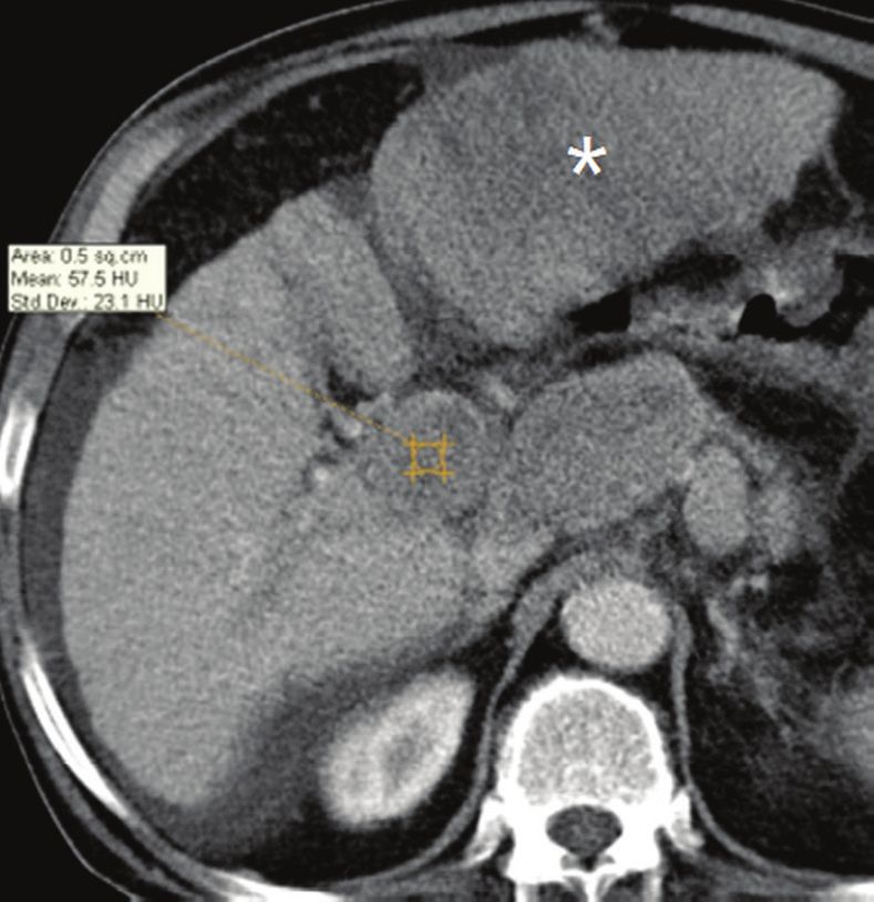
Minoda AM et al. / The ABCD of portal vein thrombosis: a systematic approach
a portal branch into the center of each hepatic segment
(Figure 1). The splenic vein originates from the junction
of its tributaries in the splenic hilum and receives the
short gastric, left gastroepiploic, inferior mesenteric, and
posterior pancreatic veins. The superior mesenteric vein
is formed by tributaries that drain the colonic segments
on the right, the small intestine, and the head of the pan-
creas. The left gastric vein connects directly to the portal
vein at its origin. Other tributaries, such as the right gas-
tric, cystic, accessory pancreatic, and superior pancreati-
coduodenal veins, are also received by the portal vein(7–9),
as depicted in Figure 2.
SYSTEMATIC RADIOLOGICAL APPROACH
TO PORTAL VEIN THROMBOSIS
A (age)
Portal vein thrombosis is classified as acute or chronic
depending on the time since its onset. However, it is dif-
ficult to define the time of onset on the basis of clinical pa-
rameters. In addition to fact that it can be asymptomatic,
the duration of symptoms has been shown to be an un-
reliable indicator of the time of onset of the condition(5).
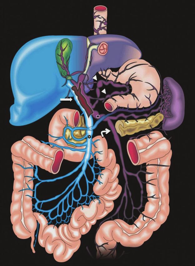
Figure 2. Schematic drawing showing the anatomy of the portal venous sys-
tem. The portal vein (straight arrow), formed by the union of the splenic vein
(black asterisk) and superior mesenteric vein (white asterisk), directly receives
the right and left gastric veins (arrowheads). The inferior mesenteric vein
(curved arrow) drains directly into the splenic vein. (Adapted from Netter(10)).
Therefore, the use of imaging methods is a feasible and
indispensable approach to making the differentiation be-
tween acute and chronic cases of portal vein thrombosis.
On ultrasound, an acute thrombus usually appears as
heterogeneous material in the vessel lumen, accompanied
by an increase in the caliber of the portal vein (to > 13 mm).
In some cases, the thrombus is hypoechoic or isoechoic,
and, on color Doppler, there is an absence of blood flow
in part or all of the lumen(9,11), as shown in Figure 3A.
As the thrombus ages and becomes chronic, its content
becomes more echogenic, with calcifications or cavernous
transformation, which is defined as the formation of collat-
eral vessels that bypass the obstruction, exhibiting multiple
serpiginous structures with flow on color Doppler, in the
periportal region(9,11), as can be seen in Figures 3B and 3C.
Despite being characteristic of the chronic phase, the ap-
pearance of a thrombus at 6–20 days after the acute event
has been reported(11). In the absence of cirrhosis or other
Figure 1. Coronal CT reconstruction showing the portal venous system. The
concomitant causes of portal hypertension, signs of severe
main portal vein (black asterisk) is formed by the union of the splenic vein portal hypertension, such as ascites, splenomegaly, and
(white arrow) and the superior mesenteric vein (white asterisk). The right and portosystemic collaterals, also suggest chronicity.
left portal branches divide within the hepatic hilum (black arrowheads). Other
tributaries include the inferior mesenteric vein (black arrow) and the gastric On CT, the acute form of portal vein thrombosis pres-
veins (white arrowhead). ents as increased portal vein attenuation in the pre-con-
Radiol Bras. 2020 Nov/Dez;53(6):424–429 425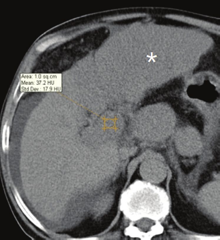
Minoda AM et al. / The ABCD of portal vein thrombosis: a systematic approach

 A                                                            B                                       C

 D                                                    E                                                   F
                                                     Figure 3. A: Patient in a hypercoagulable state with extensive acute portal vein thrombosis. Longi-
                                                     tudinal ultrasound image showing a hypoechoic thrombus (black arrow) in the main portal vein, the
                                                     caliber of which is increased, with slight peripheral blood flow on color Doppler (white arrow), the flow
                                                     being related to partial occlusion and not being a flow within the thrombus (which differentiates it from
                                                     a tumor thrombus). B: Schematic drawing showing the cavernous transformation of the portal vein.
                                                     Extensive thrombus (asterisk) in the main portal vein, with the formation of multiple collaterals (ar-
                                                     rows) around the obstruction. C: Color Doppler ultrasound in a case of chronic portal vein thrombosis,
                                                     showing serpiginous vessels (arrow) in the periportal region, indicative of cavernous transformation.
                                                     D: Unenhanced axial CT scan of the abdomen in a case of acute portal vein thrombosis, showing an
                                                     increase in the caliber of the portal vein (> 13 mm), which contains hyperdense material, best seen in
                                                     its right branch (arrow). E: Axial T2-weighted MRI scan in a case of acute portal vein thrombosis, show-
                                                     ing a hyperintense acute thrombus within the portal vein (white arrow). There is flow artifact (flow void)
                                                     in the main portal vein, suggesting partial thrombosis (black arrow). F: Magnetic resonance cholangi-
                                                     ography in a case of acute portal vein thrombosis. Note the markedly hyperintense signal, suggesting
                                                     acute portal vein thrombosis. Note also that, at first glance, it can mimic the common bile duct (arrow).
                                                     However, the diagnosis of acute portal vein thrombosis was confirmed after detailed analysis and
                                                     three-dimensional reconstructions. G: Contrast-enhanced axial T1-weighted MRI in a case of chronic
                                                     portal vein thrombosis, showing cavernous transformation of the portal vein (arrow).

                                                                               portal vein should be actively investigated, because they in-
                                                                               dicate a more fragile vessel and can complicate the surgical
                                                                               procedure(4,5).
                                                                                    Total or partial filling defects in the portal venous sys-
 G                                                                             tem can be detected on MRI, especially in the portal phase.
                                                                               On T1-weighted images, acute thrombi can be isointense
trast phase, a lack of enhancement after administration of                     to the muscle tissue or hyperintense when newly formed,
intravenous contrast, and increased portal vein caliber(4),                    whereas they continue to show high signal intensity on
as illustrated in Figure 3D. In the chronic form, the throm-                   T2-weighted images (Figure 3E). Chronic thrombi tend to
bosed vessel can be hypodense, containing linear calcifica-                    be hypointense or appear as a flow void, although a slow
tions, or obliterated, with cavernous transformation(4,11).                    flow artifact can change the signal to isointense or hyper-
In candidates for liver transplantation, calcifications in the                 intense(6), as shown in Figures 3F and 3G.

426                                                                                                       Radiol Bras. 2020 Nov/Dez;53(6):424–429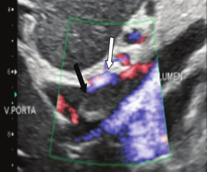
Minoda AM et al. / The ABCD of portal vein thrombosis: a systematic approach

B (biology)                                                                    C (collaterals)
     Thrombi can be differentiated, in terms of biology                             The absence of portosystemic collaterals and spleno-
or nature, as having a vascular or tumor nature. Vascular                      megaly usually indicates acute thrombosis, although the
thrombi are usually secondary to slow flow in patients with                    presence of those signs does not automatically indicate
cirrhosis, whereas tumor thrombi are related to invasion                       chronic thrombosis, because they can be attributable to
of a portal branch by a neoplasm. Despite the disparate                        intrahepatic portal hypertension secondary to cirrhosis(5).
pathophysiology, the two types can coexist, especially in                           The left gastric vein is responsible for the most com-
patients with HCC(5). Tumor thrombosis is a common,                            mon portosystemic shunt (Figure 5A), and a left gastric
well-reported complication of HCC and is considered one                        vein caliber > 6 mm is indicative of portal hypertension.
of the contraindications to liver transplantation, dramati-                    Dilated vessels in the esophageal wall are known as esoph-
cally altering the treatment options and prognosis(2,4,5).                     ageal varices, whereas those located adjacent to the esoph-
     On color Doppler, a tumor thrombus shows blood flow                       agus are known as paraesophageal varices(8), as illustrated
in its interior, with 100% specificity when the flow found                     in Figure 5B.
is arterial (neovascularization), a finding that is absent                          In patients with portal hypertension, a recanalized
in vascular thrombi(11). Neovascularization on CT is also                      umbilical vein can also be observed (Figure 5C), as can
highly specific and has been widely described as indicative                    a splenorenal shunt(9), as shown in Figure 5D, and gall-
of a tumor thrombus, which typically shows enhancement                         bladder wall varices, which are commonly seen in patients
greater than 20 HU after contrast administration(12–14),                       with cavernous transformation(8), as depicted in Figure
as depicted in Figures 4A and 4B. Vascular thrombi show                        5E. Other, less common shunts are deviation of the su-
variable density, depending on their age, and do not show                      perior mesenteric vein to the right renal vein, mesenteric
enhancement after contrast(7), as can be seen in Figures                       varices, and collaterals to the diaphragm.
4C and 4D.

                                            A                                                 B

Figure 4. A,B: CT scan showing a tumor
thrombus secondary to HCC (asterisk).
Axial sections acquired before and after
contrast administration (A and B, re-
spectively), showing 20 UH of enhance-
ment in the thrombus (i.e., 37 UH in the
pre-contrast phase and 57 UH in the
post-contrast phase). In addition, there
is expansion of the vessel, also related
to the tumor component. C: Axial CT
scan of a patient with a vascular throm-
bus, showing no enhancement of the
thrombus in the portal phase (asterisk).
D: Contrast-enhanced T1-weighted MRI
scan of a patient with a vascular throm-
bus in the left portal vein branch (black
arrow), showing no enhancement.
                                            C                                      D

Radiol Bras. 2020 Nov/Dez;53(6):424–429                                                                                              427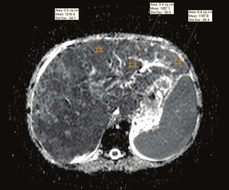
Minoda AM et al. / The ABCD of portal vein thrombosis: a systematic approach

 A                                                           B                                                 C

                  D                                                                           E
Figure 5. A: Gastroesophageal varices. Axial contrast-enhanced CT scan showing dilated vessels at the lesser curvature and at the gastroesophageal junction
(arrow). B: Contrast-enhanced MRI showing paraesophageal varices, presenting as dilated vessels around the distal esophageal portion (arrow). C: Contrast-
enhanced axial T1-weighted MRI scan showing recanalization of the paraumbilical veins, the reopening of the veins occurring in the round/falciform ligament
(arrow). D: Contrast-enhanced MRI of a splenorenal shunt. There is communication between the splenic vein and the left renal vein through serpiginous, dilated
vessels (arrow). E: Axial contrast-enhanced CT scan showing dilated and tortuous vessels in the gallbladder wall (arrow).

                  A                                                                       B
Figure 6. A: DWI of a patient with chronic liver disease and diffuse HCC, showing restricted diffusion in the right hepatic lobe (white arrow) and in the left portal
vein branch (black arrow). B: DWI of another patient with chronic liver disease and diffuse HCC, in which the ADC measured in the thrombus was 1.0 × 10⁻³
mm2/s, a value similar to that found for the tumor.

D (diffusion)                                                                       have signal intensity and apparent diffusion coefficient
    Diffusion-weighted imaging (DWI) can facilitate                                 (ADC) values similar to those of the primary tumor, the
the qualitative and quantitative evaluation of portal vein                          signal being hyperintense on DWI, which shows restric-
thrombosis. The role of DWI in the differentiation be-                              tion, and hypointense on the ADC map(5,6), as depicted
tween vascular and tumor thrombi was emphasized by                                  in Figures 6A and 6B. Vascular thrombi do not typical-
Catalano et al.(6), who demonstrated that tumor thrombi                             ly present restricted diffusion, except in cases of acute

428                                                                                                           Radiol Bras. 2020 Nov/Dez;53(6):424–429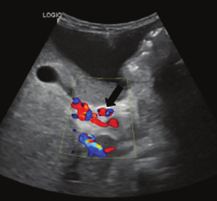
Minoda AM et al. / The ABCD of portal vein thrombosis: a systematic approach

hemorrhage, in which there is a “shine-through” effect                               vein thrombosis (neoplastic versus bland) on CT images using soft-
with different hemoglobin degradation products, limiting                             ware-based texture analysis and thrombus density (Hounsfield units).
                                                                                     AJR Am J Roentgenol. 2016;207:W81–W87.
and confounding the accurate analysis of the sequence.
                                                                                5.   Margini C, Berzigotti A. Portal vein thrombosis: the role of imaging
Therefore, DWI is an additional tool that, together with                             in the clinical setting. Dig Liver Dis. 2017;49:113–20.
the determination of other characteristics, such as the                         6.   Catalano OA, Choy G, Zhu A, et al. Differentiation of malignant
caliber of the portal vein, tumor expansion, neovascu-                               thrombus from bland thrombus of the portal vein in patients with
larization, the signal in the other (T1- and T2-weighted)                            hepatocellular carcinoma: application of diffusion-weighted MR
                                                                                     imaging. Radiology. 2010;254:154–62.
sequences, and the signs of portal hypertension, helps
                                                                                7.   Li C, Hu J, Zhou D, et al. Differentiation of bland from neoplastic
differentiate between vascular and tumor thrombi(5,13,14).                           thrombus of the portal vein in patients with hepatocellular carci-
                                                                                     noma: application of susceptibility-weighted MR imaging. BMC
CONCLUSION                                                                           Cancer. 2014;14:590.
     Because of its morbidity and the limitations of the                        8.   Melo-Leite AF, Mota Jr A, Chagas-Neto FA, et al. Acquired portosys-
                                                                                     temic collaterals: anatomy and imaging. Radiol Bras. 2016;49:251–6.
clinical evaluation, portal vein thrombosis demands care-
                                                                                9.   Martinelli ALC. Hipertensão portal. Medicina (Ribeirão Preto).
ful analysis by imaging methods. Important information                               2004;37:253–61.
should be extracted by the various imaging methods. By                         10.   Netter FH. Atlas of human anatomy. 2nd ed. East Hanover, NJ:
adopting the systematic ABCD approach, the data can be                               Novartis; 1997.
interpreted in a more careful and comprehensive manner.                        11.   Jha RC, Khera SS, Kalaria AD. Portal vein thrombosis: imaging the
                                                                                     spectrum of disease with an emphasis on MRI features. AJR Am J
REFERENCES                                                                           Roentgenol. 2018;211:14–24.
 1. Berzigotti A, García-Criado A, Darnell A, et al. Imaging in clinical       12.   Quarrie R, Stawicki SP. Portal vein thrombosis: what surgeons need
    decision-making for portal vein thrombosis. Nat Rev Gastroenterol                to know. Int J Crit Illn Inj Sci. 2018;8:73–7.
    Hepatol. 2014;11:308–16.                                                   13.   Sandrasegaran K, Tahir B, Nutakki K, et al. Usefulness of conven-
 2. Young K, Wong R. Evaluation and management of acute and chron-                   tional MRI sequences and diffusion-weighted imaging in differ-
    ic portal vein thrombosis in patients with cirrhosis. Clin Liver Dis             entiating malignant from benign portal vein thrombus in cirrhotic
    (Hoboken). 2018;10:152–6.                                                        patients. AJR Am J Roentgenol. 2013;201:1211–9.
 3. Ponziani FR, Zocco MA, Campanale C, et al. Portal vein thrombo-            14.   Gawande R, Jalaeian H, Niendorf E, et al. MRI in differentiating
    sis: insight into physiopathology, diagnosis, and treatment. World J             malignant versus benign portal vein thrombosis in patients with
    Gastroenterol. 2010;16:143–55.                                                   hepatocellular carcinoma: value of post contrast imaging with sub-
 4. Canellas R, Mehrkhani F, Patino M, et al. Characterization of portal             traction. Eur J Radiol. 2019;118:88–95.

Radiol Bras. 2020 Nov/Dez;53(6):424–429                                                                                                            429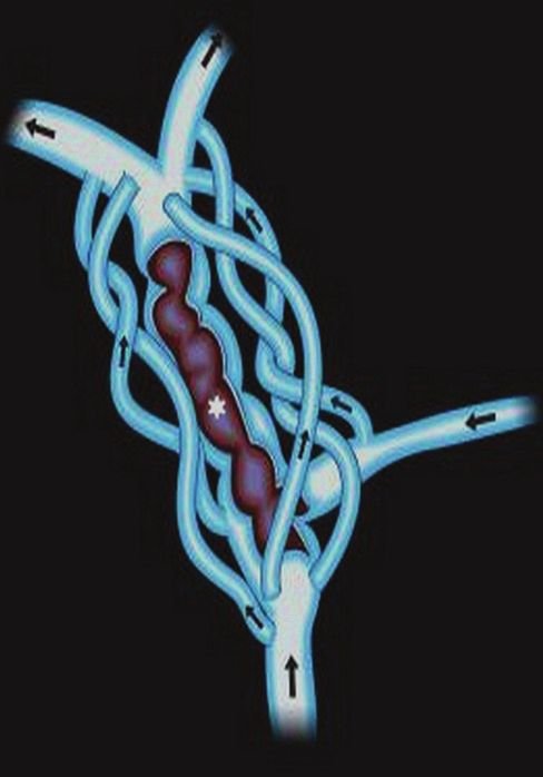
You can also read