Ractopamine-induced changes in the proteome of post-mortem beef
←
→
Page content transcription
If your browser does not render page correctly, please read the page content below
Journal homepage: http://www.sasas.co.za/journals

                                        South African Journal of Animal Science 2019, 49 (No. 3)

     Ractopamine-induced changes in the proteome of post-mortem beef
                     longissimus lumborum muscle
H.M. Kim1, S.P. Suman1#, S. Li1, C.M. Beach2, M.N. Nair3, C. Zhai3, B.M. Edenburn4, T.L. Felix5,
                                   A.C. Dilger4 & D.D. Boler4
              1
               Department of Animal and Food Sciences, University of Kentucky, Lexington, KY 40546, USA
                      2
                        Proteomics Core Facility, University of Kentucky, Lexington, KY 40506, USA
                3
                  Department of Animal Sciences, Colorado State University, Fort Collins, CO 80523, USA
                     4
                        Department of Animal Sciences, University of Illinois, Urbana, IL 61801, USA
             5
               Department of Animal Science, Pennsylvania State University, University Park, PA 16802, USA

                      (Received 10 October 2018; Accepted 26 March 2019; First published online 13 May 2019)

             Copyright resides with the authors in terms of the Creative Commons Attribution 4.0 South African License.
                                         See: http://creativecommons.org/licenses/by/4.0/za
           Condition of use: The user may copy, distribute, transmit and adapt the work, but must recognize the authors and
                                            the South African Journal of Animal Science.
______________________________________________________________________________________
Abstract
       Ractopamine is a beta-adrenergic agonist that is approved for use in beef cattle, pigs and turkeys as a
repartitioning agent to increase lean muscle deposition and decrease lipogenesis. Although the effects of
dietary ractopamine on the proteome profile of post-mortem pork muscles have been examined, its influence
on beef muscle proteome has not been studied. Therefore, the objective of this study was to examine the
effect of ractopamine on the proteome profile of post-mortem beef longissimus lumborum (LL) muscle. LL
muscle samples were obtained from the carcasses of six (n = 6) steers fed ractopamine (RAC; 400 mg
ractopamine hydrochloride for 28 days) and six (n = 6) steers fed no ractopamine (CON). The muscle
proteome was analysed using two-dimensional gel electrophoresis and tandem mass spectrometry. Five
differentially abundant spots were identified, and all the spots were over-abundant in RAC. The identified
proteins were involved in muscle structure development (F-actin-capping protein subunit beta-2; PDZ and
LIM domain protein-3), chaperone activity (heat shock protein beta-1), oxygen transport (myoglobin), and
glycolysis (L-lactate dehydrogenase A chain). These results suggested that dietary ractopamine could
influence the abundance of enzymes associated with muscle development and muscle fibre type shift in beef
LL muscle.
______________________________________________________________________________________
Keywords: growth promotants, meat quality, proteomics
#
    Corresponding author: spsuma2@uky.edu

Introduction
       Ractopamine is a beta-adrenergic agonist that is approved as a growth promotant in the pork, turkey
and beef industries in the United States. This beta-agonist enhances muscle protein synthesis, decreases
lipid deposition, and increases leanness (Johnson & Chung, 2007). The improved leanness could be
attributed to the increased feed efficiency (Avendaño-Reyes et al., 2006; Abney et al., 2007; Quinn et al.,
2016) and muscle accretion (Quinn et al., 2008; Bryant et al., 2010; Boler et al., 2012; Brigida et al., 2018).
Furthermore, several studies have documented that ractopamine feeding resulted in muscle fibre type shift
from oxidative (Types IIA, IIX) to glycolytic (Type IIB) in pigs (Depreux et al., 2002; Gunawan et al., 2007;
Almeida et al., 2015). Gonzalez et al. (2009) examined six muscles (longissimus lumborum,
semimembranosus, vastus lateralis, adductor, gracilis and rectus femoris) from ractopamine-fed steers and
observed a fibre type shift from Type I to Type IIA in all muscles except semimembranosus. Meta-analyses
of data from ractopamine feeding studies in beef animals reported that the growth promotant increased rib
eye area and hot carcass weight, but decreased tenderness and marbling (Lean et al., 2014).
       Recently, several studies have examined the influence of dietary ractopamine on the proteome profile
of post-mortem skeletal muscles. Costa-Lima et al. (2015) analysed the sarcoplasmic proteome profile of
pork longissimus thoracis and found that nine proteins were differentially abundant between control and
ractopamine-fed pigs. These results suggested that ractopamine influenced the abundance of enzymes

URL: http://www.sasas.co.za
ISSN 0375-1589 (print), ISSN 2221-4062 (online)
Publisher: South African Society for Animal Science                                             http://dx.doi.org/10.4314/sajas.v49i3.3.
Kim et al., 2019. S. Afr. J. Anim. Sci. vol. 49 425
associated with glycolytic metabolism and thus may potentially influence the conversion of muscle to meat.
Additionally, Wu et al. (2016) examined the sarcoplasmic proteome of semimembranosus from pigs fed
ractopamine and control diets and observed that five proteins (involved in oxidative metabolism, chaperone
and plasma membrane repair) were differentially abundant between the two groups.
Although the effects of ractopamine on proteome profile of pork muscles have been studied, its
influence on beef muscle proteome has not been investigated. Therefore, the objective of the current study
was to examine the influence of ractopamine on the proteome of beef longissimus lumborum (LL) muscle.
Materials and Methods
The muscle samples were obtained from a previous feeding study (Edenburn et al., 2016) that was
completed at the University of Illinois. All animal procedures were approved by the University of Illinois
Institutional Animal Care and Use Committee (IACUC protocol #12009) and followed the guidelines
recommended in the Guide for the Care and Use of Agricultural Animals in Agricultural Research and
Teaching (FASS, 2010). Steers were fed a corn-based diet for 188 days before the initiation of this study and
were implanted with Component TE-IS (80 mg trenbolone acetate, 16 mg estradiol; Elanco Animal Health,
Greenfield, IN) 104 days before the initiation of the study. Seventy-two steers were used in the study and
were allocated to 12 pens with 6 steers per pen. Pens were randomly assigned to 1 of 2 treatments on day
0: ractopamine hydrochloride (RAC) and no ractopamine (CON). Steers were fed in 3-m concrete bunks.
Steers in the RAC group received Optaflexx 45 (Elanco Animal Health, Greenfield, IN, USA) to provide 400
mg RAC/steer per day for 28 days. All steers were fed the same basal diet of 60% dry-rolled corn, 20% corn
silage (approximately 50 : 50 grain : forage), 10% dry distillers grains, and 10% supplement on a dry matter
basis. Each diet was formulated to meet or exceed NRC guidelines (NRC, 2000). Steers were fed once daily.
At the end of the 28-day ractopamine supplementation, one steer was randomly selected from each of the
six pens in CON (628 kg average body weight) and RAC (635 kg average body weight) at the end of the
28-day feeding period. This approach provided six replicates (n = 6) from RAC and six replicates (n = 6) from
CON for proteome analysis. These 12 steers were transported to a USDA-inspected commercial meat
packing facility. Cattle were fasted for approximately 16 hours, but were provided water until slaughter. At 24
th th
hours post-mortem, a 2.5-cm thick sample of LL muscle (between the 12 and 13 ribs) was collected from
each carcass, vacuum-packaged, frozen immediately at -80 °C, and transported in dry ice to University of
Kentucky. The data on growth performance, carcass traits and meat quality are presented and discussed in
Edenburn et al. (2016).
The muscle proteome from beef LL muscle was extracted as described by Lametsch et al. (2003).
One gram of frozen muscle tissue was cut and homogenized (Polytron PT 10-35 GT, Kinematica, Luzern,
Switzerland) in 5 mL extraction buffer (8 M urea, 2 M Thiourea, 2% Chaps, 65 mM DTT, and 0.5% pH 3-10
ampholyte) for 5 min. Crude extract was transferred to centrifuge tubes, vigorously shaken for 2 hours at
4 °C, and centrifuged (10000 × g) for 30 min at 4 °C. The supernatant, which consisted of muscle proteins,
was filtered and utilized for analysis. Bradford assay was used to determine the protein concentration of
sarcoplasmic extract (Bio-Rad, Hercules, CA, USA). The sarcoplasmic proteins (900 μg) were mixed with
rehydration buffer (Bio-Rad) optimized to 7 M urea, 2 M thiourea, 4% CHAPS, 20 mM DTT, 0.5% Bio-Lyte
5/8 ampholyte, and 0.001% of bromophenol blue. The mixture was loaded into immobilized pH gradient
(IPG) strips (pH 3-10, 17 cm; Bio-Rad). The IPG strips were subjected to passive rehydration for 16 hours.
First dimension isoelectric focusing (IEF) process was conducted using Protean IEF cell system. First, an
active rehydration step was conducted with low voltage (50 V) and increased by stages, with final rapid
voltage ramping to reach a total of 80 kVh. Subsequently, the IPG strips were equilibrated with equilibration
buffer I (6 M urea, 0.375 M Tris–HCl, pH 8.8, 2% SDS, 20% glycerol, 2% DTT) for 15 min, followed by
equilibration buffer II (6 M urea, 0.375 M Tris–HCl, pH 8.8, 2% SDS, 20% glycerol, 2.5% Iodoacetamide) for
15 min. The second dimension protein separation process was conducted by 12% sodium dodecyl sulphate
polyacrylamide gel electrophoresis (SDS-PAGE; 38.5:1 ratio of acrylamide to bis-acrylamide) using Protean
II XL system (Bio-Rad). The gels were stained by Colloidal Coomassie Blue for 48 hours, and destained until
the background of the gels was cleared. The CON and RAC samples were evaluated under the same
conditions (two gels/sample), resulting in 24 gels. The gels were scanned using VersaDoc (Bio-Rad) and gel
images were analysed using PDQuest (Bio-Rad, Hercules, CA, USA). First, spot detection was conducted
and matched, then normalized (Joseph et al., 2012). The spots were considered differentially abundant when
a 1.5-fold or more intensity difference was measured between CON and RAC, with 90% statistical
significance (P426 Kim et al., 2019. S. Afr. J. Anim. Sci. vol. 49
LTQ-Orbitrap mass spectrometer (Thermo Fisher Scientific, Waltham, MA) coupled with an Eksigent
Nanoflex cHiPLC™ system (Eksigent, Dublin, CA, USA) through a nano electrospray ionization source. A
reverse phase cHiPLC column (75 μm × 150 mm) was operated (300 nL/min flow rate) for separation of the
peptides. Water with 0.1% (v/v) formic acid was used for mobile phase A, and acetonitrile with 0.1% (v/v)
formic acid was used for mobile phase B. A 50 min gradient was applied. The initial 3% mobile phase B was
linearly increased to 50% in 24 min and further to 85% and 95% for 5 min each, before it was decreased to
3%, then the column was re-equilibrated. The mass analysis method consisted of eight scan events per
segment. The first scan event was an Orbitrap MS scan (100 - 1600 m/z) with 60 000 resolutions for parent
ions, and then followed by data dependent MS/MS for fragmentation of the seven most intense ions through
collision induced dissociation (CID). The LC-MS/MS data were submitted to a local Mascot server for MS/MS
protein identification through Proteome Discoverer (version 1.3, Thermo Fisher Scientific, Waltham, MA,
USA) based on the Bos taurus database from National Center for Biotechnology Information (NCBI). The
parameters of the MASCOT MS/MS ion search were trypsin digest with a maximum of two miscleavages,
cysteine carbamidomethylation, methionine oxidation, a maximum of 10 ppm MS error tolerance, and a
maximum of 0.8 Da MS/MS error tolerance. A decoy database was conducted and searched. To distribute
the confidence indicators for the peptide matches, filter settings to determine false discovery rates (FDR)
were used. Peptide matches that passed the filter associated with the strict FDR (target setting of 0.01) were
assigned as high confidence. For the MS/MS ion search, proteins with two of more high confidence peptides
were considered unambiguous identifications without manual inspection, whereas proteins identified with
one high confidence peptide were manually inspected and confirmed.
The differentially expressed proteins were matched against the STRING database (Szklarczyk et al.,
2015) to determine the protein-protein interaction network, in which the network nodes represented the
proteins and the lines indicated functional associations.
Results and Discussion
Five differentially abundant spots were identified from the image analyses of muscle proteome gels
(Figure 1). The accession number, database score, matched peptides, and sequence coverage of the
identified proteins are listed in Table 1. All the identified proteins were over-abundant (PKim et al., 2019. S. Afr. J. Anim. Sci. vol. 49 427
2010), and the ALP strengthens the association between α-actinin and actin filaments (Xia et al., 1997;
Klaavuniemi et al., 2004; Vallenius et al., 2004).
pH 3 10
MW (kDa)
100
2
5
1
3
4
10
Figure 1 Coomassie-stained two-dimensional gel of muscle proteome extracted from longissimus lumborum
of beef steers fed on ractopamine. Five protein spots differentially abundant in control and ractopamine-fed
beef steers are encircled and numbered.
Heat shock protein beta-1 (Hsp27) belongs to the family of small heat shock proteins, which are
distributed widely in various tissues and play an important role in cell survival under stress conditions
(Bakthisaran et al., 2015; Haslbeck & Vierling, 2015). This protein plays a critical role in stabilizing the
cytoskeleton, especially with protecting muscle filaments and stabilizing the muscle structure (Perng et al.,
1999a; 1999b). Furthermore, Pivovarova et al. (2005) reported that Hsp27 efficiently prevents heat-induced
aggregation of F-actin. Previous proteomic investigations indicated that Hsp27 is involved in muscle growth
and meat quality. Lametsch et al. (2006) analysed the proteome of longissimus muscles from pigs that
exhibited compensatory growth and normal growth and observed that Hsp27 was over-abundant in animals
that demonstrated compensatory growth, suggesting an important role of Hsp27 in muscle hypertrophy
during compensatory growth. Shibata et al. (2009) compared the proteome of semitendinosus muscles from
grass-fed and grain-fed cattle and found that Hsp27 was over-abundant in grain-fed cattle. These authors
speculated that Hsp27 may have some role in skeletal muscle growth in exercise-restricted cattle.
Furthermore, Hsp27 has been reported to be associated with tenderness (Kim et al., 2008; Carvalho et al.,
2014) and colour (Sayd et al., 2006; Joseph et al., 2012).428 Kim et al., 2019. S. Afr. J. Anim. Sci. vol. 49
Table 1 Identity and functional roles of differentially abundant proteins in muscle proteome of longissimus
lumborum from beef steers fed on ractopamine
ProtScore/ Sequence Over-
Accession Spot
Spota Protein Matched Coverage Function abundant c
No: ratio
peptides (%) treatmentb
1 F-actin-capping protein P79136-2 1569.72/19 62.13 Muscle RAC 2.20
subunit beta-2 development
2 PDZ and LIM domain Q3SYZ8 1346.65/20 63.92 Muscle RAC 1.80
protein-3 development
3 Heat shock protein beta-1 Q3T149 2787.86/20 80.60 Chaperone RAC 1.96
activity
4 Myoglobin P02192 2371.06/26 99.35 Oxygen RAC 1.51
transport
5 L-lactate dehydrogenase A P19858 944.61/22 56.33 Glycolysis RAC 1.62
chain
a
Spot number refers to the numbered spots in gel image (Figure 1). Spots are identified by accession number (UniProt),
ProtScore, matched peptides number, and sequence coverage of peptides
b
CON = Non-ractopamine hydrochloride diet ; RAC = 400 mg ractopamine hydrochloride diet for 28 days before
slaughter
c
Spot ratio of RAC/CON
Myoglobin is the oxygen-binding heme protein in mammalian muscle tissue, and its primary function
is storage and transport of oxygen (Lehninger et al., 2005; Schiaffino & Reggiani, 2011). It transports oxygen
from red blood cells to mitochondria within the muscles during periods of increased metabolic activity and
serves as an oxygen reservoir during anoxic and hypoxic conditions in the skeletal muscles (Ordway &
Garry, 2004). Furthermore, myoglobin plays a critical role in meat colour (Mancini & Hunt, 2005; Suman &
Joseph, 2013; Faustman & Suman, 2017). The over-abundance of myoglobin in RAC was unexpected since
dietary ractopamine is known to cause a muscle fibre shift from oxidative to glycolytic in pigs (Depreux et al.,
2002) and beef cattle (Gonzalez et al., 2009).
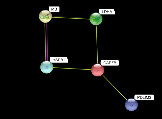
Figure 2 Protein-protein interaction network of differential abundant proteins in muscle proteome of
longissimus lumborum from CON (non-ractopamine hydrochloride diet) and RAC (400 mg ractopamine
hydrochloride diet for 28 days before slaughter) beef steers
The interacting proteins were identified using STRING 11.0 software (Szklarczyk et al., 2015).
The nodes represent proteins from Bos taurus database, whereas the lines (purple = experimental evidence; light green
= text mining evidence; black = co-expression evidence) indicate predicted functional annotations.
HSPB1 = Heat shock protein beta-1; MB = myoglobin; LDHA = L-lactate dehydrogenase A chain; CAPZB = F-actin-
capping protein subunit beta-2; PDLIM3 = PDZ and LIM domain protein-3Kim et al., 2019. S. Afr. J. Anim. Sci. vol. 49 429
L-lactate dehydrogenase A chain is an enzyme that catalyses the reversible conversion of lactate to
pyruvate (Gladden, 2004). The over-abundance of lactate dehydrogenase in RAC could be attributed to the
muscle fibre shift from oxidative to glycolytic caused by ractopamine. Previous studies have reported that
lactate dehydrogenase is more abundant and active in glycolytic muscle fibres than in oxidative muscle
fibres (Picard et al., 2002; Huber et al., 2007; Izumiya et al., 2008). Ractopamine shifts muscle fibre type
from fast oxidative-glycolytic (Type IIA and IIX) to fast glycolytic (Type IIB) in pigs (Depreux et al., 2002) and
from slow oxidative (Type I) to fast oxidative-glycolytic (Type IIA) in cattle (Gonzalez et al., 2008).
Furthermore, Costa-Lima et al. (2015) observed over-abundance of L-lactate dehydrogenase A chain in
longissimus thoracis muscles of ractopamine-fed pigs. Meta-analysis of studies on ractopamine-fed beef
reported that this beta-agonist increases shear force (Lean et al., 2014). The over-abundance of L-lactate
dehydrogenase A chain may be one of the possible biomarkers for the decrease in meat tenderness in
ractopamine-fed cattle. Guillemin et al. (2011) studied 24 protein markers related to meat tenderness in beef
longissimus thoracis (fast oxidative-glycolytic) and semitendinosus (fast glycolytic) muscles and found that
lactate dehydrogenase B chain was positively correlated with toughness in semitendinosus, but not in
longissimus thoracis. Furthermore, Maltin et al. (2003) suggested that glycolytic muscles would be less
tender than oxidative ones due to inherent variations in muscle fibre size, contractile mechanisms, and
metabolic pathways.
Conclusion
The results of the present study suggested that dietary ractopamine influenced the abundance of
proteins related to muscle structure development, chaperone activity, oxygen transport, and glycolysis in
post-mortem beef longissimus lumborum muscle. Additional studies are necessary to characterize how
ractopamine influences the proteome in ante- and peri-mortem beef skeletal muscles to characterize the
influence of the growth promotant on muscle to meat conversion and meat quality attributes.
Acknowledgements
This is publication number 18-07-090 of the Kentucky Agricultural Experiment Station and is published with the
approval of the director. This work was supported by the National Institute of Food and Agriculture, U.S. Department of
Agriculture, Hatch-Multistate Project 1014747.
Authors’ Contributions
Conception and design: SPS, TLF, ACD & DDB; data collection and analyses: HMK, SL, CMB, MNN, CZ & BME;
drafting of paper: HMK; critical revision and final approval of version to be published: SPS, TLF & DDB.
Conflict of Interest Declaration
The authors declare that they have no affiliations with any organization or entity with any financial or non-
financial interest that could bias the subject matter and outcomes discussed in this manuscript.
References
Abney, C.S., Vasconcelos, J.T., McMeniman, J.P., Keyser, S.A., Wilson, K.R., Vogel, G.J. & Galyean, M.L., 2007.
Effects of ractopamine hydrochloride on performance, rate and variation in feed intake, and acid-base balance in
feedlot cattle. J. Anim. Sci. 85, 3090-3098.
Almeida, V.V., Nuñez, A.J.C., Schinckel, A.P., Ward, M.G., Andrade, C., Sbardella, M., Berenchtein, B., Coutinho, L.L. &
Miyada, V.S., 2015. Gene expression of beta-adrenergic receptors and myosin heavy chain isoforms induced by
ractopamine feeding duration in pigs not carrying the ryanodine receptor mutation. Livest. Sci. 172, 91-95.
Avendaño-Reyes, L., Torres-Rodríguez, V., Meraz-Murillo, F.J., Pérez-Linares, C., Figueroa-Saavedra, F. & Robinson,
P.H., 2006. Effects of two β-adrenergic agonists on finishing performance, carcass characteristics, and meat
quality of feedlot steers. J. Anim. Sci. 84, 3259-3265.
Bakthisaran, R., Tangirala, R. & Rao, C.M., 2015. Small heat shock proteins: role in cellular functions and pathology.
BBA-Proteins Proteom. 1854, 291-319.
Boler, D.D., Shreck, A.L., Faulkner, D.B., Killefer, J., McKeith, F.K., Homm, J.W. & Scanga, J.A., 2012. Effect of
ractopamine hydrochloride (Optaflexx) dose on live animal performance, carcass characteristics and tenderness
in early weaned beef steers. Meat Sci. 92, 458-463.
Brigida, D.J., Antonelo, D.S., Mazon, M.R., Nubiato, K.E.Z., Gomez, J.F.M., Netto, A.S., Leme, P.R., Consolo, N.R.B.,
Pesce, D.M.C. & Silva, S.L. 2018. Effects of immunocastration and a β-adrenergic agonist on retail cuts of feedlot
finished Nellore cattle. Animal 12, 1690-1695.
Bryant, T.C., Engle, T.E., Galyean, M.L., Wagner, J.J., Tatum, J.D., Anthony, R.V. & Laudert, S.B., 2010. Effects of
ractopamine and trenbolone acetate implants with or without estradiol on growth performance, carcass
characteristics, adipogenic enzyme activity, and blood metabolites in feedlot steers and heifers. J. Anim. Sci. 88,
4102-4119.
Carvalho, M.E., Gasparin, G., Poleti, M.D., Rosa, A.F., Balieiro, J.C.C., Labate, C.A., Nassu, R.T., Tullio, R.R., Regitano,
L.C.D.A., Mourão, G.B. & Coutinho, L.L., 2014. Heat shock and structural proteins associated with meat
tenderness in Nellore beef cattle, a Bos indicus breed. Meat Sci. 96, 1318-1324.430 Kim et al., 2019. S. Afr. J. Anim. Sci. vol. 49
Clark, K.A., McElhinny, A.S., Beckerle, M.C. & Gregorio, C.C., 2002. Striated muscle cytoarchitecture: an intricate web of
form and function. Annu. Rev. Cell Dev. Bi. 18, 637-706.
Costa-Lima, B.R.C., Suman, S.P., Li, S., Beach, C.M., Silva, T.J.P., Silveira, E.T.F., Bohrer, B.M. & Boler, D.D., 2015.
Dietary ractopamine influences sarcoplasmic proteome profile of pork longissimus thoracis. Meat Sci. 103, 7-12.
Depreux, F.F.S., Grant, A.L., Anderson, D.B. & Gerrard, D.E., 2002. Paylean alters myosin heavy chain isoform content
in pig muscle. J. Anim. Sci. 80, 1888-1894.
Edenburn, B.M., Kneeskern, S.G., Bohrer, B.M., Rounds, W., Boler, D.D., Dilger, A.C. & Felix, T.L., 2016. Effects of
supplementing zinc or chromium to finishing steers fed ractopamine hydrochloride on growth performance,
carcass characteristics, and meat quality. J. Anim. Sci. 94, 771-779.
FASS, 2010. Guide for the care and use of agricultural animals in agricultural research and teaching. 3rd edition.
Federation of Animal Science Societies, Champaign, Illinois, USA.
Faustman, C. & Suman, S.P., 2017. The eating quality of meat: color In: Lawrie´s Meat Science. 8th edition. Edited by F.
Toldrá. Woodhead, Cambridge, England. pp. 329-356.
Gagaoua, M., Terlouw, E.M.C., Micol, D., Boudjellal, A., Hocquette, J.F. & Picard, B., 2015. Understanding early post-
mortem biochemical processes underlying meat color and pH decline in the longissimus thoracis muscle of young
blond d’aquitaine bulls using protein biomarkers. J. Agr. Food Chem. 63, 6799-6809.
Gladden, L.B., 2004. Lactate metabolism: A new paradigm for the third millennium. J. Physiol. 558, 5-30.
Gonzalez, J.M., Dijkhuis, R.D., Johnson, D.D., Carter, J.N. & Johnson, S.E., 2008. Differential response of cull cow
muscles to the hypertrophic actions of ractopamine-hydrogen chloride. J. Anim. Sci. 86, 3568-3574.
Gonzalez, J.M., Johnson, S.E., Thrift, T.A., Savell, J.D., Ouellette, S.E. & Johnson, D.D., 2009. Effect of ractopamine-
hydrochloride on the fiber type distribution and shelf-life of six muscles of steers. J. Anim. Sci. 87, 1764-1771.
Guillemin, N., Bonnet, M., Jurie, C. & Picard, B., 2011. Functional analysis of beef tenderness. J. Proteomics 75,
352-365.
Gunawan, A.M., Richert, B.T., Schinckel, A.P., Grant, A.L. & Gerrard, D.E., 2007. Ractopamine induces differential gene
expression in porcine skeletal muscles. J. Anim. Sci. 85, 2115-2124.
Haslbeck, M. & Vierling, E., 2015. A first line of stress defence: small heat shock proteins and their function in protein
homeostasis. J. Mol. Biol. 427, 1537-1548.
Hoshijima, M., 2006. Mechanical stress-strain sensors embedded in cardiac cytoskeleton: Z disk, titin, and associated
structures. Am. J. Physiol.-Heart C. 290, H1313-H1325.
Huber, K., Petzold, J., Rehfeldt, C., Ender, K. & Fiedler, I., 2007. Muscle energy metabolism: structural and functional
features in different types of porcine striated muscles. J. Muscle Res. Cell M. 28, 249-258.
Izumiya, Y., Hopkins, T., Morris, C., Sato, K., Zeng, L., Viereck, J., Hamilton, J.A., Ouchi, N., LeBrasseur, N.K. & Walsh,
K., 2008. Fast/glycolytic muscle fiber growth reduces fat mass and improves metabolic parameters in obese mice.
Cell Metab. 7, 159-172.
Johnson, B.J. & Chung, K.Y., 2007. Alterations in the physiology of growth of cattle with growth-enhancing compounds.
Vet. Clin. N. Am. Food A. 23, 321-332.
Joseph, P., Suman, S.P., Rentfrow, G., Li, S. & Beach, C.M., 2012. Proteomics of muscle-specific beef color stability.
J. Agr. Food Chem. 60, 3196-3203.
Kim, N.K., Cho, S., Lee, S.H., Park, H.R., Lee, C.S., Cho, Y.M., Choy, Y.H., Yoon, D., Im, S.K. & Park, E.W., 2008.
Proteins in longissimus muscle of Korean native cattle and their relationship to meat quality. Meat Sci. 80,
1068-1073.
Klaavuniemi, T., Kelloniemi, A. & Ylänne, J., 2004. The ZASP-like motif in actinin-associated LIM protein is required for
interaction with the α-actinin rod and for targeting to the muscle Z-line. J. Biol. Chem. 279, 26402-26410.
Krcmery, J., Camarata, T., Kulisz, A. & Simon, H.G., 2010. Nucleocytoplasmic functions of the PDZ-LIM protein family:
new insights into organ development. BioEssays 32, 100-108.
Lametsch, R., Karlsson, A., Rosenvold, K., Andersen, H.J., Roepstorff, P. & Bendixen, E., 2003. Postmortem proteome
changes of porcine muscle related to tenderness. J. Agr. Food Chem. 51, 6992-6997.
Lametsch, R., Kristensen, L., Larsen, M.R., Therkildsen, M., Oksbjerg, N. & Ertbjerg, P. 2006. Changes in the muscle
proteome after compensatory growth in pigs. J. Anim. Sci. 84, 918-924.
Lean, I.J., Thompson, J.M. & Dunshea, F.R., 2014. A meta-analysis of zilpaterol and ractopamine effects on feedlot
performance, carcass traits and shear strength of meat in cattle. PloS one 9, e115904. doi:
10.1371/journal.pone.0115904
Lehninger, A.L., Nelson, D.L. & Cox., M.M., 2005. Lehninger Principles of Biochemistry (4th ed.). W.H. Freeman, New
York, USA.
Maltin, C., Balcerzak, D., Tilley, R. & Delday, M., 2003. Determinants of meat quality: tenderness. Proc. Nutr. Soc. 62,
337-347.
Mancini, R.A. & Hunt, M.C., 2005. Current research in meat color. Meat Sci. 71, 100-121.
NRC, 2000. Nutrient requirements of beef cattle (7th ed.). National Academies Press, Washington, D.C., USA.
Ordway, G.A. & Garry, D.J., 2004. Myoglobin: an essential hemoprotein in striated muscle. J. Exp. Biol. 207, 3441-3446.
Perng, M.D., Cairns, L., Ijssel, P.V.D., Prescott, A., Hutcheson, A.M. & Quinlan, R.A., 1999a. Intermediate filament
interactions can be altered by HSP27 and alphaB-crystallin. J. Cell Sci. 112, 2099-2112.
Perng, M.D., Muchowski, P.J., IJssel, P.V.D., Wu, G.J.S., Hutcheson, A.M., Clark, J.I. & Quinlan, R.A., 1999b. The
cardiomyopathy and lens cataract mutation in αB-crystallin alters its protein structure, chaperone activity, and
interaction with intermediate filaments in vitro. J. Biol. Chem. 274, 33235-33243.
Picard, B., Lefaucheur, L., Berri, C. & Duclos, M.J., 2002. Muscle fiber ontogenesis in farm animal species. Reprod. Nutr.
Dev. 42, 415-431.Kim et al., 2019. S. Afr. J. Anim. Sci. vol. 49 431
Pivovarova, A.V., Mikhailova, V.V., Chernik, I.S., Chebotareva, N.A., Levitsky, D.I. & Gusev, N.B., 2005. Effects of small
heat shock proteins on the thermal denaturation and aggregation of F-actin. Biochem. Bioph. Res. Co. 331,
1548-1553.
Pollard, T.D. & Cooper, J.A., 1986. Actin and actin-binding proteins. A critical evaluation of mechanisms and functions.
Annu. Rev. Biochem. 55, 987-1035.
Pomies, P., Macalma, T. & Beckerle, M.C., 1999. Purification and characterization of an alpha-actinin-binding PDZ-LIM
protein that is up-regulated during muscle differentiation. J. Biol. Chem. 274, 29242-29250.
Pomies, P., Pashmforoush, M., Vegezzi, C., Chien, K.R., Auffray, C. & Beckerle, M.C., 2007. The cytoskeleton-
associated PDZ-LIM protein, ALP, acts on serum response factor activity to regulate muscle differentiation. Mol.
Biol. Cell 18, 1723-1733.
Ponsuksili, S., Murani, E., Phatsara, C., Schwerin, M., Schellander, K. & Wimmers, K., 2009. Porcine muscle sensory
attributes associate with major changes in gene networks involving CAPZB, ANKRD1, and CTBP2. Funct. Integr.
Genomic. 9, 455-471.
Pyle, W.G., Hart, M.C., Cooper, J.A., Sumandea, M.P., Tombe, P.P.D. & Solaro, R.J., 2002. Actin capping protein. Circ.
Res. 90, 1299-1306.
Quinn, M.J, Reinhardt, C.D., Loe, E.R., Depenbusch, B.E., Corrigan, M.E., May, M.L. & Drouillard, J.S., 2008. The
effects of ractopamine-hydrogen chloride (Optaflexx) on performance, carcass characteristics, and meat quality of
finishing feedlot heifers. J. Anim. Sci. 86, 902-908.
Quinn, M.J., Walter, L.J., Swingle, R.S., Defoor, P.J., Harper, L.B. & Lawrence, T.E., 2016. Comparison of the effects of
Actogain or Optaflexx on finishing feedlot steer performance and carcass characteristics. Prof. Ani. Sci. 32,
455-460.
Russell, B., Curtis, M.W., Koshman, Y.E. & Samarel, A.M., 2010. Mechanical stress-induced sarcomere assembly for
cardiac muscle growth in length and width. J. Mol. Cell Cardiol. 48, 817-823.
Sayd, T., Morzel, M., Chambon, C., Franck, M., Figwer, P., Larzul, C., Roy, P.L., Monin, G., Chérel, P. & Laville, E.,
2006. Proteome analysis of the sarcoplasmic fraction of pig semimembranosus muscle: implications on meat
color development. J. Agr. Food Chem. 54, 2732-2737.
Schafer, D.A., Korshunova, Y.O., Schroer, T.A. & Cooper, J.A., 1994. Differential localization and sequence analysis of
capping protein beta-subunit isoforms of vertebrates. J. Cell Biol. 127, 453-465.
Schiaffino, S. & Reggiani, C., 2011. Fiber types in mammalian skeletal muscles. Physiol. Rev. 91, 1447-1531.
Shibata, M., Matsumoto, K., Oe, M., Ohnishi-Kameyama, M., Ojima, K., Nakajima, I., Muroya, S. & Chikuni, K., 2009.
Differential expression of the skeletal muscle proteome in grazed cattle. J. Anim. Sci. 87, 2700-2708.
Suman, S.P. & Joseph, P., 2013. Myoglobin chemistry and meat color. Annu. Rev. Food Sci. T. 4, 79-99.
Szklarczyk, D., Franceschini, A., Wyder, S., Forslund, K., Heller, D., Huerta-Cepas, J., Simonovic, M., Roth, A., Santos,
A., Tsafou, K.P., Kuhn, M., Bork, P., Jensen, L.J. & von Mering, C., 2015. String v10: Protein-protein interaction
networks, integrated over the tree of life. Nucleic Acids Res. 43 (Database issue), D447-D452.
Vallenius, T., Scharm, B., Vesikansa, A., Luukko, K., Schäfer, R. & Mäkelä, T.P., 2004. The PDZ-LIM protein RIL
modulates actin stress fiber turnover and enhances the association of α-actinin with F-actin. Exp. Cell Res. 293,
117-128.
Wang, L., Lei, M., Zuo, B., Xu, D., Ren, Z. & Xiong, Y., 2010. Multiple alternative splicing and differential expression of
actinin-associated LIM protein (ALP) during porcine skeletal muscle development in vitro and in vivo. Meat Sci.
84, 655-661.
Wang, L., Xu, Y., Wang, Y., Zhong, T., Tang, G., Li, L., Zhang, H. & Xiong, Y., 2014. Identification and characterization of
a differentially expressed protein (CAPZB) in skeletal muscle between Meishan and Large White pigs. Gene 544,
107-113.
Wu, W., Yu, Q.Q., Fu, Y., Tian, X.J., Jia, F., Li, X.M. & Dai, R.T., 2016. Towards muscle-specific meat color stability of
Chinese Luxi yellow cattle: A proteomic insight into post-mortem storage. J. Proteomics 147, 108-118.
Xia, H., Winokur, S.T., Kuo, W.L., Altherr, M.R. & Bredt, D.S., 1997. Actinin-associated LIM protein: identification of a
domain interaction between PDZ and spectrin-like repeat motifs. J. Cell Biol. 139, 507-515.You can also read