Epigenetics of hematopoiesis and hematological malignancies
←
→
Page content transcription
If your browser does not render page correctly, please read the page content below
Downloaded from genesdev.cshlp.org on September 8, 2021 - Published by Cold Spring Harbor Laboratory Press

REVIEW

Epigenetics of hematopoiesis
and hematological malignancies
Deqing Hu1 and Ali Shilatifard1,2
1
 Department of Biochemistry and Molecular Genetics, 2Robert H. Lurie Comprehensive Cancer Center, Northwestern University
Feinberg School of Medicine, Chicago, Illinois 60611, USA

Hematological malignancies comprise a diverse set of                       migrate to the bone marrow, where they reside through-
lymphoid and myeloid neoplasms in which normal hema-                       out life (Fig. 1A; Moore and Metcalf 1970; Lux et al.
topoiesis has gone awry and together account for ∼10% of                   2008; Orkin and Zon 2008; Baron et al. 2012; Cullen
all new cancer cases diagnosed in the United States in                     et al. 2014). In mammalian adults, HSCs exist in a rela-
2016. Recent intensive genomic sequencing of hemato-                       tively quiescent state but retain the capabilities of both
poietic malignancies has identified recurrent mutations                    self-renewal and multipotency, ensuring their lifelong
in genes that encode regulators of chromatin structure                     maintenance in the bone marrow while, through a hierar-
and function, highlighting the central role that aberrant                  chical cascade of differentiation, giving rise to all types of
epigenetic regulation plays in the pathogenesis of these                   phenotypically distinct mature blood cells (Fig. 1B). A
neoplasms. Deciphering the molecular mechanisms for                        combination of extrinsic and intrinsic factors—including
how alterations in epigenetic modifiers, specifically                      niche-associated factors, signal transduction pathways,
histone and DNA methylases and demethylases, drive he-                     transcription factors, and chromatin modifiers—contrib-
matopoietic cancer could provide new avenues for devel-                    utes to the dynamic equilibrium between self-renewal
oping novel targeted epigenetic therapies for treating                     and the multipotent differentiation potential of HSCs.
hematological malignancies. Just as past studies of blood                  The disruption and misregulation of these processes
cancers led to pioneering discoveries relevant to other                    have the potential to lead to life-threatening hematologi-
cancers, determining the contribution of epigenetic mod-                   cal disorders (Li and Clevers 2010; Doulatov et al. 2012).
ifiers in hematologic cancers could also have a broader im-                   Hematologic malignancies can arise during any stage of
pact on our understanding of the pathogenesis of solid                     blood cell development and can affect the production and
tumors in which these factors are mutated.                                 function of blood cells with consequences that include an
                                                                           inability to fight off infections or susceptibility to uncon-
                                                                           trolled bleeding. The HSCs in the bone marrow can give
Hematopoiesis is a highly dynamic developmental pro-                       rise to immature progenitor cells of either the myeloid
cess requiring both self-renewal and a well-regulated dif-                 or lymphoid lineages. The cells of myeloid lineage include
ferentiation process of hematopoietic stem cells (HSCs)                    erythrocytes, platelets, and the white blood cells of the in-
to maintain the lifelong regeneration of the mammalian                     nate immune response such as neutrophils, eosinophils,
blood cells. The ontogeny of the mouse hematopoietic                       dendritic cells, and macrophages, while the lymphoid lin-
system involves two waves of hematopoiesis during devel-                   eage produces B and T lymphocytes involved in the adap-
opment, beginning with a transient primitive hematopoi-                    tive immune response (Fig. 1B). Perturbation of normal
esis, which originates from the embryonic mesoderm and                     hematopoietic differentiation can result in three main
progresses to the extraembryonic yolk sac to produce                       types of blood cancers in clinic: leukemia, lymphoma,
primitive erythrocytes and some myeloid cells around                       and myeloma. Leukemia is caused by an excessive produc-
embryonic day 7.5 (E7.5) of mouse development (Medvin-                     tion of abnormal white blood cells in the bone marrow, re-
sky et al. 2011). After the first wave of primitive hemato-                sulting in circulating leukemic cells in the blood. Based on
poiesis, HSCs are generated in the aorta–gonad–                            the lineage of the neoplastic cells and the clinical course,
mesonephros (AGM) around E9.5, resulting in a second                       leukemia can be categorized into acute lymphocytic leu-
wave of definitive hematopoiesis, which contributes to                     kemia (ALL), acute myeloid leukemia (AML), chronic
all hematopoietic lineages found in the fetus and adult                    lymphoid leukemia (CLL), and chronic myeloid leukemia
mice. As embryonic development progresses, HSCs colo-                      (CML).
nize the fetal liver around E12.5 and, shortly before birth,
                                                                           © 2016 Hu and Shilatifard This article is distributed exclusively by Cold
                                                                           Spring Harbor Laboratory Press for the first six months after the full-issue
[Keywords: chromatin; epigenetics; hematopoiesis; histone]                 publication date (see http://genesdev.cshlp.org/site/misc/terms.xhtml).
Corresponding author: ash@northwestern.edu                                 After six months, it is available under a Creative Commons License (At-
Article is online at http://www.genesdev.org/cgi/doi/10.1101/gad.284109.   tribution-NonCommercial 4.0 International), as described at http://
116.                                                                       creativecommons.org/licenses/by-nc/4.0/.

        GENES & DEVELOPMENT 30:2021–2041 Published by Cold Spring Harbor Laboratory Press; ISSN 0890-9369/16; www.genesdev.org                   2021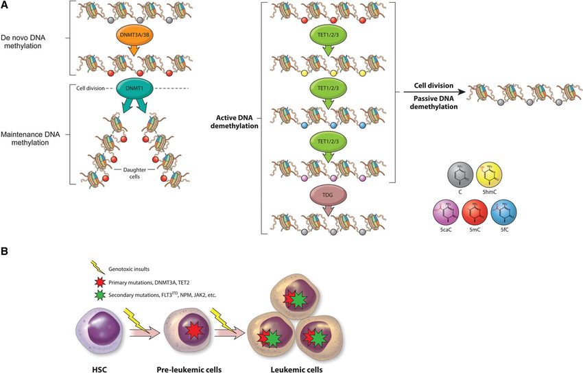
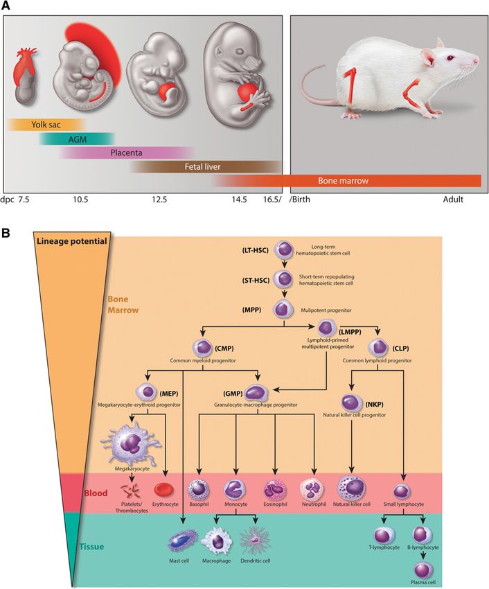
Downloaded from genesdev.cshlp.org on September 8, 2021 - Published by Cold Spring Harbor Laboratory Press

Hu and Shilatifard

Figure 1. Hematopoietic systems in mammals. (A) Embryonic tissues of mouse hematopoietic development. Mouse hematopoietic ac-
tivity arises from the mesoderm and first emerges in the extraembryonic yolk sac around E7.5 followed by the establishment of HSCs in
AGM and the placenta around E.9.5. HSCs colonize the fetal liver around E12.5, migrate to the bone marrow before birth, and reside there
throughout life. (B) Schematic overview of normal hematopoietic hierarchy in adult mice. HSCs sit at the top of the hierarchy and have
both the capacity of self-renewal and the multipotent potential to give rise to all mature hematopoietic cell lineages. After receiving a
differentiation signal, HSCs first lose self-renewing capacity and then progressively lose lineage potential, as they are restricted to a certain
lineage. (LT-HSC) Long-term HSC; (ST-HSC) short-term repopulating HSC; (MPP) multipotent progenitor; (CMP) common myeloid pro-
genitor; (CLP) common lymphoid progenitor; (LMPP) lymphoid-primed multipotent progenitor; (MEP) megakaryocyte/erythroid progen-
itor; (GMP) granulocyte-macrophage progenitor.

  Lymphomas are derived from a transformation of lym-                       aggressive). The indolent NHLs mainly are comprised of
phocytes residing in lymph nodes that has adverse effects                   follicular lymphoma (FL), mantle cell lymphoma, margin-
on the lymphatic and immune systems. Lymphomas can                          al zone lymphoma, small lymphocytic lymphoma, and
result from the transformation of B or T lymphocytes or                     cutaneous T-cell lymphoma (CTCL). Any of the indolent
natural killer (NK) cells and can be divided into two                       lymphomas can demonstrate aggressive behavior or a
main types: Hodgkin’s lymphoma and non-Hodgkin’s                            higher-grade transformation. The aggressive NHLs con-
lymphoma (NHL). The majority of NHLs (∼85%) is                              sist of diffuse large B-cell lymphoma (DLBCL), Burkitt’s
B-cell lymphomas, which can be further divided based                        lymphoma, lymphoblastic lymphoma, and various groups
on their appearance (e.g., follicular vs. diffuse) and how                  of T-cell and NK-cell lymphomas. The third major type of
quickly they are likely to grow and spread (indolent vs.                    hematologic malignancies are plasmacytic neoplasms in

2022       GENES & DEVELOPMENT
Downloaded from genesdev.cshlp.org on September 8, 2021 - Published by Cold Spring Harbor Laboratory Press

                                                                                         Hematopoiesis and hematologic malignancies

which abnormal antibodies secreting B lymphocytes,                matological malignancies, suggesting prominent roles
called plasma cells, accumulate in the bone marrow or             played by the epigenetic alterations in these diseases
within other tissues. Based on the site of involvement,           (Lawrence et al. 2014). In contrast to genetic aberrations,
the disease burden, and the presence of end organ damage,         epigenetic alterations are generally reversible and thus
these neoplasms can be divided into several categories,           may have a more therapeutic value from a clinical stand-
including plasmacytoma (of bone or extramedullary),               point. Indeed, inhibitors targeting chromatin-modifying
monoclonal gammopathy of undetermined significance                enzymes are being used in clinical trials (Cai et al. 2015;
(MGUS), smoldering (asymptomatic) myeloma, and                    Brien et al. 2016). In this review, we discuss our current
symptomatic plasma cell myeloma.                                  understanding of how epigenetic regulators function in
   Myeloid malignancies also include myeloprolifera-              normal hematopoiesis and highlight the consequences
tive neoplasms (MPNs) and myelodysplastic syndrome                of mutations in the DNA and histone lysine methylation
(MDS). MDS is a group of diverse bone marrow disorders            machineries in hematological malignancies.
characterized by disorderly and ineffective hematopoiesis,
which can lead to cytopenia, low levels of red blood cells        MLL in normal hematopoiesis and in the transcriptional
(anemia), neutrophils (neutropenia), or platelets (throm-         elongation checkpoint defect in leukemia
bocytopenia). These lower cell counts can result from a
failure of progenitor cells to mature, thereby accumulat-         The mixed-lineage leukemia (MLL or KMT2A) gene was
ing in the bone marrow, or, alternatively, progenitor cells       originally identified through cytogenetic studies of infant
could mature into blood cells with a shortened life span.         leukemia patient cells as being involved in chromosomal
Seemingly mature blood cells may not function properly            rearrangements that juxtaposed the N terminus of MLL
due to an abnormal shape (dysplasia). Around 30% of               with a variety of translocation partners. Although it was
MDS cases have the possibility of turning into AML (Lind-         suspected of being involved as a transcriptional regulator
berg 2005). MDS itself can arise as an adverse effect of ra-      based on its homology with Trithorax, a regulator of ho-
diation and chemotherapy. MPNs is a group of diseases             meotic gene expression in Drosophila, insights into its
characterized by the overproduction of one or more blood          biochemical function came from the purification of the
cell types in the bone marrow and circulating blood (Saeidi       closest yeast homolog Set1 [Su(var)3-9, enhancer of zeste,
2016). MPN can also further evolve into AML.                      and trithorax domain 1]. These studies demonstrated that
   The term “epigenetics” was coined by developmental             simple model systems such as yeast and Drosophila can
biologist Conrad Waddington to describe heritable chang-          provide fundamentally important molecular information
es in the cellular phenotype and gene expression that are         about conserved biological processes such as transcription
independent of alterations in the DNA sequence during             and epigenetics that are relevant to hematopoiesis and he-
embryonic development (Slack 2002; Berger et al. 2009).           matological malignancies.
Classic examples of the epigenetic regulation of gene               Fundamental molecular studies in yeast identified Set1
expression include genomic imprinting, position effect            as biochemically residing in a large macromolecular com-
variegation, and the regulation of homeotic (Hox) gene ex-        plex that was named the complex of proteins associated
pression. These processes turn out to be regulated by chro-       with Set1 (COMPASS). This complex harbors methyl-
matin modifiers implementing DNA methylation for                  transferase activity specifically toward Lys4 of histone
genomic imprinting, methylation of H3 Lys9 for the es-            H3 (Miller et al. 2001; Krogan et al. 2002; Shilatifard
tablishment of heterochromatin, and methylation of his-           2012; Piunti and Shilatifard 2016). The human wild-type
tone H3 at Lys4 or Lys27 for the activation or repression         MLL gene encodes a protein of 3969 amino acids that is
of Hox gene expression, respectively (Piunti and Shilati-         post-translationally cleaved by Taspase I into N-terminal
fard 2016). By controlling chromatin architecture and             and C-terminal fragments (Hsieh et al. 2003; Shilatifard
accessibility, modifications of DNA and histones can              2012). The two halves of MLL function together in a
convey this epigenetic information and influence gene ex-         COMPASS-like complex with core subunits related to
pression through favoring or antagonizing the recruitment         those found in yeast COMPASS as well as additional
of the activating or repressive complexes.                        interactors such as the tumor suppressor menin (Fig. 2A;
   The primary role of the transcription factors such as          Hsieh et al. 2003; Yokoyama et al. 2004). As in yeast
RUNX1/AML1, EVI-1, GATA3, IKAROS, and ETS in de-                  Set1, the C-terminal SET domain confers histone H3K4
termining the various stages of normal hematopoiesis is           methyltransferase activity to MLL (Milne et al. 2002;
reflected in their misregulation being the most common            Nakamura et al. 2002).
cause of hematopoietic transformation. The past several             Mll (Kmt2a) is required for normal numbers of hemato-
years have brought about an increased understanding of            poietic progenitors. Its deletion in mice causes embryonic
the biochemical and cellular functions of chromatin-mod-          lethality at E16.5, with fetal livers having dramatically re-
ifying and remodeling enzymes, specifically as coactiva-          duced numbers of HSCs, indicating an essential role for
tors and corepressors for the regulation of transcription         Mll in embryonic hematopoiesis (Hess et al. 1997; Ernst
during normal hematopoiesis and in the misregulation              et al. 2004; McMahon et al. 2007). Mll is also essential
of their activities related to hematological malignancies.        for sustaining postnatal hematopoiesis, as a conditional
Indeed, high-throughput, genome-scale sequencing has              deletion in postnatal mice with hematopoietic-specific
revealed that chromatin modifiers are among the most fre-         Vav-Cre leads to a multilineage defect in differentiation
quently mutated in cancer in general, particularly in he-         and a decrease in adult hematopoietic progenitors, with

                                                                                               GENES & DEVELOPMENT            2023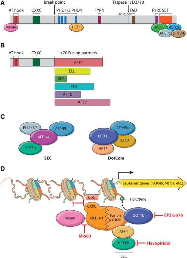
Downloaded from genesdev.cshlp.org on September 8, 2021 - Published by Cold Spring Harbor Laboratory Press

Hu and Shilatifard

                                                                              Figure 2. MLL translocation partners function in leuke-
                                                                              mogenesis. (A) Schematic representation of the domain
                                                                              structure of MLL and its stably associated cofactors in
                                                                              a COMPASS-like complex. MLL contains multiple do-
                                                                              mains essential for its biochemical and physiological
                                                                              function and is cleaved by Taspase I after aspartic acid
                                                                              2718 to generate a large N-terminal fragment and a small
                                                                              C-terminal fragment, which subsequently associate non-
                                                                              covalently through FY-rich N-terminal (FYRN) and FY-
                                                                              rich C-terminal (FYRC) domains. (AT hook) Binds to
                                                                              the minor groove of AT-rich DNA sequences; (PHD)
                                                                              Plant homology domain; (SET) Su(var)3-9, enhancer of
                                                                              zeste, and trithorax domain. Core COMPASS subunits
                                                                              WDR5, RBBP5, ASH2L, and DPY30 interact with the
                                                                              SET domain, while Menin and HCF1 associate with
                                                                              more N-terminal regions. (B) Structure of the most fre-
                                                                              quent MLL chimeras in acute leukemia. A typical MLL
                                                                              fusion protein contains the N terminus of MLL and a
                                                                              C-terminal portion of one of >70 fusion partners. The
                                                                              AT hook and CXXC domains are retained in all MLL chi-
                                                                              meras. (C ) Two distinct complexes regulating transcrip-
                                                                              tion are made up of some of the most frequent MLL
                                                                              fusion partners: SEC (super elongation complex) and
                                                                              DotCom (DOT1L complex). SEC comprises the ELL fam-
                                                                              ily members ELL1, ELL2, and ELL3; the MLL transloca-
                                                                              tion partners AF4/FMR2 family (AFF) member 1 (AFF1,
                                                                              also known as AF4) and AFF4; eleven-nineteen leukemia
                                                                              (ENL); the ALL1-fused gene from chromosome 9 (AF9);
                                                                              and the RNA polymerase II (Pol II) elongation factor P-
                                                                              TEFb (containing CDK9 and either cyclin T1 or cyclin
                                                                              T2). DotCom is composed of AF10, AF17, DOT1L, and
                                                                              AF9 or ENL. DOT1L is a methyltransferase specific for
                                                                              H3K79. (D) Therapeutic targeting of MLL translocated
                                                                              leukemia. MLL chimeras recruit SEC and/or DotCom
                                                                              to MLL target genes, which leads to their aberrant activa-
                                                                              tion through misregulation of the transcriptional elonga-
                                                                              tion checkpoint (Smith and Shilatifard 2013). Multiple
                                                                              therapeutic strategies have been developed to target dif-
                                                                              ferent steps required for the activation of MLL chimera
                                                                              target genes in leukemia: MI503 blocks the association
of Menin with MLL, potentially affecting recruitment of the MLL chimera to chromatin (indicated by a question mark); flavopiridol
(FP) inhibits the kinase activity of SEC subunit P-TEFb; and EPZ-5676 is a small molecule blocking the enzymatic activity of DOT1L.

a fatal bone marrow failure occurring at ∼3 wk of age (Jude            kemic pathogenesis, a model that is now driving the clin-
et al. 2007; Gan et al. 2010).                                         ical approaches for the treatment of leukemia associated
   Leukemia resulting from MLL translocations follows                  with MLL translocations (Cai et al. 2015). Purification of
an aggressive clinical course with a poor response to con-             some of the most common MLL translocation partners
ventional chemotherapy and often relapses very early.                  led to the identification of the super elongation complex
More than 70 translocation partners have been identified,              (SEC) that includes ELL; another previously known tran-
but they share little or no sequence similarities. All of the          scription elongation factor, P-TEFb; and additional MLL
MLL chimeras retain the N terminus but lose the majority               translocation partners AFF1, AFF4, ENL, and AF9 (Fig.
of the C-terminal portion of MLL, which contains the cat-              2C; Lin et al. 2010). ELL is also found in the little elonga-
alytic SET domain for H3K4 methylation (Fig. 2B). How-                 tion complex (LEC), which specifically regulates distinct
ever, the molecular mechanisms by which MLL                            stages of small nuclear RNA transcription, essential
chimeras could contribute to the pathogenesis of leuke-                housekeeping functions that are likely compensated for
mia were unknown until the biochemical identification                  by the wild-type allele of ELL in leukemia (Smith et al.
of the translocation partner ELL as a transcription elonga-            2011; Hu et al. 2013b).
tion factor for RNA polymerase II (Pol II) (Shilatifard et al.            A distinct protein complex that also functions in tran-
1996; Shilatifard 1998). ELL was the first MLL transloca-              scription elongation is DotCom (DOT1L complex), which
tion partner for which a molecular function was demon-                 contains the H3K79 methyltransferase DOT1L and the
strated. Based on this seminal discovery, it was proposed              MLL chromosomal translocation partners AF10, AF17,
>20 years ago that the misregulation of transcriptional                ENL, and AF9 (Fig. 2C; Okada et al. 2005; Mohan
elongation by RNA Pol II could play a central role in leu-             et al. 2010a,b). Dot1l mutant mice are embryonic-lethal

2024       GENES & DEVELOPMENT
Downloaded from genesdev.cshlp.org on September 8, 2021 - Published by Cold Spring Harbor Laboratory Press

                                                                                         Hematopoiesis and hematologic malignancies

between E10.5 and E13.5 and display a severe anemia due           on this process and determine whether such an approach
to reduced expression of the erythroid master regulator           is feasible or useful for the treatment of MLL transloca-
Gata2 (Feng et al. 2010). HoxA9 and Meis1 are the best-           tion-based leukemia.
characterized targets of the MLL chimeras, and their                 MLL translocation or PTD in leukemia patients usually
simultaneous overexpression was shown to be sufficient            occurs in one allele, leaving the second allele unaffected
for leukemic transformation (Kroon et al. 1998; Buske             in most cases. Studies have reported that the wild-type
and Humphries 2000; Zeisig et al. 2004). Both SEC and             MLL is essential for leukemic transformation by MLL chi-
DotCom have been shown to directly associate with                 meras and that its action is dependent on histone methyl-
HoxA9 and Meis1 loci and are required for their overex-           transferase activity (Milne et al. 2010; Cao et al. 2014).
pression (Okada et al. 2005; Lin et al. 2010; Neff and            The methyltransferase activity of MLL requires the for-
Armstrong 2013). Genetic deletion of Dot1l or pharmaco-           mation of a core complex with the WDR5, RBBP5,
logical inhibition of its methyltransferase activity renders      ASH2L, and DPY30 subunits (abbreviated as WRAD in
MLL chimeras unable to activate the malignant tran-               the literature) (Dou et al. 2006; Patel et al. 2008, 2009).
scriptional program in mouse models (Okada et al. 2005;           Therefore, small molecules (MM-102 and MM-401) that
Krivtsov et al. 2008; Chang et al. 2010; Bernt et al. 2011;       inhibit the methyltransferase activity of MLL via disrup-
Nguyen et al. 2011; Deshpande et al. 2013). Consequently,         tion of its association with WDR5 were developed and
a selective DOT1L small molecule inhibitor compound,              have been shown to function by blocking proliferation, in-
EPZ-5676, has been under investigation for the treatment          duction of apoptosis, and differentiation of the cells ex-
of MLL-rearranged leukemia (Cai et al. 2015). In addition         pressing MLL-AF9 (Karatas et al. 2013; Cao et al. 2014).
to translocations, partial tandem duplications (PTDs) of          However, the basis for the specificity of these inhibitors
MLL have been observed in leukemia with a normal kar-             for MLL versus other members of the COMPASS family
yotype and linked to an unfavorable prognosis after treat-        is unclear, as WDR5 is a shared component throughout
ment (Caligiuri et al. 1994, 1998; Schichman et al. 1994).        the COMPASS family. Furthermore, mouse genetic stud-
PTD most commonly occurs by insertion of either exons             ies have demonstrated that the SET domain of the wild-
5–11 or 5–12 into intron 4 of the full-length MLL gene, and       type copy of MLL is dispensable for leukemogenesis (Mis-
this leads to a duplicated N-terminal region that harbors         hra et al. 2014), raising the question of the efficacy of tar-
both the AT hooks and the CXXC domains (Whitman                   geting the wild-type copy of MLL for treating MLL
et al. 2005). A recent study showed that leukemia cells           chimera-driven leukemia.
with MLL-PTD are sensitive to DOT1L small molecule
inhibition by EPZ-5676, although the oncogenic mecha-
nisms of MLL-PTD may differ from that of MLL chimeras             Mutations of MLL3 and MLL4 members of the COMPASS
(Kuhn et al. 2015). Furthermore, since SEC contains the P-        family in enhancer malfunction and hematological
TEFb kinase module, specific targeting of this activity or        malignancies
the disruption of its biochemical integrity in leukemic
cells may represent additional therapeutic strategies for         Transcriptional enhancers were originally defined as non-
MLL translocation-based leukemia (Fig. 2D).                       coding DNA sequences that can increase the transcription
   Another therapeutic strategy for treating MLL translo-         of cognate genes in a distance-, position-, and orientation-
cation-based leukemia is to prevent the recruitment of            independent manner (Dorsett 1999; Smith and Shilatifard
MLL chimeras to chromatin. Menin associates directly              2014). A high level of H3K4 monomethylation (H3K4me1)
with an N-terminal region of MLL that is retained in all          with a relatively low level of H3K4me3 trimethylation
MLL chimeras. This interaction has been shown to be es-           (H3K4me3) has been identified as a signature of enhancers
sential for the leukemic activity of MLL chimeras through         (Heintzman et al. 2007, 2009). MLL3 and MLL4 (KMT2C
facilitating the recruitment to chromatin (Yokoyama               and KMT2D) comprise one of the three major branches of
et al. 2004, 2005; Caslini et al. 2007). Small molecule dis-      the COMPASS family in mammals, with the other two
ruptors of the MLL–menin interaction were identified              major branches represented by MLL/MLL2 (KMT2A/
through high-throughput screening and structure-based             KMT2B) and SET1A/SET1B (Mohan et al. 2011; Herz
design (Grembecka et al. 2012; Shi et al. 2012). Pharmaco-        et al. 2012; Shilatifard 2012). MLL3 (KMT2C), MLL4
logical treatment with these inhibitors (MI-2-2 and               (KMT2D; GeneID 8085), and their Drosophila ortholog,
MI503) in both leukemic cell lines and a mouse model              Trr (Trithorax-related), implement the bulk of H3K4me1
of leukemia led to apoptosis and hematopoietic differenti-        at enhancers and are required for enhancer–promoter
ation, proliferation defects, and reversal of MLL chimera-        communication during development (Fig. 3A; Herz et al.
driven leukemic transcriptional signatures (Fig. 2D;              2012, 2014; Hu et al. 2013a). Further studies by other lab-
Grembecka et al. 2012; Borkin et al. 2015). However, it           oratories have now confirmed these original findings es-
is not clear whether or how the inhibition of this mode           tablishing MLL3 and MLL4 COMPASS as major
of recruitment by MI-2-2 and MI503 discriminates be-              regulators of enhancer H3K4 function (Kanda et al. 2013;
tween the chimera, the wild-type MLL, or its closest              Lee et al. 2013; Smith and Shilatifard 2014).
homolog, MLL2 (KMT2B; GeneID 9757). Further molecu-                  Chromatin signatures can be used to further classify en-
lar studies on the inhibitory role of MI-2-2 and MI503 on         hancers as variably existing in inactive, active, or poised
endogenous MLL/MLL2/COMPASS and the changes in                    states. Histone H3K4me1 alone marks enhancers before
the pattern of H3K4 methylation should shed further light         their activation (Heintzman et al. 2007), with the addition

                                                                                               GENES & DEVELOPMENT            2025
Downloaded from genesdev.cshlp.org on September 8, 2021 - Published by Cold Spring Harbor Laboratory Press
Hu and Shilatifard
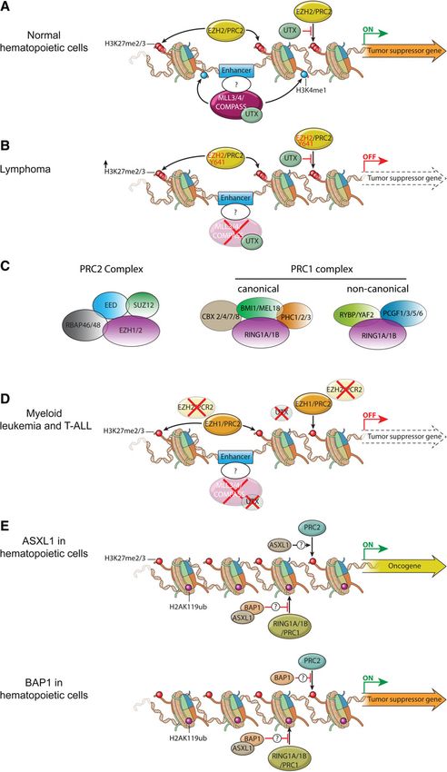
Figure 3. Misregulation of MLL3/4/COMPASS
and polycomb group (PcG) proteins in hematopoiet-
ic transformation. (A) In normal HSCs and progeni-
tors, MLL3/4/COMPASS and EZH2/PRC2 control
the proper expression of genes involved in self-re-
newal and differentiation through implementation
of H3K4me1 and H3K27me3 at enhancers. UTX, a
component of the MLL3/MLL4/COMPASS-like
complexes, harbors a demethylase activity specific
for H3K27me3 that could antagonize transcription-
al repression by the EZH2/PRC2 complex. (B) Dur-
ing lymphoma development, the catalytic subunit
of PRC2, EZH2, is frequently mutated at Y641.
This mutation results in a switch in substrate pref-
erence from nonmethylated to the monomethy-
lated and dimethylated histone H3K27, leading to
increased levels of H3K27 dimethylation
(H3K27me2) and H3K27me3 at enhancers and pro-
moters. Additionally, inactivating mutations in en-
hancer monomethyltransferases MLL3 and MLL4/
COMPASS can cause loss of H3K4me1 at enhanc-
ers. Altogether, these events independently or to-
gether could lead to repression of enhancers and
promoters of tumor suppressor genes to promote
lymphomagenesis. (C) Schematic illustration of
mammalian PRC1 and PRC2 complexes. PRC2 is
composed of four core components—EED (embry-
onic ectoderm development), SUZ12 (suppressor
of zeste 12 homolog), RBAP46, and RBAP48—and
an enzymatic subunit, EZH1 or EZH2. EED and
SUZ12 are essential for the stability of EZH1 and
EZH2 and therefore for H3K27 methylation (Pasini
et al. 2004; Xie et al. 2014). PRC1 complexes in-
clude both canonical and noncanonical forms in
mammalian cells. Both canonical and noncanonical
PRC1 complexes contain the catalytic subunit
RING1A or RING1B that implements H2AK119
monoubiquitination. Canonical PRC1 complexes
can variably include CBX2, CBX4, CBX7 or CBX8;
BMI1 (also known as PCGF4) or MEL18 (also known
as PCGF2); and PHC1, PHC2, or PHC3. The chro-
modomain found in the CBX proteins that partici-
pates in canonical PRC1 mediates the interaction
with H3K27me3 that is catalyzed by PRC2. Nonca-
nonical PRC1 complexes can contain RYBP or
YAF2 and PCGF1, PCGF3, PCGF5, or PCGF6. (D)
Inactivating mutations in MLL3, MLL4, UTX, and
EZH2 were observed in myeloid leukemias (MDS,
chronic myelomonocytic leukemia [CMML], and
primary myelofibrosis [PMF]) and T-cell ALL (T-
ALL). EZH1 can partially compensate for the loss
of EZH2 in depositing H3K27me3 at enhancers
and promoters. Loss of function of MLL3 and MLL4 removes H3K4me1 from enhancers, while UTX inactivation may increase
H3K27me3 at the enhancers and promoters. All of these epigenetic alterations can collaborate to suppress the enhancers of tumor sup-
pressor genes involved in myeloid leukemia and T-ALL. (E) In myeloid hematopoietic cells, ASXL1 (additional sex combs-like 1) promotes
the repression of PRC2 targets via a currently unknown mechanism. (Top panel) Loss-of-function mutations of ASXL1 are frequently de-
tected in CMML, MDS, and MPN, and these mutations could give rise to activation of an oncogene normally repressed by PRC2-mediated
H3K27me3. (Bottom panel) BRCA1-associated protein 1 (BAP1) can function to attenuate the activity of PRC2. BAP1 loss results in in-
creased expression of EZH2, which can result in myeloid transformation through the repression of tumor suppressor genes.
of H3K27ac indicating active enhancers. Poised enhancers et al. 2010; Rada-Iglesias et al. 2011). The MLL3, MLL4
are distinguished by the presence of H3K27me3 and are (GeneID 8085), and Trr forms of COMPASS harbor a
found primarily in stem and progenitor cells, transitioning unique subunit, UTX, which has H3K27 demethylase ac-
to active enhancers upon differentiation cues (Creyghton tivity. This raises the possibility that these complexes not
2026 GENES & DEVELOPMENTDownloaded from genesdev.cshlp.org on September 8, 2021 - Published by Cold Spring Harbor Laboratory Press
Hematopoiesis and hematologic malignancies
only are responsible for H3K4me1 at inactive, active, and HSCs by crossing Mll4 f/f mice with transgenic mice that
poised enhancers but also may facilitate enhancer transi- express interferon-inducible MxCre, expansion of HSCs
tioning from a poised to an active state during hematopoi- and common myeloid progenitors (CMPs) in the bone
etic differentiation (Fig. 3A; Cho et al. 2007; Herz et al. marrow were observed. However, these HSCs are im-
2012). paired in their self-renewing capability due to oxidative
Recent cancer genome sequencing approaches of many stress-induced DNA damage. Therefore, it has been pro-
types of primary tumors and cancer cell lines have re- posed that MLL4/COMPASS helps enforce a differentia-
vealed that MLL3 and MLL4/COMPASS are among the tion blockade of leukemic stem cells by up-regulating
most frequently mutated genes in both solid tumors and expression of antioxidant genes, with decreased cellular
hematological malignancies (Morgan and Shilatifard reactive oxygen species (ROS) protecting against oxidative
2015). The MLL3 mutation or its deletion is found in he- stress-induced DNA damage (Santos et al. 2014).
matological malignancies, including 15% of DLBCL, A potential mechanism for mutations in MLL3 and
MDS, and AML (Mrozek 2008; Pasqualucci et al. 2011; MLL4/COMPASS family members leading to lympho-
Zhang et al. 2013). MLL4 (GeneID 8085) mutations are magenesis and other hematological cancers is the mis-
prevalent in NHL (Morin et al. 2011; Pasqualucci et al. regulation of enhancer function during hematopoietic
2011; Lohr et al. 2012; Green et al. 2015), ALL (Mar differentiation (Herz et al. 2014). Aberrant transcriptional
et al. 2012; Lindqvist et al. 2015; Neumann et al. 2015), enhancer activity through either alterations of chromatin
and AML (Kandoth et al. 2013). Most of the MLL3 and modifications, mutations in enhancer-binding factors, or
MLL4/COMPASS mutations are heterozygous nonsense, mutations of enhancers themselves could potentially
frameshift, and internal insertions/deletions (indels), re- lead to oncogenesis (Herz et al. 2014). A classic example
sulting in protein truncations—which is suggestive of a of enhancer-mediated oncogenesis is the chromosomal
haploinsufficient tumor suppressor function of MLL3 translocation commonly found in Burkitt’s lymphoma
and MLL4 (GeneID 8085)—in cancer pathogenesis. A tu- that places the MYC oncogene under the control of an im-
mor suppressor function of MLL3 has been proven in a munoglobulin heavy chain (Igh) enhancer, leading to the
mouse model of AML, with a 50% reduction in gene dos- uncontrolled expression of MYC and the development of
age cooperating with other genetic alterations to promote lymphoma (Dalla-Favera et al. 1982; Park et al. 2005).
leukemogenesis (Chen et al. 2014). More recently, two independent studies of AML with a
FLs can transform from the indolent to the more aggres- chromosomal inversion between GATA2 and EVI1 re-
sive DLBCL, which has allowed the collection and analy- vealed that this inversion allows for promiscuous activa-
sis of sequential tumor lymphoid biopsies from the same tion of EVI1 by a GATA2 enhancer (Groschel et al. 2014;
patient (Okosun et al. 2014). These and related studies Yamazaki et al. 2014). Even a single-nucleotide polymor-
suggest that somatic MLL4 (GeneID 8085) mutations are phism in a PU.1 enhancer leads to reduced PU.1 expres-
likely to be early events that occur in a common progeni- sion that contributes to the development of AML (Steidl
tor cell, with additional mutations leading to the more ag- et al. 2007). Furthermore, in a subset of T-cell ALL (T-
gressive state (Green et al. 2013, 2015; Okosun et al. 2014; ALL), heterozygous acquisition of a short DNA sequence
Pasqualucci et al. 2014; Ortega-Molina et al. 2015). Mouse at a noncoding intergenic region creates a binding site for
models using Bcl2 overexpression and conditional dele- the transcription factor MYB, thereby creating a de novo
tion of Mll4 (Kmt2d; GeneID 381022) demonstrate that enhancer to activate allele-specific expression of TAL1
Mll4 loss promotes the development of lymphoma (Mansour et al. 2014). A long-range NOTCH1-dependent
through the expansion of germinal centers, likely due to oncogenic enhancer was found to undergo focal amplifica-
the loss of the expression of known tumor suppressor tions to aberrantly activate MYC expression to drive T-
genes regulating B-cell-activating pathways (Fig. 3B; Orte- ALL (Herranz et al. 2014). The pervasive contribution of
ga-Molina et al. 2015; Zhang et al. 2015). enhancer malfunction in hematological malignancies
Although occurring at a lower rate than truncating mu- and the high rate of the mutation of the MLL3 and
tations, missense mutations of MLL4 (GeneID 8085) are MLL4 (GeneID 8085) forms of COMPASS suggest that al-
found throughout the protein in lymphomas, with many terations in enhancer activity by MLL3 and MLL4 muta-
of the missense mutations located in the C-terminal do- tions help drive an oncogenic program (Fig. 3B). Further
mains impairing the in vitro histone methyltransferase studies aimed at identifying MLL4-dependent enhancers
activity of MLL4 (Zhang et al. 2015). However, these mu- and how these factors are specifically recruited to their
tations have not been characterized in vivo, and their con- cognate site using methods such as ChIP-seq (chromatin
tributions to lymphomagenesis are currently unknown. immunoprecipitation [ChIP] combined with high-
Recently developed genome-editing tools such as CRISPR throughput sequencing), genome editing, and gene expres-
will allow the exploration of these missense mutations in sion profiling will be required for testing this hypothesis.
mouse models, potentially providing insight into their
contribution to hematological malignancies.
In contrast to MLL4-inactivating mutations contribut- Polycomb group (PcG) proteins in normal
ing to lymphomagenesis, recent studies with conditional and malignant hematopoiesis
ablation of Mll4 (Kmt2d; GeneID 381022) in AML cells re-
vealed that Mll4 is essential for MLL-AF9-induced leuke- PcG proteins were initially identified in Drosophila by
mogenesis (Santos et al. 2014). When Mll4 was deleted in mutations that cause homeotic transformations, such as
GENES & DEVELOPMENT 2027Downloaded from genesdev.cshlp.org on September 8, 2021 - Published by Cold Spring Harbor Laboratory Press Hu and Shilatifard leg-to-antenna transformations or the appearance of addi- Merchan et al. 2012). Several studies have shown that tional sex combs on the second and third pairs of legs (Le- PRC2 restricts HSC activity, as a partial loss of the core wis 1978; Struhl 1981). These transformations result from components such as Ezh2, Eed, or Suz12 in mice enhances derepression of the homeotic genes (called Hox genes in HSC function (Majewski et al. 2008, 2010). However, a vertebrates) due to loss-of-function mutations in genes en- full loss of PRC2 activity has a distinct effect on HSC coding PcG proteins. In mammals, PcG proteins are also maintenance and function. Mice with an Eed deficiency critical regulators of developmental gene expression and are normal in fetal liver HSC numbers but display defects tissue homeostasis. Their misregulation has been widely in the maintenance and differentiation of adult HSCs (Xie linked to multiple disease states in a variety of contexts, et al. 2014). Homozygous deletion of Suz12 also results in particularly in cancers, including hematological malig- failures in both embryonic and adult hematopoiesis (Lee nancies (Varambally et al. 2002; Bracken et al. 2003; Schle- et al. 2015). Collectively, these studies indicate a dosage- singer et al. 2007; Kondo et al. 2008; Martinez et al. 2009). dependent impact of PRC2 activity on HSC function. In PcG proteins operate in at least two distinct multiprotein addition to HSC regulation, PRC2 also has an essential complexes, PRC1 and PRC2; each harbors an enzymatic role in lymphoid development. Conditional deletion of activity, with PRC1 implementing H2AK119 monoubi- EZH2 in the lymphoid lineage revealed its crucial role quitination, and PRC2 implementing H3K27 methylation in early B-cell development, rearrangement of the Igh (Margueron and Reinberg 2011). gene, germinal center formation, and differentiation and The mammalian PRC2 complex consists of four core survival of CD4+ T help1 (Th1) and Th2 cells (Su et al. components: EZH1 or its paralog, EZH2; embryonic ecto- 2003; Beguelin et al. 2013; Caganova et al. 2013; Tumes derm development (EED); suppressor of zeste 12 homolog et al. 2013; Zhang et al. 2014). Similarly, Suz12 deficiency (SUZ12); and RBAP46/48 (also known as RBBP7/4) (Mar- causes severe defects in B and T lymphopoiesis (Lee et al. gueron and Reinberg 2011; Aranda et al. 2015; Piunti and 2015). Future work should clarify the mechanisms under- Shilatifard 2016). EZH2 or EZH1 forms the enzymatic lying the dosage-dependent impact of PRC2 activity on core and implements H3K27 methylation. EZH1 is pre- HSC functioning and the role and mechanisms of PRC2 sent in both dividing and differentiated cells, whereas in the specification of other hematopoietic lineages. EZH2 is found only in actively dividing cells. The PCR2 PRC1 is also essential for normal hematopoiesis, with complex with EZH1 has low methyltransferase activity its core subunit, BMI1, being the most-studied component compared with the PRC2 complex containing EZH2 (Fig. of PRC1 in hematopoiesis. Ectopic expression of Bmi1 in 3C; Margueron et al. 2008). The composition of mammali- mouse embryonic stem cells has been shown to promote an PRC1 complexes is more variable and is comprised of primitive hematopoiesis (Ding et al. 2012). Bmi1 is re- the E3 ubiquitin ligases for H2A monoubiquitination quired for HSC self-renewal in both mice and humans. (RING1A or RING1B) together with either canonical The homozygous deletion of Bmi1 in mice decreases the (CBX2/4/7/8, PHC1/2/3, and BMI1/MEL18) or noncanon- number of HSCs by inducing symmetrical division, dere- ical (RYBP/YAF2 and PCGF1/3/5/6) proteins (Fig. 3C; pression of the Ink4a–Arf locus (encoding p16INK4A and Gao et al. 2012; Tavares et al. 2012; Aranda et al. 2015; p19ARF proteins), and generation of ROS and DNA damage Piunti and Shilatifard 2016). PRC1 and PRC2 co-occupy (Park et al. 2003; Iwama et al. 2004; Oguro et al. 2006; Liu many PcG target loci and play crucial roles in their et al. 2009). Bmi1 −/− cells exhibit an accelerated differen- reciprocal recruitment to chromatin (Boyer et al. 2006; tiation into the B-cell lineage due to premature transcrip- Schwartz et al. 2006; Ku et al. 2008). The chromobox tional activation of B-cell regulators Ebf1 and Pax5 (Oguro (CBX) proteins have a chromodomain that recognizes the et al. 2010). Although homologous to Bmi1, Mel18 loss H3K27me3 mark implemented by PRC2 and mediates has very little impact on the repopulating capacity of fetal the chromatin targeting of the canonical PRC1 complex HSCs, with the self-renewing capacity of adult HSCs be- (Cao et al. 2002; Fischle et al. 2003; Min et al. 2003; Agger ing enhanced in the absence of Mel18, suggesting that et al. 2007; Lee et al. 2007). Conversely, noncanonical Bmi1 and Mel18 have distinct functions in HSCs (Iwama PRC1-catalyzed H2AK119 monoubiquitination is capable et al. 2004; Kajiume et al. 2004). However, Mel18 was of targeting PRC2 to chromatin (Blackledge et al. 2014; demonstrated to have important roles in the proliferation Kalb et al. 2014). and maturation of B cells, indicating a function in lym- PcG proteins are essential regulators of hematopoiesis phoid differentiation (Akasaka et al. 1997; Tetsu et al. and can orchestrate the expression of genes that control 1998). the balance between self-renewal and the multipotency Multiple members of the CBX family have been identi- of HSCs. Transgenic and conditional knockout studies fied within the canonical PRC1 complex, and several of in mice have provided valuable information on the physi- them have been shown to be vital for hematopoietic ho- ological role of PcG proteins in the hematopoietic system. meostasis. Cbx7 is highly expressed in long-term HSCs, Ezh1 is required for HSC maintenance, and its conditional and its overexpression enhances HSC self-renewal and in- loss leads to HSC senescence and impairment of B-cell duces leukemia, whereas an ectopic expression of Cbx2, lymphopoiesis (Hidalgo et al. 2012). Forced expression of Cbx4, or Cbx8 results in differentiation and exhaustion Ezh2 was found to prevent the exhaustion of the long- of HSCs, revealed by the competitive transplantation as- term repopulating capacity of HSCs in a serial transplan- say (Klauke et al. 2013). Although the repopulating activ- tation assay and cause development of myeloproliferative ity of fetal liver cells deficient for Cbx2 is comparable with disease in recipient mice (Kamminga et al. 2006; Herrera- wild-type cells, homozygous deletion of Cbx2 leads to 2028 GENES & DEVELOPMENT
Downloaded from genesdev.cshlp.org on September 8, 2021 - Published by Cold Spring Harbor Laboratory Press

                                                                                          Hematopoiesis and hematologic malignancies

reduced cellularity in postnatal bone marrow, spleen, and          oullas et al. 2016). Therefore, the aberrantly high level of
thymus (Core et al. 1997; Iwama et al. 2004). The knock-           H3K27me3 may lead to persistent repression of some
down of each member of the CBX family in human CD34+               tumor suppressor genes (Fig. 3B), while its less focused dis-
core blood cells leads to a profound reduction in the prolif-      tribution could allow derepression of other loci contribut-
eration of hematopoietic stem and progenitor cells (van            ing to lymphomagenesis.
den Boom et al. 2013).                                                The importance of the H3K37me3 mark in promoting
   Studies on the role of the catalytic components of the          epigenetic reprogramming in hematological malignancies
PRC1 complex, RING1A and RING1B, are still limited,                is further suggested by the high frequency of inactivating
and H2A monoubiquitination function in fetal and adult             mutations of the H3K27me3 demethylase UTX that re-
hematopoiesis remains unclear. Mice with homozygous                sides within MLL3/MLL4 COMPASS-like complexes
deletion of Ring1a are viable but display skeletal abnor-          (Agger et al. 2007; Cho et al. 2007; Lan et al. 2007). Exome
malities (del Mar Lorente et al. 2000). Conditional dele-          and whole-genome sequencing has identified homozy-
tion of Ring1b in adult hematopoietic lineages revealed            gous and heterozygous inactivating mutations of UTX
its restrictive role in the proliferation of hematopoietic         in multiple myeloma and T-ALL but not in lymphomas
stem and progenitor cells and its favorable function in            that harbor the EZH2 Y641 activating mutation (van
the proliferation of differentiated progenies (Cales et al.        Haaften et al. 2009; Morin et al. 2011). Studies of Utx func-
2008). In vitro studies revealed that HSCs with a deletion         tion in T-ALL found that Utx can act as a tumor suppres-
of both Ring1a and Ring1b show a severe defect in their            sor in a Notch1-induced murine model of leukemia
self-renewing capacity (Piunti et al. 2014). Future studies        (Ntziachristos et al. 2014). More recent studies found
using mice with a conditional deletion of Ring1a and/or            that UTX can also act as an oncogene in TAL1-positive
Ring1b will shed light on the role of H2A monoubiquiti-            T-ALL, while UTX can inhibit cell growth when overex-
nation in normal hematopoiesis.                                    pressed in TAL1-negative T-ALL cells (Benyoucef et al.
   EZH2 is the most frequently mutated PcG member in               2016). Therefore, potential therapeutic options for hema-
the pathogenesis of hematological malignancies (Kroeze             tological cancers can be aimed at recovering normal cellu-
et al. 2012; Woods and Levine 2015). EZH2 overexpression           lar H3K27 methylation levels with small molecule
is commonly observed in various epithelial malignancies,           inhibitors of PRC2 or, in a subset of cases, activating mol-
including breast and prostate cancer, suggesting that              ecules of H3K27 demethylases (Fig. 3D; Knutson et al.
EZH2 functions as an oncogene (Varambally et al. 2002;             2012; McCabe et al. 2012b).
Kleer et al. 2003; Li et al. 2009a). Indeed, recurrent somatic        Although an excess of H3K27me3 activity generally has
mutations that result in increased EZH2 enzymatic                  an oncogenic role in cancer, loss-of-function mutations in
activity have been observed in lymphoma (Morin et al.              EZH2 have been detected in a subset of myeloid malig-
2011; Yap et al. 2011; McCabe et al. 2012a). Monoallelic           nancies, most commonly in MDSs, chronic myelomono-
mutations that result in substitution of Y641 within the           cytic leukemia (CMML), primary myelofibrosis (PMF),
SET domain of EZH2 lead to increased conversion of                 and T-ALL (Ernst et al. 2010; Nikoloski et al. 2010;
H3K27me1 to H3K27me2 (H3K27 dimethylation) and                     Ntziachristos et al. 2012; Score et al. 2012; Simon et al.
H3K27me3 and have been identified in 22% of patients               2012; Zhang et al. 2012). These inactivating mutations
with germinal center diffuse B-cell lymphoma (Morin                of EZH2 predict a poorer overall outcome in CMML,
et al. 2011). Transgenic mouse models initially demon-             MDS, and PMF (Grossmann et al. 2011; Guglielmelli
strated that a specific overexpression of EZH2 Y641F/N in          et al. 2011). Conditional loss of Ezh2 in a hematopoietic
lymphocytes is insufficient for lymphomagenesis on its             system contributes to the pathogenesis of MDS and accel-
own but can cooperate with Bcl2 or Myc overexpression              erates the onset of the early T-cell precursor ALL (ETP-
(Beguelin et al. 2013; Berg et al. 2014). These studies sug-       ALL) induced by oncogenic NRASQ61K (Score et al.
gested that the carcinogenic activity of EZH2 Y641 muta-           2012; Muto et al. 2013; Danis et al. 2016). Besides EZH2
tions rely on other oncogenic events to drive malignant            inactivating mutations, loss-of-function mutations of
transformation. However, a more recent study with the              the PRC2 core components SUZ12 and EED have also
Y641F mutation knocked in the endogenous Ezh2 locus                been detected in T-ALL, all of which lead to lower levels
in mice showed that Ezh2Y641F itself could cause develop-          of H3K27me3 (Ntziachristos et al. 2012; Simon et al.
ment of lymphoma and melanoma (Souroullas et al.                   2012). Intriguingly, myeloid leukemia can have inactivat-
2016). In this mouse model, Ezh2Y641F was found to coop-           ing mutations of both PRC2 components and UTX, rais-
erate with the loss of p53 or overexpression of Bcl2, but          ing the question of how simultaneous mutations of
not Myc, to accelerate lymphoma progression (Souroullas            factors with opposing enzymatic activity can contribute
et al. 2016). These discrepancies resulting from the differ-       to leukemogenesis (van Haaften et al. 2009; Jankowska
ent mouse models used in these studies demonstrate that            et al. 2011). A very recent study demonstrated that Ezh1
the oncogenic function of somatic mutations may not be             function is essential for the pathogenesis of myeloid ma-
faithfully recapitulated by transgenic mouse models that           lignancies induced by Ezh2 loss of function (Mochizuki-
introduce an extra copy of mutant alleles expressed under          Kashio et al. 2015). Therefore, inactivating mutations
the control of exogenous promoters. Despite the overall            of UTX could facilitate EZH1-dependent repression of
higher abundance of H3K27me3 observed in lymphoma                  tumor suppressor genes that would otherwise be dere-
cells induced in knock-in Ezh2 Y641F mice, many loci               pressed by EZH2 loss of function (Fig. 3D). Understanding
lose H3K27me3 and exhibit increased transcription (Sour-           the context in which PRC2 can act as an oncogene or

                                                                                                GENES & DEVELOPMENT            2029
Downloaded from genesdev.cshlp.org on September 8, 2021 - Published by Cold Spring Harbor Laboratory Press Hu and Shilatifard tumor suppressor in lymphomagenesis and leukemogene- and mesothelioma cells (LaFave et al. 2016; Schoumacher sis will undoubtedly be an active area of future research. et al. 2016). Future work to dissect the molecular mecha- Over the past decade, a series of studies has implicated nisms and potential cross-talk for the PRC1 and PRC2 PRC1 in HSC self-renewal and differentiation, which re- complexes in their hematopoietic context and in MDS quire the core subunits of the Bmi1 and Cbx family of pro- and other malignancies is another important area requir- teins (Lessard and Sauvageau 2003; Park et al. 2003; ing investigation. Klauke et al. 2013). In contrast to PRC2, somatic muta- tions in the PRC1 components have not been reported for hematopoietic malignancies. However, overexpres- The nuclear receptor-binding SET domain (NSD) sion of BMI1, an integral subunit that stimulates the ubiq- family of H3K36 methyltransferases in hematological uitinase activity of PRC1 toward H2AK119, has been malignancies observed in myeloid malignancies and has emerged as a useful indicator for the prognosis of MDS, CMML, The NSD family of histone methyltransferases comprises and AML (Mihara et al. 2006; Chowdhury et al. 2007; three proteins: NSD1, NSD2 (also known as multiple my- Mohty et al. 2007). However, it remains unclear whether eloma SET domain [MMSET] and Wolf-Hirschhorn syn- H2AK119 monoubiquitination is concomitantly in- drome candidate 1 [WHSC1]), and NSD3 (also known as creased with BMI1 overexpression and whether this his- WHSC1L1). Although members of the NSD family were tone modification contributes to myeloid malignancies. initially shown to have enzymatic activity toward multi- In Drosophila, H2AK119 monoubiquitination can be re- ple residues on histones, subsequent studies demonstrat- moved by the Polycomb-repressive deubiquitinase (PR- ed that these enzymes preferentially implement histone DUB) complex, which is comprised of the BRCA1-associ- H3K36me2 (Li et al. 2009b; Kuo et al. 2011; Qiao et al. ated protein 1 (BAP1) homolog, Calypso, and additional 2011). NSD1 and NSD2 have each been linked to human sex combs (Asx) (Scheuermann et al. 2010). A mammalian developmental overgrowth syndromes. The NSD2 gene PR-DUB complex containing BAP1 and ASX-like 1 is among a group of genes deleted in Wolf-Hirschhorn syn- (ASXL1) or ASXL2 was also biochemically isolated and drome, and loss of NSD2 is thought to be responsible for a demonstrated to harbor deubiquitinase activity in vitro subset of the clinical features characteristic of this syn- (Scheuermann et al. 2010; Dey et al. 2012). ASXL1 is fre- drome (Andersen et al. 2014). Deletion or loss-of-function quently mutated in a wide range of myeloid malignancies, mutations involving NSD1 results in Sotos syndrome, an most commonly in AML, CMML, MDS, and MPN. The autosomal dominant overgrowth syndrome characterized mutations of ASXL1 are associated with a dismal overall by a distinctive facial appearance, delayed development, prognosis in patients with MDS and AML (Abdel-Wahab and learning disabilities (Douglas et al. 2003; Turkmen et al. 2011, 2012; Bejar et al. 2011; Patel et al. 2012). How- et al. 2003). ever, and in contrast to what has been observed in Droso- The roles of members of the NSD family in normal phila, ASXL1 inactivating mutations did not cause hematopoiesis have not been investigated. However, significant changes in the H2AK119 monoubiquitination alterations of these genes are recurrently observed in he- levels in myeloid hematopoietic cells but did promote matological malignancies, implying critical roles in myeloid transformation through the loss of PRC2-medi- normal hematopoiesis. A recurring t(5;11)(q35;p15.5) dated H3K27 methylation, leading to derepression of chromosomal translocation fuses NSD1 to nucleoporin- PRC2 target genes (Fig. 3E; Abdel-Wahab et al. 2012; Inoue 98 (NUP98) to generate a NUP98–NSD1 chimera in 5% et al. 2013). of human AMLs (Cerveira et al. 2003). Transplantation BAP1 is best known for being recurrently mutated in studies in mice demonstrated that expression of the numerous cancers such as mesothelioma, renal cell carci- NUP98–NSD1 chimera sustains self-renewal of myeloid noma, and metastatic uveal melanoma (Harbour et al. progenitors and is sufficient to induce AML in vivo by pre- 2010; Bott et al. 2011; Pena-Llopis et al. 2012). Consistent venting the EZH2-dependent silencing of the Hoxa9 with a physical association with Asxl1, Bap1 loss of func- and Meis1 proto-oncogenes (Wang et al. 2007). NSD2 is tion also leads to myeloid transformation in mice (Dey commonly overexpressed in 15%–20% of multiple myelo- et al. 2012; LaFave et al. 2015). In contrast to the reduction ma cases through a chromosomal translocation t(4;14) of H3K27me3 seen with Asxl1 loss, Bap1 deletion leads to (p16.3;q32) that juxtaposes the NSD2 gene with the IgH increased Ezh2 expression, higher levels of H3K27me3, locus (Chesi et al. 1998). Aberrant up-regulation of and enhanced repression of PRC2 target genes in hemato- NSD2 and a subsequent increase of H3K36me2 lead to poietic cells. Thus, Ezh2 deletion or inhibition was shown an altered localization and overall decrease in the levels to abrogate myeloid malignancy and mesothelioma in- of H3K27me3 (Kuo et al. 2011; Martinez-Garcia et al. duced by Bap1 loss (LaFave et al. 2015). These findings 2011; Popovic et al. 2014). Recent global profiling of his- raise the possibility that myeloid transformation resulting tone modifications by mass spectrometry from 115 cancer from ASXL1 and BAP1 loss could be independent of the cell lines identified a cluster of 13 cell lines exhibiting a function of the BAP1–ASXL1 complex (Fig. 3E). EZH2 de- signature of high H3K36me2. These were divided between pendence for BAP1 mutant malignancies appears to be multiple myeloma and ALL cell lines, with all but one of context-dependent, as BAP1 mutant uveal melanoma the multiple myeloma cell lines having the t4;14 rear- cells do not exhibit sensitivity toward EZH2 inhibition, rangement with the IgH promoter, while the other multi- which is in contrast to findings observed in hematopoietic ple myeloma cell line and all of the ALL lines had an 2030 GENES & DEVELOPMENT
Downloaded from genesdev.cshlp.org on September 8, 2021 - Published by Cold Spring Harbor Laboratory Press

                                                                                          Hematopoiesis and hematologic malignancies

activating mutation in the SET domain of NSD2                         Studies have revealed that the normal distribution of
(E1099K). This activating mutation was found in 14% of             methylated cytosines is disrupted in hematological malig-
the t(12;21) ETV6-RUNX1-containing pediatric ALLs.                 nancies, and even subtypes of AML have different DNA
NSD2 knockdown selectively inhibited the proliferation             methylation patterns (Figueroa et al. 2010). Although a
and xenograft growth of ALL lines harboring the E1099K             few mutations in DNMT1 and DNMT3B have been report-
mutation in NSD2, indicating its requirement for NSD2              ed, cancer genome deep sequencing efforts have identified
mutant ALL (Jaffe et al. 2013). In addition to the therapeu-       an overwhelming prevalence of heterozygous mutations
tic targeting of NSD2’s catalytic activity, an essential           of DNMT3A (Ley et al. 2010; Yamashita et al. 2010).
function for the second plant homeodomain (PHD) finger             DNMT3A mutations have also been detected in patients
in targeting NSD2 to chromatin provides another poten-             with MDS and MPN and are associated with an increased
tial therapeutic strategy (Huang et al. 2013b). Last, the on-      risk of progression to AML (Stegelmann et al. 2011; Walter
cogenic potential of NSD3 was first suggested by the               et al. 2011). DNMT3A mutations can be truncating or
observation that it was overexpressed in breast cancer             missense mutations. The most common mutation is the
and was later found as a fusion partner of the NUT onco-           substitution of Arg882 within the catalytic domain to his-
gene in midline carcinoma (Filippakopoulos et al. 2010).           tidine, although substitutions of R882 with other amino
In addition, rare cases of AML and MDS have NUP98–                 acids are also observed (Ley et al. 2010; Yamashita et al.
NSD3 fusions, but the functional importance of these fu-           2010). R882 mutations can induce hypomethylation by
sions remains to be demonstrated (Rosati et al. 2002;              disrupting DNMT3A oligomerization (Kim et al. 2013).
Taketani et al. 2009).                                             Two independent studies identified the existence of
                                                                   DNMT3A mutations in preleukemic HSCs in patients
                                                                   with AML. These studies indicated that DNMT3A loss-
DNA methylation in hematopoiesis
                                                                   of-function mutations may confer a self-renewal advan-
and hematological cancers
                                                                   tage to HSCs and therefore lead to the development of leu-
Long before the methylation of lysine residues in histones         kemia after an acquisition of mutations in additional
were recognized as important epigenetic marks, cytosine            factors over time (Fig. 4B; Corces-Zimmerman et al.
methylation at the C5 position of mammalian genomic                2014; Shlush et al. 2014).
DNA, usually in the context of CpG dinucleotides, was                 DNA methylation was long considered to be an irre-
known to have a critical role in the epigenetic processes          versible epigenetic modification that could be removed
of genomic imprinting and X inactivation (Yang et al.              only by the passive mechanism of cell division. This
2015). The regulation of CpG methylation has since                 view was reversed by the discovery of the TET (ten-elev-
been established as being critical for stem cells and their        en translocation) family of dioxygenases that use oxygen,
differentiation potential, while aberrant DNA methyla-             Fe(II), and a-ketoglutarate as substrates in a sequential
tion is pervasive in cancer, including in blood malignan-          enzymatic reaction to convert 5-methylcytosine (5mC)
cies. Cytosine methylation is carried out by a family of           into 5-hydroxymethylcytosine (5hmC) and subsequently
DNA methyltransferases, including DNMT1, DNMT3A,                   into 5-formylcytosine (5fC) and 5-carboxylcytosine
and DNMT3B (Okano et al. 1998). DNMT1 is considered                (5caC) (Iyer et al. 2009; Tahiliani et al. 2009). 5caC is rec-
as the maintenance DNA methyltransferase that can                  ognized and replaced with a nonmethylated cytosine res-
bind hemimethylated DNA during cell division, result-              idue by TDG (thymine DNA glycosylase)-mediated
ing in the inheritance of the methylated cytosine state            base excision repair during the final step of cytosine
in the daughter strand. DNMT3A and DNMT3B function                 demethylation (Fig. 4A; He et al. 2011; Ito et al. 2011).
as de novo DNA methyltransferases, which can methyl-               In addition, oxidized derivatives of 5mC by Tet proteins
ate the unmethylated cytosines during embryogenesis                also facilitate passive demethylation by inhibiting DNA
(Fig. 4A; Stein et al. 1982; Okano et al. 1998; Jones and          binding of DNMT1 during cell division (Fig. 4A; Valin-
Liang 2009).                                                       luck and Sowers 2007; Hashimoto et al. 2012). In accor-
   DNA methylation is dynamically regulated during he-             dance with the pervasive aberrant cytosine methylation
matopoietic differentiation, and each DNA methyltrans-             in blood cancers, misregulation of the TET family of pro-
ferase plays crucial roles in physiological hematopoiesis          teins is now implicated in oncogenesis. Indeed, TET1
(Tadokoro et al. 2007; Broske et al. 2009; Trowbridge              was first identified as an MLL fusion partner in rare cases
et al. 2009; Ji et al. 2010; Bock et al. 2012; Challen et al.      of AML and ALL that have a t(10,11)(q22;q23) translo-
2012). Deletion of Dnmt1 in HSCs demonstrates its re-              cation (Ono et al. 2002; Lorsbach et al. 2003; Tahiliani
quirement for HSC self-renewal and differentiation                 et al. 2009). The chimeric protein lacks the hydroxylase
(Broske et al. 2009; Trowbridge et al. 2009). Hematopoiet-         activity, as the cys-rich domain of TET1 that is essential
ic-specific disruption of Dnmt3a in mice leads to both in-         for enzymatic activity is deleted (Tahiliani et al. 2009).
creased and decreased DNA methylation at individual                Interestingly, studies demonstrated that TET1 is over-
loci, and these together contribute to persistent self-re-         expressed in MLL-rearranged leukemia and that TET1
newal and a differentiation block of HSCs (Challen et al.          function is required for up-regulating the expression
2012). Dnmt3b knockout alone has a negligible impact               of MLL chimera oncogenic targets such as HOXA9,
on HSC function; however, its deletion along with                  MEIS1, and PBX3 (Huang et al. 2013a). Reconciling
Dnmt3a further enhances the self-renewal versus differ-            this requirement for TET1 in MLL-rearranged leuke-
entiation of HSCs (Challen et al. 2014).                           mias and the molecular consequence of the MLL-TET1

                                                                                                GENES & DEVELOPMENT            2031
You can also read