Literatur Review - mit Conclusion - updates since 15.3.20 - Univ. Prof. Dr. Wolfram Reiterer FCCP
←
→
Page content transcription
If your browser does not render page correctly, please read the page content below
SARS‐CoV Infektion ‐ Treatment Options                                                                ‐1‐
Univ. Prof. Dr. W. Reiterer – www.prof‐reiterer.at

Literatur Review – mit Conclusion – updates since 15.3.20
zur persönlichen, privaten Verwendung und Information gedacht

2020 Feb 19[Online ahead of print]

Breakthrough: Chloroquine Phosphate Has Shown Apparent Efficacy in Treatment of
COVID-19 Associated Pneumonia in Clinical Studies
Jianjun Gao 1 , Zhenxue Tian 2 , Xu Yang 2 Affiliations

Abstract
The coronavirus disease 2019 (COVID-19) virus is spreading rapidly, and scientists are endeavoring
to discover drugs for its efficacious treatment in China. Chloroquine phosphate, an old drug for
treatment of malaria, is shown to have apparent efficacy and acceptable safety against COVID-19
associated pneumonia in multicenter clinical trials conducted in China. The drug is recommended to
be included in the next version of the Guidelines for the Prevention, Diagnosis, and Treatment of
Pneumonia Caused by COVID-19 issued by the National Health Commission of the People's Republic
of China for treatment of COVID-19 infection in larger populations in the future.

Keywords: 2019-nCoV; COVID-19; SARS-CoV-2; chloroquine; pneumonia.

Features, Evaluation and Treatment Coronavirus (COVID-19)
Marco Cascella 1 , Michael Rajnik 2 , Arturo Cuomo 3 , Scott C. Dulebohn, Raffaela Di Napoli   4

In: StatPearls [Internet]. Treasure Island (FL): StatPearls Publishing; 2020 Jan–.
2020 Mar 8. Affiliations

Excerpt
According to the World Health Organization (WHO), viral diseases continue to emerge and represent a
serious issue to public health. In the last twenty years, several viral epidemics such as the severe
acute respiratory syndrome coronavirus (SARS-CoV) in 2002 to 2003, and H1N1 influenza in 2009,
have been recorded. Most recently, the Middle East respiratory syndrome coronavirus (MERS-CoV)
was first identified in Saudi Arabia in 2012. In a timeline that reaches the present day, an epidemic of
cases with unexplained low respiratory infections detected in Wuhan, the largest metropolitan area in
China's Hubei province, was first reported to the WHO Country Office in China, on December 31,
2019. Published literature can trace the beginning of symptomatic individuals back to the beginning of
December 2019. As they were unable to identify the causative agent, these first cases were classified
as "pneumonia of unknown etiology." The Chinese Center for Disease Control and Prevention (CDC)
and local CDCs organized an intensive outbreak investigation program. The etiology of this illness is
now attributed to a novel virus belonging to the coronavirus (CoV) family, COVID-19. On February 11,
2020, the WHO Director-General, Dr. Tedros Adhanom Ghebreyesus, announced that the disease
caused by this new CoV was a "COVID-19," which is the acronym of "coronavirus disease 2019". In
the past twenty years, two additional coronavirus epidemics have occurred. SARS-CoV provoked a
large-scale epidemic beginning in China and involving two dozen countries with approximately 8000
cases and 800 deaths, and the MERS-CoV that began in Saudi Arabia and has approximately 2,500
cases and 800 deaths and still causes as sporadic cases. This new virus seems to be very contagious
and has quickly spread globally. In a meeting on January 30, 2020, per the International Health
Regulations (IHR, 2005), the outbreak was declared by the WHO a Public Health Emergency of
International Concern (PHEIC) as it had spread to 18 countries with four countries reporting human-to-
human transmission. An additional landmark occurred on February 26, 2020, as the first case of the
disease, not imported from China, was recorded in the United States. Initially, the new virus was called
2019-nCoV. Subsequently, the task of experts of the International Committee on Taxonomy of Viruses
(ICTV) termed it the SARS-CoV-2 virus as it is very similar to the one that caused the SARS outbreak
(SARS-CoVs). The CoVs have become the major pathogens of emerging respiratory disease
outbreaks. They are a large family of single-stranded RNA viruses (+ssRNA) that can be isolated in
different animal species. For reasons yet to be explained, these viruses can cross species barriers and
can cause, in humans, illness ranging from the common cold to more severe diseases such as MERS
and SARS. Interestingly, these latter viruses have probably originated from bats and then moving into
other mammalian hosts — the Himalayan palm civet for SARS-CoV, and the dromedary camel for
MERS-CoV — before jumping to humans. The dynamics of SARS-Cov-2 are currently unknown, but
there is speculation that it also has an animal origin. The potential for these viruses to grow to become
a pandemic worldwide seems to be a serious public health risk. Concerning COVID-19, the WHO
SARS‐CoV Infektion ‐ Treatment Options                                                                 ‐2‐
Univ. Prof. Dr. W. Reiterer – www.prof‐reiterer.at

raised the threat to the CoV epidemic to the "very high" level, on February 28, 2020. Probably, the
effects of the epidemic caused by the new CoV has yet to emerge as the situation is quickly evolving.
World governments are at work to establish countermeasures to stem possible devastating effects.
Health organizations coordinate information flows and issues directives and guidelines to best mitigate
the impact of the threat. At the same time, scientists around the world work tirelessly, and information
about the transmission mechanisms, the clinical spectrum of disease, new diagnostics, and
prevention and therapeutic strategies are rapidly developing. Many uncertainties remain with regard
to both the virus-host interaction and the evolution of the epidemic, with specific reference to the times
when the epidemic will reach its peak. At the moment, the therapeutic strategies to deal with the
infection are only supportive, and prevention aimed at reducing transmission in the community is our
best weapon. Aggressive isolation measures in China have led to a progressive reduction of cases in
the last few days. In Italy, in geographic regions of the north of the peninsula, political and health
authorities are making incredible efforts to contain a shock wave that is severely testing the health
system. In the midst of the crisis, the authors have chosen to use the "Statpearls" platform because,
within the PubMed scenario, it represents a unique tool that may allow them to make updates in real-
time. The aim, therefore, is to collect information and scientific evidence and to provide an overview of
the topic that will be continuously updated.

Copyright © 2020, StatPearls Publishing LLC.

Drug Treatment Options for the 2019-new Coronavirus (2019-nCoV)
              1 2 3
Hongzhou Lu

Abstract
As of January 22, 2020, a total of 571 cases of the 2019-new coronavirus (2019-nCoV) have been
reported in 25 provinces (districts and cities) in China. At present, there is no vaccine or antiviral
treatment for human and animal coronavirus, so that identifying the drug treatment options as soon as
possible is critical for the response to the 2019-nCoV outbreak. Three general methods, which include
existing broad-spectrum antiviral drugs using standard assays, screening of a chemical library
containing many existing compounds or databases, and the redevelopment of new specific drugs
based on the genome and biophysical understanding of individual coronaviruses, are used to discover
the potential antiviral treatment of human pathogen coronavirus. Lopinavir /Ritonavir, Nucleoside
analogues, Neuraminidase inhibitors, Remdesivir, peptide (EK1), arbidol, RNA synthesis inhibitors
(such as TDF, 3TC), anti-inflammatory drugs (such as hormones and other molecules), Chinese
traditional medicine, such ShuFengJieDu Capsules and Lianhuaqingwen Capsule, could be the drug
treatment options for 2019-nCoV. However, the efficacy and safety of these drugs for 2019- nCoV still
need to be further confirmed by clinical experiments.
Keywords: 2019-nCoV; Coronaviruses; pneumonia.

Potential Interventions for Novel Coronavirus in China: A Systematic Review
Lei Zhang 1 , Yunhui Liu   1

Abstract
An outbreak of a novel coronavirus (COVID-19 or 2019-CoV) infection has posed significant threats to
international health and the economy. In the absence of treatment for this virus, there is an urgent
need to find alternative methods to control the spread of disease. Here, we have conducted an online
search for all treatment options related to coronavirus infections as well as some RNA-virus infection
and we have found that general treatments, coronavirus-specific treatments, and antiviral treatments
should be useful in fighting COVID-19. We suggest that the nutritional status of each infected patient
should be evaluated before the administration of general treatments and the current children's RNA-
virus vaccines including influenza vaccine should be immunized for uninfected people and health care
workers. In addition, convalescent plasma should be given to COVID-19 patients if it is available. In
conclusion, we suggest that all the potential interventions be implemented to control the emerging
COVID-19 if the infection is uncontrollable.

Keywords: 2019-CoV; COVID-19; MERS; SARS; coronavirus; potential interventions.

Learning From the Past: Possible Urgent Prevention and Treatment Options for Severe
Acute Respiratory Infections Caused by 2019-nCoV
Jared S Morse 1 , Tyler Lalonde 1 , Shiqing Xu 1 , Wenshe Ray Liu    1
SARS‐CoV Infektion ‐ Treatment Options                                                                ‐3‐
Univ. Prof. Dr. W. Reiterer – www.prof‐reiterer.at

Affiliations
Abstract
With the current trajectory of the 2019-nCoV outbreak unknown, public health and medicinal measures
will both be needed to contain spreading of the virus and to optimize patient outcomes. Although little
is known about the virus, an examination of the genome sequence shows strong homology with its
better-studied cousin, SARS-CoV. The spike protein used for host cell infection shows key
nonsynonymous mutations that might hamper the efficacy of previously developed therapeutics but
remains a viable target for the development of biologics and macrocyclic peptides. Other key drug
targets, including RNA-dependent RNA polymerase and coronavirus main proteinase (3CLpro), share
a strikingly high (>95 %) homology to SARS-CoV. Herein, we suggest four potential drug candidates
(an ACE2-based peptide, remdesivir, 3CLpro-1 and a novel vinylsulfone protease inhibitor) that could
be used to treat patients suffering with the 2019-nCoV. We also summarize previous efforts into
drugging these targets and hope to help in the development of broad-spectrum anti-coronaviral agents
for future epidemics.

Keywords: 2019-nCoV; 3CLpro; RdRp; SARS; antiviral agents; coronavirus; spike proteins.

SARS-CoV-2 Cell Entry Depends on ACE2 and TMPRSS2 and Is Blocked by a Clinically
Proven Protease Inhibitor
Markus Hoffmann 1 , Hannah Kleine-Weber 2 , Simon Schroeder 3 , Nadine Krüger 4 , Tanja Herrler      5

, Sandra Erichsen 6 , Tobias S Schiergens 7 , Georg Herrler 8 , Nai-Huei Wu 8 , Andreas Nitsche 9
, Marcel A Müller 10 , Christian Drosten 3 , Stefan Pöhlmann 11

Abstract
The recent emergence of the novel, pathogenic SARS-coronavirus 2 (SARS-CoV-2) in China and its
rapid national and international spread pose a global health emergency. Cell entry of coronaviruses
depends on binding of the viral spike (S) proteins to cellular receptors and on S protein priming by host
cell proteases. Unravelling which cellular factors are used by SARS-CoV-2 for entry might provide
insights into viral transmission and reveal therapeutic targets. Here, we demonstrate that SARS-CoV-2
uses the SARS-CoV receptor ACE2 for entry and the serine protease TMPRSS2 for S protein priming.
A TMPRSS2 inhibitor approved for clinical use blocked entry and might constitute a treatment option.
Finally, we show that the sera from convalescent SARS patients cross-neutralized SARS-2-S-driven
entry. Our results reveal important commonalities between SARS-CoV-2 and SARS-CoV infection and
identify a potential target for antiviral intervention.

Keywords: ACE2; COVID-19; SARS-CoV-2; TMPRSS2; coronavirus; entry; neutralization; priming;
spike.

Therapeutic Strategies in an Outbreak Scenario to Treat the Novel Coronavirus
Originating in Wuhan, China
                  1
Robert L. Kruse

Abstract
A novel coronavirus (2019-nCoV) originating in Wuhan, China presents a potential respiratory viral
pandemic to the world population. Current efforts are focused on containment and quarantine of
infected individuals. Ultimately, the outbreak could be controlled with a protective vaccine to prevent
2019-nCoV infection. While vaccine research should be pursued intensely, there exists today no
therapy to treat 2019-nCoV upon infection, despite an urgent need to find options to help these
patients and preclude potential death. Herein, I review the potential options to treat 2019-nCoV in
patients, with an emphasis on the necessity for speed and timeliness in developing new and effective
therapies in this outbreak. I consider the options of drug repurposing, developing neutralizing
monoclonal antibody therapy, and an oligonucleotide strategy targeting the viral RNA genome,
emphasizing the promise and pitfalls of these approaches. Finally, I advocate for the fastest strategy
to develop a treatment now, which could be resistant to any mutations the virus may have in the
future. The proposal is a biologic that blocks 2019-nCoV entry using a soluble version of the viral
receptor, angiotensin-converting enzyme 2 (ACE2), fused to an immunoglobulin Fc domain (ACE2-
Fc), providing a neutralizing antibody with maximal breath to avoid any viral escape, while also helping
to recruit the immune system to build lasting immunity. The ACE2-Fc therapy would also supplement
decreased ACE2 levels in the lungs during infection, thereby directly treating acute respiratory distress
pathophysiology as a third mechanism of action. The sequence of the ACE2-Fc protein is provided to
SARS‐CoV Infektion ‐ Treatment Options                                                                ‐4‐
Univ. Prof. Dr. W. Reiterer – www.prof‐reiterer.at

investigators, allowing its possible use in recombinant protein expression systems to start producing
drug today to treat patients under compassionate use, while formal clinical trials are later undertaken.
Such a treatment could help infected patients before a protective vaccine is developed and widely
available in the coming months to year(s).

Keywords: coronavirus; Wuhan; neutralizing antibody; ACE2; outbreak; 2019-nCoV
SARS‐CoV Infektion ‐ Treatment Options                                              ‐5‐
Univ. Prof. Dr. W. Reiterer – www.prof‐reiterer.at

Link:
https://documentcloud.adobe.com/link/track?uri=urn%3Aaaid%3Ascds%3AUS%3Abff41e8a-
0cd4-4d42-8bb0-3dfeabc49f25
SARS‐CoV Infektion ‐ Treatment Options                                          ‐6‐
Univ. Prof. Dr. W. Reiterer – www.prof‐reiterer.at

https://documentcloud.adobe.com/link/track?uri=urn%3Aaaid%3Ascds%3AUS%3Aae23673e-
3195-4de9-bb46-ba9e65c17f58
SARS‐CoV Infektion ‐ Treatment Options                                                           ‐7‐
Univ. Prof. Dr. W. Reiterer – www.prof‐reiterer.at

SARS-CoV-2 – Bindung an die Wirtszelle – 24.3.20

Die Forscher schließen, dass der neue Virus SARS-CoV-2 genauso wie SARS auch TMPRSS2 nutzt,
um über das Spike-Protein an Wirtszellen zu binden und in sie einzudringen. Camostat-Mesylat, ein
Inhibitor von TMPRSS2, kann somit die Infektion der Lungenzellen mit SARS-CoV-2 inhibieren.
SARS-Antikörper stören auch SARS-CoV-2, wenn auch weniger effektiv.
Die Forscher fanden neben diesem potenziellen Wirkstoff allerdings eine weitere relevante
Unterstützung gegen SARS-CoV-2. Frühere Arbeiten zeigten bereits, dass genesene SARS-Patienten
einen Antikörper gegen das virale Spike-Protein entwickelten. Ob diese Antikörper auch gegen das
neue SARS-CoV-2 helfen konnten, untersuchte das Team nun in seiner Zellstudie. Tatsächlich fanden
sie, dass das Serum von Patienten, die nach einer Infektion mit dem älteren SARS-Virus genesen
waren, konzentrationsabhängig sowohl SARS-Viren als auch SARS-CoV-2 dabei hemmten, neue
Zellen zu infizieren. Bei dem neueren SARS-CoV-2 geschah diese Hemmwirkung allerdings mit
geringerer Effizienz. Aus Kaninchen gewonnene Sera gegen SARS-Spike-Protein hemmten SARS-
Viren und mit geringerer Effizienz auch SARS-CoV-2.
Das Team schließt, dass Antikörper gegen SARS-Viren, speziell gegen das Spike-Protein der SARS-
Viren, auch die Infektion durch SARS-CoV-2 zumindest reduzieren können.
Zwei schnelle Chancen gegen SARS-CoV-2: Impfstoff auf SARS-Basis, Hemmstoff aus Pankreatitis
Damit bietet diese Arbeit zwei mögliche Waffen gegen das neue Virus. Einmal einen Hemmstoff,
Camostat-Mesylat, gegen die Bindung des Virus an die Wirtszellen, und schließlich einen, wenn auch
nicht optimalen, Impfstoff auf Basis des SARS-Virus. In diesem Kontext besonders relevant:
Camostat-Mesylat ist in Japan zur Behandlung von Menschen zugelassen – allerdings zur
Behandlung einer Pankreatitis. Der Off-Label-Einsatz wäre demnach bei Patienten mit SARS-CoV-2-
Infektion möglich.

Daten zur Inkubationszeit – Manifestation des Infektes:

Diagnosis and clinical management of severe acute respiratory syndrome Coronavirus
2 (SARS-CoV-2) infection: an operational recommendation of Peking Union Medical
College Hospital (V2.0)

DGP – Basierend auf den dort gewonnenen Erfahrungen aus dem aktuellen Ausbruch seit Dezember
2019 veröffentlichte ein Team des Peking Union Medical College Hospital nun eine Empfehlung für
die Vorgehensweise rund um Diagnose und klinische Behandlung von SARS-CoV-2-Infektionen.
Auszüge aus dieser Publikation zu Untersuchung und supportiver Behandlung berichten wir hier als
Checkliste.
Das Team des Peking Union Medical College Hospital hat auf den Erfahrungen aus China in
Kooperation mit weiteren Autoren ein Protokoll entwickelt, um Diagnose und Management von SARS-
CoV-2-Infektionen zu standardisieren. Teile dieser Publikation zu Untersuchung und supportiver
Behandlung vor allem in der Klinik berichten wir hier als Checkliste. Details dieser Liste sollten
allerdings anhand der neuesten Erkenntnisse laufend aktualisiert werden.
Diagnostische Kriterien

Supportive epidemiologische Historie (z. B. Kontakt zu infizierter Person)
  Klinische Manifestation
        Fieber, normale oder niedrige Werte weißer Blutkörperchen, reduzierte Lymphozytenzahl zu
        Beginn
        Im Frühstadium zeigt die radiologische Untersuchung charakteristische kleine Schattenflecken
        und interstitielle Veränderungen, besonders prominent in den extrapulmonaren Bändern.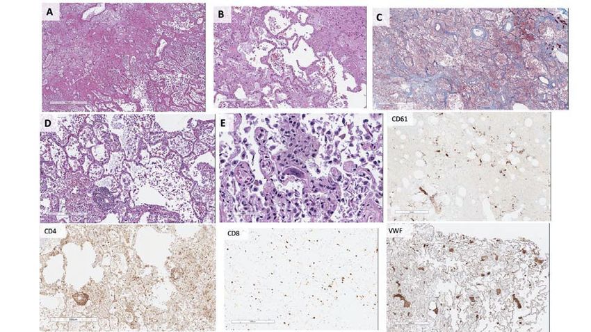
SARS‐CoV Infektion ‐ Treatment Options                                                                ‐8‐
Univ. Prof. Dr. W. Reiterer – www.prof‐reiterer.at

        Fortgeschrittene Stadien zeigen beidseitig ground-glass opacities (milchige Schatten) und
        Infiltrationen.

Diagnose
    SARS-CoV-2 Nukleinsäuren positiv in Sputum, Pharynx-Abstrich und Sekret des unteren
    respiratorischen Trakts
    Real-time reverse Transkriptase–Polymerase-Kettenreaktion (rRT-PCR)
    Für Patienten mit akutem Fieber (>37,5 °C innerhalb von 72 Stunden) und unauffälliger
    Bildgebung, wenn die absolute Zahl peripherer Lymphozyten geringer als 0,8 × 10⁹/l wird, oder
    die Zahl von CD4+ und CD8+ T-Zellen deutlich abnimmt, sollte die Isolierung und enge
    Beobachtung zu Hause durchgeführt werden, selbst wenn der erste SARS-CoV-2-Test negativ
    ausfiel. Eine Wiederholung des Tests sollte nach 24 h angedacht werden. Ebenso kann ein CT
    angefertigt werden, wenn nötig.

Screening bei Aufnahme
Tag 1: Immer
  Nukleinsäure-Test von Sputum oder naso-/oropharyngealen Abstrichen
  Großes Blutbild, Urintest, Analyse der arteriellen Blutgase
  Leber- und Nierenfunktion, C-reaktives Protein (CRP), Procalcitonin (PCT), Kreatinkinase plus
  Myoglobin, Koagulation
  Thorax-CT

Tag 1: Wenn angebracht
  Inflammatorische Zytokine: Interleukine IL-6, IL-10 und Tumornekrosefaktor (TNF)-α
  TB Lymphozyten Untergruppen (z. B. CD4+, CD8+) und Komplement
  Siehe Li et al. 2003 (Chin Med J, Engl.), Tai-sheng et al. 2003 (Chin J Lab Med), Taoran G et al.
  2020 (Chin J Internal Med)

Weitere Untersuchungen bei bestätigter Infektion

Tage 2–3 nach Aufnahme
  Röntgenaufnahmen der Brust oder CT, weitere Aufnahmen je nach Erkrankungsstatus, nicht länger
  als 5 Tage später
  Tage 3, 5. und 7 und bei Entlassung je nach Erkrankungsstatus
  Großes Blutbild, Leber- und Nierenfunktion, Kreatinkinase plus Myoglobin, Koagulation, CRP,
  PCT, TB Lymphozyten Untergruppen (z. B. CD4+, CD8+)

Wiederholung an Tagen 5–7, wenn möglich
  PCT, TB Lymphozyten Untergruppen (z. B. CD4+, CD8+)
  Siehe Li et al. 2003 (Chin Med J, Engl.), Tai-sheng et al. 2003 (Chin J Lab Med), Taoran G et al.
  2020 (Chin J Internal Med)

Bei Entlassung
  Großes Blutbild
  Röntgenbild der Brust
  Leber- und Nierenfunktion
  Alle auffälligen Ergebnisse bei Aufnahme

Behandlungselemente bei COVID-19

Supportive Behandlung
  Elektrolyten
  Flüssigkeit
  Vitalwerte?
  Sauerstoffsättigung?
Sauerstoffbehandlung
Sauerstofffraktion jeweils angepasst an Sättigung
  Hypoxämie?
     → Sauerstofftherapie
     Sättigung mind. 90 % (Männer und nicht schwangere Frauen)
     Sättigung zwischen 92–95 % (schwangere Frauen)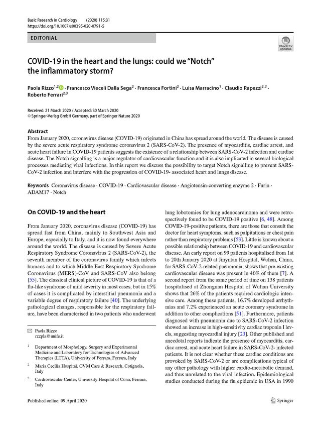
SARS‐CoV Infektion ‐ Treatment Options ‐9‐
Univ. Prof. Dr. W. Reiterer – www.prof‐reiterer.at
Milde Hypoxämie?
→ Nasenkanüle mit 5 l/min
Stärkerer Sauerstoffmangel?
→ Höherer Durchfluss mit 20 l, graduell ansteigend bis zu 50–60 l/min
Die Autoren empfehlen nicht invasive Ventilation nur für Patienten, die dies tolerieren, und Intubierung
nur durch erfahrenes Personal mit Schutzkleidung.
Li und seine Kollegen empfehlen zudem eine protektive Beatmungsstrategie für ein akutes
respiratorisches Distresssyndrom (akutes Lungenversagen, ARDS). Für Patienten mit besonders
schwerem ARDS raten sie zu extrakorporaler Membranoxygenierung (ECMO) oder Bauchlage.
Antivirale Behandlung
Li und Kollegen betonen, dass bislang unklar ist, ob existierende antivirale Therapien gegen SARS-
CoV-2 anschlagen, schlagen aber vor, Lopinavir/Ritonavir, wenn angebracht, in der Menge von zwei
Tabletten zweimal täglich für 14 Tage zu geben.
Glukokortikoid-Behandlung
Schwer erkrankte Patienten können, schreiben die Ärzte, in frühem Stadium beispielsweise intravenös
Methylprednisolon 40–80 mg erhalten (einmal täglich für 5 Tage). Die Behandlung kann je nach
klinischem Zustand und radiologischer Manifestation angepasst werden.
Intravenöses Immunglobulin
Frühe intravenöse Infusion mit humanem Immunoglobulin empfehlen Li und Kollegen für Patienten in
kritischem Zustand, je nach ihrer klinischen Verfassung in Dosierungen zwischen 0,25–0,5 g/(kg/Tag)
für 3–5 Tage.
Antibakterielle Therapie
Wenn eine bakterielle Infektion auf Basis klinischer und bildgebender Daten vermutet wird, können
Patienten mit milder Erkrankung oral antibiotisch gegen CAP (ambulant erworbene Pneumonie)
behandelt werden. Vorgeschlagen werden beispielsweise Cephalosporine oder Fluoroquinolone. Bei
schwer erkrankten Patienten sollten alle möglichen Pathogene abgedeckt werden, wenn nötig.
[DOI 10.1080/22221751.2020.1735265 ] © Alle Rechte: DeutschesGesundheitsPortal.de
Medikamentöse Therapieformen -27.3.20, cit. Amboss
Bisher ist keine nachweislich wirksame Therapieform etabliert, daher stets experimentell; ein
Einsatz kann unter Nutzen-Risiko-Abwägung in Einzelfällen erwogen werden [121][97][59]
Übersicht aktuell erprobter Wirkstoffe
Therapieversuche mit vielen Substanzen, klinische Studien laufend, es werden auch
verschiedene Kombinationsregime getestet!
o Hervorgehoben erscheinen die am intensivsten untersuchten Wirkstoffe
Wirkstoff(-gruppen), Substanzen und mögliche therapeutische Zielstrukturen [122][123]
o Inhibition der Adhäsion und Invasion
Camostat [46] (Protease-Inhibitor)
o Inhibition der Fusion
Chloroquin/Hydroxychloroquin , zusätzlich oder als alternativ führender
Mechanismus immunsuppressive Effekte [124][125][126][127]
Intensiv erprobt wird die Kombination mit Azithromycin [128]
Azithromycin wird auch zum Einsatz als Immunmodulans bei
COVID-19-Pneumonie diskutiert, Zithromax
Umifenovir [129]
o Protease-Inhibition
Lopinavir/Ritonavir [130][131][24]
Darunavir/Ritonavir (ggf. in Kombination mit Umifenovir)
Remdesivir [132][133]
o RNA-Polymerase-Inhibitoren bzw. Nukleotidanaloga
Favipiravir [134]
Remdesivir [133][135]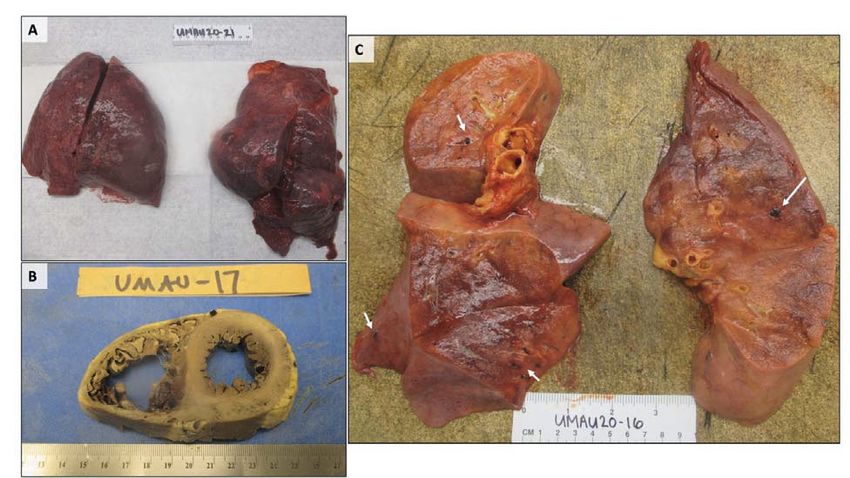
SARS‐CoV Infektion ‐ Treatment Options ‐10‐
Univ. Prof. Dr. W. Reiterer – www.prof‐reiterer.at
Baloxavirmarboxil [136]
o Antikörpertherapie und Biologicals [22]
Tocilizumab [137][138], insb. in der Phase des ARDS bei erhöhtem IL-6 und CRP
Roactemra Inj.Lsg, 4St 1380.- Interleukin-Inhibitor
Rekombinantes ACE2 (rhACE2, APN01) [47][139]
o Passive Immunisierung durch Serumtherapie: Form der Impfung, bei genügend hoher
Anzahl Immunisierter und Serumspenden im Verlauf als Option für eine breitere
Anwendung, bspw. bei Risikogruppen [140]
Interaktionen der genannten Medikamente: Zahlreich, bei erwogenem Einsatz zu beachten
(Fachinfo!) bzw. gemäß Übersicht der University of Liverpool! [141]
o Siehe auch
Risikokonstellationen einer verlängerten QT-Zeit
Torsade de pointes - Klinisches Management
Für die Wirksamkeit der Medikamente ist wahrscheinlich der Zeitpunkt des Einsatzes im
Krankheitsverlauf entscheidend – während in die Virusinvasion und -replikation eingreifende
Medikamente (z.B. Remdesivir, Hydroxychloroquin) so früh wie möglich appliziert werden müssten,
könnten andere Ansätze, die auf die Kontrolle der dysregulierten Immunantwort bei schweren
Verläufen abzielen (z.B. Tocilizumab), auch in späteren Phasen des Krankheitsverlaufes sinnvoll
eingesetzt werden!
Favipiravir
Wikipedia: Favipiravir ist Guanin-Analogon[3] und ein Inhibitor der viralen RNA-abhängigen RNA-
Polymerase von verschiedenen Viren,[4] nicht jedoch von zellulären Polymerasen. Weiterhin erhöht es
die Mutationsrate bei der Replikation des Influenzavirus[5] und des Ebolavirus.[6] Favipiravir ist ein
Prodrug, das heißt, es wird im Stoffwechsel durch die HGPRT in Favipiravir-ribofuranosyl-5'-
monophosphat (FRMP) und Favipiravir-ribofuranosyl-5'-triphosphat (FRTP) überführt,[7][8] wobei FRTP
die wirksame Form von Favipiravir bei der Hemmung der RNA-abhängigen RNA-Polymerase ist.[2]
Favipiravir ist unter anderem wirksam gegen das Influenzavirus, das Maul-und-Klauenseuche-Virus,
verschiedene Flaviviren (das West-Nil-Virus, das Gelbfieber-Virus), Arenaviren, Bunyaviren und
Alphaviren,[2] manche Enteroviren,[9] das Nipahvirus,[10] Noroviren,[11] das Ebolavirus,[12] das Lassa-
Virus,[11] das Tollwutvirus[13][14] und das Rifttalfieber-Virus.[15] Es wirkt auch gegen das Zika-Virus, aber
schlechter als MK-608.[16]
Im Februar 2020 wurde Favipiravir in China in einer ersten nicht randomisierten Doppelblindstudie an
80 Patienten als antivirale Therapie gegen das Coronavirus SARS-CoV-2 getestet.[21][22] In einer
weiteren Studie, in der Favipiravir gegen das virostatische Präparat Arbidol (Umifenovir) an jeweils
rund 120 Patienten verglichen wurde, zeigte Favipiravir eine signifikante Verbesserung. Die
Ergebnisse dieser Studie werden jedoch bezweifelt.[23] Favipiravir hat zuvor im Februar 2020 in China
die Zulassung zu klinischen Tests zur Evaluierung der Wirksamkeit bei COVID-19 erhalten.[24]
Avigan (Favipiravir) gehört zu den Arzneimitteln, für die das Bundesministerium für Gesundheit die
zentrale Beschaffung zur Behandlung infizierter und schwer erkrankter COVID-19 Patienten in
Deutschland eingeleitet hat. Da es sich bei einer Covid-19-Therapie um einen individuellen
Heilversuch ohne klinischen Wirksamkeitsnachweis handele, solle der Einsatz vorrangig bei schweren
Verlaufsformen patientenindividuell erwogen werden.[25]
WHO launches global megatrial of the four most promising coronavirus treatments – 22.3.20
Remdesivir: shuts down viral replication by inhibiting akey viral enzyme, the RNA-dependent
RNApolymerase.
chloroquine/hydroychloroquine: drug works by decreasing the acidity in endosomes, compartment
inside cells that they use to ingest outside marerial and that some viruses can coopt to enter a cell.
Doses needed are usually high and could dause serious toxicities.
Main entryway for SARS-CoV2 is a different one, using ist socalled spike protein to attach to a
receptor on the surface of human cells.SARS‐CoV Infektion ‐ Treatment Options ‐11‐ Univ. Prof. Dr. W. Reiterer – www.prof‐reiterer.at combination of two HIV drugs – lopinavir u. ritonavir and this combination with interferon-beta: first used in Saudi Arabia for MERS patients. Interferone given late in the disease it could easily lead to worse tissue damage. The influenca drug favipiravir may be added to the trial Design ist not double-blind, we have to balance scientific rigor against speed. Executive Summary Covid19 v2 – Stellungnahme zur COVID19 Krise Zusammenfassung einiger quantitativer Perspektiven, 31.3.20 Beiglböck M., Grohs Ph., Hermisson J., Nordborg M. u. Schachermayer W. https://www.oesterreich.gv.at Robert Koch Institut: Coronavirus SARS-CoV-2 https://www.rki.de/DE/Content/InfAZ/N/Neuartiges_Coronavirus/Steckbrief.html The FDA-approved Drug Ivermectin inhibits the replication of SARS-CoV-2 in vitro. - 13.4.20 Academic Journal (English) By: Caly L; Druce JD; Catton MG; Jans DA; Wagstaff KM, Antiviral Research [Antiviral Res], ISSN: 1872-9096, 2020 Apr 03, pp. 104787; Publisher: Elsevier; PMID: 32251768; Although several clinical trials are now underway to test possible therapies, the worldwide response to the COVID-19 outbreak has been largely limited to monitoring/containment. We report here that Ivermectin, an FDA-approved anti-parasitic previously shown to have broad-spectrum anti-viral activity in vitro, is an inhibitor of the causative virus (SARS-CoV-2), with a single addition to Vero-hSLAM cells 2 hours post infection with SARS-CoV-2 able to effect ∼5000-fold reduction in viral RNA at 48 h. Ivermectin therefore warrants further investigation for possible benefits in humans. Copyright © 2020. Published by Elsevier B.V. Remdesivir, lopinavir, emetine, and homoharringtonine inhibit SARS-CoV-2 replication in vitro. Academic Journal (English) By: Choy KT; Yin-Lam Wong A; Kaewpreedee P; Sia SF; Chen D; Yan Hui KP; Wing Chu DK; Wai Chan MC; Pak-Hang Cheung P; Huang X; Peiris M; Yen HL, Antiviral Research [Antiviral Res], ISSN: 1872-9096, 2020 Apr 03, pp. 104786; Publisher: Elsevier; PMID: 32251767; An escalating pandemic by the novel SARS-CoV-2 virus is impacting global health and effective therapeutic options are urgently needed. We evaluated the in vitro antiviral effect of compounds that were previously reported to inhibit coronavirus replication and compounds that are currently under evaluation in clinical trials for SARS-CoV-2 patients. We report the antiviral effect of remdesivir, lopinavir, homorringtonine, and emetine against SARS-CoV-2 virus in Vero E6 cells with the estimated 50% effective concentration at 23.15 μM, 26.63 μM, 2.55 μM and 0.46 μM, respectively. Ribavirin or favipiravir that are currently evaluated under clinical trials showed no inhibition at 100 μM. Synergy between remdesivir and emetine was observed, and remdesivir at 6.25 μM in combination with emetine at 0.195 μM may achieve 64.9% inhibition in viral yield. Combinational therapy may help to reduce the effective concentration of compounds below the therapeutic plasma concentrations and provide better clinical benefits. Copyright © 2020. Published by Elsevier B.V. Evidence that Vitamin D Supplementation Could Reduce Risk of Influenza and COVID-19 Infections and Deaths. Academic Journal (English) By: Grant WB; Lahore H; McDonnell SL; Baggerly CA; French CB; Aliano JL; Bhattoa HP, Nutrients [Nutrients], ISSN: 2072-6643, 2020 Apr 02; Vol. 12 (4); Publisher: MDPI Publishing; PMID: 32252338; The world is in the grip of the COVID-19 pandemic. Public health measures that can reduce the risk of infection and death in addition to quarantines are desperately needed. This article reviews the roles of vitamin D in reducing the risk of respiratory tract infections, knowledge about the epidemiology of influenza and COVID-19, and how vitamin D supplementation might be a useful measure to reduce risk. Through several mechanisms, vitamin D can reduce risk of infections. Those mechanisms include
SARS‐CoV Infektion ‐ Treatment Options                                                              ‐12‐
Univ. Prof. Dr. W. Reiterer – www.prof‐reiterer.at

inducing cathelicidins and defensins that can lower viral replication rates and reducing concentrations
of pro-inflammatory cytokines that produce the inflammation that injures the lining of the lungs, leading
to pneumonia, as well as increasing concentrations of anti-inflammatory cytokines. Several
observational studies and clinical trials reported that vitamin D supplementation reduced the risk of
influenza, whereas others did not. Evidence supporting the role of vitamin D in reducing risk of COVID-
19 includes that the outbreak occurred in winter, a time when 25-hydroxyvitamin D (25(OH)D)
concentrations are lowest; that the number of cases in the Southern Hemisphere near the end of
summer are low; that vitamin D deficiency has been found to contribute to acute respiratory distress
syndrome; and that case-fatality rates increase with age and with chronic disease comorbidity, both of
which are associated with lower 25(OH)D concentration. To reduce the risk of infection, it is
recommended that people at risk of influenza and/or COVID-19 consider taking 10,000 IU/d of vitamin
D3 for a few weeks to rapidly raise 25(OH)D concentrations, followed by 5000 IU/d. The goal should
be to raise 25(OH)D concentrations above 40-60 ng/mL (100-150 nmol/L). For treatment of people
who become infected with COVID-19, higher vitamin D3 doses might be useful. Randomized
controlled trials and large population studies should be conducted to evaluate these
recommendations.
.
How to reduce the likelihood of coronavirus-19 (CoV-19 or SARS-CoV-2) infection and lung
inflammation mediated by IL-1.
Editorial & Opinion
(English) By: Conti P; Gallenga CE; Tetè G; Caraffa A; Ronconi G; Younes A; Toniato E; Ross R;
Kritas SK, Journal Of Biological Regulators And Homeostatic Agents [J Biol Regul Homeost Agents],
ISSN: 0393-974X, 2020 Mar 31; Vol. 34 (2); Publisher: Biolife; PMID: 32228825;

SARS-CoV-2, also referred to as CoV-19, is an RNA virus which can cause severe acute respiratory
diseases (COVID-19), with serious infection of the lower respiratory tract followed by bronchitis,
pneumonia and fibrosis. The severity of the disease depends on the efficiency of the immune system
which, if it is weak, cannot stem the infection and its symptoms. The new CoV-19 spreads in the
population at a rate of 0.8-3% more than normal flu and mostly affects men, since immune genes are
more expressed on the X chromosome. If CoV-19 would spread with a higher incidence rate (over
10%), and affect the people who live in closed communities such as islands, it would cause many
more deaths. Moreover, people from the poorest classes are most at risk because of lack of health
care and should be given more assistance by the competent authorities. To avoid the aggravation of
CoV-19 infection, and the collapse of the health system, individuals should remain at home in
quarantine for a period of approximately one month in order to limit viral transmission. In the case of a
pandemic, the severe shortage of respirators and protective clothing, due to the enormous demand
and insufficient production, could lead the CoV-19 to kill a large number of individuals. At present,
there is no drug capable of treating CoV-19 flu, the only therapeutic remedies are those aimed at the
side effects caused by the virus, such as inflammation and pulmonary fibrosis, recognized as the first
causes of death. One of the COVID-19 treatments involves inhaling a mixture of gaseous hydrogen
and oxygen, obtaining better results than with oxygen alone. It was also noted that individuals
vaccinated for viral and/or bacterial infectious diseases were less likely to become infected. In
addition, germicidal UV radiation "breaks down" the oxygen O2 which then aggregate into O3 (ozone)
molecules creating the ozone layer, capable of inhibiting viral replication and improving lung
respiration. All these precautions should be taken into consideration to lower the risk of infection by
CoV-19. New anti-viral therapies with new drugs should also be taken into consideration. For example,
microbes are known to bind TLR, inducing IL-1, a pleiotropic cytokine, highly inflammatory, mediator of
fever and fibrosis. Therefore, drugs that suppress IL-1 or IL-1R, also used for the treatment of
rheumatoid arthritis are to be taken into consideration to treat COVID-19. We strongly believe that all
these devices described above can lead to greater survival and. therefore, reduction in mortality in
patients infected with CoV-19.
Copyright 2020 Biolife Sas. www.biolifesas.org.

Novel coronavirus 2019 (COVID-19): Emergence and implications for emergency care
Jane YeeMD Lucy UngerMD Frank ZadraveczMD Paloma CarielloMD
Allan SeibertMD Michael Austin JohnsonMD, PhD Matthew Joseph FullerMD
Ann. Emerg. Med. 2020;1-7
https://doi.org/10.1002/emp2.12034
SARS‐CoV Infektion ‐ Treatment Options                                                             ‐13‐
Univ. Prof. Dr. W. Reiterer – www.prof‐reiterer.at

Abstract
A novel coronavirus (COVID-19) causing acute illness with severe symptoms has been isolated in
Wuhan, Hubei Province, China. Since its emergence, cases have been found worldwide, reminiscent
of severe acute respiratory syndrome andMiddle East respiratory syndrome outbreaks over the past 2
decades. Current understanding of this epidemic remains limited due to its rapid development and
available data. While occurrence outside mainland China remains low, the likelihood of increasing
cases globally continues to rise. Given this potential, it is imperative that emergency clinicians
understand the preliminary data behind the dynamics of this disease, recognize possible presentations
of patients, and understand proposed treatment modalities.
KEYWORDS: global health, infectious disease, public health

Endothelial cell infection and endotheliitis in COVID-19

Zsuzsanna Varga, Andreas J Flammer, Peter Steiger, Martina Haberecker, Rea Andermatt, Annelies S
Zinkernagel, et al.
Published:April 20, 2020DOI:https://doi.org/10.1016/S0140-6736(20)30937-5

Cardiovascular complications are rapidly emerging as a key threat in coronavirus disease 2019
(COVID-19) in addition to respiratory disease. The mechanisms underlying the disproportionate effect
of severe acute respiratory syndrome coronavirus 2 (SARS-CoV-2) infection on patients with
cardiovascular comorbidities, however, remain incompletely understood.
SARS-CoV-2 infects the host using the angiotensin converting enzyme 2 (ACE2) receptor, which is
expressed in several organs, including the lung, heart, kidney, and intestine. ACE2 receptors are also
expressed by endothelial cells.
Whether vascular derangements in COVID-19 are due to endothelial cell involvement by the virus is
currently unknown. Intriguingly, SARS-CoV-2 can directly infect engineered human blood vessel
organoids in vitro.

Here we demonstrate endothelial cell involvement across vascular beds of different organs in a series
of patients with COVID-19 (further case details are provided in the appendix).
• View related content for this article

Patient 1 was a male renal transplant recipient, aged 71 years, with coronary artery disease and
arterial hypertension. The patient's condition deteriorated following COVID-19 diagnosis, and he
required mechanical ventilation. Multisystem organ failure occurred, and the patient died on day 8.
Post-mortem analysis of the transplanted kidney by electron microscopy revealed viral inclusion
structures in endothelial cells (figure A, B). In histological analyses, we found an accumulation of
inflammatory cells associated with endothelium, as well as apoptotic bodies, in the heart, the small
bowel (figure C) and lung (figure D). An accumulation of mononuclear cells was found in the lung, and
most small lung vessels appeared congested.
SARS‐CoV Infektion ‐ Treatment Options ‐14‐
Univ. Prof. Dr. W. Reiterer – www.prof‐reiterer.at
FigurePathology of endothelial cell dysfunction in COVID-19
Show full caption
View Large Image
Figure Viewer
Download Hi-res image
Download (PPT)
Patient 2 was a woman, aged 58 years, with diabetes, arterial hypertension, and obesity. She
developed progressive respiratory failure due to COVID-19 and subsequently developed multi-organ
failure and needed renal replacement therapy. On day 16, mesenteric ischaemia prompted removal of
necrotic small intestine. Circulatory failure occurred in the setting of right heart failure consequent to
an ST-segment elevation myocardial infarction, and cardiac arrest resulted in death. Post-mortem
histology revealed lymphocytic endotheliitis in lung, heart, kidney, and liver as well as liver cell
necrosis. We found histological evidence of myocardial infarction but no sign of lymphocytic
myocarditis. Histology of the small intestine showed endotheliitis (endothelialitis) of the submucosal
vessels.
Patient 3 was a man, aged 69 years, with hypertension who developed respiratory failure as a result of
COVID-19 and required mechanical ventilation. Echocardiography showed reduced left ventricular
ejection fraction. Circulatory collapse ensued with mesenteric ischaemia, and small intestine resection
was performed, but the patient survived. Histology of the small intestine resection revealed prominent
endotheliitis of the submucosal vessels and apoptotic bodies (figure C).
We found evidence of direct viral infection of the endothelial cell and diffuse endothelial inflammation.
Although the virus uses ACE2 receptor expressed by pneumocytes in the epithelial alveolar lining to
infect the host, thereby causing lung injury, the ACE2 receptor is also widely expressed on endothelial
cells, which traverse multiple organs.
Recruitment of immune cells, either by direct viral infection of the endothelium or immune-mediated,
can result in widespread endothelial dysfunction associated with apoptosis (figure D).
The vascular endothelium is an active paracrine, endocrine, and autocrine organ that is indispensable
for the regulation of vascular tone and the maintenance of vascular homoeostasis.
Endothelial dysfunction is a principal determinant of microvascular dysfunction by shifting the vascular
equilibrium towards more vasoconstriction with subsequent organ ischaemia, inflammation with
associated tissue oedema, and a pro-coagulant state.
Our findings show the presence of viral elements within endothelial cells and an accumulation of
inflammatory cells, with evidence of endothelial and inflammatory cell death. These findings suggest
that SARS-CoV-2 infection facilitates the induction of endotheliitis in several organs as a direct
consequence of viral involvement (as noted with presence of viral bodies) and of the host inflammatory
response. In addition, induction of apoptosis and pyroptosis might have an important role in
endothelial cell injury in patients with COVID-19. COVID-19-endotheliitis could explain the systemic
impaired microcirculatory function in different vascular beds and their clinical sequelae in patients with
COVID-19. This hypothesis provides a rationale for therapies to stabilise the endothelium while
tackling viral replication, particularly with anti-inflammatory anti-cytokine drugs, ACE inhibitors, and
statins.
This strategy could be particularly relevant for vulnerable patients with pre-existing endothelial
dysfunction, which is associated with male sex, smoking, hypertension, diabetes, obesity, and
established cardiovascular disease, all of which are associated with adverse outcomes in COVID-19.
ZV and AJF contributed equally as first authors, and RAS, FR, and HM contributed equally as last
authors. AJF reports fees from Alnylam, Amgen, AstraZeneca, Fresenius, Imedos Systems, Novartis,
Pfizer, Roche, Vifor, and Zoll, unrelated to this Correspondence. MRM reports consulting relationships
with Abbott, Medtronic, Janssen, Mesoblast, Portola, Bayer, NupulseCV, FineHeart, Leviticus, Baim
Institute for Clinical Research, Riovant, and Triple Gene, unrelated to this Correspondence. FR has
been paid for the time spent as a committee member for clinical trials, advisory boards, other forms of
consulting and lectures or presentations. These payments were made directly to the University of
Zurich and no personal payments were received in relation to these trials or other activities. All other
authors declare no competing interests.SARS‐CoV Infektion ‐ Treatment Options ‐15‐ Univ. Prof. Dr. W. Reiterer – www.prof‐reiterer.at Pulmonary and Cardiac Pathology in Covid-19: The First Autopsy Series from New Orleans Sharon E. Fox,1,2* Aibek Akmatbekov,1, Jack L. Harbert,1, Guang Li,3 J. Quincy Brown,3 Richard S. Vander Heide1* 1) Department of Pathology, LSU Health Sciences Center, New Orleans 2) Pathology and Laboratory Medicine Service, Southeast Louisiana Veterans Healthcare System 3) Department of Biomedical Engineering, Tulane University *To whom correspondence should be addressed: sfox@lsuhsc.edu & rvand3@lsuhsc.edu Abstract: SARS-CoV-2 has rapidly spread across the United States, causing extensive morbidity and mortality, though the histopathologic basis of severe disease cases has yet to be studied in detail. Over the past century, autopsy has contributed significantly to our understanding of numerous disease processes, but for several reasons, autopsy reports following deaths related to SARSCoV-2 have thus far been limited across the globe. We report on the relevant cardiopulmonary findings in the first series of autopsies in the United States, with the cause of death being due to SARS-CoV-2 infection. These cases identify key pathologic states potentially contributing to severe disease and decompensation in these patients. All rights reserved. No reuse allowed without permission. (which was not certified by peer review) is the author/funder, who has granted medRxiv a license to display the preprint in perpetuity. medRxiv preprint doi: https://doi.org/10.1101/2020.04.06.20050575.this version posted April 10, 2020. The copyright holder for this preprint Introduction: The first confirmed case of SARS-CoV-2 infection in the United States was reported on January 20, 2020. Since that time, the virus has spread across the country, with several cities within the United States becoming epicenters of the pandemic. As of March 31, 2020 the Louisiana Department of Health reported a total of 5,237 COVID-19 cases with 1,355 hospitalizations, and 239 COVID-19 related deaths statewide. A total of 1,834 of the 5,239 COVID-19 cases and 101 of the 239 deaths have occurred in the city of New Orleans – the highest rate of death per capita in the United States. University Medical Center in New Orleans, built following Hurricane Katrina, is equipped with an autopsy suite meeting the modern standards recommended by the CDC for performance of autopsy on COVID-19 positive patients. We report here on the cardiopulmonary findings of the first four autopsies of a series of twelve performed on patients within the United States, with relevant implications for the treatment of severe cases. Brief Clinical Summary: The four decedents included male and female patients, ages 44-76. All were African American, and had a history of obesity class 2-3, and hypertension controlled by medication. Three of the patients had insulin-dependent type II diabetes, two had known chronic kidney disease (stages 2 and 3), and one was taking methotrexate. In all cases the clinical course consisted of approximately three days of mild cough and fever to 101- 102° F., with sudden respiratory decompensation just prior to arrival in the emergency department. Chest radiographs revealed bilateral ground-glass opacities, consistent with acute respiratory distress syndrome (ARDS) which worsened over the hospital course. The patients were intubated and brought to the ICU. Treatment in the ICU included vancomycin, azithromycin, and aefepime for all patients, with one patient receiving dexamethasone. All of the patients tested positive for SARS-CoV-2 (by 2019 Novel Coronavirus Real Time RT-PCR). Notable laboratory findings were the development of elevated ferritin, fibrinogen, PT, and within 24 hours of death, an increased neutrophil count with relative lymphopenia. Glucose and AST became slightly elevated above normal, and creatinine increased above baseline for all patients. D-dimers drawn near the time of death in two patients were markedly elevated (1200-2900 ng/mL). (A detailed description of ante-mortem laboratory findings can be found in Table S1 in the Supplementary Appendix). When the patients continued to deteriorate despite support, the families elected to withdraw care. In each case, consent for autopsy was given, and nonrestricted by the next of kin. Studies performed outside of routine autopsy were determined to be exempt by the IRB at Tulane University. Gross Findings: Gross examination of the lungs at the time of autopsy revealed the tracheae to be of normal caliber and mildly erythematous. All of the lungs were heavy, the left ranging from 680g to 1030g ,
SARS‐CoV Infektion ‐ Treatment Options                                                              ‐16‐
Univ. Prof. Dr. W. Reiterer – www.prof‐reiterer.at

normal (583 +/-216); right ranging from 800g to 1050g, normal (663+/-239). They contained the
usual lobes and fissures, with exception to one decedent with prior partial lobectomy on the right
The pulmonary arteries at the hilum of each of the lungs were free of thromboemboli. The
bronchi revealed thick, white mucous in the lungs of one patient, and pink froth in the airways of
the other three. Mild to moderate serosanguinous pericardial and pleural effusions were also
present. The parenchyma of each of the lungs was diffusely edematous and firm, consistent with
the clinical diagnosis of ARDS. Notably, regions of dark-colored hemorrhage with focal
demarcation could be identified throughout the peripheral parenchyma in the lungs of all but one
of the decedents (Figure 1A). On cut sections, the areas identified as hemorrhagic on the
external surface showed frank hemorrhage. After fixation, the cut surfaces of the lung tissue
showed alternating areas of tan-grey consolidation with patchy areas of hemorrhage that ranged
from 3-6 cm in maximal diameter. In some cases, small, firm thrombi were present in sections
of the peripheral parenchyma (Figure 1C). Only in the case of the patient on
immunosuppression was there focal consolidation - the remainder of the lungs showed no
evidence of lobar infiltrate, abscess, or definitive gross inflammatory process.
Examination of the heart was performed in three cases, with the hearts ranging in size from 430g to
550g (normal: 365g +/-71). The most significant gross findings were cardiomegaly, and right
ventricular dilatation. In one case, massive dilatation could be seen, in which the right ventricular
cavity measured 3.6cm in diameter, while the left ventricle measured 3.4cm in greatest diameter
(Figure 1B). The cut surface of the myocardium was firm, red-brown, and free of significant
lesions in all cases, and the coronary arteries showed no significant stenosis or acute thrombus
formation.

FIGURE 1: Gross Findings of the Lungs and Heart. A) Lungs with bilateral pulmonary edema and
patches of dark hemorrhage, and B) A heart showing extreme right ventricular dilatation, with
straightening of the interventricular septum. C) Cut sections of lung showing thrombi present within
peripheral small vessels (white arrows).

Microscopic Findings:
Pulmonary. The lungs were extensively sampled across central and peripheral regions of each lobe
bilaterally. Histologic examination of the lungs showed bilateral diffuse alveolar damage with a
comparatively mild-to-moderate lymphocytic infiltrate, composed of a mixture of CD4+ and
CD8+ lymphocytes (Figure 2), located predominantly in the interstitial spaces and around larger
bronchioles. CD4+ lymphocytes could be seen in aggregates around small vessels, some of
which appeared to contain platelets and small thrombi. In all but one case, foci of hemorrhage
were present. Desquamated type 2-pneumocytes with apparent viral cytopathic effect consisting
of cytomegaly, and enlarged nuclei with bright, eosinophilic nucleoli, were present within
alveolar spaces (Figure 3). The largest of these cells frequently contained an eccentric clearing
of the cytoplasm with small vesicles discernible at higher power, likely representing viral
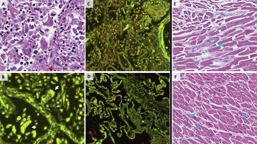
SARS‐CoV Infektion ‐ Treatment Options                                                              ‐17‐
Univ. Prof. Dr. W. Reiterer – www.prof‐reiterer.at

inclusions. Scattered hyaline membranes could be seen, as well as fibrin deposition, highlighted
by trichrome stains (Figure 2), consistent with diffuse alveolar damage. The alveolar capillaries
were notably thickened, with surrounding edema, and fibrin thrombi were present within the
capillaries and small vessels. A notable finding was the presence of CD61+ megakaryocytes
(Figure 2), possibly representing resident pulmonary megakaryocytes, with significant nuclear
hyperchromasia and atypia. These cells were located within alveolar capillaries, and could be
seen in association with, and actively producing platelets (Figure 2). The fibrin and platelets
present within small vessels also appeared to aggregate inflammatory cells, with entrapment of
numerous neutrophils. Only in the case of the patient on immunosuppression was there evidence
of a focal acute inflammatory infiltrate possibly consistent with secondary infection. The
neutrophils in this case, however, were partially degenerated and entrapped in fibers, possibly
representing neutrophil extracellular traps (Figure 3),1,2 and were present in association with
clusters of CD4+ mononuclear cells. No significant neutrophilic infiltrate was identified within
airways or the interstitium to suggest secondary infection in other cases.

FIGURE 2: Pulmonary Microscopic Findings. All patients demonstrated extensive diffuse alveolar
damage. A) Hyaline membranes and hemorrhage (H&E), with B) Fibrin thrombi present within
distended small vessels and capillaries, and C) Extensive extracellular fibrin deposition highlighted in
blue by Masson-Trichrome stain. D) Perivascular aggregations of lymphocytes, which were positive for
CD4 immunostain, with only scattered CD8 positive cells present. E) Numerous megakaryocytes were
present within the small vessels and alveolar capillaries, highlighted by CD61 and Von Willebrand
Factor immunostains.

Cardiac. The sections of myocardium did not show any large or confluent areas of myocyte necrosis.
The cardiac histopathology was remarkable, however, for scattered individual cell myocyte
necrosis in each heart examined. In rare areas, lymphocytes were adjacent to, but not
surrounding degenerating myocytes. Whether this may represent an early manifestation of a
viral myocarditis is not certain, but there was no significant brisk lymphocytic inflammatory
infiltrate consistent with the typical pattern of viral myocarditis. This may be consistent with a
recent paper by Chen et al. that hypothesizes that pericytes may be infected by the SARS-CoV-2
virus and cause capillary endothelial cell/microvascular dysfunction which may cause individual
cell necrosis.3 There was no obvious viral cytopathic effect by light microscopy, but direct viral
infection of myocytes cannot be entirely ruled out in this limited examination.
SARS‐CoV Infektion ‐ Treatment Options                                                              ‐18‐
Univ. Prof. Dr. W. Reiterer – www.prof‐reiterer.at

FIGURE 3: SARS-CoV-2 cytopathic effects. A) H&E stain of several enlarged pneumocytes within a
damaged alveolus, having enlarged nuclei, prominent nuceloli, and cytologic atypia. B) Relative
distribution of dsDNA (red) versus RNA (green) in tissue sections via DRAQ5 and SYTO RNASelect
fluorescent staining (see Supplementary Methods for staining details). Virally infected cells in
alveolar spaces show multinucleation and grouping as evidenced by DNA stain, and abundant RNA
present within the cytoplasm (white arrows), C) Entrapment of immune cells, including degeneration
neutrophils, within fibrin, and strands of extracellular material with weak DNA staining, and D) Control
lung tissue obtained at autopsy for non-pulmonary cause of death prior to the SARS-CoV-2 pandemic.
E) and F) H&E stains of cardiac myocytes with focal degeneration (blue arrows).

Discussion:
The dominant process in all cases was consistent with diffuse alveolar damage, with a mild to
moderate mononuclear response consisting of notable CD4+ aggregates around thrombosed
small vessels, and significant associated hemorrhage. Important additional mechanisms that may
have contributed to death in this initial series of autopsies include a thrombotic microangiopathy
that was restricted to the lungs. This process may involve activation of megakaryocytes, possibly
those native to the lung, with platelet aggregation and platelet-rich clot formation, in addition to
fibrin deposition. Small vessel thrombus formation in the lung periphery was in many cases
associated with foci of alveolar hemorrhage. In one case, extensive fibrin and early organization
was present, with degenerated neutrophils within the alveoli possibly representing neutrophil
extracellular traps.1,2 On RNA imaging, we were able to visualize multinucleated cells within
alveolar spaces, containing abundant RNA, likely representing virally infected cells. These may
represent the multinucleated cells previously described from a single report of post-mortem
biopsy from a decedent in China.4 Cardiac findings were significant for a lack of myocarditis,
and the rise in BNP observed in at least one of our cases was likely due to acute right ventricular
dilatation. The underlying cause of scattered atypical myocyte degeneration remains uncertain.
There is prior evidence of viral infection causing activation of both maladaptive cytokine pathways,
and platelet response, and our findings suggest that these immune functions may be related to
severe forms of Covid-19. In response to systemic and pulmonary viral infections of H1N1
influenza and dengue, megakaryocytes have been known to respond by overexpressing IFITM3,
and producing platelets with the same over-expression 5 In addition, platelets and
megakaryocytes may have receptors for viruses6–9, some of which have been specifically activated
in H1N1 influenza, often in association with lymphopenia10–12 There is even some evidence that
the earlier SARS-CoV directly infected megakaryocytes, and that platelets function was affected
in damaged lungs of those with severe SARS.14 We do not currently have evidence of direct
infection of megakaryocytes by SARS-CoV-2, but the abundance of these cells in the lungs at
autopsy is likely related to the abundance of small, sometimes platelet-rich thrombi, and foci of
hemorrhage.
You can also read