COVID-19 Pathogenesis: From Molecular Pathway to Vaccine Administration
←
→
Page content transcription
If your browser does not render page correctly, please read the page content below
biomedicines
Review
COVID-19 Pathogenesis: From Molecular Pathway to
Vaccine Administration
Francesco Nappi 1, * , Adelaide Iervolino 2                    and Sanjeet Singh Avtaar Singh 3

                                          1   Department of Cardiac Surgery, Centre Cardiologique du Nord, 93200 Saint-Denis, France
                                          2   Department of Cardiovascular Sciences, Fondazione Policlinico Universitario Agostino Gemelli IRCSS,
                                              00168 Rome, Italy; adelaide.iervolino@libero.it
                                          3   Department of Cardiothoracic Surgery, Golden Jubilee National Hospital, Glasgow G81 4DY, UK;
                                              sanjeetsinghtoor@gmail.com
                                          *   Correspondence: francesconappi2@gmail.com; Tel.: +33-149-334-104; Fax: +33-149-334-119

                                          Abstract: The Coronavirus 2 (SARS-CoV-2) infection is a global pandemic that has affected millions of
                                          people worldwide. The advent of vaccines has permitted some restitution. Aside from the respiratory
                                          complications of the infection, there is also a thrombotic risk attributed to both the disease and the
                                          vaccine. There are no reliable data for the risk of thromboembolism in SARS-CoV-2 infection in
                                          patients managed out of the hospital setting. A literature review was performed to identify the
                                          pathophysiological mechanism of thrombosis from the SARS-CoV-2 infection including the role of
                                          Angiotensin-Converting Enzyme receptors. The impact of the vaccine and likely mechanisms of
                                          thrombosis following vaccination were also clarified. Finally, the utility of the vaccines available
                                          against the multiple variants is also highlighted. The systemic response to SARS-CoV-2 infection is
                                          still relatively poorly understood, but several risk factors have been identified. The roll-out of the
                                          vaccines worldwide has also allowed the lifting of lockdown measures and a reduction in the spread

                                   of the disease. The experience of the SARS-CoV-2 infection, however, has highlighted the crucial role
Citation: Nappi, F.; Iervolino, A.;
                                          of epidemiological research and the need for ongoing studies within this field.
Avtaar Singh, S.S. COVID-19
Pathogenesis: From Molecular              Keywords: SARS-CoV-2; COVID-19; thromboembolism; ACE inhibition; pathophysiology
Pathway to Vaccine Administration.
Biomedicines 2021, 9, 903. https://
doi.org/10.3390/biomedicines9080903
                                          1. Introduction
Academic Editor: Byeong Hwa Jeon
                                               Patients with severe acute respiratory syndrome Coronavirus 2 (SARS-CoV-2) infection
                                          may develop associated arterial and venous thrombotic complications. Data reported in
Received: 2 June 2021
                                          the 2019 U.S. Coronavirus Disease Patient Registry (COVID-19) recorded a 2.6% rate of
Accepted: 26 July 2021
                                          thrombotic complications in the 299 patients who required non-critical hospitalization
Published: 27 July 2021
                                          compared to the rate of 35.3% of the 170 patients hospitalized in critical care units [1,2].
                                          Klok et al. confirmed a remarkably high 31% incidence of thrombotic complications in
Publisher’s Note: MDPI stays neutral
                                          ICU patients with COVID-19 infections2 . These results supported the recommendation
with regard to jurisdictional claims in
                                          to use drug prophylaxis for thrombosis in all COVID-19 patients admitted to the ICU.
published maps and institutional affil-
iations.
                                          The data strongly support increased drug prophylaxis dosage, even in the absence of
                                          randomized trials.
                                               To date, there are no reliable data to establish the risk of thromboembolism in SARS-
                                          CoV-2 infection in patients whose clinical conditions do not require hospitalization. Several
                                          studies reported that patients admitted to the hospital with COVID-19 disease experienced
Copyright: © 2021 by the authors.
                                          thrombotic complications involving the heart, brain, and peripheral vascular system, which
Licensee MDPI, Basel, Switzerland.
                                          mainly led to myocardial infarction (MI), ischemic stroke, and venous thromboembolism
This article is an open access article
                                          (VTE) [3–5].
distributed under the terms and
                                               During the initial months of the pandemic acceleration, several autopsy studies [6,7]
conditions of the Creative Commons
Attribution (CC BY) license (https://
                                          highlighted the presence of systemic microthrombi in many organs, including lungs, heart,
creativecommons.org/licenses/by/
                                          and kidneys, thereby suggesting how thrombosis could contribute to the frequent and
4.0/).                                    often fatal multisystem organ failure in patients with severe COVID-19 disease [8,9].

Biomedicines 2021, 9, 903. https://doi.org/10.3390/biomedicines9080903                                 https://www.mdpi.com/journal/biomedicines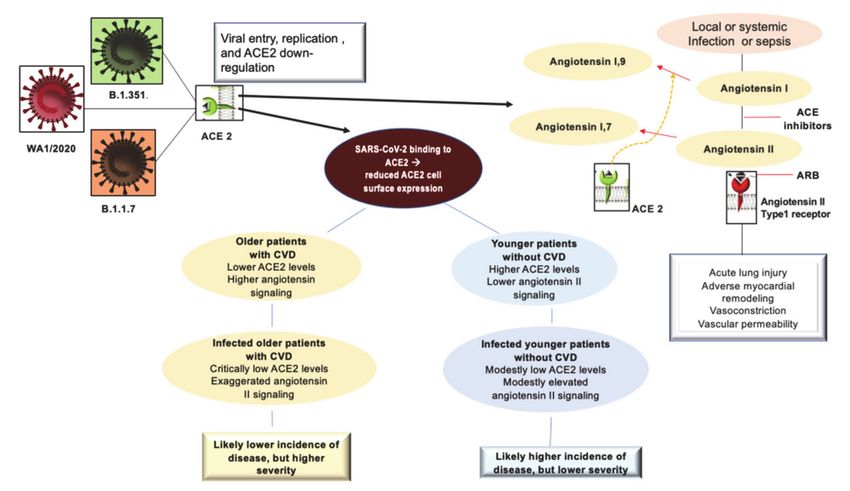
Biomedicines 2021, 9, 903                                                                                            2 of 28

                            1.1. Angiotensin-Converting Enzyme (ACE) 2 Receptor: The Evolutionary Stage of Infection from
                            Himalayan Palm Civet and Bat Coronavirus to SARS-CoV2 Infection
                                The gateway for SARS-CoV-2 to target cells is the angiotensin-converting enzyme
                            (ACE) 2 receptor, which is mostly expressed by epithelial cells of the lung, heart, blood
                            vessels, kidneys, and intestines. The ACE family of receptors includes both ACE and
                            ACE2 which, although they both are dipeptidyl mono-carboxydipeptidases have distinct
                            physiological functions.

                            1.2. Structure of the ACE as Ligand-Binding Receptors
                                  SARS-CoV-2 uses common cellular transmission which is based on the binding of
                            ligands to specific cell surface receptors. ACE2 is a G protein-coupled receptor (GPCR) and
                            belongs to a category of receptors that play a central role in the initiation and regulation
                            of cellular processes [10]. The GPCR constitutes the most prominent class of receptors
                            implicated in pathological disorders of the cardiovascular, respiratory, endocrine, immune,
                            and neural systems. Activation of GPCRs is also common in neoplastic pathologies. The
                            function that GPCRs exert is mediated by responses to specific interactions with hormones,
                            neurotransmitters, pathogens, metabolites, ions, fatty acids, and drugs [11,12].
                                  GPCRs are crucial modulators of transmission between the internal and external
                            environment of cells. GPCRs are integral membrane proteins with an extracellular N-
                            terminal and seven transmembrane (TM) helical domains, from TM1 to TM7, connected
                            via link regions. Evidence suggested that GPCRs have a more complex role than originally
                            considered. The binding of GPCRs to very different types of extracellular stimuli leads to
                            conformational changes of the TM domain with the consequent structural remodeling of the
                            protein [13–17]. Inter alia, these conformation changes induce coupling with cytoplasmic
                            proteins and subsequently the activation of enzymes that lead to the generation of a second
                            messenger. Once the second messenger is formed it can activate a sequence of signals
                            inside the cell [14]. This specific role of GPCRs results in increasing levels of intracellular
                            cyclic Adenosine Monophosphate (cAMP) and represents the pivotal pathway in response
                            to ligands, such as signaling of the renin–angiotensin system (RAS). It is important to
                            underline that the levels of cAMP production in the cellular domain are modulated by
                            several factors. Multidrug Resistance Proteins (MRPs) allow the efflux of cAMP from
                            the inside of the cell to the extracellular fluid, thus maintaining homeostatic intracellular
                            concentrations. The role of transporters activated by MRPs serves to regulate the balance
                            of cAMP within the cell.
                                  Lu et al. [15] reported concern about the structural conformation of the ACE/GPCR
                            complex and its interaction with SARS-CoV by focusing on lipid rafts. The structure,
                            activation, and signaling of the ACE/GPCR complex are strongly influenced by the bilayer
                            domain with specific membrane-GPCR interactions [16]. It has been shown that some
                            subsets of GPCR are preferentially isolated in distinct regions of the membrane defined as
                            lipid rafts [17–19]. Cholesterol partitions preferentially into lipid rafts which contain 3 to
                            5-fold the amount of cholesterol found in the surrounding bilayer. Evidence has shown
                            that lipid rafts serve as an entry site for SARS-CoV. For example, lipid rafts in Vero E6 cells
                            were involved in the “entry” of the coronavirus of the severe acute respiratory syndrome
                            (SARS-CoV). As has been clarified by the tests after SARS-CoV infection, the integrity of the
                            lipid rafts was a necessary requirement to produce the pseudotyped SARS-CoV infection.
                            If plasma membrane cholesterol depletion was induced using the relocalized MbetaCD
                            marker on the caveolin raft the SARS-CoV, ACE2 receptor was not significantly modified.
                            Although the surface expression of ACE2 still allowed binding to the virus, treatment
                            with MbetaCD inhibited the infectivity of the pseudotyped SARS-CoV by 90%. The
                            observed data concern the ectodomain of the SARS-CoV protein S (S1188HA) which can be
                            associated with lipid rafts. The spike protein, after binding to its receptor, colocalized with
                            the ganglioside marker GM1 residing on the raft. The study found that S1188HA binding
                            was not affected by plasma membrane cholesterol depletion supporting the conclusion that
                            lipid rafts serve as a gateway for SARS-CoV [20–24].
Biomedicines 2021, 9, 903                                                                                          3 of 28

                            1.3. Function of ACE Receptor
                                 The function of ACE is to split angiotensin I into angiotensin II, which binds and
                            activates the type 1 angiotensin II receptor. This activation triggers a series of patho-
                            physiological mechanisms that ultimately have vasoconstrictor, proinflammatory, and
                            pro-oxidative effects. It is important to underline that among the functions of ACE2 is
                            the hydrolytic degradation of angiotensin II to angiotensin 1–7 and angiotensin I to an-
                            giotensin 1–9. Once angiotensin 1–9 is generated, it binds to the Mas receptor, producing
                            anti-inflammatory, antioxidant, and vasodilatory reactions. From a pathophysiological
                            point of view, it is important to distinguish the two forms of ACE2 receptors. The first is
                            a type 1 integral transmembrane protein with structural features representing the extra-
                            cellular domain that acts as a receptor for the SARS-CoV-2 spike protein. The second is
                            a soluble form representing circulating ACE2. To date, our knowledge is limited on the
                            relationship that is established between SARS-CoV-2 and the two forms of the receptor.
                            A better understanding of this relationship may more precisely define the operational
                            adaptive or maladaptive processes that sustained COVID-19 infection [25,26].

                            1.4. ACE Receptor and Binding to Human Coronary Viruses
                                  The knowledge we have on the interaction between the human ACE2 receptor (hACE2)
                            and the Himalayan palm civet receptor (cACE2) with SARS-CoV derives from the usage of
                            the receptor by the Human (hSARS-CoV) and Himalayan palm civet coronavirus (cSARS-
                            CoV) [27]. The hSARS- CoV can bind both hACE2 and cACE2 receptors while the palm
                            civet coronavirus has no interaction with the ACE2 receptor expressed in humans. It
                            is known that the adaptation of c SARS-CoV to humans was determined by two-point
                            mutations, recognized as K479N and S487T, in the binding domain of the SARS-CoV spike
                            protein (SARS-CoV-S) [26].
                                  The mutations that have recently characterized SARS-CoV-2 led to more aggressive
                            variants of the virus and the concept of adaptive mutations (as noted by Wu et al. [28]) with
                            strengthened receptor binding and tropism (RBT). The authors demonstrated that adaptive
                            mutations of RBT led to the identification of genetic mutations of the virus that enhanced
                            interaction with human or palm civet ACE2. The genetic adaptation processes that took
                            place between hSARS-CoV and cSARS-CoV could also be recorded in SARS-CoV-like
                            viruses that have been isolated in bats [28]. A previous study found that the pathways
                            in which bat coronaviruses infected host cells did not occur through the interaction of
                            the ACE2 receptor with expressed SARS-CoVS and remained a mystery. However, the
                            important finding remains that the substitution of the amino acid sequence found between
                            residues 323 and 505 of the corresponding sequence of the SARS-CoV-S/RBT is sufficient
                            to allow the activation of the human ACE2 receptor [28].
                                  Coronaviruses can enter target cells effortlessly due to their ability to exploit many
                            cell surface molecules such as proteins and carbohydrates. Lectins play a fundamental role
                            in this process. For example, host calcium-dependent (type C) lectins have been recognized
                            to play a central role in SARS-CoV-2 infection. Evidence suggests a specific intercellular
                            role exerted by non-integrin 3-grabbing adhesion molecule (DC-SIGN) of dendritic cells.
                            This is a type C lectin expressed on macrophages and dendritic cells that functions to
                            recognize the high-mannose glycosylation patterns commonly found on viral and bacterial
                            pathogens. Coronavirus protein S is highly glycosylated, thus, providing the virus with
                            the opportunity to interact with host lectins such as Dendritic Cell (DC)/Liver/lymph
                            node-specific intercellular adhesion molecule-3-grabbing integrin (L-SIGN). L-SIGN, which
                            is expressed on liver and lung endothelial cells and has been reported as an alternative
                            receptor for SARS-CoV and bat coronavirus type HCoV-229E [29,30].
                                  The first demonstration of the possibility that SARS-CoV-2 interacts with the human
                            ACE2 receptor is reported in the landmark study from the University of North Carolina
                            at Chapel Hill [31,32]. The authors reported the substantially high risk of SARS-like bat
                            coronavirus disease named SHC014-CoV circulating in Chinese horseshoe bat populations.
                            This type of coronavirus has a high binding affinity with the ACE2 receptor [33,34]
Biomedicines 2021, 9, 903                                                                                              4 of 28

                                  The new SARS-CoV-2 virus expressed the bat coronavirus SHC014 spike in mouse-
                            adapted SARS-CoV backbones.
                                  Menachery et al. created a chimeric virus starting with the RsSHC014-CoV sequence
                            that was isolated from Chinese horseshoe bats [34]. The chimeric virus encoded a different,
                            zoonotic CoV spike protein in the context of the SARS-CoV mouse-adapted backbone. This
                            new SARS-CoV-2 virus expressed the bat coronavirus SHC014 spike. Through the hybrid
                            virus, the authors were able to evaluate the ability of the new spike protein to cause disease
                            independently of other necessary adaptive mutations in its natural backbone [32].
                                  The evidence showed at least two very interesting findings. The first was that group
                            2b viruses encoding the SHC014 peak in a wild-type backbone could efficiently use more
                            orthologs of the human angiotensin II converting enzyme (ACE2) receptor than the un-
                            modified SARS virus. The second was that group 2b viruses could replicate efficiently
                            in primary human airway cells and that it was also possible to obtain in vitro viral titers
                            equivalent to the epidemic strains of SARS-CoV. Once these results were translated in vivo,
                            replication of the chimeric virus in the mouse lung demonstrated considerable pathogene-
                            sis. This led to the trials of immunotherapeutic and prophylactic modalities to cope with
                            the SARS-CoV infection which had poor outcomes. In fact, both monoclonal antibodies
                            and the vaccine approach failed to neutralize and protect against CoV infection using the
                            new SARS-CoVS. Based on these results, the authors synthetically re-derived an infectious
                            full-length recombinant SHC014 virus and demonstrated robust viral replication both
                            in vitro and in vivo settings. This landmark report suggested 6 years ago that there was a
                            potential risk of SARS-CoV re-emergence from viruses circulating in bat populations.
                                  Recently the same group coordinated by Ralph Baric [35] studied the critical determi-
                            nants of the ACE2 receptor that support SARS-CoV-2-ACE2 interactions during infection
                            and replication of the preemergent 2B coronavirus (WIV). The Authors identified the key
                            changes that lead to infection by creating a humanized murine ACE2 receptor (hmACE2)
                            and provided evidence for the potential pan-virus capabilities of this chimeric receptor.
                                  SARS-CoV-2 cannot infect mice due to incompatibility between its receptor-binding
                            domain (RBD) and the murine ACE2 (mACE2) receptor. Since the mouse models of human
                            ACE2 (hACE2) and viruses adapted to mice have shown limitations, the researchers
                            developed another model that would allow evaluation of the pathogenetic phenomena
                            that occur in human SARS-CoV-2 infection. For example, hACE2 transgenic mice are
                            susceptible to unadapted SARS-CoV-2 viruses, but the pathogenesis observed in these mice
                            showed that virus-induced encephalitis and multi-organ infection were not comparable to
                            that observed in humans. Thus, Adams et al., to map the SARS-CoV-2 RBD and mACE2
                            interaction network, created a panel of mACE2 receptors, which have increasing levels
                            of humanizing mutations. The study used predictive structural models that allowed
                            identification of the minimal changes needed to restore replication [35].
                                  The ACE2 receptor has structurally critical sites whose integrity determines its activity.
                            The investigators worked at the level of three hot spots that determine ACE2 interaction:
                            position K353 interconnects with SARS-CoV-2 binding residues G496, N501, and Y505,
                            position K31 which forms a salt bridge with ACE2 residue K353 and links with SARS-CoV-2
                            Q493 and Y489, and position M82 which interconnects with RBD residues F486, N487,
                            and Y489. These aforementioned interface hotspots are the critical molecular sites for
                            the interaction between SARS-CoV2 and the receptor leading to virus entry. The authors
                            demonstrated that divergent residue modifications in these hot spots significantly reduce
                            the binding between humanized murine ACE2 (hmACE2) [33] and SARS-CoV-2 RBD.
                            They recorded that five amino acid changes (N30D, N31K, S82M, F83Y, and H353K) in the
                            SARS-CoV-2 RBD-ACE2 interaction hot spots lead to the modulation of infection and can
                            re-establish infection in the hmACE2 models [35].
                                  This study is crucial for the following reasons. The first is related to the fact that mouse
                            models are essential for understanding the pathogenesis of coronaviruses and are a key
                            resource for the preclinical development of vaccines and antiviral therapies. The second is
Biomedicines 2021, 9, 903                                                                                            5 of 28

                            that a detailed analysis of this study will allow the development of model systems to screen
                            for emerging coronaviruses and to develop new treatments to combat infections [35].

                            1.5. The Role of ACE2 in COVID-19 Pathogenesis
                                  The ACE2 receptor has been implicated in the pathogenesis of COVID-19, especially
                            with regards to its potential effects on the most vulnerable patients presenting with cardio-
                            vascular co-morbidities. COVID-19 does not have the same impact on all members of the
                            population. An exponential increase in the severity of the disease as well as mortality, due
                            to devastating thromboembolic complications, occurs in patients over the sixth decade of
                            life with comorbidities such as cardiovascular disease and diabetes.
                                  The angiotensin-converting-enzyme 2 receptors (ACE2) serve as the attachment site
                            of the SARS-CoV-2 spike protein to enter the lung epithelial cells [36]. Upregulation
                            with increased ACE2 expression has been demonstrated in patients with cardiovascular
                            disease and diabetes treated with angiotensin-converting enzyme (ACEI) inhibitors and
                            angiotensin receptor blockers (ARBs). However, whether treatment with these agents can
                            lead to greater COVID-19 severity has not been fully clarified.
                                  Discussions related to the use of ACEI/ARBs have surfaced regarding the need to
                            continue therapy in patients taking these drugs. The current recommendations are to
                            discontinue the administration of these drugs, despite diverging opinions, which were not
                            universally endorsed by experts due to the lack of strong evidence [37]. ACE2 not only plays
                            a role in the pathogenesis of COVID-19 but also as a component of renin–angiotensin system
                            signaling (RAS) localized throughout the body. Although the evidence has conclusively
                            revealed that ACE2 receptors allow SARS-CoV-2 to enter cells, ACE2 plays a central
                            anti-inflammatory role in RAS signaling by converting angiotensin II, responsible for the
                            inflammatory process, into angiotensin 1–7, which leads to its anti-inflammatory effects [38].
                            A study performed on rodent lungs [39] showed that the reduced expression of ACE2 leads
                            to a sequence of major proinflammatory processes, that are exacerbated by age, and result
                            in dysregulation of RAS signaling throughout the body [40]. It is important to note that
                            this typical inflammatory profile, even in accentuated forms, supports pathophysiological
                            processes that represent the main feature of hypertension and diabetes, as well as being
                            very widespread in old age [36]. The upregulation of the ACE2 receptor in subjects with
                            diabetes and hypertension treated with ACI/ARBs must be seen as a restorative substrate
                            that has a physiological function. The process that unfolds during SARS-CoV2 infection
                            sees ACE2 receptors as a gateway for the virus to enter cells, while the reduction of ACE2
                            protective features in older people and those with CVD can potentially predispose them
                            to more severe forms of the disease. The ACE2 receptor facilitates SARS-CoV2 infection
                            while the fundamental anti-inflammatory function, linked to RAS signaling, is reduced
                            because it is compromised in patients who develop COVID-19. In fact, data provided by
                            the first SARS epidemic in 2003 demonstrated the double role of the ACE2 receptor, thus
                            delineating the factors predisposing to the occurrence of the disease and its severity [41,42].
                                  In SARS-CoV2 infection it is plausible that the higher expression of ACE2 leads to
                            a greater predisposition to experience the disease. Epidemiological data from the South
                            Korean population, where genetic testing has been widely used in individuals, reported
                            higher numbers of infected among young adults [41] and those with increased ACE2 levels.
                            In this regard, an Italian study, examining the severity of COVID-19 disease in the elderly
                            population with CVD, hypothesized that a reduction in ACE2 levels due to aging and CVD
                            coupled to the upregulation of the proinflammatory angiotensin II pathway are factors
                            that likely predispose older individuals to severe forms of COVID-19. Therefore, younger
                            people are more susceptible to viral infection, but older people are more likely to have
                            severe disease manifestations [42].
                                  SARS-CoV2 uses the ACE2 receptor in carrying out its infectious manifestation,
                            thereby leading to a reduced expression of ACE2 on the cell surface and an upregula-
                            tion of angiotensin II signaling in the lungs which results in the development of acute
                            damage [38]. The consequence of these morphofunctional and biochemical changes can
Biomedicines 2021, 9, 903 6 of 28
predispose elderly individuals with CVD, who have reduced levels of ACE2 compared
to young people, to exaggerated inflammation and further reduction of ACE2 expression
in the context of COVID-19. In these cases, the disease manifests itself with greater sever-
ity [43]. Observations suggest that older individuals, especially those with hypertension
and diabetes, have reduced ACE2 expression and upregulation of proinflammatory an-
giotensin II signaling. Therefore, the morphofunctional and biochemical changes can be
corrected by the increase in ACE2 levels induced by ACEI/ARB treatment [43].
First, it is possible to hypothesize that in COVID-19 disease, the binding of SARS-CoV-
2 to ACE2 receptors acutely exacerbates this proinflammatory background, predisposing
these subpopulations to greater severity and mortality of COVID-19 disease. Second, con-
sidering this hypothesis credible, a protective role of the antagonistic action of angiotensin
II against acute lung injury associated with sepsis could be effective. This supports the use
of continuous therapy with ACEI/ARB [1,44,45] (Figure 1).
Figure 1. Depicts the interaction of SARS-CoV2 with the ACE2 receptor and the inflammatory profile
pattern before and after Coronavirus 2019 (COVID-19) infection in patients with or without CVD.
The initial entry of severe acute respiratory syndrome coronavirus 2 (SARS-CoV-2) into cells is shown
with involvement mainly of type II pneumocytes. SARS-CoV-2 binds to its functional receptor, the
angiotensin-converting enzyme 2 (ACE2). After endocytosis of the viral complex, surface ACE2 is
further down-regulated, resulting in unobstructed accumulation of angiotensin II. Local activation of
the renin–angiotensin–aldosterone system may mediate lung injury responses to viral insults. The
elderly and the young may present with different pathophysiological profiles. The simplified scheme
of the pre-infection inflammatory profile among predisposed older individuals compared to their
younger counterparts is illustrated. Abbreviations: ACE2, angiotensin-converting enzyme 2; ARB,
angiotensin-receptor blocker; CVD, cardiovascular disease; SARS-CoV-2, severe acute respiratory
syndrome coronavirus 2.
Third, the aforementioned biomechanical modifications of the receptor, plausible with
aging, should be investigated. Therefore, experiments on the functioning, the regulatory
mechanisms of RAS, and the biomechanics of the receptors involved in these functions
should be implemented. Specifically, the biophysical mechanisms underlying the asso-
ciated remodeling of the lipid membrane remain to be clarified. They may be useful
in the prevention of fatal lung complications caused by genetic variants of the Wuhan
virus [26–28,32,34,35,46].
1.6. ACE Inhibitors and Angiotensin Type II Blockers Role in COVID-19 Severity
Tetlow et al. [47] did not identify any associations between ACE-I/ARB use and AKI,
macrovascular thrombi, or mortality. Other studies [48,49] also supported the continuationBiomedicines 2021, 9, 903 7 of 28
of these drugs during hospitalization from COVID-19. Among those hospitalized, a
large percentage are likely being administered either ACE inhibitors or Angiotensin II
blockers, since epidemiological data reveal that cardiomyopathies, diabetes mellitus, and
hypertension are the most frequent comorbidities found among those patients [50].
Although the upregulation of ACE2 expression, which can be altered by drug admin-
istration, has not been defined, it has nevertheless been associated with disease severity.
Several preceding studies have demonstrated that the risk of developing COVID-19 after
the administration of ACEi and ARBs increased significantly. This could be an indirect
effect of overproduction of the circulating ACE2 transcripts in the cells [51,52]. As an exam-
ple, Enalapril, which is a frequently used ACEi, was reported to increase ACE2 expression
in the kidney [53].
Concerning possible therapeutical targets, ACE2 blockers have been developed, such
as the small synthetic inhibitor N-(2-aminoethyl)-1aziridine-ethanamine (NAAE) [54]. It
is able to bind ACE2 in its closed conformation so that molecular interaction between the
viral particle and the receptor cannot be possible and fusion does not happen. Thus, NAAE
could exert dual inhibitory effects: one on ACE2 catalytic activity and another on SARS
binding [55]. Despite this, current research drives opinions towards a cautionary use of
this agent.
2. Pathophysiology of Arterial and Venous Thrombosis
To date, the complete pathophysiology profile of arterial and venous thrombosis
during COVID-19 disease has not yet been fully clarified. The literature reports prothrom-
botic abnormalities in patients with COVID-19. In a Chinese study [56] performed in the
first phase of the SARS-CoV2 epidemic, 19 patients with COVID-19, who presented with
critical clinical conditions, had elevated levels of markers of hypercoagulability such as
D-dimer found in 100%, fibrinogen in 74%, and factor VIII in 100%. The dysregulation of
the coagulation process included the presence of antiphospholipid antibodies in 53% of the
population studied. Reduced levels of protein C, protein S, and antithrombin were noted
in all patients. Complications such as stroke, arterial ischaemia, and VTE accompanied the
coagulation disorder.
Zaid et al. studied 115 patients with COVID-19 disease reporting that SARS-CoV2
directly interferes with platelets. Viral RNA and high platelet-associated cytokine levels
were found in the platelets of all study participants. These abnormalities were not related
to the severity of the disease because in 71 infected individuals the disease manifested
in a non-serious manner while for 44 patients’ hospitalization was required for critical
clinical conditions. Specific tests performed on platelets showed aggregation at lower than
expected thrombin concentrations [57].
Nicolai et al. examined the autopsy findings of 38 individuals who died with COVID-
19 which showed that histopathological changes in coagulation were marked in the vessel
microcirculation. The abnormalities recorded were microvascular thrombotic formations,
and neutrophil extracellular traps characterized by networks of extracellular neutrophil-
derived DNA and polymorphonuclear neutrophil (PMN)-platelet aggregates [58]. The
authors compared the peripheral blood of patients with COVID-19 with that of healthy
patients. In vitro responses on peripheral blood samples from the three infected patients
exhibited excessive platelet and neutrophil activation, as assessed by degranulation and
integrin IIb-IIIa activation and immunofluorescence, compared to healthy control pa-
tients’ samples.
2.1. The Inflammatory Response during SARS-CoV2 Infection and Thrombotic Complication
Histopathology of SARS infection Cov2 is distinguished from that caused by other
viruses with tropism for the respiratory tract. SARS-CoV2 leads to direct damage of en-
dothelial cells characterized by dense perivascular infiltration of T lymphocytes combined
with aberrant activation of macrophages. The excessive and uncontrolled inflammatory
response, endothelial cell apoptosis, thrombotic microangiopathy, and angiogenesis areBiomedicines 2021, 9, 903 8 of 28
other distinctive histopathological features that denote the aggressiveness of SARS-CoV2,
which may be responsible for clinically severe forms of COVID-19 thus conferring disease
characteristics not comparable to any other viral respiratory disorder [59]. One significant
finding that emerges in the evaluation of the pathophysiology of thromboembolism in
COVID-19 versus non-COVID-19 disorders is the possibility that the coagulation alter-
ations are mediated more by platelet-dependent activation and intrinsically related to
viral-mediated endothelial inflammation. As noted a distinguishing feature of thrombo-
sis during SARS-CoV2 infection is the exacerbated hypercoagulability associated with
increased concentrations of coagulation factors, acquired antiphospholipid antibodies, and
reduced concentrations of endogenous anticoagulant proteins [56].
Patients with COVID-19 who develop more severe systemic inflammation and more
critical respiratory dysfunction have a higher prevalence of thrombotic complications.
Lodigiani et al. reported 388 patients hospitalized with COVID-19 including 16% with
serious clinical conditions. Despite the use of low molecular weight heparin (LMWH) for
thromboprophylaxis in all patients in the ICU and 75% of those not in the ICU, symptomatic
VTE occurred in 4.4% of patients, ischemic stroke in 2.5%, and MI in 1.1% [60].
Given the knowledge we have, there is still no clarity on the extent to which SARS-
CoV-2 increases the risk of thromboembolism. A study performed in the United Kingdom
compared 1877 patients discharged from hospital after COVID-19 disease and 18,159 hos-
pitalized for a non-COVID-19 disease reported no difference in hospital-associated VTE
rates (4.8/1000 vs. 3.1/1000; odds ratio, 1.6 [95% CI, 0.77–3.1]; p = 0.20) [61]. One point
to clarify is whether the high rate of VTE is specific to patients who develop COVID-19
or if VTE is mainly occurring in patients as a complication associated with severe critical
disease [61]. These results are in line with a recent meta-analysis that included 41,768 pa-
tients in whom VTE was assessed in COVID-19 versus non-COVID-19 cohorts. The authors
did not record a significant statistical difference for overall risk of VTE (RR 1.18; 95%CI
0.79–1.77; p = 0.42; I2 = 54%), pulmonary embolism (RR 1.25; 95%CI 0.77–2.03; p = 0.36;
I2 = 52%) and deep venous thrombosis (RR 0.92; 95%CI 0.52–1.65; p = 0.78; I2 = 0%). A
difference was reported after analyzing the subgroups of patients who were admitted to
the intensive care unit (ICU). Critically ill patients had an increased risk of VTE in the
COVID-19 cohort compared to non-COVID-19 patients admitted to the ICU (RR 3.10; 95%
CI 1.54–6, 23), which was not observed in cohorts of non-ICU patients (RR 0.95; 95% CI
0.81–1.11) (P interaction = 0.001) [62].
2.2. Management of Thrombosis in COVID-19 Patients
There are no international guidelines that direct the prevention and treatment of
thrombotic complications in COVID-19 patients. Both published and ongoing studies
testing interventions to prevent thrombosis complications in COVID-19 are based on the
evidence reported in current clinical guidelines about VTE prophylaxis in acute COVID-19
infections. Therefore, pending the results to be provided by the completion of ongoing
trials, guidelines for the treatment of thrombotic complications in patients with COVID-19
disease are derived from medical recommendations in the coagulation disorder populations
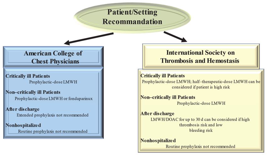
(Figure 2). However, the crucial point that remains to be clarified is whether these guidelines
are also optimal for the treatment of thrombosis due to COVID-19 [63–65].
Guidelines from the American College of Chest Physicians (ACCP) suggest (in the
absence of contraindications) prophylaxis with LMWH or fondaparinux rather than un-
fractionated heparin or direct oral anticoagulants (DOACs) for all hospitalized COVID-19
patients [63] Clearly the optimum choice of the drug to be taken is constrained by the incom-
plete knowledge of the possible interference of CoV 2 SARS with the medicament. So, the
40 mg dose of LMWH for injection once a day and the 2.5 mg dose of fondaparinux are pre-
ferred over the administration of unfractionated heparin injected subcutaneously 2–3 times
a day thus limiting the caregiver’s contact with infected patients. In addition, these drugs
are preferred over DOACs because of drug–drug interactions with antiviral agents. Both
are substrates of the P-glycoprotein and/or cytochrome P450-based metabolic pathways.Biomedicines 2021, 9, 903 9 of 28
Thus, concomitant administration of DOACs and antiviral drugs has the potential to
sharply increase DOAC anticoagulant plasma levels, thus increasing hemorrhagic risk.
Figure 2. Current Guideline Recommendations for Venous Thromboembolism Prevention in patients With Coronavirus
Disease 2019. Abbreviations: DOAC, direct oral anticoagulant; LMWH, low-molecular-weight heparin.
Given the high incidence of VTE, the proposed therapeutic dose to be used for stan-
dard thromboprophylaxis in critically ill patients with COVID-19 was either double or
single-dose administration of LMWH. The ACCP guidelines suggest the standard dose
LWMH in the absence of new clinical trial data [64]. Guideline-Directed Medical Ther-
apy (GDMT), which was established by the International Society on Thrombosis and
Hemostasis (ISTH), suggested that half-therapeutic-dose LMWH (1 mg/kg daily) can be
considered for prophylaxis in high-risk patients with COVID-19. A 50% higher dose can
be considered in patients with severe obesity (BMI ≥ 40 kg/m2 ). However, it remains
to be clarified which is the best dosage for optimal prophylactic therapy [65]. The re-
sults of ongoing randomized controlled trials (REMAP-CAP, ACTIV-4, and ATTACC),
comparing therapeutic-intensity anticoagulation with prophylactic-intensity anticoagula-
tion for patients with COVID-19-related critical illness, are awaited to establish optimal
antithrombotic prophylactic therapy [66]. Considering the pathophysiology of throm-
boembolism in COVID-19 is characterized by platelet hyperreactivity, another point under
discussion with RCTs initiated is the evaluation of administering an antiplatelet agent for
therapeutic prophylaxis.
High-risk patients hospitalized for COVID-19 have a high possibility of developing
VTE that persists after discharge [65]. However, for the latter, no specific recommendations
for post-discharge thromboprophylaxis have been established by the ACCP [64]. In con-
trast, the ISTH recommendations for post-discharge thromboprophylaxis suggest the use of
LMWH or a DOAC for all high-risk hospitalized patients with COVID-19 who have a low
risk of bleeding. Patients with COVID-19 considered to be at high risk include those with
age ≥ 65 years, presence of critical illness, cancer, previous VTE, thrombophilia, severe
immobility, and elevated D-dimer (>2 times the upper limit of normal). ISTH recommenda-
tions suggest a duration of 14 to 30 days for post-discharge thromboprophylaxis, although
the ideal administration period remains to be clarified [65].Biomedicines 2021, 9, 903 10 of 28
For patients with COVID-19 disease, no diagnostic protocols have been established
for thromboembolic complications, such as pulmonary embolism and MI, so the methods
to be used should be those validated for patients without COVID-19. Given the lack of
evidence to support the benefit, routine ultrasound checks for VTE surveillance are not
recommended. For patients with COVID-19 diagnosed with arterial or venous thrombosis,
we recommend treatment according to current established guidelines. These recognize the
benefits of LMWH administration in hospitalized patients. In the outpatient setting, DOAC
administration is recommended [58]. There are currently no recommendations issued by
ISTH and ACCP to support measuring the D-dimer to screen for VTE or to establish the
intensity of prophylaxis or treatment [64,65].
3. COVID-19 Vaccines Administration vs. Thrombosis and Variant. The New Challenge
3.1. Nucleoside-Modified RNA Encoding the SARS-CoV-2 Spike
Several studies have demonstrated the substantial role of the SARS-CoV-2 spike
protein that binds to ACE2 receptors on target cells during viral entry. Studies performed
on convalescent patients have highlighted the central role that the spike protein plays as
an immunodominant antigen triggering the host response, mediated by both antibody and
T lymphocytes [67].
Concerns about the rapid spread of the COVID-19 pandemic have favored the regis-
tration of numerous randomized clinical trials (RCTs) using different vaccination platforms
in order to evaluate their efficacy and safety. Evidence has shown that the use of rapid
response genetic platforms mRNA [67,68], adenoviral vector vaccines [69,70], inactivated
viruses [71,72], and adjuvanted spike glycoprotein [73] resulted in neutralizing antibody
responses after immunization.
The particular biological characteristics of mRNA synthesized in vitro may explain
the superior efficacy of an ideal non-viral gene replacement tool leading to many intrinsic
benefits [67,68]. First, quick protein assembly and well-regulated primary cell transduction.
Second, mRNA-based therapy avoids harmful side effects, such as its incorporation into
the cellular genetic substrate, which can ultimately limit the clinical application of most
virus- and DNA-based vectors [67,68].
Although the use of in vivo gene transfer therapy was first applied almost twenty
years ago [74,75], its usage as a vector for introducing genetic material into animals or even
into cultured cells has been very limited. Indeed, the reports of Gilboa [76] and Pascolo [77]
focused on the use of mRNA for vaccination purposes mainly directed at the development
of cellular and humoral immune responses through antigen-encoding transcripts that were
administered in vivo or delivered to dendritic cells (DC) ex vivo.
Several studies conducted at the beginning of the year 2000 [78–82] have shown
that RNA interferes with the cell-mediated adaptive immune response (pathogen and
antigen-specific response) by activating the cells of the innate immune system (non-specific
response). In particular, the action of the mRNA is directed towards the Toll-like receptors
(TLR) and specifically for the cellular subgroups TLR3, TLR7, and TLR8. It should be
noted that compared to gene replacement, RNA showed greater immunogenicity and was
associated with greater efficacy, highlighting a key role in the immune response.
Only 1 published study compared the in vitro immune response between nucleosides
and modified nucleosides. The use of incorporated pseudouridine (Ψ), 5-methylcytidine
(m5C), N6-methyladenosine (m6A), 5-methyluridine (m5U) or 2-thiouridine (s2U) in the
transcript affected the immune response of most TLRs with a substantial loss of their activa-
tion [83]. Progressing to testing nucleoside-modified mRNAs to evaluate their translation
potentials and immune characteristics in vivo, Hornung et al. [83] demonstrated that the
50 -triphosphate end of RNA produced by viral polymerases is accountable for retinoic
acid-inducible protein I (RIG-I)-mediated detection of RNA molecules. Identification of
50 -triphosphate RNA is repealed by capping the 50 -triphosphate end or by nucleoside
modification of RNA, including the use of s2U and Ψ.Biomedicines 2021, 9, 903 11 of 28
The major implication of these findings led to in vitro transcripts containing nucleoside
modifications not only translatable but also capable of activating an immune response
in vivo. Therefore, it was possible to subsequently develop mRNA with the function of a
dual therapeutic tool for both gene replacement and vaccination. Evidence reported by
Kariko et al. [84,85] on the in vitro incorporation of pseudouridine, a modified nucleoside
present in mRNA, has suggested that it improves RNA translation capacity but also
suppresses RNA-mediated immune activation in vitro and in vivo.
3.2. RNA Vaccine Platform
In January 2020 the RNA sequence of the new coronavirus SARS-CoV2 was introduced
in the RNA vaccine platform to allow rapid development of the vaccine in response to the
worsening spread of the pandemic. The advantages of vaccines that use RNA are manifold
and related to the incorporation of pseudouridine [84,85]. They offer both greater flexibility
during vaccine antigen design and expression as well as the ability to imitate viral antigen
structure and expression during native infection. One of the characteristics that make them
innovative is the lack of integration of viral RNA, which is necessary for protein synthesis,
in the cell’s genome. In fact, the viral genome is transiently expressed, then metabolized
and eliminated by the natural mechanisms of the organism, giving these vaccines greater
safety [68–72]. As a rule, vaccination with RNA can stimulate a vigorous innate immune
response eliciting B and T cell-dependent activity. RNA leads to the expression of the
vaccine antigen in host cells and, as demonstrated in specific mRNA vaccines, could
address considerable medical demand in the area of influenza prophylaxis [86].
The immunogenic benefits associated with the in vivo administration of 1-methyl
pseudouridine-containing mRNA including superior translational capacity and biological
stability were established in a landmark paper from Kariko et al. [84,85]. The same research
paper demonstrated lower serum levels of interferon-α (IFN-α) elicited by modified mRNA
in mice models with respect to unmodified ones, thus potentially reducing exaggerated
systemic inflammation and increasing safety. The improved efficacy of nucleoside-modified
RNA (modRNA) encoding the SARS-CoV-2 full-length spike modified by two proline
mutations is almost certainly due to its superior immune response [85]. Studies conducted
in the United States and Germany have reported substantially higher elicited SARS-CoV-2
neutralizing antibody titers and robust antigen-specific CD8+ and Th1-type CD4+ T-cell
responses against nucleoside modified mRNA delivered in lipid nanoparticles [86–88].
There are two widely administered mRNA vaccines, BNT162b2 (Pfizer–BioNTech)
and mRNA-1273 (Moderna). Administration of a two-dose regimen of BNT162b2 conferred
95% protection against COVID-19 in phase 3 trial participants (n = 21,720), aged 16 years
and older (95%; confidence interval, 90.3 to 97.6). The group of participants assigned to
receive BNT162b2 recorded 8 cases of COVID-19 disease with onset at least 7 days after the
second dose while in the group of individuals assigned to placebo there were 162 cases
of COVID-19 disease. Evidence observed from the analysis of subgroups defined by age,
sex, race, ethnicity, baseline body mass index, and the presence of coexisting conditions
reported similar efficacy of the vaccine with percentages between 90 and 100%. It is
important to note that among 10 cases of severe COVID-19 with onset after administration
of the first dose, 9 were reported in recipients of the placebo dose and 1 recipient with
the BNT162b2 dose. The safety profile of BNT162b2 was very high as evidenced by the
short-term appearance of mild-to-moderate pain at the injection site, fatigue, and headache.
The side effects were low and comparable to those recorded in the placebo dose recipients.
Furthermore, they were equivalent for a median of 2 months when comparing BNT162b2
with that of other viral vaccines [89].
Similar results were reported in phase 3 of the randomized, observer-blind, placebo-
controlled trial after administration of mRNA-1273 (100 µg in 15,210 participants) [90].
The efficacy of the mRNA-1273 vaccine was recorded at 94.1% (95% CI, 89.3 to 96.8%;
p < 0.001) in the prevention of COVID-19 disease, including severe disease. A double
dose of mRNA-1273 was administered to more than 96% of vaccine recipients and onlyBiomedicines 2021, 9, 903 12 of 28
2.2% had serological, virological, or both evidence of SARS-CoV-2 infection. Symptomatic
COVID-19 disease was confirmed in 185 recipients of the placebo dose versus 11 recipients
of mRNA-1273 dose.
Recipients of mRNA-1273 reported only transient local and systemic side effects and
no safety concerns were recorded. A critical illness from COVID-19 occurred in the 30 re-
cipients of the placebo dose (with one death) and in no participant who was administered
the mRNA-1273. It is important to clarify that current data on the messenger, derived
from laboratory studies, have demonstrated the efficacy of RNA (mRNA) vaccines against
SARS-CoV-2 variants. Researchers exposed serum samples from immunized individuals to
genetically modified versions of related variants and then measured neutralizing antibody
titers [90].
3.3. SARS-CoV-2 and Vaccine
We searched PubMed for research articles published by the launch of the database
until April 30, 2021, indicating no language restrictions and using the terms “SARS-CoV-
2”, “vaccine”, and “clinical trial”. We identified published clinical trial data on seven
SARS-CoV-2 studies and vaccines.
Four recombinant viral vectored vaccines have been tested in phase I/II clinical
trials [91–98]. Phase I and II trials were represented in the same study by two parts
with different patients subsets. The vaccine ChAdOx1 nCoV-19 (AZD1222), known as
AstraZeneca vaccine, was developed by the Oxford University and is constituted with
an adenoviral vector inactivated (unable to replicate) chimpanzee ChAdOx1 replication,
containing the antigen glycoprotein gene structural surface SARS-CoV-2 (protein spike;
nCoV-19). This vaccine is one of the more extensively studied following the first UK Phase
1 clinical trial published on 23 April 2020 [92]. To date three more randomized controlled
trials of the candidate vaccine have been initiated in the UK (COV002), Brazil (COV003),
and South Africa (COV005). Recently a further phase 1/2 study was carried out in Kenya.
A pooled interim analysis of four trials (COV004) showed the safety and efficacy of
the ChAdOx1 nCoV-19 vaccine (Oxford-AstraZeneca COVID-19 vaccine), (Covishield or
Vaxzevria). In recipients of two standard doses of Vaxzevria, the vaccine efficacy was 62.1%
and in recipients given a low dose followed by a standard dose, the efficacy was 90.0%. The
overall efficacy of the vaccine after administration of both doses in the population studied
was 70.4%. There were ten cases of COVID-19 that required hospitalization 21 days after
the first dose, all in the control population. Two patients were in serious condition and one
died. The authors recorded 175 serious adverse events that occurred in 168 participants, of
which 84 events occurred in recipients of the Oxford-AstraZeneca COVID-19 vaccine and
91 in the control group. Concern relating to clot formation or the occurrence of bleeding
episodes were not suggested across the analysis of these 4 RCTs [91].
The immune response after vaccine administration in participants who received two
doses of the vaccine was very effective. In particular, the specific objectives of phase 3
RCT were the evaluation of humoral and cellular safety and immunogenicity concerning
both a single dose and two-dose regimen in adults over 55 years of age. Median peak
anti- SARS-CoV-2 IgG responses 28 days after the boost dose were similar across the three
cohorts (including two groups of patients aged 18–55 and one group enrolling >55 years
old patients). Furthermore, neutralizing antibody titers after a boost dose were similar
across age groups. Within 14 days of boost dose administration, a total of 208 of 209
(>99%) recipients of the booster dose of ChAdOx1 nCoV-19 had neutralizing antibody
responses. T cell responses peaked on day 14 following a standard single dose of ChAdOx1
nCoV-19 [90,91].
The Ad26.COV2. S vaccine, known as Johnson&Johnson/Janssen COVID-19 vac-
cine, is composed of a recombinant, replication-incompetent human adenovirus type 26
(Ad26) vector that encodes a full-length, membrane-bound SARS-CoV-2 spike protein in a
prefusion-stabilized conformation [95–97]. At least 14 days after single-dose administration,
Ad26.COV2. S conferred protection against both symptomatic COVID-19 infection andBiomedicines 2021, 9, 903 13 of 28
asymptomatic SARS-CoV-2 infection. The level of efficacy remained stable at 28 days after
administration with an efficacy of 66.1% (adjusted 95% CI, 55.0 to 74.8). COVID-19 disease
occurred in 66 recipients of the administration dose of Ad26.COV2. S compared to 193
for the placebo dose [98]. The results of the administration of Ad26.COV2. S vaccine has
demonstrated efficacy against COVID-19 clinical disease with severe-critical manifestation,
including hospitalization and death. Evidence suggested a high level of efficacy after
administration of Ad26.COV2, which was greater against severe-critical COVID-19 disease
and reached a rate of 76.7% for onset at ≥14 days [adjusted 95% CI, from 54.6 at 89.1]. At
28 days in participants receiving the single dose the reported efficacy was 85.4% [adjusted
95% CI, 54.2 to 96.9] for onset at ≥28 days).
However, the unexpected data were related to the immunogenic response to Ad26.COV2
vaccine against the South African variant 20H/501Y.V2. Out of 91 cases of patients in
which the virus variant was sequenced and confirmed, 86 (94.5%), showed vaccine efficacy
against moderate to severe-critical COVID-19 that reached 52.0% and 64.0% with onset of
at least 14 days and at least 28 days after dosing, respectively. The efficacy against severe-
critical COVID-19 disease reached 73.1% and 81.7% at 14 days and 28 days respectively
after the single dose of Ad26.COV2. Evidence supported a level of safety comparable to
that of other COVID-19 vaccines that progressed to phase 3 studies. The reactogenicity
was greater with the administration of Ad26.COV2. S compared to the placebo dosage;
however, it was generally mild to moderate and transient. Note that the incidence of
severe adverse events was similar between the two populations of participants (vaccine
and placebo group) with three deaths occurring in the vaccine group (but none of them was
related to COVID-19 infection). No episodes attributable to thrombotic or haemorrhagic
phenomena were reported [98].
Vector-based adenovirus (Ad) 5 (CanSino Biological/Beijing Institute of Biotechnol-
ogy, China) [69,93] was administered in a single dose and resulted in the production of
neutralizing antibodies which increased significantly on day 14 and peaked 28 days after
vaccination. A specific T-cell response in a dose-dependent manner peaked at day 14 after
vaccination. However, of note, the vaccine demonstrated lower immunogenicity in partici-
pants over the age of 55. Administration of adenovirus type-5 vectored COVID-19 recorded
no serious adverse event within 28 days post-vaccination. An equal rate of side effects
was reported in the three groups studied. Reactogenicity was evident in the first 7 days
after administration in 30 (83%) recipients of a low dose vaccine, in 30 (83%) recipients of a
medium dose, and in 27 (75%) recipients of a high dose, respectively [93].
A heterologous recombinant adenovirus (rAd26 and rAd5)-based vaccine, Gam-
COVID-Vac (Sputnik V) [70,94] showed efficacy and safety from the interim analysis of a
phase 3 RCT. Gam-COVID-Vac is a combined vector vaccine because it consists of rAd type
26 (rAd26) and rAd type 5 (rAd5). Both adenoviruses carry the gene for the SARS-CoV-2
full-length glycoprotein S (rAd26-S and rAd5-S). The administration of rAd26-S and rAd5-S
is carried out (intramuscularly) separately with an interval of 21 days. The results of the
Phase 1/2 clinical trials showed that the Gam-COVID-Vac vaccine was well tolerated and
highly immunogenic in healthy participants. Vaccine efficacy of Gam-COVID-Vac reached
91.6% (95% CI 85.6–95.2) with few tolerable side effects (7485 [94.0%] of 7966 total events).
Although 45 (0.3%) of recipients of this vaccine (n = 16,427) and 23 (0.4%) of recipients of
the placebo dose recorded serious adverse events; however, none were deemed associated
with vaccination. There were four deaths during the study. Three (Biomedicines 2021, 9, 903 14 of 28
18–59 and 60 years and older. However, the latter vaccine showed lower neutralizing
antibody titers in older adults after two doses.
This phenomenon is related to the presence of antibodies before vaccination which
was present, albeit with variable vaccine titrations, in the three study groups. Only 25% of
participants in the low dose group, 37% of participants in the medium-dose group, and
63% of the high dose recipients, who had pre-existing high immunity to Ad5, had at least a
fourfold increase in neutralizing antibody titer on day 28 after vaccination. Multivariable
analysis showed that the pre-existing high Ad5 neutralizing antibody titers impaired post-
vaccination neutralizing antibody seroconversion and highlighted a different immune
response in relation to the age of the recipients. The impairment of serum conversion was
independent of the dose of vaccine administered in the three groups (the low-dose, the
medium-dose, and the high-dose ones). However, recipients aged 45–60-year-old appeared
to have lower neutralizing antibody seroconversion than younger recipients. In the latter,
Ad5 neutralizing antibodies were significantly enhanced after vaccination [71,72]
Finally, a clinical study of a vaccine NVX-CoV2373 (Novavax Inc. (NVAX), Gaithers-
burg, MD, USA) assembling nanoparticles consisting of adjuvant trimeric spike glyco-
proteins from severe acute respiratory syndrome coronavirus 2 (SARS-CoV-2) recorded
preliminary results. The vaccination schedule included the administration of two doses
3 weeks apart in healthy adults less than 60 years of age. Evidence suggested good toler-
ance for this vaccine which induced neutralization responses greater than those measured
in serum samples from convalescent symptomatic susceptible patients [64].
3.4. Vaccines and Immunogenicity Against Genetic Variants
Given the worldwide spread of the genetic variants of SARS-CoV-2 and the increasing
number of cases of COVID-19 disease, the recurring question today is whether the vaccines
currently administered will be effective against the mutated viral variants. Since in the case
of vaccines efficacy is based on immune responses, it is evident that the patient may have a
reduced immune response to viral variants.
Concerns related to less immunogenicity of vaccines emerged at the end of January
2021, simultaneously with the effects of SARS-CoV-2 mutagenic potential including the
strong spread of South African variant B.1.351 [99].
Despite the many mistakes that viruses can make by replicating, we are not aware of
any vaccines against viral diseases, other than seasonal influenza, which have required
regular updates on the basic constitution due to changes in the viral genome. For example,
despite the frequent mutations recorded by the hepatitis B virus, the vaccine continues to
guarantee safety and efficacy in the vaccinated population.
The progression of vaccination is more rapid in high-income countries. Unfortunately,
the majority of the world population lives in low-middle-income countries where the mass
vaccination programs remain restricted or exclusive. As persistent infections and viral
replication create the possibility of high-frequency mutations of the SARS-CoV-2 genome,
we must seek to homogeneously extend vaccine administration without any economic or
social class distinctions.
3.5. Current Knowledge
Although the term vaccine resistance has been used by experts in the field to describe
the reduced efficacy of COVID-19 vaccines against some variants, this term can be inaccu-
rate. In fact, the concept of drug resistance is more commonly aimed at antibiotics that are
used to kill or are capable of inhibiting the growth or reproduction of bacteria. In the case
of vaccines, the administration has not taken place, so the person cannot be resistant but
can have a reduced immune response.
Vaccines administered against COVID-19 are engineered from the SARS-CoV-2 spike
protein of the original virus called Wuhan-hu-1 [100] which is used by the virus to bind
and infect host cells. Emerging data from COVID-19 disease suggest that variants of
the “parent” virus appear to be more transmissible or more lethal than Wuhan-hu-1 andYou can also read