A DISSECTION OF SARS COV2 WITH CLINICAL IMPLICATIONS (REVIEW)
←
→
Page content transcription
If your browser does not render page correctly, please read the page content below
INTERNATIONAL JOURNAL OF MOlecular medicine 46: 489-508, 2020

    A dissection of SARS‑CoV2 with clinical implications (Review)
                     Felician Stancioiu1, Georgios Z. Papadakis2, Stelios Kteniadakis3,
                             Boris Nikovaevich Izotov4, Michael D. Coleman5,
                             Demetrios A. Spandidos6 and Aristidis Tsatsakis4,7

    1
     Bio‑Forum Foundation, 030121 Bucharest, Romania; 2Department of Radiology, Medical School, University of Crete,
             71003 Heraklion; 3Emergency Department, Venizeleion General Hospital, 71409 Heraklion, Greece;
             4
              Department of Analytical and Forensic Medical Toxicology, Sechenov University, 119991 Moscow,
                 Russia; 5School of Life and Health Sciences, Aston University, B4 7ET Birmingham, UK;
                    6
                      Laboratory of Clinical Virology, Medical School, 7Department of Forensic Sciences
                     and Toxicology, Faculty of Medicine, University of Crete, 71003 Heraklion, Greece

                                           Received May 15, 2020; Accepted June 9, 2020

                                                  DOI: 10.3892/ijmm.2020.4636

Abstract. We are being confronted with the most consequen-          1. Clinical aspects of COVID‑19 infections; acute respi‑
tial pandemic since the Spanish flu of 1918‑1920 to the extent      ratory distress syndrome (ARDS); known and potential
that never before have 4 billion people quarantined simultane-      treatments
ously; to address this global challenge we bring to the forefront
the options for medical treatment and summarize SARS‑CoV2           In China, the first comprehensive analysis published on the
structure and functions, immune responses and known treat-          COVID‑19 (1) included 44,672 cases and 1,023 deaths, with an
ments. Based on literature and our own experience we propose        overall case‑fatality rate (CFR) of 2.3%. There were 0 deaths
new interventions, including the use of amiodarone, simv-           in patients 9 years old or younger, and the CFR increased with
astatin, pioglitazone and curcumin. In mild infections (sore        advancing age (70‑79 years: 8.0%, 80 years and older: 14.8%)
throat, cough) we advocate prompt local treatment for the           and also with comorbid conditions: 5.6% for cancer, 6.0% for
naso‑pharynx (inhalations; aerosols; nebulizers); for moderate      hypertension, 7.3% for diabetes, 10.5% for cardiovascular
to severe infections we propose a tried‑and‑true treatment: the     disease; for intensive care patients CFR was 49.0%. Similar data
combination of arginine and ascorbate, administered orally          were published by other authors on the China epidemic (2,3);
or intravenously. The material is organized in three sections:      another analysis (4) identified 3 biological markers associated
i) Clinical aspects of COVID‑19; acute respiratory distress         with the severity of the infection and progression to ARDS
syndrome (ARDS); known treatments; ii) Structure and func-          and death: i) neutrophilia; ii) increased lactate dehydrogenase
tions of SARS‑CoV2 and proposed antiviral drugs; iii) The           LDH and iii) coagulation dysfunction (D‑dimer). There were
combination of arginine‑ascorbate.                                  2 unexpected findings: i) patients with fever ≥39˚C were
                                                                    more likely to develop ARDS but also less likely to die; and
                                                                    ii) administering methylprednisolone to patients with ARDS
Contents                                                            was associated with lower risk of death.
                                                                        In Europe there was a higher rate of severe cases in
1. Clinical aspects of COVID‑19 infections; acute respiratory       Italy (5,6) where 12% of positive patients were admitted
   distress syndrome (ARDS); known and potential treatments         to ICU vs. 5% in China; this difference was attributed to a
2. SARS‑CoV2 molecules and proposed antiviral drugs                 higher proportion of elderly and increased social contact (7,8).
3. The combination of arginine‑ascorbate                            Data from Germany (9) showed that ~1/3 of admitted patients
                                                                    had leucopenia and most of these (80%) had lymphopenia;
                                                                    C‑reactive protein (CRP) was often increased and very high
                                                                    CRPs were associated with a less favorable outcome for
                                                                    the patient. Approximately 40% of admitted patients had
                                                                    increased LDH, D‑dimer and/or thrombocytopenia, and an
Correspondence to: Dr Felician Stancioiu, Bio‑Forum Foundation,     LDH value of >400 IU/ml was associated with more severe
Splai Unirii Nr 8, Parter, 030121 Bucharest, Romania                disease. Troponin was increased in a small number of patients,
E‑mail: felicians@bio‑forum.net                                     with unclear significance.
                                                                        In the USA, a recent publication showed that a majority (71%)
Key words: SARS‑CoV2, COVID‑19, antiviral treatment, ARDS,          of COVID‑19 patients with ARDS and in intensive care, also
sepsis, arginine, ascorbate, coronavirus                            had hypotension (10,11). There is already an extensive clinical
                                                                    experience with ARDS in the USA, where >180,000 patients
                                                                    annually are admitted with this pathology and ARDS mortality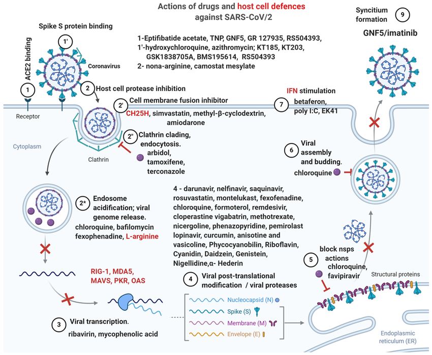
490                          Stancioiu et al: SARS‑CoV2: A Dissection and Clinical Implications

has improved from 40 to 50% 10 years ago to 20‑25% in recent        pathways and dual‑role modulators means that they do not
clinical trials. A group of USA intensive care specialists led by   always conform to mechanistic determinism, and this is espe-
Dr Calfee has analyzed >30 biomarkers and clinical variables        cially true in patients with chronic diseases or co‑morbid
from two large clinical trials on ARDS (12); subsequently an        conditions, where modifications of physiological pathways
ARDS patient population was identified which comprised              translate into unknown but important factors with unexpected
~30% of total ARDS cases, had higher levels of IL‑6 and IL‑8,       consequences. One such example is a clinical trial for sepsis
more patients with sepsis and in need for vasopressor treat-        which tested a recombinant IL‑1 receptor antagonist (IL‑1Ra)
ment, with higher mortality but also responding better to high      with negative results, but at the same time benefited patients
positive expiratory‑end pressure (PEEP) therapy. With data          with higher baseline levels of IL‑1Ra (21).
from more clinical trials (13), two ARDS populations were               Another important aspect is that multiple cellular pathways
identified, the hypoinflammatory and hyperinflammatory              besides inflammation are simultaneously affected in ARDS,
subphenotypes, with distinctive and opposite characteristics.       including coagulation, endothelial and epithelial injury path-
The former has low levels of IL‑6, IL‑8, tumor necrosis factor      ways (apoptosis) and healing (fibrosis) in lungs. A unique
receptor 1 (TNFr1), lower mortality as well as higher number        molecular signature of lethal infection with another respira-
of ventilator‑free days, high bicarbonate and protein C; the        tory virus (PR8, an influenza virus) showed over‑activation of
latter has high levels of IL‑6, IL‑8, TNFr1, low levels of bicar-   pro‑inflammatory pathways, NF‑κ B, IL‑6, TNF and neutro-
bonate and protein C, low number of ventilator‑free days and        phil chemotaxis (22), associated decreased activation of genes
higher mortality.                                                   involved in lung homeostasis and repair. Neutrophils with high
    Treatment‑wise, the same group showed that in the ARDS          pro‑inflammatory activity were numerous in lung infiltrates,
hyperinflammatory subphenotype better outcomes were                 their own chemokines further promoted neutrophil influx in
obtained with administration of high PEEP, liberal fluid            a positive feedback loop; a dose‑dependent survival was seen
strategy (vs. conservative fluid administration) and simvastatin,   with partial neutrophil depletion in this type of lethal infection.
while rosuvastatin administration was of no benefit (12,14‑16);     Comparing viral replication in non‑lethal vs. lethal infection
Table I summarizes the data from different clinical trials on       they had similar viral replication rates, but the lethal infection
ARDS subphenotypes. The observed difference between                 had increased viral titers in the lungs, and also increased early
statins in ARDS may be explained by their solubility: while         pro‑inflammatory neutrophil activation, resulting in pathologic
simvastatin is lipid‑soluble, rosuvastatin is water‑soluble, with   neutrophil infiltration and fatal lung damage (23). Similar
important consequences on cell membrane interaction.                observations increased neutrophil- and neutrophil‑attracting
    It is safe to say that COVID‑19 patients who develop ARDS       chemokines, delay in induction of IFN in airway epitlelial cells
(with high levels of interleukins, low blood pressure) belong       resulting in vascular leakage and lung damage were made in
to the hyperinflammatory subphenotype and may benefit               SARS and MERS suggesting a common pathogenical mecha-
from the therapeutic insight associated with this group of          nism (24,25). Indeed, the significant role of neutrophils in the
patients (higher PEEP, liberal fluid strategy, and simvastatin).    promotion of ARDS‑related alveolal damage was supported
In critically‑ill COVID‑19 patients, increased LDH levels           by a report in sepsis ARDS patients, where high neutrophil
are likely associated with acidosis and if low blood pressure       counts in bronchiolar lavage material harvested late in ARDS
is also present, before vasopressors they are likely to benefit     progression was associated with reduced patient survival (26).
from administration of sodium bicarbonate (increments of            Hence, it has been proposed that the sulphone dapsone may
100 ml of 8.4% solution).                                           show efficacy in COVID‑19 related ARDS, as the drug is
    Treatment‑wise, after the SARS and MERS epidemics a             highly and rapidly effective at inhibiting cytokine‑mediated
few antivirals were proposed and tested and among these are         neutrophil chemotaxis and respiratory burst in a variety of
mycophenolic acid, cyclosporine at low concentrations, chlo-        therapeutic contexts (27). Efficacy of Dapsone in the treatment
roquine; chlorpromazine, loperamide, and lopinavir, found           of Pneumocystis pneumonia suggests it will penetrate the lung
to be broad‑spectrum coronavirus inhibitors (17). Recently          tissue sufficiently to attenuate neutrophil activity. The haemo-
FDA has approved remdesivir (GS5734 ‑ an RNA polymerase             toxicity of the drug can be ameliorated with concomitant
inhibitor) as specific treatment for COVID‑19; different proto-     cimetidine administration (27,28).
cols use chloroquine (500 mg q 12 h); hydroxychloroquine                Overall, the major clinical manifestations: sore throat,
(200 mg q 12 h); lopinavir/ritonavir (400/100 mg every 12 h),       cough, breathing difficulties, malaise, fever, chills, diarrhea,
α‑interferon (aerosol inhalation 5 million IUbid), and there are    generalized myalgia, drowsiness, dyspnea, and pneumonia are
>50 treatments tested in clinical trials (18).                      similar in SARS‑CoV2, MERS‑CoV, and SARS CoV infec-
    For ARDS, as the most severe form of COVID‑19 infection,        tions (29), but COVID‑19 differentiates itself from MERS and
there is ample data from proteomics showing highly activated        SARS in infectivity (R0), with much higher human‑to‑human
pathways of inflammation, increased levels of eosinophil‑ and       transmission rates owing much to the fact that infectivity is
neutrophil‑derived proteins, epithelial and endothelial injury,     not restricted to symptomatic patients (30); however, we can
matrix metalloproteinase 7 (MMP7), α‑ and β‑hemoglobin,             improve therapeutic strategies with inferences from structural
apolipoprotein A1, osteopontin and chemokines (19,20). The          comparisons of these viruses.
context of increased inflammation in ARDS justifies the use
of biological agents, antagonists of the IL‑6 receptor (IL‑6R),     2. SARS‑CoV2 molecules and proposed antiviral drugs
and TNF‑ α, such as infliximab, rituximab, ustekinumab,
etanercept, adalimumab and tocilizumab (18). However,               SARS‑CoV2 consists of two types of molecules: RNA and
the complexity of living biological systems with redundant          proteins; RNA encodes for 27 viral proteins, of which 16 are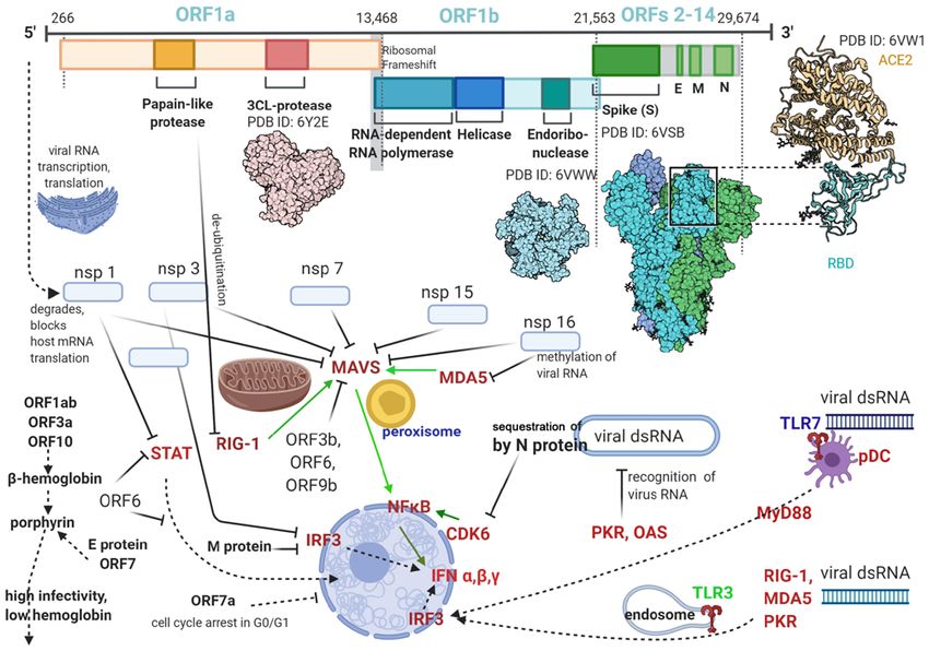
INTERNATIONAL JOURNAL OF MOlecular medicine 46: 489-508, 2020                                    491

Table I. ARDS subphenotypes (data from Wilson and Calfee) (13).

                                                                       Hypoinflammatory                      Hyperinflammatory
Parameters                                                                  ARDS                                  ARDS

IL‑6, IL‑8, TNFr1 (plasma levels)                           Low                                                      High
Bicarbonate, CRP (plasma levels)                            High                                                     Low
Mortality                                                   Low                                                      High
Ventilator‑free days                                        High                                                     Low
PEEP (positive end‑expiratory pressure) associated          Low                                                      High
with better 90‑day mortality (ALVEOLI study)
Fluid strategy with better outcome for 90‑day	Conservative                                                  Liberal administration
mortality (FACCT study)                            administration of fluids                                        of fluids
Simvastatin administration has benefit on 28‑ and            No                                                      Yes
90‑day survival (HARP‑2)
Rosuvastatin administration has benefit on 90‑day            No                                                       No
survival (HARP‑2)

ARDS, acute respiratory distress.

non‑structural proteins (nsps), and 11 are accessory and            has no known functional domain or motif and is different
structural proteins. There are 4 major structural proteins:         from the orf8 or orf8b of SARS‑CoV, which does not have
nucleocapsid protein (N), spike surface glycoprotein (S), matrix    orf10 at all. However, ORF10 binds to heme and ORF8 to
protein (M), and small envelope protein (E). The N‑protein          porphyrins (37); heme inactivation increases viral infectivity
binds to viral RNA, encloses it in a capsid and assists RNA         through inhibition of the host innate antiviral response (38)
synthesis and folding during viral replication; it also modifies    which relies on heme‑containing enzymes such as nitric oxide
host responses, cell cycle and gene translation, and after RNA      synthase (detailed below); also increases viral permeation of
replication it guides the viral membrane proteins for viral         tissues via porphirin binding and tissue destruction (39).
assembly. The S‑protein is essential for attachment and fusion          The spike (S) protein of the new virus contains 1,273 amino
of the virus to host cells (31).                                    acids; compared to SARS‑CoV it has 27 amino acid substitu-
    Sequencing the SARS‑CoV2 genome in February 2020 (32)           tions, 8 in the heptad repeat domain, 6 in the RBD, and 4 in
showed that phylogenetically it belongs to the genus                the S1 subunit, modifying two peptides which are antigens
Betacoronavirus, subgenus Sarbecovirus, and has closer              for SARS‑CoV (40). The binding energy between the viral
genetic similarity to SARS‑CoV than MERS‑CoV. The                   S protein and host ACE2 receptor is higher in the new
genome consists of a single, positive‑stranded RNA with             virus versus SARS‑CoV (‑50.6 kcal/mol vs. ‑78.6 kcal/mol)
~30 k (29,811) nucleotides; from these only 5 nucleotides were      suggesting a more stable bond (41); at the same time the
found to be different between SARS‑CoV2 and SARS‑CoV.               affinity of the S protein of the new virus for the ACE2 receptor
Similar sequencing results (>99.9% similarity) were obtained        is in the 15 nM range, which is about 10‑20 times higher than
by different teams on different patient samples (33,34):            SARS‑CoV, an important factor for the differential infec-
GenBank sequences MN988668, NC_045512, MN938384.1,                  tivity (42).
MN975262.1, MN985325.1 and MN994468.1.                                  The S protein is a class I viral fusion protein like the
    Coronaviruses have the largest known RNA genomes and            influenza virus hemagglutinin (HA); SARS‑CoV2 also has an
their RNA is transcribed via open reading frames (ORFs);            S1/S2 cleavage site which is recognizable by furin, a ubiqui-
this peculiarity increasing the efficacy of viral replication and   tous cell protease. When similar modifications (insertion of
mutagenesis, and making it more difficult to be eliminated by       a polybasic furin cleavage site) occur in the hemagglutinin
the immune system. The SARS‑CoV2 genome has 14 ORFs of              proteins of avian and human influenza viruses, their virulence
different lengths; first ORF has ~10,000 nucleotides (~2/3 of       is greatly increased (43).
viral RNA) and encodes nsps 1‑16, with the structural and               Functionally the S protein has a receptor‑binding
accessory proteins being translated from the remainder 1/3 of       subunit (S1), an S2 fusion domain (S2), separated by a cleavage
RNA (35) (Fig. 1).                                                  site (S1/S2) and another cleavage site (S2') located within S2.
    Comparing the amino acid sequence of SARS‑CoV and               The multiple cleavage sites are activated by a wide variety of
SARS‑CoV2 reveals identical sequences in the envelope,              proteases from the host cell: cathepsins, serine proteases from
matrix, nsp7, nsp13 and accessory proteins p6 and 8b, while         the trypsin‑like transmembrane serine protease family (TTSP),
nsp3 and nsp2 had 102 and 61 amino acid substitutions,              and furin‑like proprotein convertases.
respectively.                                                           Both SARS‑CoV and SARS‑CoV2 need S protein
    Importantly, orf8 and orf10 are present in SARS‑CoV2 but        priming through cleavage by a host cellular protease (trans-
not in SARS‑CoV (36). The orf8 protein from SARS‑CoV2               membrane protease serine 2 ‑ TMPRSS2, TMPRSS11a,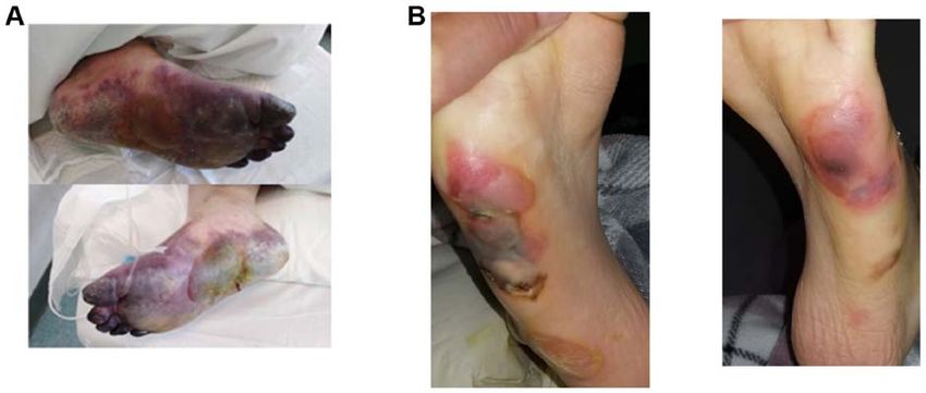
492                          Stancioiu et al: SARS‑CoV2: A Dissection and Clinical Implications

                          Figure 1. SARS‑CoV2 genome, translated proteins and some of their known pro‑viral actions.

hypoxanthine‑aminopterin‑thymidine, trypsin, elastase,                       The nsps perform essential functions in immune antago-
cathepsin L) (44,45), and the cell surface from respiratory              nism through formation of the viral replication complex and
epithelial cells has abundant TMPRSS2 and trypsin‑like                   double membrane vesicles which shield the viral RNA, viral
protease (46). Interestingly, human airway epithelial cells              RNA proofreading, binding of nucleic acid and helicase
can be infected by SARS‑CoV even when proteases from cell                activity (48).
surface are absent (44), meaning that a cell fusion process
used by SARS‑CoV involves endosomal membranes and a                      Immunosuppressive functions of the SARS‑CoV2 proteins.
treatment which blocks only the cell surface proteases or the            Besides needing to ensure the entry of viral material into
endosomal protease will not prevent, but only depress viral              cells and subsequent replication, the viral proteins and RNA
cell entry; however, a combination of two substances, camostat           also need to evade the host immune system, and because the
and EST, a cathespsin inhibitor effectively blocked the viral            success of viral infection depends on the impairment of the
entry into cells.                                                        host antiviral response, this aspect deserves special attention.
     SARS‑CoV2‑infected cells aggregate to form syncy-                       Airway epithelial cells are able to prevent viral infections
tiums; the S protein of SARS‑CoV2 can mediate a cell‑cell                through multiple mechanisms. The airway surface liquid (ASL)
syncytium formation without a proteolytic enzyme (trypsin),              is secreted by the submucosal glands, goblet and club cells and
while SARS‑CoV S protein cannot; thus the membrane                       mobilized by cilium cells, and contains the gel‑forming mucins
fusion capacity of SARS‑CoV2 is much higher than that of                 MUC5B and MUC5AC, glycosaminoglycan keratin sulfate,
SARS‑CoV (46).                                                           and antimicrobial peptides and proteins (AMPs). ASL forms
     The S protein of both coronaviruses has another pecu-               a pericilliary layer with volume, fluidity, pH and microbiome
liarity: its dependency on Ca2+ (47). Significant cell membrane          maintained in an optimal range for toxin elimination and the
ordering prior to membrane fusion is needed, and it requires             actions of AMPs, and facilitating mobilization of the innate
the presence of Ca2+ ions because of the negatively charged              immune response by macrophages, monocytes, dendritic cells,
residues in the fusion platform. Consequently the entry of               innate lymphoid cells, and γ/δ T cells. Finally an adaptive,
SARS‑CoV2 is highly dependent on the Ca2+ concentration in               specific immune response is triggered in response to antigens
its cell environment, and calcium chelators inhibit viral fusion         involving T and B lymphocytes, chemokines, cytokines and
and cell entry. Thus it was observed that SARS‑CoV entry is              antigen‑neutralizing IgAs (49).
blocked by amiodarone, which blocks calcium channels in the                  After evading or overwhelming this first line of defense
endosome and lysosome (47).                                              and entering host cells, the virus activates the cellular antiviral
INTERNATIONAL JOURNAL OF MOlecular medicine 46: 489-508, 2020 493
defenses effectuated by hundreds of proteins (cytokines, move to the mitochondria and block the interaction between
chemokines, host restriction factors) which block different RIG‑I and MAVS to block IFN induction; iv) the papain‑like
steps in the viral replication and are activated by specific protease (PlPro) removes ubiquitin from RIG‑I and down-
cell sensors/receptors. Interferons (IFNs) via IFN‑stimulated regulates type‑I IFN response; v) cytoskeleton perturbations
genes (ISGs) initiate innate and adaptive immune responses in mitochondria, with modification of the mitochondrial
which alter host cell cycle, translation and apoptosis, virus membrane potential and impairment of MAVS activity;
entry, viral RNA availability, stability and translation, particle vi) blocking IFN‑β production by de‑ubiquitination (DUB) of
and budding (50). RIG‑1 and other viral sensors and effectors (56).
Molecules involved in virus detection in the cytoplasm Microarrays helped identify the proteins through which
of host cells are: the retinoic acid‑inducible gene‑I (RIG‑I), a viral particles can antagonize host immune responses:
RNA helicase with the respective cell receptors ‑ RIG‑I‑like 13 proteins were inhibitors of MAVS (LGP2, A20, SMURF2,
receptors (RLR); and the melanoma differentiation‑associated etc.) 14 for RIG‑1 (USP3, ARL16 and RNF122) and 6 for
protein 5 (MDA5), both of which have the same downstream MDA5 (USP3, ARL5B, TRIM59); RNF125 inhibited RIG‑1,
effector: the mitochondrial antiviral signaling protein (MAVS). MDA5 and MAVS (57).
Besides RIG‑1 and MDA5, other cellular viral RNA sensors
are known: the protein kinase RNA‑activated (PKR), oligoad- Proposed therapeutic interventions. The current therapeutic
enylate synthetase (OAS), latent endoribonuclease (RnaseL), interventions for COVID‑19 (18) are derived from SARS treat-
cyclic GMP‑AMP (cGAMP) synthase (cGAS) (51); PKR stops ments; the first drugs shown in 2005 to be effective in vitro
host cell translation and helps activate the NF‑κ B. for SARS, both pre‑ and post‑exposure, are chloroquine and
Early in infection the antiviral response is driven by the hydroxychloroquine; since they were extensively studied and
peroxisomal MAVS, while later the mitochondrial MAVS shown to exert pleiotropic antiviral actions (58). Chloroquine
triggers an interferon (IFN)‑dependent, sustained immune is a weakly basic substance and upon entering cells it is
response. MAVS signaling ultimately results in NF‑κ B (and protonated and concentrated in acidic organelles: lysosomes,
other pro‑inflammatory pathways) activation which translo- Golgi vesicles and endosomes; the subsequent increase in
cates to the nucleus and activates the transcription of genes endosomal pH interferes with the terminal glycosilation of
encoding IFN‑ α /β, cytokines, many antiviral proteins, and the ACE2 receptor. Besides inhibiting the S protein‑induced
RIG‑I/MDA5 in a positive feedback loop (52). viral fusion (59), and the PlPro protease (60) chloroquine
Following activation, IFN‑α/β‑type I IFNs are produced can benefit patients with porphirin extravasation (Fig. 2) by
in and activate most cells, while type III IFN (IFN‑λ) mostly preventing orf1ab, orf3a, orf10 attack on hemoglobin (37) with
mucosal cells; they activate very similar IFN‑stimulated porphirin formation.
genes (ISGs). IFN signaling occurs very fast since it does Protease inhibitors target viral nsps needed for its replica-
not require new protein synthesis; all IFNs use the Janus tion, such as the RNA‑dependent RNA polymerase (RdRp)
kinase (JAK) ‑ signal transducer and activator of transcrip- inhibited by ribavirin (which also inhibits viral mRNA
tion (STAT) pathway. pDCs produce most of the IFN‑α during capping); the 3C‑like protease (3CLpro) is inhibited by the
an infection; IFN‑γ bridges the innate and adaptive immune lopinavir‑ritonavir combination (48), which seems to have
responses, helps regulate immune function, and is mostly better results compared to ribavirin, which was associated
secreted by activated T cells and natural killer (NK) cells (51). with anemia, hypoxia and increased risk of death in SARS
TLRs from endosomes recognize viruses, while TLRs patients (61).
from cell membrane typically recognize bacteria. MAVS and FDA‑approved drugs and agents re‑purposed for
the TLR adaptor MyD88 activate cell‑specific transcriptional COVID‑19, such as ribavirin, IFNs and corticosteroids, were
pathways, with pro‑ or anti‑inflammatory profiles (ex macro- shown, however, to be ineffective especially in severe CoV
phages vs. fibroblasts) (53). infections (48); recent data on SARS‑CoV2 suggest that more
All nucleated cells are thought to have RLR and PKR recep- specific and efficacious treatments can be administered to
tors for viral nucleic acids, while toll‑like receptors (TLRs) COVID‑19 patients; Table II summarizes such proposed
are present in myeloid dendritic cells (mDCs) ‑ TLR3/7 and FDA‑approved drugs and Fig. 3 offers a graphic presentation.
plasmacytoid dendritic cells (pDC) TLR7/8, and also in endo- Among COVID‑19 proposed drugs already approved for
somes of most cells (54). other pathologies, GR 127935, a known potent entry inhibitor
TLR4 interacts with with the adaptor protein MyD88 of Ebola and Marburg viruses, binds the SARS‑CoV‑2 receptor
and activates the mitogen‑activated protein kinase (MAPK) ACE2 and also is a selective 5‑HT1B/1D receptor antagonist,
and the NF‑κ B signaling pathway. The c‑Jun NH2‑terminal making it useful in patients with hypotension and tachyar-
kinases (JNK), the signal‑regulated kinases (ERKs), the rhythmia, when administration of vasopressors is questionable.
p38 MAPKs, are major MAPKs with important roles in innate Agents that bind the SARS‑CoV‑2 spike protein and are
immunity (55). known to reduce lung inflammation, include GSK1838705A,
SARS blocks antiviral defenses and the IFN production BMS195614, GSK1838705A, which inhibit the insulin like
in multiple ways, at multiple sites, many involving RIG‑I and growth factor‑1 receptor and are used in cancer.
MAVS (50): i) after transcription the coronaviral dsRNA is For blocking the ACE2 receptor, three FDA‑approved
protected from detection by storage in double‑membrane drugs are proposed: TNP, an inhibitor of tyrosine kinase,
vesicles; ii) M protein of SARS virus localizes in membranes IP6K and Akt pathways, which can also shown to inhibit
associated with the Golgi complex and binds to the host RIG‑I, MERS infection; eptifibatide acetate, an inhibitor of
thus impeding activation of MAVS; iii) ORF3b and ORF6 platelet aggregation already tested in septic shock; and the494 Stancioiu et al: SARS‑CoV2: A Dissection and Clinical Implications
Figure 2. Severe COVID‑19, interstitial hemorrhage, porphyria, necrosis. (A) Bullous porphyria and necrosis in literature (39). (B) Our own experience.
Figure 3. Antivirals and their actions.
Abelson (Abl) kinase inhibitor GNF5, known to inhibit repli- of SARS/MERS CoV (66) as the virion and endosomal
cation of Dengue virus, which deserves further attention. It membranes fuse via a cathepsin L‑dependent mechanism;
was shown that Abl2, but not Abl1 is necessary for replication the Abl2 kinase inhibitors GNF5/GNF2/imatinib inhibit theINTERNATIONAL JOURNAL OF MOlecular medicine 46: 489-508, 2020 495
Table II. FDA‑approved drugs with SARS‑CoV2 antiviral actions.
Viral molecule inhibited; Method and/or software used for testing
Substance analyzed (Ref.) mechanism FDA approved drugs
Hydroxychloroquine and Spike (S) Protein ‑ binding Virtualized quantum mechanical modeling
azithromycin (59) motifs of ACE2 receptor;
Eptifibatide acetate, ACE2 receptor protein High‑throughput virtual screening used to
TNP, GNF5, GR 127935, of host cells investigate LOPAC library drugs
RS504393 (62) binding site
KT185, KT203 S‑protein (RBD of spike protein) Software: PyRx, Open Babel, AutoDock Vina,
GSK1838705A, binding site PyMol, GROMACS; online resources
BMS195614, RS504393 (62) anti‑inflammatory SWISS MODEL, HADDOCK, RCSB PDB,
NCBI, ProCheck at RCSB validation server,
ProSA‑web, SAVES‑Verify3D server
Darunavir, Nelfinavir, Protease: Structure‑based drug repositioning
Saquinavir, Rosuvastatin (63) Mpro, central site
Montelukast, Fexofenadine (63) Protease: Structure‑based drug repositioning
Mpro, terminal site
Chloroquine, Protease: Homology model of the protease based on
formoterol, 16 other FDA‑ papain‑like protease SARS‑coronavirus PLpro structure; drugs
approved drugs (60) (PLpro) docked in S3/S4 pockets of active site
Remdesivir (‑13.1 KJ/mol) Protease Connectivity map and the docking configurations used
cloperastine (‑10.4) 3CLpro (PDB ID 6LU7) to simulate the docking bonding energy between
vigabatrin (‑10.2) antiviral and the respective protease in KJ/mol
methotrexate (‑6.9)
Remdesivir (‑18.6 KJ/mol) Protease Used PyRx for loading and visualising the SDF files for
vigabatrin (‑12.1) 6Y84 ‑ main protease with the ligands and the AutoDock Vina tool for testing the
cloperastine (‑10.1) nicer‑ unliganded active site docking on two target proteins 6LU7 and 6Y84 from PDB
goline (‑9.0) phenazo‑
pyridine (‑8.4) (64)
Pemirolast (65) Protease; RNA Replicase Using COVID‑19 Docking Server
inhibitor
Chloroquine (37) Accessory proteins Simulated molecular docking of viral proteins with
prevents binding of orf1ab, human heme or porphyrins with (LibDock tool) of
ORF3a, ORF10 to heme, Discovery‑Studio 2016
ORF8, surface
glycoproteins to porphyrins
Favipiravir (37) Accessory proteins: Simulated molecular docking of viral proteins with
inhibits binding of E protein, human heme or porphyrins with (LibDock tool) of
ORF7a to porphyrin, prevent Discovery‑Studio 2016
virus entering host cells
fusion of the viral and endosomal membrane, an early stage of The third drug is probably the best viral fusion inhibitor,
viral infection. GNF5/GNF2/imatinib also inhibit the forma- RS 504393, which can bind both the SARS‑CoV‑2 S protein
tion of cell syncitia before the hemifusion step, effectively and its receptor ACE2 and is used for treating lung injury
blocking membrane fusion and viral entry (67). and bronchial wall thickening; moreover, it is also a selective
Finally, 3 drugs deserve special attention for COVID‑19 antagonist of the monocyte chemoattractant protein‑1 (MCP‑1)
treatment: KT185 and KT203 which bind the S protein and receptor CCR2, blocks the upregulation of pronociceptive,
are potent, selective inhibitors of a/b‑hydrolase domain pro‑inflammatory interleukins IL‑1β, IL‑18, IL‑6, and as such
containing 6 (ABHD6), a transmembrane serine which are very useful for decreasing the ‘cytokine storm’ and the
hydrolases the endogenous cannabinoid 2‑arachidonoyl- associated neuropathic pain (patients hypersensitive to touch).
glycerol (2‑AG), and more importantly decrease macrophage Besides FDA‑approved drugs, a variety of natural
activation. compounds have been investigated for their antiviral actions496 Stancioiu et al: SARS‑CoV2: A Dissection and Clinical Implications
Table III. Natural compounds with antiviral actions.
Viral molecule inhibited;
Substance analyzed (Ref.) mechanism Method and/or software used for testing
Anisotine and vasicoline of Protease; Using COVID‑19 Docking Server
Justicia adhatoda (65) RNA Replicase
inhibitor
Phycocyanobilin, Riboflavin, Cyanidin, Protease (Mpro); COVID‑19 Docking Server to
Daidzein, Genistein (69) RNA Replicase inhibit Mpro and RdRp
inhibitors
Curcumin (70) Protease Structure of metabolite and COVID_19 protease
inhibitor from PubChem and Protein Data Bank (PDB);
molecular docking by MVD (Molegro Virtual Docker)
For 6LU7: Nigellidine ‑6.29 Kcal/mol; Proteases Docking of compounds from Nigella sativa
chloroquine ‑6.29; OH‑chloroquine ‑5.57; (3CLpro and and drugs performed with Molecular Operating
favipiravir ‑4.23; For 2GTB: Mpro) Environment software (MOE)
α‑Hederin ‑6.50 kcal/mol; 6LU7 and 2GTB
chloroquine ‑6.20; OH‑chloroquine ‑5.51; inhibitors
favipiravir ‑4.12 (68)
on SARS‑CoV2, one advantage being that some plants by S protein with IC50 of 4.3 nM; its protective effect mani-
contain more than one active ingredient (65); in the case fested half an hour after and 6 hours before exposure to
of Nigella sativa its main active substances, nigellidine coronavirus (46).
and α‑Hederin compare positively to either chloroquine or IFN can also be administered directly to patients via inha-
hydroxychloroquine based on their binding energy with viral lations, however, affinity amino acids have showed limited
proteases (68), more information is given in Table III. benefits depending on the timing of administration and
prompts further analysis of IFN actions.
Local treatment and IFNs. Based on the observation that the Similarly to the neutrophilia and lymphopenia observed
host IFN response is delayed in CoV infections, with subse- in severe COVID‑19 patients, fatal SARS infection is associ-
quent over‑activation of neutrophils in the lung and vascular ated with high IFN and ISGs expression and low T cell and
leakage (54), IFN‑stimulating substances administered by antibody activity, while viral clearance needs increased T cell
inhalation showed efficacy against SARS and MERS in vitro responses (25).
and in vivo. Inhaled substances have the advantage of lower The immune response of CoV patients is imbalanced,
volume of distribution (ASL has a volume of 20‑30 ml) and with over‑stimulation of the monocyte‑macrophage line and
can act early in COV infection during the viral fusion process, decreased T cell activation; while early administration of
inhibiting the binding of its S protein to the host receptor (71). IFN‑1 has protective effects, in later stages administration of
One such substance is polyI:C, a TLR3 agonist shown to IFN, with the possible exception of IFN‑γ, is associated with
induce differentiation of mDC, promote Th1 activation with increased neutrophil infiltration in the lungs. This is due to
IL‑12 and type I IFN production, the activation of the innate the selective activation of ISGs in macrophages, but not in
immune response and reversing the PGD2 effects in the B or T cells, since signaling in these cells requires the myeloid
lungs (72,73). A shortcoming of the polyI:C may be its require- differentiation factor 88 (MyD88), a key molecule used in
ment for either RIG‑1 or MDA5 activity, which are known to all TLR signaling pathways of adaptive immunity except for
be inactivated by coronaviruses and render polyI:C ineffective TLR3 which is present in all endosomes (77).
post‑infection, limiting it to a prevention drug (74). In animal models of lethal COV infection, the immu-
A very potent inhibitor of CoV fusion is nona‑L‑arginine nopathological events are mostly IFN‑I-dependent and
with high affinity for furin (effective at concentrations of independent of viral replication; IFN‑α/β receptor (IFNAR)
40 nM); hexa‑ and hepta‑peptides of the basic aminoacids receptor ablation or neutrophil depletion avoided lethal infec-
arginine and lysine also showed excellent inhibition of viral tion but did not affect the viral load. TNF inhibition improved
fusion (75). lymphopenia, which is not unexpected since IFN‑I sensitizes
Another intranasal‑administered peptide, EK1, which T cells to apoptosis.
contains the heptad‑repeat HR2 from the binding motif of the It was shown that an important role in modulating the
S protein has inhibited infection by many human coronavi- inflammatory response is played by two proteins containing the
ruses (46); its administration in mice reduced lung viral titers Toll or interleukin (IL)‑1 receptor (TIR): MyD88 and the TIR
by 1,000‑fold 2014 (76) and its lipidation, EK1C4, resulted in domain‑containing adaptor‑inducing interferon (TRIF) (78).
much greater potency, inhibiting the cell‑cell fusion mediated For controlling inflammation both MyD88 and TRIF proteinsINTERNATIONAL JOURNAL OF MOlecular medicine 46: 489-508, 2020 497
are required, since the release of the anti‑inflammatory IL10, IL‑8 and IL‑17) there was also an increase in the metabolic
IL19, endothelial lipase, metalloproteinase inhibitor 1 (Timp1) pathways of complex lipids (inositol phosphate, nicotinate,
and plasminogen activator inhibitor 1 (Serpin1) is prevented in nicotinamide), intermediates needed in SARS‑CoV infection
the absence of either one, resulting in excessive pro‑inflamma- and probably linked to prenylation. The strongest proviral
tory modulation. activation was seen in the β2 subunit of the coatomer protein
During early CoV replication in non‑lethal models, pDCs complex (COPB2) followed by inositol hexaphosphate
are the major IFN‑I producers, and pDCs sense coronaviral kinase 1 (IHPK1); abelson kinase 1 (ABI1); diacylglycerol
RNA via TLR7 within endosomes (54). The same author kinase epsilon (DGKE); cyclin‑dependent kinase 5 regulator
shows that the coronavirus endonuclease (EndoU) from (CDK5R2); protein kinase C ι (PRKCi). Among antiviral
nsp 15 has a key role in preventing early induction of innate molecules were found the diacylglycerol kinase δ (DGKD),
responses, by removing the viral dsRNA that would otherwise the double‑stranded RNA‑activated protein kinase (PKR);
trigger host cell dsRNA responses, including IFN, PKR and cyclin‑dependent kinase 6 (CDK6) and most MAPKs with
OAS/RNase L, and delaying the viral RNA cytoplasmic the exception of MAP3K11, which is weakly proviral;
sensing mediated by MDA5. ABl1 inhibitors and molecules acting on lipidic and glucidic
During early stages of SARS infection, IFN‑γ presence metabolism are thus important.
is associated with viral clearance and infection resolution; Another factor influencing IFN actions is the hemo-
IFN‑γ is synthesized mainly by activated T cells and natural globin β‑chain which was shown to stimulate IFN‑β production
killer (NK) cells (79). IFN‑γ (type II) and IFN‑α/β (type I) via RIG‑1, including potentiating of RIG‑I ubiquitination;
have synergistic actions; given alone neither inhibits viral hemoglobin is present in lung, kidney, peripheral blood mono-
replication, but administration of both inhibited viral replica- nuclear and other cell lines (38,85), and SARS‑CoV2 nsps can
tion by 3000‑fold at 24 h, with stronger inhibition at 48 and bind to and degrade hemoglobin (37).
72 h. Indoleamine 2,3‑dioxygenase (IDO), stimulated by
Oligonucleotide arrays were used to study the ISGs regu- IFN‑γ and inhibited by TLR activation and allergens plays
lated by IFNs; all IFNs induced complement component C1r, an important role in immune suppression (normally prevents
IFN‑induced nuclear phosphoprotein, vascular endothelial hyper‑inflammation and autoimmunity) by inhibiting T cell
growth factor (VEGF)‑related protein (VRP or VEGF‑C), activation and polyclonal proliferation of T cells, mostly
and phospholipid scramblase, an apoptosis promoter; its through degradation of tryptophan and kynurenines; its
levels increased 8‑fold by IFN‑ α, 10‑fold by IFN‑ β, and role in SARS‑CoV2 infection was not explored but likely is
3‑fold by IFN‑γ (80). Additionally each IFN preferentially important (86). Activation of IDO is strictly dependent on
induced specific molecules: IFN‑ α, somatic cytochrome c; prior expression of Arginase 1 and the resulting polyamines,
hepatitis C‑associated p44; pyridoxal kinase; and lysosomal with immunosuppressive actions (87); while NO is an IDO
acid lipase (LIPA); IFN‑ β, clathrin‑like protein, PKR, inhibitor; the role of IDO in the SARS‑CoV2 infection and its
hypoxia‑inducible factor‑1 (HIF‑1α) which regulates apop- immune evasion has not yet been explored.
tosis and cell response to hypoxia via expression of VEGF, The data above confirm that in severe CoV infection the
p53, p21; IFN‑ γ, mitochondrial 3‑ketoacyl‑CoA thiolase; host immune system is mounting an imbalanced response to
poly (ADP‑ribose) polymerase; proton‑ATPase‑like protein. infection which powerfully favors neutrophils/macrophages
Both type I and type II IFNs increase expres- and inhibits the adaptive cellular response; early events
sion of the CH25H gene which codes for the enzyme include hindered pDCs activities and over‑activation of
Cholesterol‑25‑hydroxylase, which converts cholesterol into macrophages with induction of IFN‑I expression and autocrine
25‑hydroxycholesterol (25HC) (50), an oxysterol with antiviral over‑stimulation; this may be due to 25‑hydroxycholesterol or
effects via alteration of membrane properties, inhibition of IDO overactivation, Myd88 inactivation by the virus, hemo-
sterol biosynthesis and prenylation (priming) of virus and host globin degradation and/or simultaneous actions of other viral
proteins. However, 25HC production by macrophages also has molecules (EndoU).
a central role in regulating the immune system, and at nM However, it is possible that the delayed IFN response
concentrations it suppresses the IL‑2 mediated proliferation of followed by neutrophil over‑activation is simply due to the lack
B cells and inhibits IgA production (81). of activation in the early stage of infection of the peroxisomal
Another possible pathological modification induced by the MAVS necessary for early IFN production (52), and this obser-
CoV in susceptible patients is the activation of alternatively vation gains more weight as peroxisome function declines
activated macrophages, M2, which normally have anti‑inflam- significantly in tandem with cellular senescence, advancing
matory actions and are involved in the wound‑healing age, and age‑related co‑morbidities (88). Peroxisome senes-
processes (82), but were also found in the lungs of mice lacking cence is associated with dramatic changes in their protein
STAT1 signaling pathway (STAT1 knockouts), which after the import capabilities, functional integrity, ability to process
SARS‑CoV infection, had an increased number of M2 macro- ROS, regulation of growth and division of other organelles,
phages in their lungs, activated via the STAT6 pathway, and decreased ability to oxidize fatty acids, accumulation of free
developed pulmonary fibrosis (83). fatty acids and diacylglycerol, followed by reorganization of
The modifications induced by the SARS‑Cov infection in protein kinase C (PKC) signalling (89).
host cells were also investigated via a library screen of small This fact is related to the important metabolic functions
interfering RNA (siRNA) targeting the human kinome (84). of peroxisomes, which have >50 enzymes in their matrix
Besides the expected activation of pro‑inflammatory pathways performing critical metabolic functions besides produc-
(production of NO and ROS in macrophages, IL‑2, IL‑6, tion and degradation of H2O2 including fatty acid oxidation,498 Stancioiu et al: SARS‑CoV2: A Dissection and Clinical Implications
synthesis of specialized lipids, including anti‑inflammatory (also a weak base) impair the glycosylation of ACE2, an addi-
docohexaenoic acid and degradation of pro‑inflammatory tional mechanism for blocking viral fusion.
leukotrienes and recently it was shown that multiple viruses The pH‑dependent fusion of various viruses endocytosed
interfere with antiviral signaling dependent on peroxisomes, into host cells by non‑coated vesicles (caveolas) or in associa-
examples being the accessory protein Vpu of HIV‑1 which tion with clathrin is well‑documented (96); additionally CoV
annihilates peroxisomes (90), the VP16 protein of HSV‑1 and influenza viruses can fuse with host cell membranes
which blocks the peroxisomal MAVS (91), also the hepatitis C via anchoring cell receptors and internalization in the lipid
virus protein NS3‑4A, the capsid of the Dengue and West Nile bi‑layer, a process blocked by constituent hydrolaze CH25H,
virus, and the vMIA protein of cytomegalovirus (92). methyl‑β‑cyclodextrin, a cholesterol‑sequestering drug, simv-
Peroxisomal MAVS stimulates ISGs via IRF1 and IRF3 in astatin and possibly other lipophilic statins.
an IFN‑independent manner and does not induce IFN produc- Low pH is affecting essential aspects of the native immune
tion in macrophages, which is dependent on mitochondrial response ‑ neutrophil activation, chemotaxis, production of
MAVS. Interestingly, cells lacking either peroxisomal or mito- ROS, NO and phagocytosis, apoptosis, cytokine production,
chondrial MAVS had an unusual biphasic behavior, restricting activation of transcription factors by binding to DNA, and
viral replication in the first 24 h, but not at 72 h, and showing also decrease of lymphocyte function, lymphokine secre-
that both organelles are necessary for viral clearance (53). The tion and activation, while hypoxia is potentiating these
functional status of peroxisomes can be correlated both with actions (97‑101); acidosis is associated with increased need
age and the severity of SARS‑Cov2 infection, and is impaired for intensive care and hospitalization (102). In addition, a low
by poor oxidative status, low cellular pH (acidosis) and/or extra‑ and intra‑cellular pH not only favors viral infection, but
hypoxia which is also seen in patients with chronic pathologies also impedes the antiviral action of known medication.
or genetic deficiencies.
pH modifies the activity of antiviral medications. Many
pH‑dependent viral actions. At the surface of the respira- proteins undergo conformational changes of their tertiary and
tory epithelium low pH favors viral entry and replication by quaternary structure induced by ionic changes (Ca2+, H+, Fe2+
impairing the actions of the AMPs from the airway surface and Mn+) and pH modifications. Such modifications are docu-
liquid (ASL), which have pH‑sensitive antimicrobial functions, mented for the main protease (Mpro) of SARS‑CoV2 during its
with the pH of ASL being actively regulated by bicarbonate binding to protease inhibitors at pH 4.0‑8.0, and it was shown
transport (49); another pH‑lowering viral activity is the forma- that they were pH‑dependent, with different binding energy
tion of cell syncytiums. Lower intracellular pH favors the (stability of protein complex) and specific bonding type/atom
activity of the proteases needed for viral replication, decreases interaction (103).
the mitochondrial membrane potential and inactivates MAVS, Small modifications in pH can have critical consequences
which is essential for IFN and NFκ B activation and produc- as was shown with arbidol, a broad‑spectrum antiviral which
tion of antiviral protein switches. In addition, low pH promotes binds to the influenza virus haemagglutinin (HA), induce a
oxidative phosphorylation to anaerobic glycolysis with 0.2 unit pH modification and blocks viral fusion; consequently
increased mitochondrial ROS production via related cellular some influenza strains developed a HA which disrupted the
pathways (93). drug‑HA bond with loss of medication effectiveness (104).
The modifications in the cellular redox state, ionic balance More aspects of pH modifications in viral infections are
and pH which are simultaneous with the actions of specific discussed below; re‑establishing physiological pH levels
molecules in a pathway are not often measured or considered (7.35‑7.45) is an important therapeutic consideration in
during lab experiments, but in the context of dual or multiple ALI/ARDS.
regulators they may have important consequences, one such
example being the effect of hemoglobin (Hb) on IFN produc- Genetic and translational factors involved in severe
tion (85). While exerting a direct inhibitory effect on MDA5 COVID‑19. An important question is whether the host genetic
which inhibits IFN production, Hb also modifies a cell redox factors significantly impact the immune functions and the
state, promotes the RIG‑I pathway and stimulates IFN produc- host answer to viral infection, as it was found that individual
tion; the increase in ROS by Hb is stimulated only by the variations exist in expression of RIG‑1 and other antiviral
microbial and not by the host protease. molecules (57).
Additionally, the infectivity of CoV and other enveloped Studying the bronchoalveolar lavage fluids from ARDS
viruses is known to be impaired by an increase in endosomal patients by proteomic analysis during the initial 4 days of care,
pH in a dose‑dependent manner; mild bases such as lipophilic survivors and non‑survivors showed differences: non‑survivors
amines or amphiphilic drugs with a tertiary amino group can had decreased expression of proteins involved in immune acti-
accumulate in acidic organelles via protonation, where they vation, coagulation, iron metabolism, and increased protein
neutralize internal pH, alter the properties of the adjacent expression of glycolysis and collagen pathways (105).
membranes and inhibit multiple processes occurring in the Also, an association was found between four SNPs and
respective compartment (94); this was also observed with ARDS patients: rs78142040 of ARSD ‑ an arylsulfatase
administration of bafilomycin or ammonium chloride (NH4Cl) gene involved in sphingolipid metabolism and associated
in cell cultures at 5 mM concentrations (58,72,95). In case of with 60‑day mortality; rs9605146 in XKR3 gene affecting
CoV the elevated endosomal pH blocks the cysteine proteases membrane transport and red cells, neuromuscular and
cathepsin B and L (CatB/L) (45) and inhibits the S protein central nervous systems, was associated with susceptibility to
entry into host cells. Furthermore, NH4Cl and chloroquine ARDS; rs3848719 of ZNF335, zinc finger protein regulatingINTERNATIONAL JOURNAL OF MOlecular medicine 46: 489-508, 2020 499
lymphoblast proliferation, was associated with increased Angiotensin II activates NF‑κ B in monocytes with proin-
ARDS severity (APACHE II score); nicotinamide phosphori- flammatory effects, and RAS inhibitors also decrease NF‑κ B
bosyltransferase (NAMPT) with the ‑1535T allele associated activation and neutrophil chemotaxis (115). Moreover, angio-
with decreased susceptibility to ARDS, and the ‑1001G variant tensin converting enzyme (ACE) affects pulmonary vascular
with higher susceptibility to ARDS and mortality in a tone and permeability, epithelial cell apoptosis, and lung
Caucasian population; other pathways showing differential fibrosis via fibroblast activation so that RAS inhibitors may be
activation in ARDS patients were T lymphocyte signalling, beneficial in ARDS.
calcium‑induced apoptosis of T lymphocytes, B‑cell develop- ACE2 inactivates AngII and was shown to protect mice
ment, and autoimmune thyroid disease (106). from sepsis‑induced ALI, by downregulating RAS which is
Other genes associated with the pulmonary injury type involved in pulmonary hypertension and pulmonary fibrosis;
of ARDS are POPDC3 (expressed in cardiac and skeletal ACE2 expression increases after IFN‑I administration.
muscle, role in glioma and muscular dystrophy), PDE4B SARS‑CoV infection produces a rapid downregulation of
(phosphodiesterase), ABCC1 (organic anion transporter ACE2 expression at cell surface, ACE2 protein levels and
of oxidized glutatione, cysteinyl leukotrienes and antiviral RNA expression (83,116).
drugs), and TNFRS11 (TNF receptor superfamily member 11) Another observation is that ACE inhibitors increase
the rs1190286 allele of POPDC3 is associated with decreased levels of the peptide Ang‑(1‑7), which inhibits allergic
ARDS risk (107). inflammation by suppressing ERK‑ and NF‑κ B‑dependent
Using bioinformatic analysis for genetic associations pathways (117,118).
with ARDS, a total of 201 genes were found predominantly Translating data from animal models to clinical practice
involved in pathways modulating inflammation, especially shows sometimes important differences, and we have contra-
linked to innate immunity, reactive oxygen species, and endo- dicting results in clinical trials involving RAS and ARDS.
thelial vascular signaling (108); it is worth noting that all these A South Korean study with 182 patients (115) showed that
pathways are modulated by NOS. mechanically ventilated patients on RAS inhibitors had better
survival rates at 30 days than patients not receiving them;
ACE2, RAS and ACE inhibitors. First genetic markers however at 90 days there was no significant difference in
analyzed as risk factors for ARDS pathogenesis were found mortality between the 2 groups, and patients on RAS inhibitor
in 2002 to be correlated with ACE polymorphism, with the required longer ICU stay and mechanical ventilation.
D allele increasing its actions (109). The frequency of the More studies are needed to inform decisions on the use of
DD genotype was significantly increased in the ARDS group, RAS inhibitors in CoV infected patients; we should note that
suggesting a role for the renin‑angiotensin system in ARDS ACE inhibitors increase bradykinin levels, responsible for the
development. A 2004 study in a small Vietnamese popula- associated cough and angio‑edema and also a potent vasode-
tion with SARS found an increase frequency of D allele in pressor (119), restricting their use in patients with COVID‑19
patients with hypoxia (n=22) versus no hypoxia (n=22) (110). ARDS and hypotension. The risk for bradykinin‑mediated
However, a larger study (n=168) evaluated the influence of adverse reactions was found to be linked to the levels of
ACE I/D polymorphism on SARS‑CoV infection susceptibility aminopeptidase P, which catabolizes bradykinin and was
and development of ARDS in a Chinese population and found significantly lower in hypertensive patients with a history
no association between the frequency of the D allele and pres- of angio‑oedema during ACE inhibitor treatment (120); the
ence of infection or severe disease; the only factors associated aminopeptidase N is also a receptor for some strains of coro-
with requirement of intensive care was age and male sex (111). naviruses (121).
Measuring serial ACE levels in a small number of ARDS
patients (n=36) revealed that during the first 24 h the ACE levels 3. The combination of arginine‑ascorbate
were normal or decreased, with no difference between septic
and aseptic ARDS, and after an initial decrease during the There is ample data suggesting that the severity of the
first 96 h ACE levels normalized by day 7; persisting low COVID‑19 is less related to the viral replication itself than
ACE levels tended to be associated with ARDS aggravation to the host responses to the infection: delayed IFN produc-
and fibrosis (112). tion, increased neutrophils and cytokines in the lung creating
ACE2 is the main host receptor used by SARS‑CoV2, and a pro‑inflammatory, pro‑apoptotic milieu, combined with
together with its homologue ACE, also a non‑specific protease, lymphopenia, acidosis, coagulation and vascular endothe-
modulate the activity of RAS, with ACE2 inactivating lium modifications and defective tissue repair with fibrosis
angiotensin II (AngII). In animal models, administration in the lungs. Most treatments are focused on blocking viral
of recombinant ACE2 protects mice from lung injury and replication, however, in fatal cases irreversible changes and
ACE‑deficient mice had much better outcome in acute lung deterioration occur even though viral replication is essentially
injury (113). Also in animal models with acute inflamma- blocked. Therapeutic interventions which combine agents
tion it was shown that the renin angiotensin system (RAS) for pleiotropic actions rather than single agents acting on
regulates neutrophil influx and pre‑treatment of mice with well‑defined pathways are more likely to improve patient
the RAS inhibitor enalapril and losartan decreased lung outcomes.
neutrophil infiltration, concomitant with bradykinin increase Analyzing genes with modified expression in ARDS
and angiotensin II decrease; agonists of bradykinin receptor evidenced 201 genes, predominantly from pathways modu-
also inhibited neutrophil chemotaxis induced by IL‑8 and lating inflammation, innate immunity, reactive oxygen species,
pulmonary recruitment of neutrophils (114). and endothelial vascular signaling (108); all these pathways500 Stancioiu et al: SARS‑CoV2: A Dissection and Clinical Implications
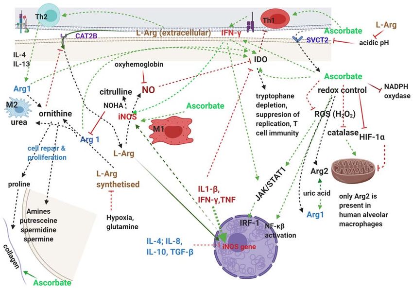
are modulated by NO generated from arginine, the only physi- Ascorbate stimulates the synthesis and actions of antiviral
ological substrate for NO (122). NO indirectly in multiple ways, one via IFN‑ γ stimula-
Arginine‑ascorbate has several important benefits in CoV tion (132), another via inhibition of HIF‑1α and correction
infection: firstly, direct antiviral actions, secondly, improve- of hypoxia, which significantly inhibits the availability of
ment of leukocyte function and number, especially important intracellular arginine and NO production (133); and also
in patients with neutrophilia and lymphopenia and finally it by synergizing with the apoptotic effects of NO (134) in
provides essential components and stimulates the mechanisms virus‑infected cells, and thus inhibiting viral replication.
of tissue repair. It is also important to note that all three of NOS as well as
IDO (135) are heme‑containing proteins which require ascor-
a) Antiviral actions. During the 2003 SARS epidemic bate or another intracellular reducing agent for their activation.
medicinal NO gas, a mixture of 0.8% NO and 99.2% N2 was In this context, the observation that three accessory proteins of
administered by Keyaerts et al (123) and followed by prompt SARS‑CoV2 ‑ orf1ab, ORF3a, ORF10 ‑ bind to heme followed
improvement in oxygenation and sustained patient benefit; by Fe inactivation and sequestration (37) deserve to be further
following‑up on this observation it was shown that NO donor investigated for specific anti‑NOS and other IDO activities.
compounds inhibited SARS‑CoV replication in vitro, a In the case of SARS‑CoV2 infection which directly antag-
finding replicated by another team who also observed onizes NO production, arginine provides both the substrate
that besides S‑nitroso‑N‑acetylpenicillamine (SNAP), for iNOS and activates its synthesis; together with ascorbate,
IFN also inhibited SARS‑CoV replication in vitro (124), which is necessary for iNOS activation, it prepares a new
confirming results obtained by another team who showed functional enzyme necessary for NO production.
that IFN stimulates iNOS and NO production for prompt
antiviral action (125). b) Improving lymphocyte function and number. Lymphopenia
Interestingly, IFN‑ γ stimulates NO production, and is present in a significant number of severe COVID‑19
inhibition of NOS in mice resulted in conversion of a patients (9) and its causes are strongly linked to arginine and
resolving infection by the ectromelia virus into fulminant ascorbate deficiencies.
mousepox (126). Arginine is known to improve lymphocyte‑based immu-
NO production from arginine by the inducible NO nity (136). Arginine depletion due to upregulation of Arg1
synthase (iNOS) was shown to be essential for native immune in myeloid‑derived suppressor cells induces T‑cell anergy,
responses in many infections and arginine availability is a crit- decreased proliferation of T cells, low expression of CD3 ζ‑chain
ical factor for host resistance to infection (122). NO can pass T‑cell receptor, and impairments of production of T cell cyto-
through membranes, unlike complement and antibody, and kines and of upregulation of cyclin D3 and cyclin‑dependent
is especially useful in syncytia, it can act on multiple targets kinase 4 (cdk4) (137). L‑arginine is essential for maturation
increasing efficacy and preventing developing of resistance, of CD3+ and the proliferation of CD8+ T lymphocytes (138).
and do not require recognition of infected cell by the immune Arginine is also critical for B‑lymphocyte differentiation; in
system, which can be limited by pathogens or tissue‑specific transgenic mice over‑expressing arginase in intestinal cells
differences (127). there was an impaired transition of pro‑ to pre‑B cells in
Reducing arginine availability either by nutrient depriva- the bone marrow with marked decrease in B cell cellularity,
tion or by specific pathogens significantly blunts the immune serum IgM levels and number and size of Peyer's patches; this
response, the former via impaired TLR4/MAPK pathway was reversed by arginine supplementation (139).
signalling, the latter by inducing arginase 1 via other TLRs Noting the essential role of arginine for immunity, an
in macrophages in an autocrine‑paracrine manner involving arginine deficiency syndrome (ADS) was proposed, defined
cytokines, an immune evasion mechanism shown with myco- by pathological increase in arginase, decreased NO produc-
bacteria (122) and probably also in SARS‑CoV2 infection. tion, decreased arginine availability, abnormal T cell function
Arginine also directly stimulates transcription/translation including loss of ζ‑chain, and presence of one or more of:
of the iNOS gene leading to de novo NOS protein synthesis, trauma, cancer, chronic infection, liver necrosis, pulmonary
and this sheds light on the ‘arginine paradox’ where intracel- hypertension; treatment for ADS include L‑arginine and the
lular NO production is directly related to the extracellular arginase inhibitors N‑hydroxy‑arginine (NOHA) and COX‑2
arginine concentration even though endogenous synthesis of inhibitors.
arginine provides approximately half of the arginine needed by Ascorbate is also important for adaptive immune responses,
cells (128). To ensure the increased need for intracellular argi- starting with the observations that reducing environments
nine, the cationic amino acid transporter CAT2B is induced make immune responses more efficient (140); it increases the
by IFN‑γ, which is stimulated by ascorbate (129). Reciprocally, levels of IFN produced by activated fibroblasts (141); improves
the pH buffering action of arginine is increasing the cellular neutrophil function by preserving/restoring function of their
uptake of ascorbate by improving the activity of the cellular myeloperoxidase; is essential for lymphocyte development,
transporter of ascorbate, SVCT2, which has a pH optimum stimulating the proliferation of NK cells (142); and has ample
of 7.5 and is reduced to ~50% when pH is 5.5 (130). epigenetic effects through dioxygenases TET (ten‑eleven
Ascorbate is actively accumulated in phagocytic cells translocases) and Jumonji C (JmjC)‑domain‑containing
where its concentration is 70‑100 times higher than in plasma; histone demethylases (131).
it enhances chemotaxis, ROS generation, phagocytosis and A comprehensive review of the actions of ascorbate
viral clearance, important for minimising necrosis and tissue including those on the immune system (131) concludes that
damage (131). enhancement of B‑ and T‑cell differentiation and proliferationYou can also read