High-Grade Serous Ovarian Cancer: Basic Sciences, Clinical and Therapeutic Standpoints - MDPI
←
→
Page content transcription
If your browser does not render page correctly, please read the page content below
International Journal of
           Molecular Sciences

Review
High-Grade Serous Ovarian Cancer: Basic Sciences,
Clinical and Therapeutic Standpoints
Michael-Antony Lisio, Lili Fu, Alicia Goyeneche, Zu-hua Gao               and Carlos Telleria *
 Experimental Pathology Unit, Department of Pathology, McGill University, Montreal, QC H3A 2B4, Canada;
 michael-antony.lisio@mail.mcgill.ca (M.-A.L.); Lili.Fu@muhc.mcgill.ca (L.F.); alicia.goyeneche@mcgill.ca (A.G.);
 zu-hua.gao@mcgill.ca (Z.-h.G.)
 * Correspondence: carlos.telleria@mcgill.ca

 Received: 18 January 2019; Accepted: 19 February 2019; Published: 22 February 2019

 Abstract: Among a litany of malignancies affecting the female reproductive tract, that of the ovary is
 the most frequently fatal. Moreover, while the steady pace of scientific discovery has fuelled recent
 ameliorations in the outcomes of many other cancers, the rates of mortality for ovarian cancer have
 been stagnant since around 1980. Yet despite the grim outlook, progress is being made towards
 better understanding the fundamental biology of this disease and how its biology in turn influences
 clinical behaviour. It has long been evident that ovarian cancer is not a unitary disease but rather
 a multiplicity of distinct malignancies that share a common anatomical site upon presentation.
 Of these, the high-grade serous subtype predominates in the clinical setting and is responsible for a
 disproportionate share of the fatalities from all forms of ovarian cancer. This review aims to provide a
 detailed overview of the clinical-pathological features of ovarian cancer with a particular focus on the
 high-grade serous subtype. Along with a description of the relevant clinical aspects of this disease,
 including novel trends in treatment strategies, this text will inform the reader of recent updates to the
 scientific literature regarding the origin, aetiology and molecular-genetic basis of high-grade serous
 ovarian cancer (HGSOC).

 Keywords: high-grade serous ovarian cancer; cortical inclusion cysts; serous tubular intra-epithelial
 carcinoma; ovarian surface epithelium; homologous recombination; BRCA; mutant p53;
 genetically-engineered mouse models; debulking surgery; chemotherapy

1. Prevalence and Mortality
      Ovarian cancer is a salient public health concern, which, in spite of its infrequent incidence,
remains the deadliest form of gynaecological malignancy. According to the WHO, each year an
estimated total of 225,500 cases of ovarian cancer will be diagnosed and 140,200 patients will succumb
to this disease, representing the 7th most common form of cancer and the 8th leading cause of
cancer-related death among women worldwide [1,2]. These figures, taken together, underline the
status of ovarian cancer as significant source of morbidity and mortality in the global population.
In Western nations, ovarian cancer is the 5th most frequent cause of cancer-related death in women [3].
The Surveillance, Epidemiology and End Results (SEER) program of the American National Cancer
Institute (NCI) records an annual incidence of 11.6 cases/100,000 women per year according to the latest
statistical cohort, with an estimated 224,940 women living with the disease in 2015 [4]. In Canada, the
Canadian Cancer Society predicted in 2017 an average of 2800 cases diagnosed and 1800 deaths/year [5].
Whereas the survival rates for a number of solid tumours have improved significantly in the last
50 years, a recent meta-analysis drawing upon survival data from numerous countries concluded that
the 5-year overall survival from ovarian cancer had remained virtually unchanged since about 1980 [6].

Int. J. Mol. Sci. 2019, 20, 952; doi:10.3390/ijms20040952                            www.mdpi.com/journal/ijms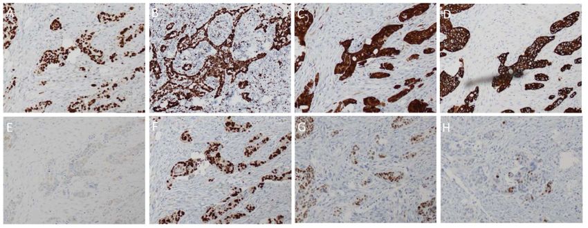
Int. J. Mol. Sci. 2019, 20, 952                                                                   2 of 33

According to the most recent figures published by the SEER (2008–2014), the current 5-year survival
rate in the US is approximately 47.4% [4].

2. Subtype Classification
      Although the term “ovarian cancer” implies a unitary disease, from the perspective of the
pathologist it was apparent as early as the 1930s that it was more appropriate to classify ovarian
neoplasms as multiple distinct entities through the lens of histopathology [7]. This culminated
in the 1973 WHO guidelines, which signified the first systematic attempt to delineate the many
ovarian cancer subtypes [7]. Histologically, about 90% of ovarian tumours are deemed to have
occurred through the transformation of epithelial cells as opposed to those originating from germ
cells or sex-cord-stromal tissues [8]. These are thus designated as epithelial ovarian cancers (EOC).
That nomenclature itself applies to a broad category of disease with a whole range of taxonomy therein
contained. This notably includes the four well-defined histological subtypes, which have constituted
the basis for EOC diagnosis over the past few decades. These are referred to as: serous, mucinous,
clear-cell and endometrioid—appellations deriving from their morphology and tissue architecture
as observed through microscopy. Furthermore, the assignment of a tumour grade, based on the
apparent degree of cytological aberration, allows for an additional degree of stratification for serous
and endometrioid EOCs [3]. Thus, despite sharing some similarity in histological appearance and
terminology, high-grade and low-grade serous carcinomas of the ovary are now considered to be
two entirely different neoplasms, with distinct modes of carcinogenesis, molecular-genetic features
and sites of origin [9]. While the majority of cases observed clinically belong to one of the four major
histotypes, a number of rarer types have been noted. These include malignant transitional cell (Brenner)
tumours as well as cases of mixed type and undifferentiated carcinoma [10].
      Although referred to as ovarian cancer, it has long been observed that the histology of these
tumours resembles non-ovarian tissues. For example, endometrioid ovarian carcinoma, as its name
suggests, features a glandular architecture similar to the endometrium, while mucinous tumours
can resemble either endocervical glands or the gastrointestinal epithelium [11]. Recent studies have
supported the notion of an extra-ovarian origin for many mucinous tumours along with the carcinomas
of the clear-cell and endometrioid subtypes, which likely derive from metastatic intestinal tumours
and endometriotic lesions respectively [12,13]. The origin of the serous subtype was long debated but
in the case of high-grade serous neoplasms, it is now widely acknowledged that the majority originate
from the epithelium of the fallopian tube.
      Recent efforts to study EOC from the molecular and genetic perspective have led to a paradigm
shift in the classification of this disease via the introduction of the dualistic model of ovarian
carcinogenesis. This model was first proposed by Kurman and Shih in 2004 and has since garnered
widespread acceptance, being officially recognized in 2014 by the WHO in their updated classification
guidelines for tumours of the female reproductive organs [9,14]. This model segregates the variety of
EOC subtypes into two broad categories called Type 1 and Type 2. The Type 1 neoplasms typically
develop along a step-wise progression from pre-malignant or borderline lesions in a manner common
to many other epithelial cancers [10]. From the genetic perspective, these tumours display frequent
oncogenic alterations to many cellular signalling pathways such as RAS-MAPK and PI3K-AKT but
are otherwise genomically stable and P53 wild type [10]. From a clinical perspective, these tumours
typically present as large, unilateral, cystic neoplasms that grow in an indolent fashion and when
confined to the ovary they have an excellent prognosis [10]. This category includes low-grade serous,
clear-cell, mucinous and transitional cell (Brenner) subtypes [10].
      By contrast, the Type 2 category is marked by a far more aggressive pattern of disease behaviour.
Tumours develop rapidly and usually are disseminated widely at the time of presentation, resulting in
poor overall prognosis [10]. From a genetic viewpoint, these tumours are characterized by P53
mutations and genomic instability due to defects in pathways contributing to DNA repair [10].
The prototypical Type II neoplasm, HGSOC, is by far the dominant subtype diagnosed clinically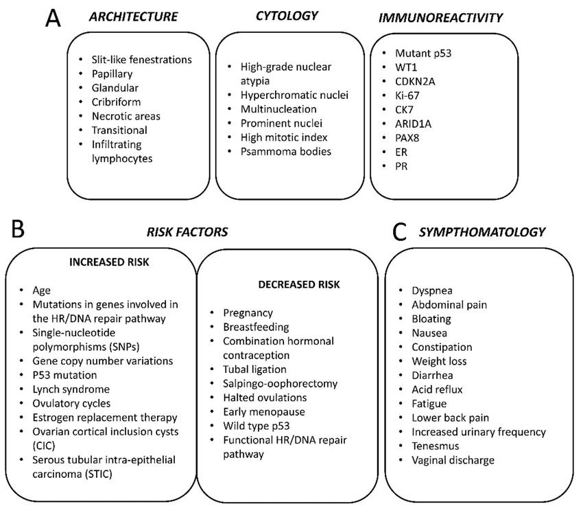
Int. J. Mol. Sci. 2019, 20, 952 3 of 33
and accounts for 70–80% of deaths from all forms of ovarian cancer [10,15]. In summary, the term
ovarian cancer is a broad designation for a myriad of distinct diseases sharing an anatomical site
Int. J. Mol.
upon Sci. 2019, 20, x FOR PEER REVIEW
presentation. 3 of 32
3. Histopathology
3.
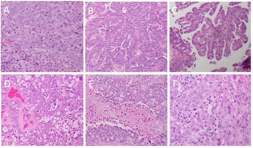
From the
From the perspective
perspective of of aa pathologist
pathologist visualizing
visualizing stained tissue sections under a microscope,
HGSOC tumours typically feature
HGSOC tumours typically feature solid masses solid masses of cells (Figure
of cells 1A) with
(Figure 1A)slit-like fenestrations
with slit-like (Figure
fenestrations
1B) [9]. 1B)
(Figure In some
[9]. In areas, the tumours
some areas, often often
the tumours have have
a papillary (Figure
a papillary 1C),1C),
(Figure glandular
glandular(Figure
(Figure 1B)1B)
or
cribriform (Figure 1D) architecture that is said to resemble the surface
or cribriform (Figure 1D) architecture that is said to resemble the surface epithelium epithelium of the fallopian
fallopian
tube [9,11].
tube [9,11]. The regions of solid growth growth areare frequently
frequently accompanied
accompanied by by areas
areas of
of extensive
extensive necrosis
necrosis
(Figure1E)
(Figure 1E)[9].
[9].InIncertain
certaincases,
cases,HGSOC
HGSOC maymay present
present withwith areas
areas displaying
displaying a solid
a solid growth
growth patternpattern
that
that simulates the appearance of endometrioid or transitional cell carcinoma
simulates the appearance of endometrioid or transitional cell carcinoma (Figure 1D) [10]. Although (Figure 1D) [10].
Although morphologically
morphologically distinct, thesedistinct,
tumours these
show tumours show an immunoreactivity
an immunoreactivity identical
identical to typical HGSOC to and
typical
are
HGSOC
thus and are thus
not considered as anot considered
separate entityas[10].
a separate entity
Researchers [10].recently
have Researchers
namedhave recently
a group namedasa
of HGSOC
group
the SETof(“Solid,
HGSOC as the SET (“Solid, pseudo-Endometrioid
pseudo-Endometrioid and/or Transitional cell and/or Transitional cell
carcinoma-like”) carcinoma-like”)
tumours [16]. It was
tumours
found that[16].
SETIttumours
was found that SETassociate
frequently tumours withfrequently
BRCA1 associate
mutations with
and BRCA1
containmutations
a greaterand contain
number of
a greater number of tumour-infiltrating lymphocytes (Figure
tumour-infiltrating lymphocytes (Figure 1F) compared to typical HGSOC [16]. 1F) compared to typical HGSOC [16].
Figure 1. Heterogeneity of the histopathological architecture of high-grade serous cancer. (A): solid
Figure 1. Heterogeneity of the histopathological architecture of high-grade serous cancer. (A): solid
architecture (original magnification × 20); (B): glandular architecture with slit-like spaces (original
architecture (original magnification × 20); (B): glandular architecture with slit-like spaces (original
magnification × 10; (C): papillary architecture (original magnification × 5); (D): cribriform and
magnification × 10; (C): papillary architecture (original magnification × 5); (D): cribriform and
pseudoendometroid architecture (original magnification × 20); (E): solid architecture with “geographic”
pseudoendometroid architecture (original magnification × 20); (E): solid architecture with
necrosis (original magnification × 10); (F): solid architecture with tumour infiltrating lymphocytes
“geographic” necrosis (original magnification × 10); (F): solid architecture with tumour infiltrating
(original magnification × 20).
lymphocytes (original magnification × 20).
From the cytological perspective, HGSOC is characterized by high-grade nuclear atypia; with
large,From the cytological
hyperchromatic and perspective,
pleomorphicHGSOC is characterized
nuclei with the potential byforhigh-grade nuclear
multinucleation atypia;
(Figure 2A)with
[9].
The nucleoli are usually prominent and might appear large and eosinophilic (Figure 2B) [9]. There[9].
large, hyperchromatic and pleomorphic nuclei with the potential for multinucleation (Figure 2A) is
The nucleoli are usually prominent and might appear large and eosinophilic (Figure 2B) [9].
usually a high mitotic index with an abundance of visible mitotic figures (Figure 2C) that may be of There is
usually aappearance
atypical high mitotic
[9].index with anbodies,
Psammoma abundance
whichof are
visible mitotic
areas figures (Figure
of calcification 2C)associated
typically that may be of
with
atypical appearance [9]. Psammoma bodies, which are areas
papillary tumours, are also typically present (Figure 2D) [9]. of calcification typically associated with
papillary tumours, are also typically present (Figure 2D) [9].
A number of immunological markers are used to differentiate HGSOC from other subtypes of
EOC. Unlike low-grade serous tumours, HGSOC is almost invariably P53-mutant and will usually
stain with a strong diffuse nuclear-positivity in nearly all cells (Figure 3A) [9]. This, however, is
dependent on the type of mutation present. Missense mutations in TP53 typically correlate with
positive staining due to the mutant protein accumulating owing to a lost capacity for degradation by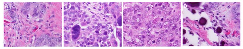
Int. J. Mol. Sci. 2019, 20, 952 4 of 33
A number of immunological markers are used to differentiate HGSOC from other subtypes of
EOC. Unlike low-grade serous tumours, HGSOC is almost invariably P53-mutant and will usually stain
with a strong diffuse nuclear-positivity in nearly all cells (Figure 3A) [9]. This, however, is dependent
on the type of mutation present. Missense mutations in TP53 typically correlate with positive staining
Int. J. Mol. Sci. 2019, 20, x FOR PEER REVIEW 4 of 32
due to the mutant protein accumulating owing to a lost capacity for degradation by the proteasome [9].
Int. J. Mol. Sci. 2019, 20, x FOR PEER REVIEW 4 of 32
If, however,
form the genemight
of the protein containsnot abenonsense
detectable mutation, then the resultant
by the antibody [9]. In thattruncated
case, theform of the
staining protein
would be
might
almost not be detectable
totally negative. by the antibody
Compared to [9].alternative
the In that case,forms
the staining
of EOC, would
HGSOC be almost
is totally negative.
frequently found to
form of the protein might not be detectable by the antibody [9]. In that case, the staining would be
Compared
stain positive to for
the WT1
alternative
(Figure forms
3B) of EOC,
and CDKN2A HGSOC (a.k.a.is P16)
frequently
(Figure found
3C) toThe
[9]. stain positive forindex,
proliferation WT1
almost totally negative. Compared to the alternative forms of EOC, HGSOC is frequently found to
(Figure
assessed 3B) and CDKN2A
through the (Figure
number(a.k.a.
of P16)
cells (Figure
positive3C) for [9]. The would
Ki-67, proliferation
be 3C) index,
higher assessedtothrough
compared low-gradethe
stain positive for WT1 3B) and CDKN2A (a.k.a. P16) (Figure [9]. The proliferation index,
number of
serous lesions cells positive
(Figure for Ki-67, would
4A, rightofpanel) be higher
[9]. The for compared
epithelial to low-grade serous lesions (Figure 4A,
assessed through the number cells positive Ki-67, marker
would be CK7 is positive
higher comparedin HGSOC (Figure
to low-grade
right
3D) but panel)
CK20 [9].
is The epithelial
usually negative marker
(Figure CK7 is positive in HGSOC (Figure 3D) but CK20 is usually
serous lesions (Figure 4A, right panel) [9].3E)
The[11]. Furthermore,
epithelial markerwhenCK7 compared
is positive to in ovarian
HGSOCclear-cell
(Figure
negative
carcinoma, (Figure
HGSOC 3E) [11].
is Furthermore,
HNF1β negative when
and compared
ARID1A to ovarian
positive clear-cell
(Figure 4A, carcinoma,
right panel)HGSOC is
[17]. In
3D) but CK20 is usually negative (Figure 3E) [11]. Furthermore, when compared to ovarian clear-cell
HNF1β
common withnegative and ARID1A
most other positive
forms negative (Figure
of EOC isand 4A, right
the expression panel) [17]. In
of PAX8(Figurecommon
(Figure 4A, with
3F), aright most
marker other forms
of tissues of
carcinoma, HGSOC is HNF1β ARID1A positive panel) [17]. In
of EOC is
Müllerianwith the expression
origin including of PAX8 (Figure
the fallopian 3F), a marker of tissues of Müllerian origin including the
common most other forms of EOC istube [18]. The expression
the expression of the oestrogen
of PAX8 (Figure 3F), a marker receptor (ER)of
of tissues is
fallopian
detectable tube
in [18]. The
about 80% expression
of cases of the 3G),
(Figure oestrogen
whereas receptor
the (ER) is detectable
progesterone receptorin about
(PR) 80%
is onlyoffound
cases
Müllerian origin including the fallopian tube [18]. The expression of the oestrogen receptor (ER) is
(Figure 3G), whereas
to be positive in around the30%progesterone
of HGSOC receptor samples
(PR) is only found 3H)to[9].
be positive in around 30% of
detectable in about 80% of cases (Figure patient
3G), whereas the(Figure progesterone receptor (PR) is only found
HGSOC patient samples (Figure 3H) [9].
to be positive in around 30% of HGSOC patient samples (Figure 3H) [9].
Figure 2. Cytological features of high-grade serous cancer. (A): Multinucleated tumor giant cells; (B):
Figure 2. Cytological features of high-grade serous cancer. (A): Multinucleated tumor giant cells;
severe pleomorphism
Figure 2. and prominent
Cytological features nucleoli;
of high-grade (C):cancer.
serous frequent mitotic
(A): figures; (D):
Multinucleated psammoma
tumor bodies.
giant cells; (B):
(B): severe pleomorphism and prominent nucleoli; (C): frequent mitotic figures; (D): psammoma
All original magnifications × 40.
severe pleomorphism and prominent nucleoli; (C): frequent mitotic figures; (D): psammoma bodies.
bodies. All original magnifications × 40.
All original magnifications × 40.
Figure
Figure 3. Immunologicalmarkers
3. Immunological markerstypically
typicallyseen
seen
in in high-grade
high-grade serous
serous ovarian
ovarian cancer.
cancer. (A): (A):
p53;p53;
(B):
(B):
WT-1;WT-1;
(C): (C):
p16;p16; (D):
(D): CK7;
CK7; (E): (E): CK20;
CK20; (F): (F): PAX8;
PAX8; (G):
(G): ER; ER; (H):
(H): PR.PR.
All All original
original magnifications
magnifications × × 10.
10.
Figure 3. Immunological markers typically seen in high-grade serous ovarian cancer. (A): p53; (B):
4. Epidemiology
WT-1; (C): p16;and(D):Risk
CK7;Factors
(E): CK20; (F): PAX8; (G): ER; (H): PR. All original magnifications × 10.
4. Epidemiology and Risk Factors
The distribution of ovarian cancer incidence worldwide is not even, with a substantial variation
4. Epidemiology
The and Risk
distribution Factors
of ovarian cancer
on the basis of geography, ethnicity andincidence
the level worldwide
of economicisdevelopment.
not even, withThe a substantial variation
peak age-adjusted
on the basis of geography,
The distribution
incidence rates are inof ethnicity
ovarian and
Northern cancer and the level of
incidence worldwide
Central/Eastern economic development.
Europe;iswith
not even, The peak
with a substantial
intermediate age-adjusted
rates seenvariation
in North
incidence
on the rates
basis of are in
geography,Northern and
ethnicity Central/Eastern
and the level of Europe;
economic with intermediate
development.
America, Western Europe and Australia; and lower rates in Asia and Africa [19]. Recent trends The rates
peak seen in North
age-adjusted
seem
America,
incidence Western
to show a rates
stableare Europe
in Northern
or slight and Australia; and
andinCentral/Eastern
reduction lower
age-standardized rates in
Europe; Asia
rates inwithand Africa
mostintermediate[19].
high-incomeratesRecent trends
seen in
countries, seem
North
whereas
to show
America,
they aWestern
appearstable
to beorrising
slightin
Europe reduction
and
many low-in age-standardized
Australia; andand lower ratesrates
middle-income in most
incountries
Asia high-income
and [19].
AfricaAs[19].
such, countries,
Recent
there trendswhereas
is far seem
less of
they
to appear
show a stable
a disparity to be rising in many
or slight compared
in incidence reduction inlow- and middle-income
to age-standardized
30 years ago [19].rates countries [19]. As
in most high-income
In countries such, there is
countries,
with a multi-ethnic far less
whereas
population, of
a disparity in incidence compared to 30 years ago [19]. In countries with a multi-ethnic
they appear to be rising in many low- and middle-income countries [19]. As such, there is far less of population,
athe incidence
disparity may depend
in incidence on ethnicity.
compared For example,
to 30 years ago [19].inInthe US the disease
countries is more frequent
with a multi-ethnic among
population,
non-Hispanic, Caucasian women compared to Hispanic, Asian or African
the incidence may depend on ethnicity. For example, in the US the disease is more frequent among American women [4].
Like mostCaucasian
non-Hispanic, other epithelial
women cancers,
compared EOCtotends to be Asian
Hispanic, diagnosed more frequently
or African as a function
American women [4]. of
increasing age. other
Like most As such, the number
epithelial cancers, of cases
EOC occurring
tends to be each year is expected
diagnosed to increase
more frequently as aasfunction
global life-
of
Int. J. Mol. Sci. 2019, 20, 952                                                                   5 of 33

the incidence may depend on ethnicity. For example, in the US the disease is more frequent among
non-Hispanic, Caucasian women compared to Hispanic, Asian or African American women [4].
      Like most other epithelial cancers, EOC tends to be diagnosed more frequently as a function of
increasing age. As such, the number of cases occurring each year is expected to increase as global
life-expectancies continue to improve [19]. In the US, the median age of diagnosis is at 63 years [4].
Epithelial ovarian cancer subtypes are infrequently seen in pre-menopausal women (≤45 years of
age) while ovarian germ cell tumours occur mainly in younger women [19]. The total lifetime risk of
developing ovarian cancer has been estimated to be only about 1.3% for American women; however,
there are a number of known risk-factors that may modify the risk in individuals [4,19] (Figure 4B).
For example, there is a substantial heritable component of risk due to genetic factors. The risk
for women with an affected first-degree relative is threefold greater than that of women without
any affected relatives [20]. Familial cases usually are found to be due to germline mutations in the
tumour-suppressor genes BRCA1 and BRCA2, which also contribute increased risk of developing
breast cancer in these same families [19]. A recent study found that 3.6% of ovarian cancer patients
have germline mutations in BRCA1 while 3.3% have germline mutations in BRCA2 [21]. Overall, it was
estimated that germline BRCA1 and BRCA2 mutations contribute to the development of 10–20% of
EOCs [22]. Compared to the normal population, BRCA1 mutation carriers have an estimated 44% risk
of developing ovarian cancer by age 70, while this risk is up to 27% for BRCA2 mutant individuals [23].
The cancers occurring in these women are usually high-grade serous carcinomas, which manifest at an
earlier age than in sporadic cases [19]. The contribution of high-penetrance alleles of BRCA1/2 can
only account for a small part of the heritable component of ovarian cancer [22]. Many other genes
bearing low penetrance mutations are thought to play an important role. For instance, women with
mutations in the genes BRIP1, RAD1C and RAD1D have estimated lifetime risks of developing ovarian
cancer of 5.8%, 5.2% and 12%, respectively [24,25]. Other gene variations that have been linked with
greater risk include BARD1, CHEK2, MRE11A, RAD50, PALB2 and ATM [25,26]. The common link
between all these genes is their role in the homologous recombination (HR)-mediated pathway of
DNA repair, which is known to play a prominent role in the pathophysiology of HGSOC. Women with
Lynch syndrome bearing mutations in genes involved in DNA mismatch repair also have a greater
risk of developing EOC, mostly of the clear-cell and endometrioid subtypes [27].
      In recent years, genome-wide association studies (GWAS) have also been used to search for
single-nucleotide polymorphisms (SNPs) correlating with greater risk of developing ovarian cancer [22].
Several of these loci have been identified, and, while each is associated with only a miniscule increase
in absolute risk, the combination of multiple alleles has been demonstrated to considerably impact an
individual’s polygenic risk score [28].
      Endometriosis is known to predispose individuals towards developing EOC, particularly the
clear-cell and endometrioid subtypes, which are known to derive from endometriotic lesions [13,19].
      Many modifiable or lifestyle factors have also been viewed as influencing an individual’s risk
of developing ovarian cancer. Generally, ovarian cancer has been associated with women having
experienced a greater number of ovulatory cycles in their lifetime [19]. As such, factors tending to
reduce a woman’s ability to ovulate have been linked with a reduced lifetime risk of developing this
disease. For example, both the early occurrence of menarche and an older age at menopause have
been connected with a possible increased risk [29,30]. Likewise, women who have given birth have a
lower risk than nulliparous women, with a risk reduction of 10–20% associated with each additional
birth [30]. Studies have also found that women who breastfeed have lower risk compared to those
who do not [30,31]. The use of hormone (oestrogen plus a progestin)-containing oral contraceptives
has been robustly associated with a reduced risk of developing ovarian cancer, with users or former
users having up to 30% lower risk compared to never-users [19,32]. This apparent protective effect
was more apparent in long-term users and extended to all major subtypes of EOC [30,32]. One study
claimed that the use of these compounds might have even prevented up to 200,000 worldwide cases
of EOC over the past several decades [32]. Procedures aimed at reducing fertility, such as by tubal
Int. J. Mol. Sci. 2019, 20, 952 6 of 33
ligation, have been shown to reduce the risk of developing certain forms of EOC, while the use of
oestrogen hormone therapy during menopause has been linked with a heightened risk in the women
undergoing such treatment [30,33,34] (Figure 4B).
Other potential risk factors include obesity, diabetes, smoking and usage of perineal talc [19].
Unfortunately, many of the modifiable factors that have been associated with ovarian cancer are not
Int. J. Mol.
easily Sci. 2019, 20,
amenable to xchange,
FOR PEER REVIEW
while 6 of 32
others, including pregnancy and oral contraceptives use, cannot
be recommended as a cancer prevention strategy [19]. Moreover, a recent study of an Australian
concluded that only about 7–11% of ovarian cancer cases could be attributed to these modifiable
cohort concluded that only about 7–11% of ovarian cancer cases could be attributed to these modifiable
factors [35].
factors [35].
Figure 4. Histopathological features (A), risk factors (B) and symptoms (C) of high-serous ovarian cancer.
Figure 4. Histopathological features (A), risk factors (B) and symptoms (C) of high-serous ovarian
5. Origin
cancer.
The precise cell and tissue of origin for HGSOC has long been a matter of contention: unlike
5. Origin
low-grade serous tumours, which arise from pre-existing lesions such as serous cystadenomas or serous
The precise
borderline tumours,celllocating
and tissue of origin for component
a precancerous HGSOC hasinlong the been
case ofa matter
HGSOC ofhas
contention: unlike low-
proven difficult [36].
grade
In fact,serous
HGSOC tumours,
remains which ariseepithelial
the only from pre-existing lesions such
cancer without as serousprecancerous
a recognized cystadenomas or serous
lesion [11].
borderline
Because mosttumours,
HGSOClocating
patients,a even
precancerous
at an earlycomponent in the
stage, feature thecase of HGSOC
cancerous has proven
involvement difficult
of the ovary,
it[36].
wasInnatural
fact, HGSOC
to assumeremains only epithelial
that the disease cancer
originated fromwithout a recognized
that location. precancerous
The tissue of originlesion
was
[11]. Because
reputed to be most HGSOC
the ovarian patients,
surface even at an
epithelium earlyastage,
(OSE), simple,feature the cancerous
uncommitted layer involvement of the
of flat-to-cuboidal
ovary, it wasderived
epithelium naturalfrom
to assume that the disease
the coelomic mesoderm originated from that
and related location.
to the The tissue
mesothelial of origin
covering of was
the
reputed to be the ovarian surface epithelium (OSE), a simple, uncommitted layer
peritoneal cavity [11,37]. As early as 1971, Fathalla put forward the “incessant ovulation” hypothesis of flat-to-cuboidal
epithelium
that derived
would attain from the acceptance
widespread coelomic mesoderm
in subsequentand years
related to theHis
[36,38]. mesothelial coveringthat
theory contended of the
the
peritoneal
constant cavity
cycle [11,37].
of repair Asregeneration
and early as 1971,theFathalla put forward
OSE experiences as athe “incessant
result ovulation”
of ovulation mighthypothesis
contribute
that
to would attain widespread
carcinogenesis by creating theacceptance
kind of in subsequent yearsand
pro-inflammatory [36,38]. His theory microenvironment
pro-oxidative contended that the
constant cycle
conducive to DNAof repair
damage and regeneration
[36–38]. the OSE
The inability experiences
to repair as a result
such damage of ovulation
adequately might
was thought
contribute
to be at the to rootcarcinogenesis by creating[36].
of HGSOC carcinogenesis the Indeed,
kind ofit has pro-inflammatory
since become known and pro-oxidative
that patients
microenvironment
harbouring germlineconducive
mutationstoin DNA damage [36–38].
BRCA1/2—which encode Theproteins
inability to repairinsuch
implicated damage
the pathway
adequately was thought to be at the root of HGSOC carcinogenesis [36]. Indeed, it has since become
known that patients harbouring germline mutations in BRCA1/2—which encode proteins implicated
in the pathway responsible for the homologous recombination-mediated repair of double strand
breaks (DSBs)—are at much greater risk of developing HGSOC [36]. According to this theory, the
total number of ovulatory cycles a woman experiences would be related directly to her risk of
acquiring HGSOC [36,38]: numerous studies concluded that factors suppressing ovulation, such asInt. J. Mol. Sci. 2019, 20, 952 7 of 33
responsible for the homologous recombination-mediated repair of double strand breaks (DSBs)—are
at much greater risk of developing HGSOC [36]. According to this theory, the total number of
ovulatory cycles a woman experiences would be related directly to her risk of acquiring HGSOC [36,38]:
numerous studies concluded that factors suppressing ovulation, such as pregnancy, breastfeeding and
the use of hormone-containing oral contraceptives, reduced an individual’s risk of developing the
disease [36,37,39] (Figure 4B), thus reinforcing the theory.
Furthermore, it was observed that ovulatory repair resulted in the tendency for certain sections
of the OSE to invaginate and become trapped beneath the surface of the ovary in structures called
cortical inclusion cysts (CICs) [37]. Within the ovary, CICs are exposed to several hormones capable of
promoting growth and differentiation, something thought sufficient to engender the transition to a
state of metaplasia in the OSE lining of these CICs [37]. In cells harbouring pre-existent mutations or
DNA damage, this would create the ideal scenario for neoplastic transformation [37].
The theory for an ovarian origin of HGSOC nevertheless remained questionable in a number of
key areas. Histologically, HGSOC is said to resemble more closely tissues developmentally derived
from the Müllerian duct during embryogenesis [11,39,40]. It was not known how the coelomic cells
of the OSE could differentiate into a Müllerian-like tissue during carcinogenesis [11]. Some have
postulated that the relatively undifferentiated nature of the OSE would allow it to more readily
undergo metaplasia to resemble a Müllerian phenotype [39,40]. One study shows that the ectopic
activation of the gene HOXA9 in cultured mouse OSE was sufficient to induce the formation of tumours
histologically resembling serous carcinoma [41]. Although this study provided a potential mechanistic
framework for an OSE origin of HGSOC, doubt nevertheless persisted due to the enduring absence of
any identifiable precursor lesion or in-situ carcinoma in patients’ ovaries [11,39]; at the time, many
speculated that it was due to their destruction during the process of tumorigenesis [42].
In 1999, Dubeau cast doubt on the dogma that HGSOC originated from the OSE and instead
advocated for a source derived from the Müllerian epithelium [43]. His argument invoked the
differences in histogenesis between the OSE and the Müllerian epithelium, of which the latter appears
to resemble more closely the histological phenotype of ovarian carcinomas. Furthermore, he raised
the lack of evidence supporting the theory of the OSE being capable of metaplastic transformation to
resemble the Müllerian phenotype and emphasized on the continued absence of observable precursor
lesions in the ovary. Moreover, he argued that cases of primary peritoneal carcinoma, which arise
without ovarian involvement and which are indistinguishable clinically and histopathologically from
HGSOC, were proof of an extra-ovarian cell of origin for this neoplasm [43]. He advanced the theory
that serous ovarian carcinomas were derived from the secondary Müllerian system, the vestigial
remnants of Müllerian epithelium present ectopically outside of the cervix, endometrium and fallopian
tube [43]. Nonetheless, the belief that HGSOC could originate from a tissue of extra-ovarian origin
was not widely held until the introduction of risk-reducing salpingo-oophorectomy in patients with
inherited BRCA mutations [10]. In 2001, Piek et al. described the presence of small dysplastic lesions
similar to HGSOC within the fallopian tubes of suspected BRCA mutation carriers [39,44]. Stratified,
disorganized and enlarged epithelial cells with highly atypical nuclei morphologically characterized
these lesions [39]. The examination of samples from a cohort of non-mutant individuals failed to locate
any such lesions [36,44]. This discovery was aided by the introduction of a new histological approach
for sampling the fallopian tube in which the entire fallopian tube, with particular attention to the
fimbria, was sectioned [10]. Previously, studies examining the ovary for precursor lesions failed to
completely examine the fallopian tube [7]. These lesions, which later became known as serous tubular
intra-epithelial carcinomas (STICs), featured the virtual absence of ciliated cells with a shift in favour
of the alternative secretory population [37]. Later studies have established that these lesions are far
more common at the ciliated end of the fallopian tube, which is the section directly adjacent to the
ovary [37,45]. An analysis using immunohistochemistry found that these lesions stained strongly for
P53 compared to the surrounding epithelium, suggesting that these cells were P53 mutant [11,46].
They also over-expressed γH2AX, a marker of DNA double strand breaks [11,46]. One study foundInt. J. Mol. Sci. 2019, 20, 952 8 of 33
that 38% of a cohort of BRCA mutant women having undergone salpingo-oophorectomy harboured
STIC lesions in their fallopian tubes, without any corresponding abnormalities being found in the
ovaries of such patients [45]. The existence of these microscopic intra-epithelial carcinomas suggested
that the secretory epithelial cells of the distal fallopian tube (FTSEC) were the preferred cells of origin
for HGSOC, at least in women bearing BRCA1/2 mutations. This was supported strongly by a study
by Kuhn et al. showing that STICs possessed the identical P53 mutation to that present within the
concurrent HGSOC in women with this disease [47]. Furthermore, it has been shown that STICs contain
shortened telomeres compared to the co-existent cancer within the same patient [48]. The presence of
telomere shortening has become an established hallmark of the early stages of carcinogenesis [10].
While STICs were known to occur in women with BRCA mutations, it was not known if they
could contribute to carcinogenesis in sporadic cases of HGSOC. A 2007 study by Kindelberger et al.
showed that STICs were found in 52% of patients with sporadic advanced-stage HGSOC [46]. STICs
also have been reported in the fallopian tubes of women undergoing hysterectomy and bilateral
salpingo-oophorectomy for non-prophylactic reasons [10,49]. Recent genetic studies have established
that HGSOC and paired STICs have many other shared genetic alterations including changes in gene
copy number [22]. One study established that, in cases of HGSOC featuring the amplification of
CCNE1, a similar copy number change for this gene was present in the STICs isolated from the same
patient [50]. The tubal origin for HGSOC also has been reinforced by a study that used gene expression
profiling to show that the pattern of gene expression in HGSOC more closely resembles that of the
fallopian tube epithelium rather than that of the OSE [51]. Transformation of cultured human fallopian
tube epithelium in-vitro also results in cells that resemble the morphology, immunophenotype and
gene expression profile of human HGSOC [52]. In addition, a novel mouse model that features induced
mutations in the same genes commonly affected in human patients also develops serous carcinoma
from the fallopian tube [53].
Why the overwhelming majority of STICs are found to occur at the fimbriated end of the fallopian
tube remains a mystery [42]. Some studies claim to show that the fimbriae are enriched in cells
with “stem like” properties [54]. Wang and colleagues [55] used lineage tracing in a murine model
to identify a population of quiescent, label-retaining cells in the distal oviduct of female animals.
There were subsequently shown to be endowed with enhanced spheroid forming capacity along with
the ability to differentiate into structures resembling multiples tissues of Müllerian origin, including
the endometrium and distal/proximal oviduct. The authors theorize that their results point to the
existence (in humans) of a stem-like cell of origin in the distal fallopian tube that may underlay all the
various subtypes of ovarian carcinoma. It has also been argued that the distal fallopian tube represents
a developmental “transition zone,” analogous to that present in the cervix, which is prone to malignant
transformation [42].
Notwithstanding the convincing evidence that the fallopian tube is the major site of origin for
HGSOC, it remains established that ovulation is a consistent risk factor in epidemiological studies.
To explain this, it has been proposed that the proximity of the fimbriae to the ovarian surface might
subject it to many of the same pro-inflammatory mediators and ROS thought to contribute to the
development of genotoxic stress in the OSE following ovulation [37]. Yet despite the most diligent
examination, a significant percentage of HGSOC cases present without fallopian tube involvement [15].
This has led some to suggest that there still might be precursor cells in the ovary that underlie such
cases [15]. A new unifying theory contends that these cases arise from the early implantation of
FTSECs in the OSE through a process called “endosalpingiosis” [42]. In this scenario, the fallopian
tube epithelium might become incorporated into the same CICs that are the preferred site of origin for
HGSOC in the ovary [40,42]. As has already been highlighted, the microenvironment of the ovary is
more favourable for inducing neoplastic transformation compared to that of the fallopian tube [37,42].
Although endosalpingiosis has been demonstrated in mouse ovaries, the same process has never been
observed in humans [42]; thus, the precise progenitor for a substantial percentage of HGSOC cases
remains obscure.Int. J. Mol. Sci. 2019, 20, 952 9 of 33
6. Dissemination
High-grade serous ovarian carcinoma notably does not require the blood or lymph in order
to metastasize. For most other epithelial cancers to spread, tumour cells must typically undergo a
sequence of cellular transformations to traverse the basement membrane, migrate to and invade the
vasculature, survive in suspension, extravasate and re-establish themselves as a colony at a distant site.
By contrast, HGSOC typically spreads by direct extension to the adjacent organs within the peritoneal
cavity or through the detachment of cells from the primary tumour [11]. For a tumour growing on the
surface of the ovary or fallopian tube, there are no anatomical barriers capable of restricting the spread
of tumour cells throughout this fluid-filled space between the body’s visceral organs [56]. Once the
cells have exfoliated from the primary tumours site, either singly or in clusters, they become suspended
in the peritoneal fluid and are spread by a passive process that follows the physiological flow of this
fluid around the peritoneal cavity [11]. These cells then can implant and seed distant organs or tissues
with nests of cancer cells, which develop rapidly into secondary tumour nodules.
Although virtually every organ or structure within the peritoneal cavity may be involved
in secondary dissemination, HGSOC cells are known to exhibit a particular predilection for the
omentum [11]. In fact, 80% of patients with HGSOC present with omental metastases [57]. Composed
largely of energy-dense adipocytes, this large fat-pad extends from the stomach and covers the
intestines. It has been hypothesized that HGSOC preference for the omentum stems from a cellular
metabolic requirement for fatty-acid-based catabolism (β-oxidation) [57]. It has been shown that
adipocytes produce pro-inflammatory cytokines such as IL-8 that promote the homing and invasion of
tumour cells [57]. In the same study, co-culturing adipocytes with ovarian cancer cells was seen to
promote greater lipolysis in adipocytes and β-oxidation in cancer cells [57]. This co-culturing also led
to increased proliferation of ovarian cancer cells in-vitro and rapid growth of transplanted tumours
in-vivo [57].
While HGSOC spreads readily within the peritoneal cavity, its metastatic growths only invade
the surface of affected organs [11]. Secondary tumours typically colonize the mesothelial cell layer but
invade no further, leaving the deeper lamina largely intact [11]. Spread outside the peritoneal cavity is
uncommon, although certain pelvic and/or para-aortic lymph nodes sometimes can be involved [58].
Hematogenous spread is thought to be largely precluded, based on the observation that patients
treated with peritoneovenous shunts—having received a large number of tumour cells into the
circulation—mostly failed to develop any distant metastases even two years after the procedure [59].
There is the potential, however, for metastasis to the liver, while in the most advanced stage of HGSOC
tumour cells may also cross the diaphragmatic barrier and enter the pleural space, where they can
cause pleural effusions or even implant in the parenchyma of the lung [8].
Patients with late-stage disease frequently develop ascites featuring a prominent cellular
component, which are commonly referred to as “malignant ascites”. HGSOC cells might participate in
the formation of these ascites either by blocking the lymphatic drainage or by secreting vasoactive
and angiogenic factors which promote vascular permeability [11]. A lingering mystery is the role of
multicellular structures in the pathogenesis of HGSOC. These structures frequently appear in the form
of spheroids or aggregates of suspended tumour cells and commonly are isolated from the ascites
of patients with advanced disease. They have been proposed to represent a fundamental unit of
metastatic spread, while also forming a chemo-resistant niche to allow for the HGSOC cancer cells to
survive therapy [11]. Importantly, the formation of multicellular structures might allow the cells to
survive in anchorage-independent conditions by preventing anoikis [11].
7. Symptomatology, Diagnosis and Staging
One of the principal factors influencing the elevated mortality of HGSOC patients is the inability
to diagnose the disease at an early, localized stage. Only about 13% of cases of serous ovarian carcinoma
are diagnosed at stage I or stage II [60]. In fact, the vast majority of cases usually are diagnosed at
the stage of distant metastasis, which greatly prejudices an individual’s prognosis [4]. The 10-yearInt. J. Mol. Sci. 2019, 20, 952 10 of 33
survival of patients diagnosed with early-stage HGSOC is 55%, compared to only 15% for those having
presented with an advanced-stage disease [60].
There are currently no effective screening strategies for the early detection of ovarian cancer [8].
A recent trial evaluating the utility of transvaginal ultrasonography in combination with serum
CA125 levels demonstrated some promise in terms of early detection but failed to improve patient
outcomes [61,62]. Genetic tests might be useful to detect heritable BRCA mutations in patients with
a known family history of breast and ovarian cancer [8]. In such cases, the at-risk individual might
elect to undergo risk-reducing prophylactic surgery such as bilateral salpingo-oophorectomy, typically
upon completion of childrearing or by the age of 40 at the latest [22]. This technique has proven to be
effective in preventing the emergence of ovarian cancer in as much as 85–90% of cases [63]. Finally,
a very recent study shows promise in the early detection of ovarian cancers based on the genetic
analysis of mutations detected in the DNA recovered from liquid biopsies obtained during routine
Papanicolaou tests [64].
Because the symptoms associated with HGSOC are often diverse and non-specific, there is usually
little likelihood that a patient will encounter the appropriate medical specialist in time for an early
diagnosis to be made [8,65]. Symptoms typically are gastrointestinal and include abdominal pain,
bloating, nausea, constipation, anorexia, diarrhoea and acid reflux [8,9]. Other symptoms include
fatigue, back pain, tenesmus, as well as elevated urinary frequency [8,9]. At an advanced stage,
respiratory symptoms might be present such as cough and dyspnoea [9] (Figure 4C).
Unfortunately, by the time a patient becomes symptomatic her disease is found to be at an
advanced stage between 75–80% of cases [9]. This differs from other forms of EOC such as clear-cell
carcinoma that typically become symptomatic at a far earlier stage [8]. If a diagnosis of EOC is
suspected, the patient will typically be subjected to a pelvic and rectovaginal examination along
with radiographic imaging such as transvaginal or abdominal ultrasonography, CT, MRI or PET [8].
Blood levels of CA125 also might be measured, which in combination with other tests, might be
of diagnostic value [8]. Imaging will typically reveal complex, hyper-vascular pelvic masses and
omental/peritoneal nodules [9]. Serum CA125 levels often are elevated, especially in advanced cases,
with average values that can range between 500–1000 U/mL [66]. Advanced disease typically will
feature extensive peritoneal carcinomatosis, involving most of the major abdominal organs and may
be associated with the accumulation of large volumes of ascites [11].
To aid in diagnosis, laparoscopic surgery usually is performed to obtain a tumour sample for
biopsy and to aid in the staging of the disease [8]. The most recent (2014) FIGO staging system is based
on the degree of dissemination of the disease at diagnosis. At stage I, the cancer still is confined to
the ovaries or fallopian tubes [67]. By stage II, the disease already has spread to other pelvic organs
such as the uterus [67]. Stage III involves spread beyond the pelvis to organs or tissues within the
peritoneal cavity or to the retroperitoneal lymph nodes [67]. Stage IV results from spread beyond
the peritoneal cavity, including to the lungs and involvement of inguinal and other extra-abdominal
lymph nodes [67]. The end stage of the disease is characterized by malignant bowel obstruction due
to the formation of fibrous adhesions between loops of the bowel by the metastatic tumours [56].
This impedes the patient from normal alimentation, leading to cachexia, malnutrition, and, eventually,
death from factors which may include intercurrent infection [56].
8. Genetics
A major milestone in the understanding of HGSOC occurred as the result of a 2011 study by the
cancer genome atlas (TCGA) network which sought to evaluate the disease’s genetic features through
whole exome sequencing of samples from 316 patients [68]. This revealed that the genomic landscape
of HGSOC is characterized by profound genomic instability with few recurrent gene mutations other
than in TP53 [68]. This was in contrast to Type-1 EOCs which are characterized by frequent oncogenic
mutations in genes such as BRAF, KRAS, PTEN, CTNNB1 and PIK3CA while being P53 wild type [68].
In the case of HGSOC, it was found that upwards of 96% of the samples contained somatic TP53Int. J. Mol. Sci. 2019, 20, 952 11 of 33
mutations, seemingly suggesting that this mutation is a defining feature of HGSOC and one likely to
be required for disease initiation [22]. Retrospective studies established that the small percentage of
P53 wild type samples from the aforementioned TCGA study derived from patients whose disease
had likely been misdiagnosed as HGSOC [69]. Thus, one might conclude that TP53 mutations are
virtually ubiquitous in HGSOC. Because P53 commonly is found to be mutated even in the STIC
precursor lesions, it is likely that this event is one of the earliest in the sequence of carcinogenesis for
HGSOC [22].
Further studies have shed light on the precise nature and function of the P53 mutations present in
patients with HGSOC. One study, using data from the International Agency for Research on Cancer
(IARC) P53 database, reported that 70.4% of TP53 mutations were in fact missense mutations, which
encode for a protein with an amino-acid substitution [70]. There was a much smaller contribution
from frameshift, nonsense and splice mutations, which affect 12%, 8.67% and 5.1% of patients [70].
These mutations encode for truncated or malformed proteins, which are equally likely to confer a
total abolition of function. The missense mutations can result in three distinct phenotypes depending
on their effects on P53 protein function: there is the potential for either a loss of function, dominant
negative or a gain of function mutation [70].
The P53 protein consists of multiple structural domains, each specifying one aspect of the protein’s
functional behaviour. Around 80% of total mutations are located in the central DNA binding domain
of the P53 protein. These likely result in a loss of function due to the inability to serve as a transcription
factor to modulate the expression of target genes [70]. Because P53 functions as a tetramer, many
missense mutations also may result in the formation of a dominant-negative protein which can inhibit
tetramerization, even in the presence of residual P53 wild type protein [70]. Mutant p53 protein is far
more stable in the cell than its wild type counterpart due to the inability to interact with its inhibitor
HDM2, which normally ensures its timely degradation by the proteasome [70]. This increased stability
and thus, the higher protein levels of mutant P53, may enable it to possess an additional, oncogenic
gain of function activity [70]. Studies have shown that the type and location of a patient’s TP53
mutation have implications on the individual’s prognosis [71,72].
Besides TP53, few other gene mutations are common between patients with HGSOC. The TCGA
study found BRCA1 mutations in about 12.5% of patients (9% germline mutations and 3.5% somatic
mutations), BRCA2 mutations in about 11.5% of cases (8% germline mutations and 3.3% somatic
mutations) and a smaller number of mutations involving CSMD3 (6%), NF1 (4%), CDK12 (3%),
GABRA6 (2%) and RB1 (2%) [68]. By contrast, there appears to be a much more prominent role for gene
copy-number variation in HGSOC due to genomic instability, resulting in the amplification or loss of
many genes. The most frequent focal amplifications involve the genes CCNE1, MYC and MECOM.
Each is involved in more than 20% of the samples analysed [68].
An analysis integrating mutational frequency, copy number alterations and changes in gene
expression has provided evidence of the main pathways involved in HGSOC pathogenesis.
This method revealed that the homologous recombination pathway of DNA repair is defective in
51% of cases [68]. This involved mutations to BRCA1 and BRCA2 (germline and somatic) in 20% of
patients, with a further 11% having BRCA1 silencing by DNA hypermethylation [68]. In patients
with hereditary BRCA1/2 mutations, the other allele almost always displays loss of heterozygosity
(LOH) [22]. One study determined that 91% patients with BRCA1 germline mutation displayed
locus specific LOH compared to 72% of non-carriers [73]. Another group estimated that 100% of
patients with germline BRCA1 mutations have LOH along 76% of those with inherited mutations in
BRCA2 [74]. Moreover, the type of mutation present may be significant, especially in the case of BRCA2
for which patients with truncating mutations in the RAD15 binding domain had a survival advantage
over individuals with other mutations in the same gene [75]. Other genes encoding proteins in the
homologous recombination pathway are affected recurrently in HGSOC, including PTEN, RAD51C,
ATM and ATR, as well as many of the Fanconi anaemia genes [68]. The involvement of these genes
would suggest that defects in homologous recombination DNA repair play a major role in the aetiologyInt. J. Mol. Sci. 2019, 20, 952 12 of 33
of HGSOC. Intriguingly, CCNE1-amplification cases segregated from those with BRCA mutation,
suggesting that there could be two distinct pathways driving the pathogenesis of HGSOC with a
heterogeneity among patients [68]. BRCA-mutant patients notably possessed a significant overall
survival advantage over those with CCNE1 amplification [68]. In general, it has been seen that patients
with BRCA mutations initially respond better to chemotherapy and therefore have a better 5-year
survival than non-mutant individuals [76]. This survival advantage, however, is not maintained in the
long-term in the case of BRCA1 but persists with a small advantage in the case of BRCA2 [76].
Other pathways frequently altered include: RB1 (67%), PI3K/Ras (45%) and NOTCH (22%) [68].
The TCGA study also identified activation of the FOXM1 transcription factor pathway as another
important hallmark of HGSOC, with 87% of patients presenting with overactivation of the transcription
network downstream of this protein [68]. Importantly, FOXM1 normally is suppressed by P53 in the
context of DNA damage, suggesting that P53 mutation might contribute to the overactivity of this
pathway [68].
Many studies have highlighted the important role of PI3K-AKT pathway in HGSOC [22].
The TCGA study linked amplifications of PIK3CA, PIK3CB and PIK3K4 with decreased overall
survival [68]. Another study found that, in patients with advanced HGSOC, PI3KCA mutations were
found in 5% of samples, along with amplification of PI3KCA and AKT2 in 12% and 10% of samples [77].
It also has been argued that most genetic studies underestimate the percentage of cases with the loss
of expression of PTEN [78]. Using an immunohistochemical analysis by tissue microarray, one study
showed that around 50–75% of cases are characterized by PTEN loss or under-expression [78]. Also,
GAB2, an adaptor protein between the Ras-MAPK and PI3K-AKT signalling pathways, was shown by
one study to be amplified recurrently in 44% of cases [79].
Analysis of haploinsufficiency has suggested that the autophagic pathway also is significantly
disrupted through gene deletion [80]. One study reported that the genes BENC1 and LC3 were
mono-allelically deleted in 94% of patients with HGSOC [80]. In addition, it was shown that HGSOC
cell lines were hypersensitive to treatment in-vitro with autophagy inhibitors [80].
The downregulation of the Let-7 family of micro-RNA also is thought to play an important role in
the aetiology of HGSOC, leading to the translational overexpression of many proteins, including the
DNA-binding factor HMGA2 [81]. This protein is an important regulator of chromatin conformation
and functions as a structural factor regulating the assembly of the enhanceosome complex, thus
participating in the expression of many genes. HMGA2 was found to be overexpressed in 64% of
HGSOC tumours by immunohistochemistry and its expression correlated with a less-differentiated
phenotype and poor patient prognosis [81,82].
Although genomic instability is a feature of HGSOC, only a few recurrent recombination events
have been identified to result in the creation of fusion genes [22]. One such event is the formation of the
inter-chromosomal fusion gene CDKN2D-WDFY2, which was detected in 20% of cases of HGSOC [83].
CDKN2D (a.k.a P19) is a cell-cycle specific regulator of AKT signalling and the fusion protein was
demonstrated to be sufficient to activate the PI3K-AKT pathway in transfected cells [83].
9. Gene Expression Profiling and Molecular Subtypes
Although referred to as a singular class of malignancy, a recent line of evidence from studies
employing gene expression profiling has revealed that HGSOC actually is characterized by a whole
spectrum of molecular diversity. One such influential study, by the group of Tothill et al., succeeded
in delineating four distinct molecular subtypes of HGSOC with significant correlations to patient
outcome [84]. Using 285 predominantly high-grade serous tumour samples, their analysis of
differential gene expression segregated the pooled data into 6 robust clusters, which were accorded
the names C1-C6 [84]. Of these, clusters C3 and C6 were deemed unlikely to represent HGSOC [84].
Cluster C1 is marked by its association with a reactive stromal signature and the upregulation of
genes associated with extracellular matrix production/remodelling, cell adhesion, cell signalling
and angiogenesis [22,84]. At the histopathological level, this subtype is distinguished by extensiveYou can also read