Cancer-associated mutations in endometriosis: shedding light on the pathogenesis and pathophysiology
←
→
Page content transcription
If your browser does not render page correctly, please read the page content below
Human Reproduction Update, pp. 1–27, 2020
doi:10.1093/humupd/dmz047

                                                                                                                                                                             Downloaded from https://academic.oup.com/humupd/advance-article-abstract/doi/10.1093/humupd/dmz047/5802383 by Dokuz Eylul University Library (DEU) user on 10 April 2020
                                   Cancer-associated mutations in
                                   endometriosis: shedding light on the
                                   pathogenesis and pathophysiology
                                   Sun-Wei Guo                      *,1,2
                                   1
                                     Shanghai Obstetrics and Gynecology Hospital, Fudan University, Shanghai 200011, China, and 2 Shanghai Key Laboratory of Female
                                   Reproductive Endocrine-Related Diseases, Shanghai 200011, China

                                   *Correspondence address. Research Institute, Shanghai Obstetrics and Gynecology Hospital, Fudan University, 419 Fangxie Road, Shanghai
                                   200011, China. Fax: 86-21-6345-5090; E-mail: hoxa10@outlook.com https://orcid.org/0000-0002-8511-7624

                                   Submitted on May 31, 2019; resubmitted on October 22, 2019; editorial decision on November 19, 2019

                                   TABLE OF CONTENTS
                                   .......................................................................................................................................
                                   • Introduction
                                   • Methods
                                   • Mutations and CAMs in endometriosis
                                         Somatic mutations and cancer-driver mutations
                                         Detection of somatic mutations
                                         Somatic mutations in tumours and in normal tissues
                                         CAMs in endometriosis: what do they tell us?
                                   • Shedding light on pathogenesis and pathophysiology
                                         Pathogenesis
                                         From distinct developmental trajectories to partners in crime
                                         Implications for treatment
                                         CAMs and fibrogenesis
                                         CAMs and malignant transformation
                                         Drivers of CAMs and fibrogenesis
                                         Limitations of published studies and future research
                                   • Summary answers
                                         Why is there such a wild discrepancy in reported mutation frequencies? How can we reconcile such a
                                         discrepancy?
                                         Why does ectopic endometrium have a higher mutation rate than that of eutopic endometrium?
                                         Would the occurrence of CAMs in endometriotic lesions increase the risk of cancer in their carriers?
                                         How often do CAMs occur?
                                         Will all patients with endometriosis, deep or otherwise, have CAMs in the lesions sooner or later?
                                         OE is now well documented to be linked with OVCA, but why does extraovarian endometriosis seldom lead
                                         to cancer?
                                         What clinical implications, if any, do the CAMs have for the bearers?
                                         When a patient with endometriosis is found to have CAMs, should she be concerned or worried?
                                         Do these CAMs tell us anything about the pathogenesis and/or pathophysiology of endometriosis?
                                         Are there any limitations in these studies?
                                         What kind of future research is needed so that we can build upon our knowledge and further unveil some
                                         long-standing mysteries and conundrums in endometriosis?
                                   • Conclusions
                                         Post scriptum note

© The Author(s) 2020. Published by Oxford University Press on behalf of the European Society of Human Reproduction and Embryology. All rights reserved.
For permissions, please e-mail: journals.permission@oup.com.
                                                                                         .
                                                                                         .
                                                                                         .
                                                                                         .
                                                                                         .
                                                                                         .
                                                                                         .
                                                                                         .
                                                                                         .
2 Sun-Wei Guo
BACKGROUND: Endometriosis is a benign gynaecological disease. Thus, it came as a complete surprise when it was reported recently that the
majority of deep endometriosis lesions harbour somatic mutations and a sizeable portion of them contain known cancer-associated mutations
Downloaded from https://academic.oup.com/humupd/advance-article-abstract/doi/10.1093/humupd/dmz047/5802383 by Dokuz Eylul University Library (DEU) user on 10 April 2020
(CAMs). Four more studies have since been published, all demonstrating the existence of CAMs in different subtypes of endometriosis. While
the field is still evolving, the confirmation of CAMs has raised many questions that were previously overlooked.
OBJECTIVE AND RATIONALE: A comprehensive overview of CAMs in endometriosis has been produced. In addition, with the recently
emerged understanding of the natural history of endometriotic lesions as well as CAMs in normal and apparently healthy tissues, this review
attempts to address the following questions: Why has there been such a wild discrepancy in reported mutation frequencies? Why does
ectopic endometrium have a higher mutation rate than that of eutopic endometrium? Would the presence of CAMs in endometriotic lesions
increase the risk of cancer to the bearers? Why do endometriotic epithelial cells have much higher mutation frequencies than their stromal
counterpart? What clinical implications, if any, do the CAMs have for the bearers? Do these CAMs tell us anything about the pathogenesis
and/or pathophysiology of endometriosis?
SEARCH METHODS: The PubMed database was searched, from its inception to September 2019, for all papers in English using the term
‘endometriosis and CAM’, ‘endometriosis and cancer-driver mutation’, ‘somatic mutations’, ‘fibrosis’, ‘fibrosis and epigenetic’, ‘CAMs and
tumorigenesis’, ‘somatic mutation and normal tissues’, ‘oestrogen receptor and fibrosis’, ‘oxidative stress and fibrosis’, ‘ARID1A mutation’, and
‘Kirsten rat sarcoma mutation and therapeutics’. All retrieved papers were read and, when relevant, incorporated into the review results.
OUTCOMES: Seven papers that identified CAMs in endometriosis using various sequencing methods were retrieved, and their results were
somewhat different. Yet, it is apparent that those using microdissection techniques and more accurate sequencing methods found more CAMs,
echoing recent discoveries that apparently healthy tissues also harbour CAMs as a result of the replicative aging process. Hence endometriotic
lesions, irrespective of subtype, if left intact, would generate CAMs as part of replicative aging, oxidative stress and perhaps other factors
yet to be identified and, in some rare cases, develop cancer. The published data still are unable to paint a clear picture on pathogenesis of
endometriosis. However, since endometriotic epithelial cells have a higher turnover than their stromal counterpart due to cyclic bleeding, and
since the endometriotic stromal component can be formed by refresh influx of mesenchymal cells through epithelial–mesenchymal transition,
endothelial–mesenchymal transition, mesothelial–mesenchymal transition and other processes as well as recruitment of bone-marrow-derived
stem cells and outflow due to smooth muscle metaplasia, endometriotic epithelial cells have much higher mutation frequencies than their stromal
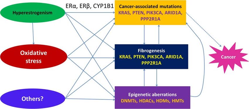
counterpart. The epithelial and stromal cellular components develop in a dependent and co-evolving manner. Genes involved in CAMs are likely
to be active players in lesional fibrogenesis, and hyperestrogenism and oxidative stress are likely drivers of both CAMs and fibrogenesis. Finally,
endometriotic lesions harbouring CAMs would conceivably be more refractory to medical treatment, due, in no small part, to their high fibrotic
content and reduced vascularity and cellularity.
WIDER IMPLICATIONS: The accumulating data on CAMs in endometriosis have shed new light on the pathogenesis and pathophysiology
of endometriosis. They also suggest new challenges in management. The distinct yet co-evolving developmental trajectories of endometriotic
stroma and epithelium underscore the importance of the lesional microenvironment and ever-changing cellular identity. Mutational profiling
of normal endometrium from women of different ages and reproductive history is needed in order to gain a deeper understanding of the
pathogenesis. Moreover, one area that has conspicuously received scant attention is the epigenetic landscape of ectopic, eutopic and normal
endometrium.
Key words: bone-marrow-derived stem cells / cancer-associated mutation / developmental trajectory / endometriosis / endothelial–
mesenchymal transition / epithelial–mesenchymal transition / fibrogenesis / mesothelial–mesenchymal transition / pathogenesis /
pathophysiology
.
Introduction .
.
. into cancer is near zero (Saavalainen et al., 2018; Bulun et al., 2019).
Endometriosis is a benign gynaecological disease characterized by the .
. Thus, it came as a complete surprise when Anglesio et al. reported in
ectopic deposition of endometrial-like tissues outside of the uterine .
. 2017 that the majority (79%) of DE lesions harbor somatic mutations
.
cavity. It has three major subtypes, namely ovarian endometrioma . and a sizeable portion of them (26%) contain known cancer-driver
.
(OE), deep endometriosis (DE) and superficial peritoneal endometrio- . mutations on genes coding for AT-rich interactive domain-containing
.
sis (PE) (Nisolle and Donnez, 1997). Featuring elevated local oestrogen . protein 1A (ARID1A), phosphatidylinositol-4,5-bisphosphate 3-kinase,
.
production and inflammation, endometriosis is one of the major con- . catalytic subunit α (PIK3CA), Kirsten rat sarcoma (KRAS), or protein
.
tributors to dysmenorrhea, infertility and chronic pelvic pain, impacting .
. phosphatase 2 scaffold subunit Aα (PPP2R1A) in the epithelial, but not
.
negatively on the quality of life in afflicted women (Vercellini et al., . the stromal, component (Anglesio et al., 2017). Although the authors
.
2014). Although OE is reported to be linked with increased risk of . were careful and did not say that the cancer-driver mutations in DE are
.
ovarian cancer (OVCA) of certain histotypes (Kurman and Shih Ie, . synonymous with increased risk of developing cancer, the connotation
.
2010; Pearce et al., 2012; Saavalainen et al., 2018), the magnitude . of ‘cancer-associated mutations’ (CAMs) is nonetheless unnerving and
.
of elevated risk is fairly moderate and the resultant absolute risk . somewhat unsettling or even alarming.
.
.
of OVCA is still low (Pearce et al., 2012; Guo, 2015; Saavalainen . Since the report by Anglesio et al. (2017) four more studies
.
et al., 2018). For extraovarian endometriosis, the risk of developing . have been published. While the study by Suda et al. reportedCancer -driver mutations in endometriosis 3
.
that 100% of OE lesions harboured somatic mutations and the .
. Mutations and CAMs
majority of them also carried CAMs (Suda et al., 2018), another .
. in endometriosis
Downloaded from https://academic.oup.com/humupd/advance-article-abstract/doi/10.1093/humupd/dmz047/5802383 by Dokuz Eylul University Library (DEU) user on 10 April 2020
study reported only 3% of OE had CAMs (Zou et al., 2018). Lac .
.
et al. reported 10% of iatrogenic endometriosis (IE) and 22% of . Somatic mutations, defined as permanent and irreversible changes of
.
DE carried CAMs (Lac and Huntsman, 2018). Noe et al. reported . the nucleotide sequence that are different from the host’s germline,
.
.
that all six non-superficial endometriosis carried somatic mutations, . in endometriotic lesions are nothing new. Various forms of muta-
.
and that the mutations were significantly enriched in epithelial but . tions, such as chromosomal aneuploidy (Shin et al., 1997; Kosugi et al.,
.
not stromal components of all lesions, suggesting that epithelium . 1999), loss of heterozygosity (LOH) (Sato et al., 2000) and copy
.
is clonal and its development is independent of stroma (Noe et al., . number changes/genomic alterations (Gogusev et al., 1999; Wu et al.,
.
2018). . 2006a,b,c; Yang et al., 2013) have been reported since the 1990s.
.
Collectively, these studies shed new light on the pathogenesis and .
. Negative findings also have been reported (Rai et al., 2010; Saare et al.,
.
pathophysiology of endometriosis. As the circle of our knowledge . 2012).
.
expands, however, more questions can be raised. .
.
Why is there such a large discrepancy in reported mutation .
. Somatic mutations and cancer-driver
frequencies? How can we reconcile such a discrepancy? Why does .
.
ectopic endometrium have a higher mutation rate than that of eutopic .
.
mutations
.
endometrium? Would the occurrence of CAMs in endometriotic . Developed from a single fertilized egg, which is essentially just one
.
lesions render an increased risk of cancer to their carriers? How . single cell, a human being consists of 1013 –1014 cells in his/her body
.
often do CAMs occur? Do all patients with endometriosis, deep . thanks to successive mitoses or cell divisions (Bianconi et al., 2013).
.
or otherwise, have CAMs in lesions sooner or later? What clinical . In its entirety, the 3.1 billion basepairs of human genome comprise
.
implications, if any, do the CAMs have for the bearers? When a patient . the entire nucleic acid sequence encoded as DNA within the 23
.
with endometriosis is found to have CAMs, should she be concerned or . chromosome pairs in the cell nuclei and a small amount of DNA
.
.
worried? OE is now well documented as being linked with OVCA, but . sequence (0.054 of the genome) in the mitochondria.
.
why does extraovarian endometriosis seldom lead to cancer? Do these . In normal physiological conditions, cells in different organs/tissues
.
CAMs tell us anything about the pathogenesis and/or pathophysiology . in humans are constantly experiencing turnover. This renewal process
.
of endometriosis? Are there, if any, limitations in these studies? Finally, . maintains the cellular homeostasis and is vital to the health of an organ-
.
what kind of future research is needed so that we can build upon . ism. Depending on the worn-out rate of the organs/tissues, some
.
our knowledge and further unveil some long-standing mysteries and . organs/tissues, such as intestinal epithelium, have a faster turnover,
.
.
conundrums in endometriosis? . with new cells replenished within days. Other organs/tissues, such as
.
In this paper, I will provide a comprehensive overview on the cur- . the brain, have a much slower turnover and remain mostly in dormancy.
.
rent status of somatic mutations, especially CAMs, in endometriosis, . During development or in adulthood, mitosis entails DNA replica-
.
identify possible causes of the CAMs, explain why there is discrepancy . tion. While the replication mechanism has a very high fidelity, with
.
among studies, describe what these reported CAMs tell us about the . the mutation rate in the order of 10−8 –10−7 per basepair per cell
.
.
pathogenesis and/or pathophysiology, and elaborate on the possible . division (Nachman and Crowell, 2000; Araten et al., 2005), mutation
.
clinical implications of CAMs. Above all, I shall address the questions . is still bound to occur given the enormous size of the genome and
.
raised above and expose areas in need of further research so that . the number of replications. In just one cell division, the chance that
.
we can learn more about the pathogenesis and/or pathophysiology .
. no mutation occurred is ∼3.4 × 10⁻¹⁴ assuming a mutation rate of
of endometriosis. . 10−8 per basepair per division. This miniscule probability amounts to
.
. tossing a fair coin consecutively 45 times and getting the head up all
.
.
. the time.
Methods .
. Hence, mutagenesis is essentially stochastic in normal physiological
.
PubMed was searched for all peer-reviewed original and review . conditions and inevitable. Because mutation occurs as a replication
.
articles related to CAMs in endometriosis published in English from . error, aging, which is intimately linked with an increasing number of cell
.
its inception to September 2019. The literature search was performed . divisions, is an important factor in causing genomic mutations (Rozhok
.
using main terms ‘endometriosis and CAM’, and ‘endometriosis and . and DeGregori, 2016). In fact, it has been shown that the total number
.
.
cancer-driver mutation’. In addition, PubMed was also searched . of stem cell divisions, which varies greatly among different tissue types,
.
using the keywords and MeSH terms ‘somatic mutations’, ‘fibrosis’, . is highly correlated with cancer risk. This explains why the incidence of
.
‘fibrosis and epigenetic’, ‘CAMs and tumorigenesis’, ‘somatic mutation . certain types of cancer, such as colorectal cancer, is much higher than
.
and normal tissues’, ‘oestrogen receptor and fibrosis’, ‘oxidative . others, such as stomach cancer (Tomasetti and Vogelstein, 2015). Since
.
stress and fibrosis’, ‘ARID1A mutation’, and ‘KRAS mutation and . human endometrium is a highly regenerative tissue undergoing monthly
.
.
therapeutics’. All retrieved papers were carefully assessed and, when . cycles of growth, differentiation and shedding during a woman’s repro-
.
relevant, incorporated into the review results. Their reference lists . ductive life, and since stem cells are involved in endometrial regen-
.
were also checked to identify any other study that could be relevant . eration (Gargett et al., 2016), spontaneous mutations are bound to
.
to this review. The eligibility of the retrieved studies was based . occur in normal endometrium, and this may explain, at least in part,
.
mainly on abstract. The decision as whether or not the study was . the mutations found in eutopic endometrium (Suda et al., 2018).
.
included in this review was made after careful assessment of its . In addition, when the organism/organs/tissues are exposed to an
.
.
content. . adverse environment, such as mutagenic chemicals/agents, UV light,4 Sun-Wei Guo
.
radiation, oxygen radicals, persistent inflammation and other deleteri- . corrected NGS technologies indicate that the CAMs prevalence in
.
ous factors, mutagenesis may be further accelerated (Cogliano et al., . adults is nearly 100% (Krimmel et al., 2016; Young et al., 2016).
.
Downloaded from https://academic.oup.com/humupd/advance-article-abstract/doi/10.1093/humupd/dmz047/5802383 by Dokuz Eylul University Library (DEU) user on 10 April 2020
2011) leading eventually to DNA damage, inactivation of tumour . In addition, since different cell types in the same tissue often have
.
suppressor genes and oncogene activation. Moreover, similar to the . distinct developmental trajectories and the proportions of different
.
evolution of species, cells with different genetic variants are under . cell types in the same tissue vary greatly in different people, the use
.
.
selective pressure: those with a high explicative fitness would eventually . of microdissection in harvesting the desired cell type greatly increases
.
outnumber cells with lower fitness. As a result, the existence of somatic . the signal-to-noise ratio and helps to detect the true mutations in a
.
mosaicism is fully documented in humans, and has been viewed as an . particular cell type. For example, before the use of microdissection,
.
aging phenotype (De, 2011; Risques and Kennedy, 2018). . the clonality of endometriotic epithelial cells could not be unequivocally
.
Not all mutations are deleterious. In fact, the majority of mutations . determined in about 18–40% of the cases due to cell contamination,
.
are harmless and have no impact on functionality or behaviours of . but with microdissection the clonality can be determined in all cases
.
.
the cell that bears the mutation and, as such, these mutations are . (Wu et al., 2003).
.
accumulated passively (Martincorena et al., 2017). But every now and . This can explain why studies that did not use microdissection often
.
then, an important gene is mutated, and the cells bearing the mutation . reported a much lower somatic mutation rate. For example, Vester-
.
have a competitive edge over other cells, such as higher proliferative or . gaard et al. reported that only 1 (4.3%) out of 23 patients with
.
survival propensity, resulting in the gradual and progressive domination . endometriosis was found to harbour mutations (Vestergaard et al.,
.
.
of the cells with that mutation. Such cells, called the mutant clones, may . 2011) (Table I). Similarly, Zou et al. reported that 3 (3.0%) of 101
.
acquire and accumulate further mutations and are the origin of cancer . OEs were found to have CAMs (Zou et al., 2018). Consequently,
.
cells (Fialkow, 1976; Nowell, 1976). . while these data appear to be genuine, the fact that the endometriotic
.
In contrast to ‘passenger’ mutations, which occur randomly and . epithelium and stroma apparently have different mutational profiles
.
confer no fitness to their bearers (Muller et al., 2012), cancer ‘driver’ . (Noe et al., 2018) renders their conclusions questionable.
.
mutations or CAMs are implicated in pathways that are critical in . Moreover, it also explains why studies using low-resolution detection
.
.
determining the proliferative, survival and metastatic propensity of . methods reported many fewer mutations. For example, using PCR in
.
tumour cells (Kato et al., 2016). Thus, CAMs are thought to be rare . combination with denaturing gradient gel electrophoresis (DGGE) on
.
in benign conditions such as endometriosis, are present mostly in pre- . nine cancer-associated genes, Vestergaard et al. reported that only 1
.
malignancy and are most frequent in metastatic cancer or those with a . (4.3%) out of 23 patients with endometriosis was found to harbour
.
metastatic potential (Kato et al., 2016). . mutations (Vestergaard et al., 2011) (Table I). In contrast, using the
.
. whole-exome sequencing method (much more accurate than DGGE),
.
.
. Li et al. found all 16 (100%) patients with OE harboured various somatic
Detection of somatic mutations .
. mutations (Li et al., 2014).
.
Somatic mutations are traditionally detected by many, mostly low- . Therefore, whether or not microdissection is used, the accuracy
.
resolution, methods. For detecting copy number changes, cytogenetic . of the detection method (so that low-abundance mutations can be
.
approaches such as multi-colour fluorescence in situ hybridization . detected) and the scope of detection (detecting specific mutations
.
.
(FISH), conventional or array-based comparative genomic hybridiza- . or unbiased whole-exome or whole-genome sequencing) should
.
tion (CGH) and LOH analysis are often used. For detecting gene . largely determine the mutation frequency. The difference in detection
.
mutations, genotyping and Sanger sequencing are often used. FISH, . methodology accounts for the discrepancy in reported mutation rates.
.
CGH, LOH, genotyping and Sanger sequencing are of low resolution .
.
and of higher error rate, and, as such, can only detect mutations in .
. Somatic mutations in tumours and in
a few predetermined loci or in large chromosomal segments, and are .
.
restricted to detecting variant allele frequencies typically higher than
.
. normal tissues
.
10% (Strom, 2016; Risques and Kennedy, 2018). . Somatic mutations used to be thought to occur exclusively in patho-
.
With the advent, and particularly the increasing affordability, of . logical tissues such as cancer, pre-neoplastic tissues, or normal tissue
.
the next generation sequencing (NGS) technologies, the resolution . adjacent to tumours. As mutation detection techniques become more
.
of detection has increased dramatically and the error rate has been . accurate, affordable and higher resolution, it becomes evident that
.
reduced substantially. The new error-correction NGS can further . spontaneous somatic mutations or genomic alterations can, and do,
.
.
increase the resolution and reduce the error rate, making the detection . occur in apparently normal tissues and benign conditions (Kato et al.,
.
of low-frequency mutations much easier and more accurate. Currently, . 2016; Risques and Kennedy, 2018). Notably, use of the microdissection
.
error-corrected NGS technologies can detect mutations in the 0.001– . technique and elimination of sequencing errors greatly increases the
.
0.1% range (Risques and Kennedy, 2018). Consequently, as the detec- . detection accuracy, permitting detection of low-frequency mutations
.
tion resolution increases, higher and higher somatic mutation rates . in apparently normal or healthy tissues/people (Krimmel et al., 2016;
.
.
have been reported in some benign diseases and even in tissues that . Dong et al., 2017) that would otherwise be missed using the older
.
are physiologically normal (Risques and Kennedy, 2018). This dramatic . sequencing methods. In addition, mutations are found to be increas-
.
improvement in detection accuracy has fundamentally changed our . ingly more abundant in aging tissues, indicating that aging or the number
.
views on somatic mutation burdens, mutational signatures, structural . of replications is a major driving force in generating mutations (Kinde
.
variants and the frequency of CAMs in apparently normal individuals. . et al., 2011; Schmitt et al., 2012; Hsieh et al., 2013; Blokzijl et al., 2016;
.
For example, older studies using NGS technology reported CAMs in . Hoang et al., 2016; Nair et al., 2016; Mattox et al., 2017; Martincorena
.
.
about 10% of individuals older than 65 years, but studies using error- . et al., 2018). Within the same individual, there is a substantial variationTable I Summary results of studies reporting CA mutations in endometriosis.
Study Type of Sample size Patient age Detection method Names of mutated Percentage of Major findings Microdissection
endometriosis (years) genes patients who had (epithelium)
mutations
...........................................................................................................................................................................................................................................
Vestergaard Not reported N = 23 31.0 ± 5.2 9 CA genesPCR in BRAF, HRAS, NRAS, 1/23 = 4.3% No. Mixed cell
et al. (2011) combination with CTNNB1, CDK4, populations
denaturing gradientgel FGFR3, PIK3CA, TP53
electrophoresis (DGGE) and PTEN
Sensitivity: ∼5%
Cancer -driver mutations in endometriosis
Li et al. (2014) OE N = 16 32.4 ± 6.2 Whole-exome 100% C-T Mutations sig. Yes
sequencing (Estimated to higher Eu and Ec have
be able to detect 76% of distinctive mutation
existing mutation) profiles
Anglesio et al. DE N = 24 36.7 ± 7.4 Exome-wide sequencing various 19 (79.2%) 5 (20.8%) Only in epithelium Yes
(2017) cancer-driver
Suda et al. OE N = 13 42.9 ± 11.0 whole-exome Various 13 (100.0%) Driver Discordant mutational Yes
(2018) sequencing mutations: 10 (76.9%) profiles between
eutopic and ectopic
endometrium is found
Lac et al. Iatrogenic N = 40 N = 36 36.5 ± 5.5 Targeted sequencing 33 genes (exons and hot 4 (10.0%) 8 (22.2%) Yes
(2018) endometriosis 33.9 ± 7.0 spots)
and DE
Zou et al. OE N = 101 32 (median) PCR amplification of the KRAS, PPP2R1A, 3 (3.0%) KRAS p.G12 V, No
(2018) potential mutational PIK3CA, BRAF, NRAS, PPP2R1A p.S256F,
hotspot regions of HRAS, ERK1, ERK2 and ARID1A
KRAS, PPP2R1A, PTEN genes, as well as
PIK3CA, BRAF, NRAS, the entire coding region
HRAS, ERK1, ERK2 and and corresponding
PTEN genes intron/exon boundaries
of the ARID1A gene.
Then sequencing and
compared against the
DNA derived from
PBMC.
Noe et al. Mixed DE: n = 5 OE: 40.3 ± 8.8 Exome sequencing 6 (100%) Significantly enriched Yes
(2018) n=1 mutations in epithelial
but not in stromal
components
CA: cancer associated; DE: deep endometriosis; Ec: ectopic endometrium; Eu: eutopic endometrium; OE: ovarian endometrioma; PBMC: peripheral blood mononuclear cell, ARID1A: AT-rich interactive domain-containing protein
1A; BRAF: B-Raf proto-oncogene, serine/threonine kinase; CDK4: cyclin-dependent kinase 4; CTNNB1: catenin β 1; ERK1: extracellular signal-regulated kinase 1; ERK2: extracellular signal-regulated kinase 2; FGFR3: fibroblast growth
factor receptor 3; HRAS: HRas proto-oncogene, GTPase; KRAS: KRAS proto-oncogene, GTPase; NRAS: NRAS proto-oncogene, GTPase; PIK3CA: phosphatidylinositol-4, 5-bisphosphate 3-kinase, catalytic subunit α ; PPP2R1A: protein
phosphatase 2 scaffold subunit Aα ; PTEN: phosphate and tension homology deleted on chromsome ten; TP53: tumour protein p53.
5
Downloaded from https://academic.oup.com/humupd/advance-article-abstract/doi/10.1093/humupd/dmz047/5802383 by Dokuz Eylul University Library (DEU) user on 10 April 20206 Sun-Wei Guo
.
in genomic alterations among different tissues, with more alterations . iatrogenic procedures reports that 51–64% of women carry CAMs in
.
seen in tissues with a fast turnover and in genes involved in cell regula- . their endometrium without any evidence of malignancy or even subtle
.
Downloaded from https://academic.oup.com/humupd/advance-article-abstract/doi/10.1093/humupd/dmz047/5802383 by Dokuz Eylul University Library (DEU) user on 10 April 2020
tion (O’Huallachain et al., 2012). Moreover, these age- or replication- . pathology, with KRAS (28.2%), PIK3CA (12.7%), and phosphate and
.
related mutations are often CAMs (Martincorena et al., 2015; Krimmel . tension homology deleted on chromsome ten (PTEN), PTEN (27.3%)
.
et al., 2016; Martincorena et al., 2018). That is, somatic mutation and . being the most common (Lac et al., 2019). Consistently, the results
.
.
even CAMs can, and do, occur in apparently healthy or normal tissues. . showed that the mutation rate is a linear function of age, with the
.
Noteworthy is that massive mutations in physiologically normal . likelihood of harbouring a mutation in endometrial tissue increased by
.
tissues have been detected when deep sequencing methods are used . 5% per year, independent of menstrual phase (Lac et al., 2019).
.
(Martincorena et al., 2015; Martincorena et al., 2018). Healthy cells in . Using microdissection and NGS whole-genome sequencing method,
.
the oesophageal epithelium, for example, carry at least several hundred . another study reports mutation burden, and signatures and CAMs on
.
mutations per cell in people in their 20s or over 2000 mutations in . 215 histologically normal endometrial glands isolated from 18 women
.
.
older people (Martincorena et al., 2018), suggesting that replicative . (Moore et al., 2018). Remarkably, it finds that, in normal endometrial
.
mutations accumulate with age. In addition, in tissues exposed to . glands, there is an average of 1324 base substitutions and an average
.
the sun, large numbers of mutant clones under positive selection . of 85 insertions/deletions (indels) per woman (Moore et al., 2018).
.
are found, and the signatures of the mutations are consistent with . Again, the mutation rate correlated linearly with age, accumulating
.
the DNA damage induced by UV rays (Martincorena et al., 2015), . ∼28 base substitutions per gland per year during adult life and an
.
.
highlighting the impact of environmental exposure on somatic muta- . extra 20 substitutions with each increasing unit of BMI (Moore et al.,
.
tions. Many of these mutations are CAMs, which confer selective . 2018). Many of these mutations occur early in life, and different
.
advantages. Surprisingly, the prevalence of NOTCH1 (a cancer-driver . mutational processes appear to be operative (Moore et al., 2018).
.
gene) mutations in normal oesophagus is found to be several times . Nearly 95% of women evaluated are found to harbour various CAMs in
.
higher than in oesophageal cancers (Martincorena et al., 2018). While . their endometrial glands, including KRAS, PIK3CA, phosphoinositide-
.
these clones appear to be a result of normal aging, it is likely that . 3-kinase regulatory subunit 1 (PIK3R1), Rho GTPase activating protein
.
.
they may acquire more genetic advantages over time and eventually . 35 (ARHGAP35), PPP2R1A and F-box and WD repeat domain con-
.
transform into malignancy (Martincorena et al., 2018). Indeed, a fitness . taining 7 (FBXW7), which have been reported to be present in ectopic
.
benefit of merely 0.4% over the time course of 20 years might be . endometrium (Anglesio et al., 2017, Suda et al., 2018) as well as in
.
enough to lead to malignant transformation (Bozic et al., 2010). Hence, . endometriosis-associated ovarian cancer (EAOC) (Kuo et al., 2009).
.
the presence of CAMs is not necessarily synonymous with cancer or .
.
tumour development. .
.
Of course, the mutational processes underlying normal aging also
.
. CAMs in endometriosis: what do they tell us?
.
are operative in tumorigenesis in a given organ or tissue (Kinde et al., . Given the above discussion, it seems evident that we can disregard,
.
2011; Schmitt et al., 2012; Hoang et al., 2016; Mattox et al., 2017; . without loss of much information, those studies reporting mutations in
.
Risques and Kennedy, 2018). So much so that half or more of somatic . endometriosis that did not use microdissection to harvest cells of the
.
mutations in tumours are estimated to have arisen before initiation of a . desired type and/or used low-accuracy detection methods. After this
.
.
tumour (Tomasetti et al., 2013), and that replication-induced mutations . screening, only five studies were considered to be trustworthy.
.
have been proposed to account for up to two-thirds of the mutations . Using whole-exome sequencing of endometriotic epithelial cells,
.
in human cancers even after adjustment for an environmental and . Li et al. reported all 16 patients with OE harbour various somatic
.
hereditary propensity for malignancy (Tomasetti et al., 2017). . mutations and identified frequent alterations in genes involved in cell
.
Extensive sequencing of adult stem cells of various organs with . adhesion and chromatin-remodelling complexes (Li et al., 2014). This
.
different cancer incidences has shown that these organs gradually . is the first study to show that all OE lesions seem to have somatic
.
.
accumulate mutations at very similar rates, but their mutation profiles . mutations (Table I). In addition, their pathway analyses using genes
.
vary from tissue to tissue (Blokzijl et al., 2016). Hundreds to thousands . found to be recurrently mutated identified chromatin remodelling as
.
of mutations are present in tumour cells and are shared by most or . one of the enriched functional groupings. In particular, their data sug-
.
all tumour cells, but the mutation burden correlates with patient’s age . gest that mutated genes encode a histone methyltransferase involved in
.
(Welch et al., 2012; Milholland et al., 2015). Consistently, the cancer . histone H3 lysine 4 (H3K4) modification, echoing previous reports of
.
incidence seems to be correlated with the number of stem cell divisions . aberrant H3K4 methylation in endometriosis (Xiaomeng et al., 2013;
.
.
across a wide variety of cancer types (Tomasetti and Vogelstein, . Monteiro et al., 2014; Sun et al., 2016). Their results thus lend support
.
2015). In fact, these age-associated or replication-driven mutations . for the notion that endometriosis can be characterized with epigenetic
.
appear to have particular mutational signatures (Alexandrov et al., . aberrations (Guo, 2009a,b).
.
2013; Alexandrov et al., 2015). The acquisition of CAMs apparently . The study by Anglesio et al. showed that in women with DE a sizeable
.
confers an explicative advantage, resulting in the clonal expansion of . portion of lesions contain known CAMs in the epithelial component
.
.
the founder cell (Vogelstein et al., 2013; Martincorena et al., 2018). . only (Anglesio et al., 2017) (Table I). Since DE is rarely reported to be
.
Consistent with the reported massive mutations in various physio- . associated with malignancy, this study presents results that are quite
.
logically normal tissues (Martincorena et al., 2015; Blokzijl et al., 2016; . unnerving. However, the clinical significance remains largely unclear.
.
Franco et al., 2018; Lee-Six et al., 2018; Martincorena et al., 2018), . The same group recently published another study on mutations in IE
.
a recent study employing targeted sequencing of hotspot regions . as well as DE (Lac et al., 2018). Here, IE refers to endometriotic lesions
.
in cancer-related genes in combination with immunohistochemistry . resulting from the surgical scars of previous obstetric or gynaecological
.
.
analysis on 110 women who had undergone either hysterectomy or . procedures. Using microdissection and a hypersensitive cancer hotspotCancer -driver mutations in endometriosis 7
.
sequencing panel, they found that 10% of IE and 22% of DE lesions . lesions that were in close physical proximity appeared to have similar
.
harbour CAMs. In addition, 18% of IE and 14% of DE lesions exhibited, . mutations, yet lesions located on the right and left ovaries displayed
.
Downloaded from https://academic.oup.com/humupd/advance-article-abstract/doi/10.1093/humupd/dmz047/5802383 by Dokuz Eylul University Library (DEU) user on 10 April 2020
by immunoreactivity, loss of PTEN, a tumour suppressive gene, in the . entirely different mutations, suggesting different developmental origins
.
epithelial component (Lac et al., 2018). Combining sequencing data and . (Suda et al., 2018). Individual endometrial glands within the normal
.
immunohistochemistry results, they reported an overall rate of CAMs . uterus of the same individual carried distinct somatic mutations. In
.
.
in 28% of IE and 36% DE lesions (Lac et al., 2018) (Table I), confirming . many ways, the study by Suda et al. (2018) has provided a greater
.
their previous study (Anglesio et al., 2017). In addition, it demonstrates . understanding of the spatiotemporal evolution of OE and a much-
.
the similarity of the two types of endometriosis (harbouring con- . needed glimpse at the unique mutational profiles of endometriotic
.
siderable CAMs) and their differences (somewhat different mutation . epithelium and uterine endometrial epithelium, and also demonstrated
.
profiles) (Lac et al., 2018). That is, CAMs are not exclusively confined . a clear explicative advantage of acquiring these CAMs.
.
to DE but can actually be seen in other subtypes of endometriosis . Based on these findings, Suda et al. offered a plausible explanation for
.
.
as well. . the pathogenesis of OE, which originates from eutopic endometrium
.
Noe et al. recently reported that, among 19 mutations sequenced in . that already carries CAMs that confer selective advantages once regur-
.
six patients with OE or DE, all were significantly enriched in epithelial . gitated into the peritoneal cavity through retrograde menstruation,
.
cells but not in stromal cells, suggesting that the evolution of non- . resulting in clonal expansion and ultimately causing symptoms (Suda
.
superficial endometriosis is not straightforward: epithelium is clonal and . et al., 2018). In other words, endometriosis originates from a defective
.
.
its development is independent of stroma (Noe et al., 2018). Using . endometrium that is harbouring CAMs.
.
droplet digital PCR analysis of microdissected epithelium- and stroma- .
.
enriched endometriosis tissues, they report that the 19 somatic pas- .
.
senger mutations analyzed were predominantly found in the epithelial .
compartment, in contrast to very few mutations in the stromal one
.
. Shedding light on pathogenesis
.
(Noe et al., 2018) (Table I). These findings are consistent with the .
. and pathophysiology
.
previous report that the endometriotic epithelial cells are monoclonal .
. Pathogenesis
(Wu et al., 2003), whereas stromal cells may be continuously regen- .
.
erated or recruited during lesional progression and development. This . In endometrium, each menstrual cycle is analogous to classic tissue
.
led to the conclusion that the evolution of endometriosis is complex, . injury and repair, which include inflammation, its resolution, angiogen-
.
in that epithelium is clonal and its development is independent of . esis, tissue formation and remodelling or re-epithelialization (Maybin
.
stroma (Noe et al., 2018). In the authors’ own words, the results . and Critchley, 2015). Each gland in the endometrium appears to be
.
.
‘do not support the views that endometriosis originate from a single . regenerated from a committed endometrial stem cell (Tanaka et al.,
.
stem/progenitor cell, which differentiate to both epithelial and stromal . 2003). Similar to eutopic endometrium, the ectopic endometrium
.
cells, or the epithelial cells differentiate into stromal cells through . sheds glandular epithelial cells during menstruation, but considerably
.
epithelial–mesenchymal transition at the site of endometriosis’ (Noe . less so in the endometriotic stromal cells, which also house recruited
.
et al., 2018). Lac and Huntsman (2018) further elaborated this point, . progenitor cells and transdifferentiated cells (see below).
.
.
proposing that endometriotic epithelium and stroma may have distinct . The study by Suda et al. has demonstrated beautifully and convinc-
.
developmental trajectories. In particular, they argue that, given the . ingly the power of sequencing in establishing the phylogenetic relation-
.
data, the most likely scenario is that progenitor cells undergo clonal . ship between two clones of cells. Based on their results, Suda et al.
.
expansion to give rise to epithelial cells, whereas stromal cells come . proposed that the endometrium of women with OE contains, prior
.
into being without clonal expansion (Lac and Huntsman, 2018). . to the formation of OE lesions, endometrial glands with pre-existing
.
By using a combination of microdissection, independent discovery . CAMs that may have selective advantages, which subsequently acquire
.
.
and validating samples, whole-exome and target-gene sequencing, mul- . more CAMs after successfully implanting onto the ectopic sites, which
.
tiregional sequencing of several sites (multiple lesions as well as eutopic . then go through clonal expansions (Figure S7 in (Suda et al., 2018)).
.
endometrium) from the same individuals, and single endometrial gland . However, caution should be exercised here. First, the finding of
.
sequencing, the study by Suda et al. is by far the most informative one . CAMs in endometrial glands is based on target-gene sequencing of
.
to date on CAMs in endometriosis, especially for OE. They reported . 109 single endometrial glands from the uteri of three women, aged
.
that epithelial cells within OE lesions exhibit extensive CAMs and . 38, 47 and 49 years old, respectively (Suda et al., 2018). Small sample
.
.
clonal expansion, and that the genomic architecture of epithelial cells . size aside, the three patients were older than the mode of the age
.
in uterine endometrium is heterogeneous (Table I). While overall the . at first surgery in women with OE (Liu et al., 2008). OE is frequently
.
OE lesions and normal endometrium had a similar number of somatic . diagnosed before 38 years of age and in some cases in adolescent girls
.
mutations per Mb sequenced, the mutational profiles in ectopic and . (Saridogan, 2017) even though a diagnostic delay is well documented
.
eutopic endometrium from the same patient were discordant (Suda . in endometriosis (Hadfield et al., 1996; Arruda et al., 2003). Given
.
.
et al., 2018). In addition, single endometrial glands carry distinct CAMs, . the somewhat ubiquitous age-related somatic mutations in healthy
.
even though the tissues appeared to be histologically benign and . organs/tissues (Risques and Kennedy, 2018), and since endometrium
.
normal (Suda et al., 2018). In general, the distributions of mutant allele . is a highly regenerative tissue that displays monoclonality in each
.
frequency (MAF) in endometriotic epithelium were higher than those . endometrial gland yet the entire endometrium exhibits a mosaic pat-
.
in uterine endometrial epithelium, and some endometriotic epithelium . tern of clonal distribution (Tanaka et al., 2003; Wu and Guo, 2008), it is
.
harboured arm-level allelic imbalances that are consistent with LOH . conceivable that normal endometrium, especially from older women,
.
.
in regions harbouring CAMs. One of their interesting findings is that . may harbour somatic mutations or even some CAMs. In fact, it has8 Sun-Wei Guo
.
been shown recently that the most common CAMs in apparently . glands with pre-existing CAMs, which are responsible for the genesis of
.
normal endometrium are KRAS and PIK3CA (Lac et al., 2019). The . endometriosis is questionable at least, especially because these CAMs
.
Downloaded from https://academic.oup.com/humupd/advance-article-abstract/doi/10.1093/humupd/dmz047/5802383 by Dokuz Eylul University Library (DEU) user on 10 April 2020
whole-genome sequencing study by Moore et al. also shows that many . are detected post hoc, after endometriosis has been diagnosed.
.
CAMs detected in the glands of endometrium, such as KRAS, PIK3CA, .
.
.
PIK3R1, ARHGAP35, FBXW7, fibroblast growth factor receptor 2 .
.
From distinct developmental trajectories to
(FGFR2), PP2R1A, PTEN, zinc finger homeobox 3 (ZFHX3), and . partners in crime
.
AT-rich interaction domain 5B (ARID5B), are all present in histo- .
.
logically normal endometrium from women without endometriosis . One question left unanswered in Noe et al. (2018) is: if endometriotic
.
or uterine fibroids (Moore et al., 2018). Thus, the proposal, based . stromal cells are under the same pressure of replication error as
.
on the post hoc evidence of CAMs in eutopic endometrium from . epithelial cells, why do they harbour much less mutations than the
.
women with endometriosis, that women with endometriosis or OE . latter? Lac and Huntsman speculate that ‘epithelial cells may be an
.
. integral process in the pathogenesis of endometriosis. Stromal cells,
have pre-existing CAMs before the genesis of endometriosis is simply .
.
premature, especially in the absence of any data suggestive of a phy- . in contrast, may play a more-supportive role in endometriosis, and
.
logenetic relationship between endometriotic lesions and the eutopic . are likely to be continuously regenerated or recruited to the site of
.
endometrium. . endometriotic lesions. It is possible that stromal cells may arise from
.
Second, while the suspicion that endometriosis may originate from . the continuous induction of metaplasia of surrounding cells to become
.
. endometrium-like stroma’ (Lac and Huntsman, 2018). They are correct
defective endometrium has long been raised (Vinatier et al., 2000), .
. in general, but perhaps a more complete account will help to shed
we need to understand that most, if not all, data on the endometrial .
.
aberrations are collected from patients who have already been surgi- . light on the development of endometriosis, probably more on its
.
cally and histologically diagnosed with endometriosis. In fact, data from . pathophysiology than on its pathogenesis.
.
several well designed animal studies of endometriosis consistently and . First, it turns out that, aside from the resident fibroblasts/stro-
.
unequivocally indicate that, once endometriosis is induced artificially, . mal cells, the stromal component in endometriotic lesions consists
.
. of fibroblasts and myofibroblasts, which can be recruited or trans-
the eutopic endometrium then acquires various molecular and histo- .
.
logical aberrations (Kim et al., 2007; Lee et al., 2009; Sherwin et al., . differentiated from several sources. First, they can be differentiated
.
2010; Naqvi et al., 2016; Kim et al., 2019). More remarkably, the extent . from endometriotic epithelial cells through epithelial–mesenchymal
.
of endometrial aberrations appears to depend on the proximity of . transition (EMT), currently an area of active research with over 80
.
endometriotic lesions to the uterus (Naqvi et al., 2016). These data . PubMed-indexed papers and counting (Matsuzaki and Darcha, 2012;
.
strongly suggest that the numerous endometrial aberrations in women . Zhang et al., 2016a,b). This may explain why the stromal component
.
. still shares some, but much fewer, mutations at some loci with the
with endometriosis may be more likely to be the consequence, rather .
.
than the cause, of endometriosis. . epithelial component, as observed in Noe et al. (2018). It could also
.
In fact, the data presented in the Suda et al. (2018) study actu- . explain why in some DE lesions the glandular epithelium is absent,
.
ally support this view: one, the mutation profiles between ectopic . yielding what is termed ‘stromal endometriosis’, that is, endometri-
.
endometrium and their eutopic counterpart are different (Fig. 1B; . otic lesions without glandular epithelium (Mai et al., 1997; Clement,
.
. 2007), which is reported to be seen in 27–45% of cases of PE
Tables S2 and S3 in (Suda et al., 2018)); and, two, the number of .
.
high MAF mutations per Mb is higher in endometriotic epithelium . (Abrao et al., 2003; Boyle and McCluggage, 2009; Kamergorodsky
.
than that in endometrial epithelium, even though KRAS and PIK3CA . et al., 2009), 0–13% of OE (Abrao et al., 2003; Kamergorodsky et al.,
.
are the most frequently mutated genes in both ectopic and eutopic . 2009) and 12–15% of DE (Abrao et al., 2003; Kamergorodsky et al.,
.
endometrium (Fig. 1B and Fig. S1B in (Suda et al., 2018)). Should . 2009).
.
mutations in eutopic endometrium be responsible for the genesis and . Second, the bone-marrow-derived stem cells (BMDSCs) can be
.
. recruited into the stroma. In fact, in a mouse model of endometriosis
formation of ectopic endometrium, the endometriotic lesions can be .
.
considered as a de facto clonal expansion of its eutopic ancestry, and . that received bone marrow transplantation, it is found that approxi-
.
the MAF of the mutated genes in eutopic endometrium would be . mately 0.1% of stromal cells and 0.04% of epithelial cells in lesions are
.
no lower than that of the ectopic endometrium. Of course, since . of donor origin (Du and Taylor, 2007). In other words, the stroma
.
endometrium is polyclonal (Tanaka et al., 2003), and since only a few . and epithelium recruited BMDSCs in the ratio of 2.5:1. In another
.
endometrial samples were harvested and sequenced in the study (Suda . study, most BMDSCs were found in endometriotic stroma in mice with
.
.
et al., 2018), it is possible that the eutopic endometrial tissue samples . induced endometriosis (Ersoy et al., 2017). Thus, the lesional stromal
.
sequenced may not be the clone that was descended from the one . component recruits more BMDSCs that naturally contain less somatic
.
that caused endometriosis. Regardless, however, the data presented in . mutations than the epithelial component.
.
Suda et al. (2018) are insufficient to conclude that endometrial CAMs . Third, endometriotic stromal cells also can be transdifferentiated
.
predate the genesis of endometriosis. . from other cells, such as endothelial cells through endothelial–
.
.
Hence, based on the very recent data on the mutational burden and . mesenchymal transition (EndoMT) or mesothelial cells through
.
CAMs in histologically normal endometrium (Moore et al., 2018; Lac . mesothelial–mesenchymal transition (MMT), as shown in a recently
.
et al., 2019) and the evidence that eutopic endometrium apparently . established mouse model of deep endometriosis (Yan et al., 2017)
.
acquires molecular aberrations after the induction of endometriosis . (also Yan et al., unpublished data). They might also be transdifferen-
.
(Kim et al., 2007; Lee et al., 2009; Sherwin et al., 2010; Naqvi et al., . tiated from cells other than endothelial and mesothelial cells, such as
.
2016; Kim et al., 2019), the notion that the endometrium of women . pericytes, or fibrocytes and perhaps monocytes (Mack and Yanagita,
.
with endometriosis contains, prior to the lesion formation, endometrial .
. 2015).Cancer -driver mutations in endometriosis 9
.
. However, the seemingly independent developmental trajectories
.
. should not be construed to mean that the two components go separate
.
Downloaded from https://academic.oup.com/humupd/advance-article-abstract/doi/10.1093/humupd/dmz047/5802383 by Dokuz Eylul University Library (DEU) user on 10 April 2020
. ways, leaving the other completely alone. On the contrary, it is well
.
. known that the function and morphogenesis of endometrial epithelial
.
. cells are regulated by paracrine effectors secreted by stromal cells
.
.
. (Arnold et al., 2001).
.
. One particularly important player involved in the paracrine effect is
.
. the exosomes secreted by endometriotic stromal and epithelial cells.
.
. For example, exosomes secreted by epithelial cells induce myofibrob-
.
. last transformation in stromal cells and also neovascularization (Han
.
. et al., 2017). Likewise, stroma-derived exosomes can also promote
.
.
. epithelial wound healing (Samaeekia et al., 2018). Exosomes derived
.
. from endometriotic stromal cells are reported to enhance angiogenesis
.
. (Harp et al., 2016).
.
. Endometriotic epithelial cells, on the other hand, express many
.
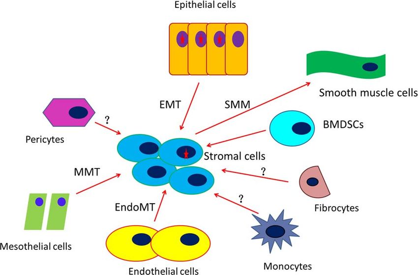
Figure 1 Schematic illustration that explains the distinct . growth factors and chemokines that are responsible for the migration,
.
developmental trajectories of endometriotic stroma and .
. proliferation and activation of fibroblasts. For example, transforming
epithelium. In essence, in contrast to endometriotic epithelial cells .
. growth factor (TGF)-β1 is most prominently expressed in the epithelial
that experience turnover, the stromal component of endometriotic .
. component of lesions (Chegini et al., 1994; Tamura et al., 1999). The
lesions recruits other cells, such as endothelial cells, mesothelial cells .
. increased levels of TGF-β1, the archetypal profibrotic molecule, may
and epithelial cells, through the endothelial–mesenchymal transition .
. induce fibroblast-to-myofibroblast transdifferentiation (FMT) in stro-
(EndoMT), mesothelial–mesenchymal transition (MMT) and epithe- .
. mal cells (Zhang et al., 2016a,b,c). Therefore, the passenger mutations
lial–mesenchymal transition (EMT). In addition, the stromal compo- .
.
nent may recruit bone marrow-derived stem cells (BMDSCs), peri- . exclusively enriched in endometriotic epithelial component, as found
.
cytes, fibrocytes and monocytes. Moreover, outflow of stromal cells . in Noe et al. (2018), do suggest distinct developmental trajectories of
.
due to smooth muscle metaplasia (SMM) can cause the loss of older . endometriotic epithelium and stroma, but the trajectories are by no
.
stromal cells. Cells with a red start indicate that these cells carry . means independent. They are actually dependent and co-evolve in the
.
cancer-associated mutations (CAMs). The red arrows indicate the . development of endometriosis.
.
direction of transdifferentiation and/or recruitment. The question . But why do normal and eutopic endometrial epithelium have much
.
mark indicates the uncertainty towing to lack of evidence. .
. lower mutation rates, as in ectopic endometrial epithelium, as shown in
.
. Suda et al. (2018)? There are several possibilities. First of all, BMDSCs
.
. have been shown to give rise to multiple endometrial cell types,
.
Lastly, in addition to the inflow of cells that are typically devoid of . including stromal, glandular and luminal epithelial cells (Taylor, 2004;
.
CAMs, there is also an outflow of cells from the stromal component . Du and Taylor, 2007). These BMDSCs presumably harbour much less
.
.
due to smooth muscle metaplasia, i.e. stromal cells eventually transdif- . mutations when recruited to the endometrial epithelial component.
.
ferentiated into smooth muscle cells. This would effectively move those . Second, in normal endometrium, the tissue regeneration in each
.
older, originally stromal cells, out of the stromal component since these . menstrual cycle involves mesenchymal–epithelial transition (MET); i.e.
.
cells now become smooth muscle cells. . stromal cells are transdifferentiated into epithelial cells and contribute
.
As myofibroblasts are the major effector cells of fibrogenesis, the . to the ‘re-epithelialization’ as part of the repair process (Huang et al.,
.
ever-increasing stromal component would result in progressive fibro- . 2012; Patterson et al., 2013; Cousins et al., 2014). Hence, the epithelial
.
.
genesis, which, in turn, leads to increased matrix stiffness but reduced . component in eutopic endometrium receives new recruits in each and
.
vascularity and cellularity (Liu et al., 2018a,b). The increased matrix . every cycle. In contrast, the MET in ectopic endometrium appeared to
.
stiffness has been shown to attenuate EMT (Matsuzaki et al., 2017), . be much diminished (Matsuzaki and Darcha, 2012). Since the endome-
.
which reduces the number of stromal cells differentiated from epithelial . trial stromal component may have recruited BMDSCs and thus have
.
cells (which may or may not carry CAMs already). The reduced EMT . much lower mutations than their epithelial counterpart, as eluded
.
effectively cuts the supply of CAM-carrying epithelial cells to the . to above, the transformed epithelial cells through MET in eutopic
.
.
stromal component. This, coupled with supplementation of other cells . endometrium presumably should have less mutations than the original
.
that are devoid of CAMs to the component, may explain why there . epithelial cells in ectopic endometrium, which are the descendants of
.
is a much lower mutation frequency in the stromal component (Noe . successive cell divisions, essentially devoid of any replenishment from
.
et al., 2018) as the component also recruits endothelial, mesothelial . other sources.
.
and other cells. . Third, ectopic endometrium faces a much harsher microenviron-
.
.
In view of the above, the stromal component of endometriotic . ment, comprising a DNA-damaging milieu. Fuelled by increased local
.
lesions is formed from multiple sources: endometriotic epithelial cells, . oestrogen production and chronic inflammation, endometriotic lesions
.
endothelial cells, mesothelial cells, BMDSCs and perhaps other cells as . are known to have excess proliferative potential (Hapangama et al.,
.
well. These cells may intrinsically have much lower mutation rates than . 2010). Yet chronic inflammation is intimately linked with increased
.
that of endometriotic epithelial cells, giving rise to the results reported . oxidative stress, which is manifested by the increased production of
.
in Noe et al. (2018). A diagram depicting the scenario described here . reactive oxygen species (ROS) as well as reduced ROS detoxification
.
.
is shown in Figure 1. . (Alexandre et al., 2006). Cellular proliferation also is closely correlatedYou can also read