Surgical Aspects and Prognostic Factors in the Management of Rectal Cancer
←
→
Page content transcription
If your browser does not render page correctly, please read the page content below
Digital Comprehensive Summaries of Uppsala Dissertations
                from the Faculty of Medicine 1764

                Surgical Aspects and Prognostic
                Factors in the Management of
                Rectal Cancer

                KEVIN AFSHARI

    ACTA
UNIVERSITATIS
 UPSALIENSIS    ISSN 1651-6206
                ISBN 978-91-513-1268-2
  UPPSALA       URN urn:nbn:se:uu:diva-450799
    2021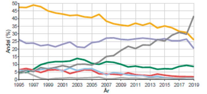
Dissertation presented at Uppsala University to be publicly examined in Centrum för klinisk
forskning Västerås,, Ingång 29, Västmanlands sjukhus, Västerås, Friday, 8 October 2021 at
13:00 for the degree of Doctor of Philosophy (Faculty of Medicine). The examination will
be conducted in Swedish. Faculty examiner: Associate Professor Per J. Nilsson (Karolinska
Institute).

Abstract
Afshari, K. 2021. Surgical Aspects and Prognostic Factors in the Management of Rectal
Cancer. Digital Comprehensive Summaries of Uppsala Dissertations from the Faculty of
Medicine 1764. 73 pp. Uppsala: Acta Universitatis Upsaliensis. ISBN 978-91-513-1268-2.

Survival among patients with stage IV rectal cancer is poor and surgical treatment for this disease
is associated with morbidities such as small bowel obstruction, complications with a diverting
loop ileostomy, and functional bowel disturbances. The overall aim of this thesis was to assess
risk factors and morbidity after surgery for rectal cancer and to evaluate factors affecting survival
in patients with stage IV rectal cancer.
   Paper I a prospective study on patients with rectal cancer with loop ileostomy who
underwent stoma closure in a 23-hour hospital stay setting. Results were compared with a
group who underwent standard in-hospital stoma closure prior to the start of the study, selected
retrospectively as controls. No differences were found in the number of complications or the
frequency of re-hospitalization or re-operation, indicating that ileostomy closure in a 23-hour
hospital stay setting in these selected patients was feasible and safe with high patient satisfaction.
   Paper II a population-based study with data gathered prospectively. In total, 11% of the
patients developed small bowel obstruction (SBO), mostly during the first year after rectal
cancer surgery. Surgical treatment for SBO was performed in 4.2% of the patients, and
the mechanism was stoma-related in one-fourth. Rectal resection without anastomoses, age,
morbidity, and previous radiotherapy (RT) was not associated with admission to the hospital
or surgery for SBO. Re-laparotomy due to complications after rectal cancer surgery was an
independent risk factor for admission for treating SBO.
   Paper III a population-based study with data gathered prospectively on bowel function at
1 year after anterior resection or stoma reversal. No associations were found between any
defecatory dysfunction and the part of the colon used for anastomosis, the level of the vascular
tie, or gender. An association was observed between higher anastomotic level and a lower risk
of incontinence and clustering. At 1 year after loop ileostomy closure, the risks of incontinence,
clustering, and urgency increased by up to fourfold.
   Paper IV a case-control study aiming to identify patient-, tumor-, and treatment-related
prognostic factors for 5-year survival in patients with rectal cancer with synchronous stage IV
disease. Patient-related factors did not differ between groups. Among the tumor-related factors,
multiple site metastases, bilobar liver metastases, and increasing numbers of liver metastases
were associated with poor survival. Prognostic treatment-related factors were preoperative RT,
metastasectomy, and radical resection of the primary tumor. The most important prognostic
factor for long-term survival was metastasectomy.

Keywords: Loop ileostomy, Small bowel obstruction, Defecatory dysfunction, Stage IV rectal
cancer

Kevin Afshari, Centre for Clinical Research, County of Västmanland, Västmanlands sjukhus
Västerås, Uppsala University, SE-72189 Västerås, Sweden.

© Kevin Afshari 2021

ISSN 1651-6206
ISBN 978-91-513-1268-2
URN urn:nbn:se:uu:diva-450799 (http://urn.kb.se/resolve?urn=urn:nbn:se:uu:diva-450799)
To my wife Pernille and daughter Ellie
List of Papers

This thesis is based on the following papers, which are referred to in the text
by their Roman numerals.

      I.   Afshari K, Nikberg M, Smedh K, Chabok A. Loop-ileostomy re-
           versal in a 23-h stay setting is safe with high patient satisfaction.
             Scand J Gastroenterol. 2021;5;1–5.

      II. Afshari K, Chabok A, Smedh K, Nikberg M. Risk factors for small
          bowel obstruction after open rectal cancer resection.
            BMC Surg. 2021;21(1):63.

      III. Afshari, K, Smedh K, Wagner P, Chabok A, Nikberg M. Risk fac-
           tors for developing anorectal dysfunction after anterior resection.
             Int J Colorectal Dis. 2021; Accepted.

      IV. Afshari K, Chabok A, Naredi P, Smedh K, Nikberg M. Prognostic
          factors for survival in stage IV rectal cancer: a Swedish nationwide
          case-control study.
            Surg Oncol. 2019;29:102–106.

Reprints were made with permission from the respective publishers.
Contents
Introduction ................................................................................................... 11
Epidemiology ........................................................................................... 11
Etiology and risk factors .......................................................................... 11
Anatomy ................................................................................................... 12
Clinical presentation and preoperative investigations .............................. 12
Clinical symptoms and assessment of local tumors............................. 12
Assessment of metastases .................................................................... 14
Staging of rectal cancer ............................................................................ 15
T-stage ................................................................................................. 15
N-stage ................................................................................................. 15
M-stage ................................................................................................ 15
Postoperative histopathology staging ....................................................... 16
Resection margin ................................................................................. 16
Residual tumor status........................................................................... 17
Vascular and perineural invasion......................................................... 17
Differentiation ..................................................................................... 17
Multidisciplinary team (MDT) conference .............................................. 18
Swedish ColoRectal Cancer Registry....................................................... 18
The Swedish National Patient Registry .................................................... 19
Local rectal cancer registry in Västmanland ............................................ 19
Neoadjuvant treatment and adverse effects .............................................. 20
Surgical treatment .................................................................................... 23
Anterior resection ................................................................................ 24
Hartmann’s procedure ......................................................................... 24
Abdominoperineal excision ................................................................. 24
Minimally invasive surgery ................................................................. 25
Temporary ileostomy ............................................................................... 26
Adjuvant chemotherapy ........................................................................... 27
Short-term postoperative complications ................................................... 27
Long-term complications and functional bowel disturbance ................... 28
Small bowel obstruction ........................................................................... 29
Liver metastasis and prognostic scoring systems ..................................... 30
Aims of this thesis......................................................................................... 32
Paper I ...................................................................................................... 32
Paper II ..................................................................................................... 32Paper III .................................................................................................... 32
Paper IV ................................................................................................... 32
Materials and methods .................................................................................. 33
Paper I ...................................................................................................... 33
Paper II ..................................................................................................... 33
Paper III .................................................................................................... 34
Paper IV ................................................................................................... 34
Ethical considerations ................................................................................... 35
Statistical analysis ......................................................................................... 36
Paper I ...................................................................................................... 36
Paper II ..................................................................................................... 36
Paper III .................................................................................................... 36
Paper IV ................................................................................................... 36
Results and discussion .................................................................................. 37
Paper I ...................................................................................................... 37
Paper II ..................................................................................................... 39
Paper III .................................................................................................... 41
Paper IV ................................................................................................... 44
General Discussion ....................................................................................... 47
Paper I ...................................................................................................... 47
Paper II ..................................................................................................... 48
Paper III .................................................................................................... 49
Paper IV ................................................................................................... 51
Conclusions ................................................................................................... 53
Paper I ...................................................................................................... 53
Paper II ..................................................................................................... 53
Paper III .................................................................................................... 53
Paper IV ................................................................................................... 53
Future perspectives ....................................................................................... 54
Paper I ...................................................................................................... 54
Paper II ..................................................................................................... 54
Paper III .................................................................................................... 54
Paper IV ................................................................................................... 54
Sammanfattning på svenska .......................................................................... 56
Acknowledgments......................................................................................... 60
References ..................................................................................................... 62Abbreviations AL Anastomotic leakage APE Abdominoperineal excision AR Anterior resection ASA American Society of Anesthesiologists CI Confidence interval CR Complete response CRC Colorectal cancer CRM Circumferential resection margin CRT Chemoradiotherapy CT Computed tomography DFS Disease–free survival DRM Distal resection margin EMVI Extramural venous invasion ESMO European Society for Medical Oncology HP Hartmann’s procedure HR Hazard ratio LARS Low anterior resection syndrome LOS Length of hospital stay LRT Long-course RT LTS Long-term survivors LR Local recurrence OR Odds ratio PET Positron emission tomography PME Partial mesorectal excision MDT Multidisciplinary team MRF Mesorectal fascia MRI Magnetic resonance imaging RT Radiotherapy SCRCR Swedish Colorectal Cancer Registry SBO Small bowel obstruction SRT Short-course RT STS Short-term survivors TaTME Transanal TME TME Total mesorectal excision TNM Tumor/Node/Metastasis
Introduction
Epidemiology
Colorectal cancer (CRC) is the third most common type of cancer worldwide.
There are geographical differences in CRC, with the highest incidence in Eu-
rope, Northern America, Australia, and New Zealand, and the lowest inci-
dence in Africa, Asia, and the Middle East1. Rectal cancer accounts for ap-
proximately 30% of all cases of CRC. In Sweden, about 2000 new cases are
diagnosed annually, and of these, about 40% are women2. Although the over-
all incidence of CRC is increasing, in Sweden, the age-standardized incidence
rates have remained stable over the last 15 years, but mortality is decreasing
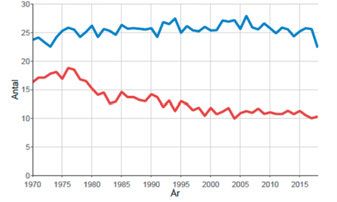
(Figure 1)2. The overall 5-year survival rate is approximately 60 % for both
men and women (although men have slightly lower survival)2.
Figure 1. Age-standardized incidence rates (upper blue lines) and mortality (lower red
lines) per 100,000 inhabitants in Sweden for 1970–2018 among men (left) and women
(right)2. Reprinted with permission
Etiology and risk factors
CRC develops through multiple mutational processes, from adenomas via ac-
tivation of oncogenes and inactivation of tumor suppressor genes3, but only
10% of adenomas become malignant4. Sporadic tumors arise in 80% of all
cases of CRC, with the remaining 20% caused by hereditary factors. About
5% of patients with CRC have cancer as a result of genes predisposing for
colorectal syndromes such as Lynch syndrome, familial adenomatous polypo-
sis, and MYH-associated polyposis5. Other well-known risk factors for the
development of CRC include inflammatory bowel disease (Crohn’s colitis and
11ulcerative colitis)6, obesity7, and high intakes of red and processed meat, to- bacco, and alcohol8. Protective factors that has been suggested include high fiber intake8, dairy products9, physical activity7, and the long-term use of as- pirin10. Anatomy The most distal part of the gastrointestinal tract is called the anal canal, and the rectum is considered as the distal 15 cm of the large bowel. The upper border begins at the sacral promontory from which the rectum extends down to the anal verge. The dentate line marks the transition of columnar glandular to squamous epithelium. According to the European Society for Medical On- cology (ESMO) guidelines, rectal tumors are defined as lesions up to 15 cm from the anal verge when diagnosed using a rigid rectoscope11. The rectum is surrounded by fatty lymphovascular tissue called the mesorectum, which in turn, is enveloped with an avascular layer called the mesorectal fascia (MRF). The upper third of the rectum is covered anteriorly and laterally by the peritoneum down to the peritoneal reflection, and the distal part below the pouch of Douglas (in women) and the rectovesical pouch (in men) is completely retroperitoneal. Organs adjacent to the rectum are anteri- orly the prostate, seminal vesicles, vas deferens, and urinary bladder in men, and the vagina and uterus in women. The rectum is limited laterally by vital structures such as the ureters, iliac vessels, and lateral pelvic wall, and poste- riorly the sacrum, coccyx, and hypogastric nerve plexus. Arterial blood supply to the rectum arises from the inferior mesenteric ar- tery, which continues as the superior rectal artery and from the internal iliac artery, which continues as the inferior rectal artery; some individuals also have a middle rectal artery. Lymphatic drainage follows the arteries, and knowledge of this is important because cancers can spread thorough this pathway12. There are three main lymphatic pathways: from the upper rectum along the superior rectal artery to the lymphatic nodes along the inferior mesenteric artery; from the lower rec- tum along the internal iliac artery to the lateral pelvic nodes; and from the anal canal to inguinal nodes. Clinical presentation and preoperative investigations Clinical symptoms and assessment of local tumors Common symptoms of rectal cancer include altered bowel habits, incomplete bowel evacuation, and rectal bleeding. Symptoms of more advanced disease 12
include weight loss, fatigue, abdominal-, rectal-, and anal pain, anal inconti-
nence, and anemia. The clinical investigation includes digital palpation of a
distal tumor with a description of its relation to the anal sphincters and assess-
ment of its mobility. Using a rigid rectoscope, the distant border is measured
from the anal verge, but a flexible sigmoidoscope is typically used to assess
the tumor. Biopsies are taken to establish the diagnosis by histopathology. Co-
lonoscopy or computed tomography (CT)-based colonography is performed
to rule out synchronous CRC, which occur in 4%–7% of patients13,14. Accord-
ing to the ESMO guidelines, rectal cancers should be classified into three cat-
egories based on the lower border of the tumor: low (0–5 cm), median (6–10
cm), or high (11–15 cm)11.
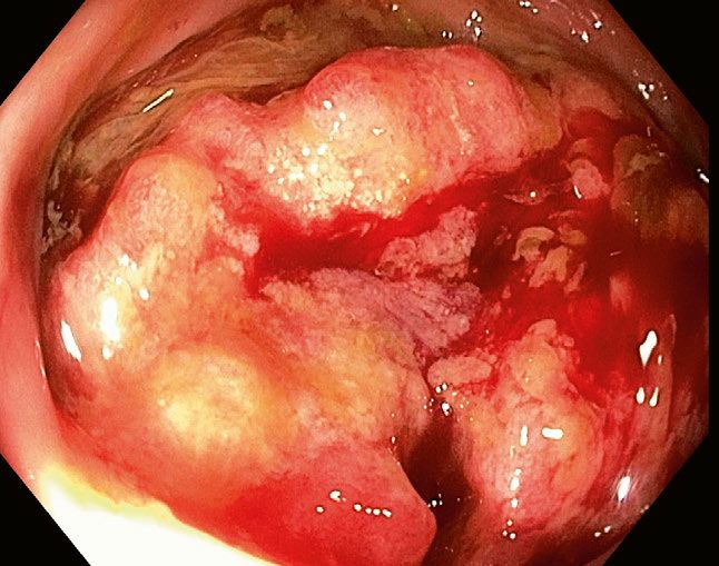
Figure 2. Endoscopical view of a rectal cancer
Magnetic resonance imaging (MRI) is also used to stage rectal cancers accord-
ing to tumor stage/depth (T-stage), lymph node involvement (N-stage) based
on the morphologic criteria of the nodes15, possible tumor deposits, the tumor
and node distance to the MRF, and extramural venous invasion (EMVI). A
positive (+) or threatened MRF seen on MRI is considered in the guidelines
to be an indication for chemoradiotherapy (CRT)11. Patients with positive
EMVI findings are associated with an increased risk of having synchronous
or developing metachronous metastasis16, and tumor deposits have been re-
ported to have prognostic value17. This staging is important because it guides
physicians in the selection of neoadjuvant treatment18-20.
13Figure 3. Magnetic resonance image of tumor in the rectum with adjacent organs in
women (left) and men (right)
Assessment of metastases
At the time of diagnosis, about 25% of patients with CRC have synchronous
metastases21, the most common locations for which are the liver (70%), fol-
lowed by the lungs (24%), peritoneum (15%), and non-regional lymph nodes
(16%). Metastases to the bones (4%) and brain (2%) are uncommon22. Syn-
chronous colorectal metastases at a single site are seen in 14% of cases and at
multiple sites in 8%21.
Contrast-enhanced CT of the thorax and abdomen is often used to detect
synchronous metastases to the liver and lungs. Compared with chest X-rays,
the introduction of CT to detect lung metastasis has led to the increased iden-
tification of lung lesions, but most (up to 42%) are too small and unspecific
for diagnosis, and only about one-fourth of these are metastases20. For detect-
ing liver metastasis with CT, the sensitivity and specificity are 74%–84% and
95%–96%, respectively, for smaller (Staging of rectal cancer
Different staging systems have been used over the years. The previously used
Dukes classification has been replaced by the tumor/node/metastasis (TNM)
classification system developed and revised by the Union for International
Cancer Control and the American Joint Committee on Cancer. In the Swedish
national guidelines for CRC, the 8th edition is recommended26 (Table 1). In
addition to the staging classification, prefixes are used to indicate preoperative
clinical stage with c and postoperative/pathological stage with p, and if neo-
adjuvant (presurgical) treatment has been given, the prefix y is used.
T-stage
This stage describes the depth of invasion of the primary tumor. The prognosis
deteriorates with higher T-stage tumors27,28.
N-stage
The number of regional lymph nodes with possible metastatic involvement is
the basis for the N-stage. Tumor deposits are also included in the N-stage and
are metastatic foci in the perirectal fat.
M-stage
The M-stage is defined as the presence of metastatic foci in distant organs,
nonregional lymph nodes, or peritoneal carcinomatosis.
Table 1a. Tumor classification by TNM staging
Primary tumor (T)
Tx Primary tumor cannot be assessed
T0 No evidence of a primary tumor
Tis Carcinoma in situ, intramucosal
T1 Tumor has invaded the submucosa
T2 Tumor has invaded the muscularis propria
T3 Tumor has invaded the subserosa or pericolorectal tissue
T4a Tumor has perforated the visceral peritoneum
T4b Tumor has invaded or adheres to other organs or structures
Regional lymph nodes (N)
Nx Regional lymph nodes cannot be assessed
N0 No regional lymph node metastases
N1 Metastases in 1–3 regional lymph nodes
N1a Metastases in 1 regional lymph node
N1b Metastases in 2–3 regional lymph nodes
N1c No regional lymph node metastases, but tumor deposits in the
subserosa or pericolorectal tissue
N2 Metastases in 4 regional lymph nodes
15N2a Metastases in 4–6 regional lymph nodes
N2b Metastases in 7 regional lymph nodes
Distant metastases (M)
M0 No distant metastases
M1 Distant metastases
M1a Metastases confined to one organ or site without peritoneal
metastases
M1b Metastases in more than one organ
M1c Metastases to the peritoneum with or without other organs
being involved
Table 1b. Tumor stage
Stage TNM grade
0 Tis N0 M0
I T1–T2 N0 M0
IIA T3 N0 M0
IIB T4a N0 M0
IIC T4b N0 M0
IIIA T1–T2 N1 M0
T1 N2a M0
IIIB T3–T4a N1 M0
T2–T3 N2a M0
T1–T2 N2b M0
IIIC T4a N2a M0
T3–T4a N2b M0
T4b N1-2 M0
IVA Any T Any N M1a
IVB Any T Any N M1b
IVC Any T Any N M1c
Postoperative histopathology staging
Postoperative histopathology staging after rectal cancer surgery means evalu-
ation of the resected specimen macroscopically and thereafter microscopi-
cally. The pathologist examines the tumor (TN-stage), resection margins (cir-
cumferential resection margin [CRM] and distal resection margin [DRM]),
and surgical quality (R-residual tumor). The quality of total mesorectal exci-
sion (TME) is judged complete, nearly complete, or incomplete, and this has
been shown to influence the prognosis29. In addition to these factors, the pa-
thology report should also state tumor differentiation and tumor infiltration in
venous and lymphatic vessels and nerves.
Resection margin
The resection surface examined is divided into the CRM and DRM. The CRM
is defined as the distance between the lateral part of the rectal tumor, tumor
16deposit, or metastatic node and the lateral resection margin. Numerous studies
have shown that a positive CRM, i.e., tumor cells within 1 mm30-36 or 2 mm37
from the margin, is a negative prognostic factor for local recurrence (LR)30-37,
systemic disease, and overall survival36,38. However, some studies have re-
ported no correlations with LR owing to modern surgical and oncological
treatments39. Today, a positive CRM is defined as 1 mm and a negative CRM
as >1 mm. A DRM of 1 cm with a complete TME is accepted in middle-to-
low rectal cancers, and a partial TME with a DMR of 5 cm is accepted in high
rectal tumors40,41.
Residual tumor status
The residual tumor (R) status depicts the involvement of a tumor at the CRM
or DRM. The classification is R0 if no residual tumor is found, R1 if a micro-
scopic residual tumor is present, and R2 if there is a macroscopic residual tu-
mor in the margin. R status is correlated with distant metastasis, LR, and pa-
tient survival42.
Vascular and perineural invasion
Vascular involvement is reported as venous and/or lymphatic. Venous inva-
sion can be either intramural (within the submucosa or the muscularis propria,
EMVI–) or extramural (beyond the muscularis propria, EMVI+). In a pathol-
ogy report, V0 means the lack of venous involvement, V1 marks venous in-
volvement detected only microscopically, and V2 marks venous invasion al-
ready detectable during the macroscopic assessment of the specimen. The ab-
sence (L0) or presence (L1) of lymphatic invasion is reported separately. In-
vasion of the lymphatic vessels might predict future lymph node metastasis,
and venous involvement is associated with decreased survival and an in-
creased risk of distant metastases43-45. Perineural invasion has been defined as
tumor growth in, around, and through peripheral nerves, and is a prognostic
factor for inferior patient outcomes46,47.
Differentiation
The most common type of CRC is an adenocarcinoma of epithelial origin. The
second most common form of adenocarcinoma is the mucinous type, defined
by >50% extracellular mucin, and thereafter, the more uncommon signet ring
cell adenocarcinoma. Other types of CRC include neuroendocrine,
adenosquamous, and medullar carcinomas48. Adenocarcinomas were previ-
ously classified as well, moderately or poorly differentiated, and this was later
changed, so that well and moderately differentiated tumors were classified as
low grade and the poorly differentiated ones as high grade49. Less well-differ-
entiated carcinomas indicate poorer patient survival outcomes50.
17Multidisciplinary team (MDT) conference When the preoperative assessment is completed and all information is gath- ered, all patients with CRC should be discussed at an MDT conference. At these conferences, different specialties, including colorectal surgeons, oncol- ogists, radiologists, pathologists, and nurses, gather together. Patients are of- ten discussed both pre-and postoperatively and sometimes during treatment, when neoadjuvant treatment is given. In Sweden, it is mandatory to discuss all patients with CRC at an MDT conference, and at present, 98% of all such patients are discussed51. After the implementation of MDTs, more patients were seen undergoing preoperative radiology assessment and reduced rates of positive CRM were observed52,53. Patients with liver and lung metastases are discussed at a separate MDT conference with hepatic and thoracic surgeons. Swedish ColoRectal Cancer Registry The Swedish Rectal Cancer Registry was established in 1995 and the Swedish Colon Cancer Registry in 2007; these were later merged into the Swedish Col- oRectal Cancer Registry (SCRCR). Data are gathered prospectively based on reports from surgeons, oncologists, and pathologists, and include information on pre- and postoperative staging, surgery, postoperative course, neoadjuvant- , adjuvant-, and palliative treatments, treatment of metastases, recurrence, and follow-up. The national coverage is estimated to be >97%54. The SCRCR has been validated on several occasions54-58. Jörgren et al.56 reported overall high validity with
The Swedish National Patient Registry
The Swedish National Patient Registry is maintained by the Swedish National
Board of Health and Welfare. Registration started in 1964, but the information
registered at that time covered only some hospitals in the country. Since 1987,
registration became nationwide and included information on inpatient care
from all hospitals in Sweden. Surgical procedures were registered from 1997
and outpatient visits from 2001. The codes used conform to the International
Classification of Disease system; the register was validated in 2011 and
showed positive predictive values of 85%–95% for diagnosis60,56.
Local rectal cancer registry in Västmanland
Since 1996, a comprehensive registry covering a catchment area of 270,000
people has been set up at the Colorectal Unit, Västmanland’s Hospital, Väs-
terås. All patients diagnosed with rectal cancer have been registered. Data re-
garding demographics, radiology, surgery, pathology, postoperative follow-
up, oncological treatment, and bowel function are collected prospectively. All
data are registered at each follow-up. All patients who have undergone surgery
for rectal cancer were scheduled for follow-up at 1, 6, 12, 24, 36, 48, and 60
months after surgery. Preoperative screening for metastases is performed rou-
tinely. Until 2002, chest radiography and liver ultrasonography were used, and
thereafter, CT of the thorax and abdomen. MRI of the pelvis was used for local
staging in almost all patients. At the 12- and 36-month follow-ups, the patients
underwent control CT scans of the thorax and abdomen.
The main differences between the local rectal cancer registry in Västman-
land and the SCRCR is the minimal number of missing variables and more in
depth information regarding preoperative factors, i.e., comorbidities, World
Health Organization performance status, preoperative bowel symptoms and
anorectal function, RT to any tumor in the abdomen prior to rectal cancer, type
of palliative treatment offered to patients with rectal cancer who will not un-
dergo surgery, and type of stoma deviation prior to rectal cancer surgery. The
following surgical and perioperative details are collected: type of closure af-
ter abdominoperineal excision (APE) (with Permacole or a gluteus maximus
muscle flap), metastasis in the abdomen, nerve sparing during dissection, mo-
bilization of the splenic flexure and potential damage to the spleen, type of
anastomosis and height, part of the bowel used for anastomosis or stoma, an-
esthesia, perioperative complications, and macroscopic evaluation of the
bowel specimen according to the surgeon. In addition, there is extensive in-
formation about the pathology and postoperative course, and at every outpa-
tient visit, particular information is collected on oncological treatment and
complications regarding the anastomosis, anorectal, urinary, sexual function,
and stoma.
19Incisional and parastomal hernias are observed and recorded through either
a clinical examination during follow-up or CT scans. Small bowel obstruction
(SBO) is registered if the patient had been admitted or received surgery for it.
Anorectal function is registered as incontinence (if leakage of any of the fol-
lowing occurs more than once a week: gas, fluid, loose stool, or firm stool),
evacuatory dysfunction (if defecating for longer than 15 min), clustering (if
needing to defecate 30 min after prior defecation), frequency (number of def-
ecations), and urgency (feeling an urgent need to defecate).
No validation study has been performed on the register, which up to Janu-
ary 2020, included 1345 patients diagnosed with rectal cancer; however, the
internal validity is good because over the time, there have always been only
three or four dedicated colorectal surgeons performing rectal cancer surgery
in Västmanland. These surgeons have reached consensus on the definitions of
different variables and also been responsible for outpatient follow-up and reg-
istration. A research nurse together with the head of the unit are responsible
for the registry and the research nurse continuously registers data using IBM
SPSS statistics software. Regarding external validity, there have been several
publications based on the registry where the medical records have been scru-
tinized, confirming the validity of the data.
Neoadjuvant treatment and adverse effects
Neoadjuvant treatment is the treatment given to patients before surgery either
as RT alone or in combination with chemotherapy, also called CRT.
Blomqvist and Glimelius61 classified rectal cancer into three categories based
on preoperative MRI scans: ‘good’, ‘bad’, and ‘ugly’. The proportions of pa-
tients in each group were: ‘good’ 20%–40%; ‘bad’ 40%–60%; and ‘ugly’
10%–20%61. This stratification remains a good basic tool in decision-making
for neoadjuvant treatment (Figure 4). The purpose of preoperative treatment
is to destroy potential tumor cells near the rectal tumor and outside the surgical
dissection plane and to downstage/or shrink the tumor from being unresectable
to resectable.
20Figure 4. Magnetic resonance imaging-based preoperative evaluation of rectal cancers
for neoadjuvant treatment61
The Uppsala Trial62 randomized patients as either preoperative short-course
RT (SRT) (5 5 Gy) or postoperative long-course RT (LRT) (total 60 Gy)
and showed lower rates of LR in the SRT group, but no difference in survival.
Later, in the Stockholm I trial63, SRT followed by surgery was compared with
surgery alone; the results showed that the LR rate was reduced by almost half
when surgery was combined with SRT, although increased postoperative mor-
tality was observed. Because of the mortality rate, the Stockholm II64 and Swe-
dish Rectal Cancer trial65 were initiated with changed RT protocols and a lim-
ited target area; the results showed lower postoperative morbidity and mortal-
ity rates apart from the low LR rate. The Dutch TME trial66 compared pre-
operative SRT and surgery within 1 week with surgery alone; the results
showed that the LR rates could be reduced to 5%. The hypothesis that chang-
ing the timing of surgery after RT could improve survival was explored in the
Stockholm III trial67. The results showed a reduced risk of postoperative com-
plications after SRT with delayed surgery for 4–8 weeks, but similar oncolog-
ical results. Since then, the RAPIDO trial68 has had a breakthrough in Sweden,
showing that SRT in combination with chemotherapy instead of LRT with
chemotherapy had low treatment-related morbidity in patients with advanced
rectal cancer, and that 19% of patients had complete tumor remission.
21Tumor response (downsizing and downstaging) with preoperative treat- ment has led to interest in an organ-preserving option to surgery named “watch and wait” in the literature. A pathological complete response (pCR) is based on an available resected specimen to verify that there are no tumor cells re- maining. A clinical complete response (cCR) is based on three factors: digital rectal examination, endoscopic evaluation, and MRI. As the pCR and cCR are not always in accordance with each other, a diagnosis of cCR carries some uncertainty. The Brazilian researcher Habr-Gama and colleagues were the pioneers of organ-preserving treatment. In 2004, a series of 265 resectable patients with rectal cancer treated with CRT were assessed for cCR at 8 weeks after CRT69. As a result, 71 (27%) had cCR and entered an intense surveillance program. Patients with an incomplete clinical response underwent TME. The mean fol- low-up duration was 57.3 months and during that time, two patients had tumor regrowth and underwent successful salvage surgery, and three patients had metachronous metastases. Five-year overall survival was 100% and disease- free survival (DFS) was 92%. These results indicated that a nonsurgical alter- native is safe in selected patients. There is large heterogenicity in studies regarding patient selection, which makes assessment of the “watch and wait” strategy difficult. Older patients may benefit from this option70. Hence, possible complications to surgery such as AL and low anterior resection syndrome (described later) can be avoided if AR is planned. For patients with distal tumors, APE can be avoided and the inherent morbidity that a stoma provides. In Sweden, early rectal tumors (i.e., T1–T3a,b, N0, and MRF–) are sup- posed to be treated with surgery alone, and neoadjuvant CRT is given to pa- tients with advanced rectal tumors (any cT or any cN and MRF+ and/or lateral lymph nodes). Therefore, including early tumors in a “watch and wait” pro- gram can lead to overtreatment. In locally advanced rectal cancers, cCR can be seen in 10%–20%71-73 and tumor regrowth in 15%–22%74,75. A “watch and wait” trial including patients with locally advanced tumors who received ne- oadjuvant CRT or short-course RT with delayed surgery according to the Swe- dish guidelines, with the first evaluation of cCR status after 6–8 weeks, is on- going in Sweden since 201776. Treatment with RT is beneficial in patients with rectal cancers, but is asso- ciated with side effects. The acute adverse effects depend on the RT dose and target volume. Typical acute symptoms include urogenital and gastrointestinal symptoms such as diarrhea, urgency, bloating, abdominal pain, nausea, and vomiting77,78. In a Cochrane systematic review including five trials, more acute toxicity symptoms were observed following LRT or CRT compared with SRT79. The late adverse effects of RT include symptoms such as fecal incontinence and increased stool frequency and urgency. These symptoms are the same as those associated with rectal cancer surgery, but RT has an additive effect80. Other described late effects of RT include increased rates of adverse 22
gastrointestinal symptoms such as bowel obstruction/ileus81,82, symptoms of
urinary dysfunction83, and sexual dysfunction84. The risk of RT induced can-
cers has been investigated in several studies, with conflicting results. No in-
creased risk of secondary cancer was found in a recent Swedish study that
included data from over 13,000 patients registered in the SCRCR85.

Surgical treatment
In 1982, Heald86 introduced the TME technique—dissection along the MRF—
to resect the rectum together with the whole mesorectum as an intact specimen
without damaging the MRF. By using this technique, they were able to de-
crease the LR to 4% without RT87, from previous LR rates of approximately
30%88. Presently, the LR rate in Sweden is around 5%51. TME is currently
considered the “gold standard” for the surgical treatment of rectal cancer89 in
the middle or low rectum. The same technique is used regardless of the surgi-
cal approach, whether it is AR, APE, or Hartmann’s procedure (HP), and
whether it is open or minimally invasive. Regarding high rectal tumors, partial
mesorectal excision (PME) is sometimes performed, meaning transecting the
bowel 5 cm below the tumor to achieve better functional outcomes90-92. Most
patients who undergo AR will have oral colon cleansing and those treated with
APE or HP will receive a rectal enema.

Figure 5. Distribution of operations from 1995 to 2019 for all patients with rectal
cancers. Key: AR (yellow), APE (purple), HP (green), local excision (red), laparot-
omy without resection (light blue), other interventions (light grey), or no surgery (dark
grey). The figure is reprinted with permission from reference2

                                                                                      23
Anterior resection In Sweden, approximately 35% of patients with rectal cancer diagnosis un- dergo AR2, which is the most common approach for rectal tumors located in the middle (6–10 cm) or upper (11–15 cm) rectum. It is termed AR because the approach is from the abdomen. In TME surgery, mesorectal excision is performed down to the pelvic floor while identifying and preserving the hy- pogastric nerves. In cases with overgrowth of the rectal tumor to other organs, these are resected together with the bowel specimen. A high ligation of the inferior mesenteric artery is performed just distally to the origin of the left colic artery. The splenic flexure is frequently taken down and the inferior mes- enteric vein divided just below the pancreas to achieve a tension-free anasto- mosis. Before dividing the rectum, distally to the tumor, the distal part is washed out with an alcohol solution. Using a circular staple device, the colo- rectal or coloanal anastomosis is created as end-to-end, side-to-end, or a J- pouch configuration. AR can be subclassified into low anterior resection (LAR) with TME or high anterior resection with PME, depending on the level of rectal transection. Because of the high risk of AL93 and the severe conse- quences, most patients subjected to LAR receive a temporary diverting loop ileostomy (see below). Hartmann’s procedure Patients not fit for AR, with weak sphincter function, or in a palliative care situation are candidates for HP, which includes TME or PME without an anas- tomosis. These patients receive a permanent end-colostomy. Abdominoperineal excision In Sweden, APE is usually performed for rectal rumors measuring
In ELAPE, dissection extends to the coccyx, keeping the mesorectum at-
tached to the levator ani muscle. The perineal part of the dissection runs along
the external sphincter, continues along the levator ani muscle to its attachment
onto the pelvis, with or without excision to the distal coccyx, resulting in a
cylinder-shaped specimen with wider lateral resection marginals. The defect
in the pelvic floor is closed using either a musculocutaneous flap or biological
mesh. Studies comparing conventional APE with ELAPE have shown lower
rates of CRM positivity and bowel perforation100-102.
    Ischioanal APE is similar to ELAPE, but instead, the perineal dissection
follows the fascia of the internal obturator muscle. This approach is used
mainly for locally advanced tumors. The most challenging part of the surgery
is the anterior dissection. In patients with anteriorly lying tumors, en bloc re-
section of a part of the prostate or vagina is indicated to increase the CRM and
minimize the risk of tumor perforation103.

Minimally invasive surgery
Open surgery was the only technique used up to the 1990s, when a laparo-
scopic approach entered colorectal surgery. In 2010, the first randomized mul-
ticenter trial comparing laparoscopic and open rectal cancer surgery—the
COREAN trial104—was initiated. This trial reported similar short-term out-
comes between the two surgical techniques. In 2013, the COLOR II trial105
reported faster return of bowel function and a shorter length of hospital stay
(LOS) for laparoscopic surgery. The 3-year follow-up studies of the trials
showed that, for the former, noninferiority of the laparoscopic approach was
met regarding DFS106 and for the latter, similar 5% recurrence rates107. Two
other trials, the ACOSOG108 and ALaCaRT109, had clear margins as primary
end points, but failed to demonstrate noninferiority when laparoscopic surgery
was compared with open surgery. However, their 2-year follow up did not
show differences in overall survival or DFS110,111. In the early 2000s, robotic
surgery was introduced to colorectal surgery. A large randomized multicenter
trial—ROLARR—compared laparoscopic with robotic surgery and had con-
version rates to open surgery as the primary end point112. The trial could not
prove significant differences between the two surgical techniques for rectal
cancer surgery. No long-term outcomes are yet available. Due to its numerous
advantages, including advanced three-dimensional vision with better nerve
discrimination, lack of tremor, more complex movements with the instru-
ments, reduced discomfort for the surgeon, and a lower learning curve, robotic
surgery is expected to overcome the limitations of laparoscopic surgery113.
    In 2010, a new technique combining laparoscopic abdominal and transanal
TME (TaTME) gained popularity for overcoming difficult pelvic dissections,
especially for distal tumors in patients with a narrow pelvis and a high body
mass index. A Dutch trial compared TaTME with laparoscopic surgery and
reported equal CRM positivity, at 4%114. In a multicenter study of TaTME and

                                                                              25
robotic surgery, no overall difference was found in pathological margins115. However, in a Norwegian trial comparing TaTME with established surgical techniques, higher AL and LR were reported, so TaTME is currently sus- pended in Norway116. More evidence is needed to determine the benefits and safety of TaTME. Temporary ileostomy A temporary diverting ileostoma is usually established after an AR to mini- mize the consequence and reduce the frequency of AL, which occurs in up to 28% of patients after AR93. In the Swedish RECTODES trial, the results showed a threefold increased risk of AL in patients without a diverting ileos- tomy93. Leaks can be associated with devastating consequences, such as ab- scess formation, sepsis, peritonitis, a higher prevalence of LR, and poor sub- sequent neorectal function117-119. However, disagreements remain regarding the routine use of a diverting ileostomy, and it is still being debated120,121. Nev- ertheless, there are high-risk patients and situations that might benefit from a loop ileostomy, such as those with colorectal or coloanal anastomoses, those undergoing technically difficult operations, malnourished patients, and obese men120. The benefit of creating a loop ileostomy is to avoid complications such as AL, which could delay or exclude adjuvant treatment. On the other hand, closure of a loop ileostomy is also associated with morbidity and mortality. In a systematic review including 6107 patients, overall morbidity following the closure of a loop ileostomy was 17.3%, with a total mortality rate of 0.4%122. Another review of complications after loop ileostomy closure demonstrated SBO in up to 15% of cases, wound infections in up to 18.3%, and AL in up to 8%123. Furthermore, having a stoma is associated with morbidity for patients such as those readmitted for dehydration and renal failure122,124,125, as well as those with skin irritation, stomal prolapse retraction, or hernia126. It is well known that many patients live with stomas for a long time before reversal, and in a Swedish single-center study, up to 75% of stomas (in colos- tomy and ileostomy) were reversed after 4 months, usually because of adju- vant chemotherapy or surgical complications; however, in one third, no health reason was identified127. In the same study, 18% of patients with a defunction- ing stoma ended up with a permanent stoma, of which, 10% were loop ileos- tomies. Following the reversal of a loop ileostomy, most patients remain in hospital until bowel function returns, despite no evidence supporting this ap- proach. The mean LOS in hospital was reported as 5.1 days after stoma clo- sure122,127. There have also been studies supporting early discharge following ileostomy reversal128-133. Most studies have been retrospective, and to our knowledge, there are only four prospective studies with a total of 72 patients who were discharged within 23 hours, suggesting that this concept is safe and feasible129-131,133. 26
Early closure of ileostomy could be beneficial, as recently shown in the
Swedish EASY trial, where significantly fewer complications were observed
when closing the ileostomy within 13 days of primary surgery134. A recent
meta-analysis also suggested the possibility of early closure for selected pa-
tients without signs of AL and an uneventful postoperative course135.

Adjuvant chemotherapy
Adjuvant chemotherapy is given to eliminate potential micro-metastases after
tumor resection. There is evidence of benefits with an increase in DFS in pa-
tients with stage II colon cancer with high-risk features, as well as in those
with stage III colon cancer136-138; however, evidence of the need for adjuvant
chemotherapy in cases of rectal cancer is not convincing. In one meta-analy-
sis, no benefits in overall survival or DFS were found; however in sub-group
analyses, DFS was improved in patients with upper rectal cancers (hazard ra-
tio [HR] 0.59; 95% confidence interval [CI] 0.40–0.85)138. What factors are
considered high risk for patients with stage II rectal cancer are not clearly de-
fined, but, as in those with colon cancer, perforations, low tumor differentia-
tion, serosal extension, venous, lymphatic, or perineural invasion, and a low
number of examined lymph nodes have been associated with a poor progno-
sis12,139-141. The advantages of receiving adjuvant chemotherapy might be seen
in patients with positive pathological risk factors. In Sweden, adjuvant chem-
otherapy is mainly considered for patients with stage II rectal cancer with risk
factors who have not received RT and for those with stage III rectal cancers,
especially in the upper rectum25. Adjuvant chemotherapy should be started no
later than 8 weeks after surgery, since starting later decreases overall survival
according to studies on patients with stage III colon cancers142,143. The same
limitations apply for those with rectal cancer in Sweden144.

Short-term postoperative complications
Rectal cancer surgery puts the body under major stress and is associated with
considerable morbidity. There are discrepancies in the definitions of different
complications, which makes reporting difficult. The Clavien–Dindo classifi-
cation of surgical complications in 2004 has been accepted in the surgical
community145 and was introduced into the SCRCR in 2011.
   Short-term complications include AL and its consequences, wound infec-
tion, intra-abdominal infection, ileus, and cardiopulmonary adverse events.
One of the most severe complications is AL, described above. There is no
consensus on the definition of AL in the literature. One way to define AL is
clinically, with peritonitis caused by leakage from any staple line, rectovaginal

                                                                              27
fistula, and pelvic abscess, even if leakage is not seen radiologically. The leak-
age should be verified by digital palpation and inspection of drain contents,
endoscopically or radiologically (e.g., rectal contrast studies, CT scans). How-
ever, if leakage is seen on the radiological investigation without clinical symp-
toms, it is not defined as leakage93. Another way to define AL after AR is “a
communication between the intra- and extraluminal compartments at the site
of anastomosis”, and this can be graded accordingly as follows: A, no active
intervention; B, active intervention such as drainage without surgery; and C,
active intervention requiring surgery146. About 60% of patients with AL end
up with a permanent stoma147. About 10% of patients with a defunctioning
stoma after AR develop AL93. After APE, the most common complication is
a perineal wound, seen in approximately 40% of cases101.
Long-term complications and functional bowel
disturbance
Rectal cancer surgery is associated with considerable long-term complica-
tions, so patients might suffer from sexual and urinary dysfunction due to the
RT or TME surgery148-151. Even though TME is nerve-sparing surgery, the hy-
pogastric nerves might be damaged to some degree, depending on the size of
the tumor and surgical technique used. Besides, rectal cancer surgery causes
the highest patient readmission rates152-154 from SBO (see below).
In the case of stoma formation, there are long-term complications, such as
prolapse, stenosis, and hernia126, in patients undergoing APE or HP receiving
a permanent colostomy or in patients undergoing AR and receiving a tempo-
rary ileostomy. As described above, having a stoma is associated with mor-
bidity, hospital readmission for dehydration and renal failure, and peristomal
skin problems or leakage126.
Following AR, most patients have some degree of anorectal dysfunction
and bowel disturbance. About 40%–80% of patients undergoing AR experi-
ence severe bowel disturbances155-158. The symptoms can arise immediately
after surgery or when the temporary ileostomy is closed. This dysfunction is
also referred to as low anterior resection syndrome (LARS). To characterize
the symptoms and each patient’s experience, a LARS score has been created
by a Danish group159 based on a combination of the following five symptoms:
flatulence and fecal incontinence, stool frequency, clustering, and urgency.
The LARS score is classed as low, minor, or major depending on the total
scores of 0–20, 21–29, and 30–42, respectively. Risk factors for developing
LARS are pre-and postoperative RT, low anastomotic height when perform-
ing TME, temporary ileostomy, AL, age 64 years, and female sex92,156,158,160-
162
. LARS is usually measured pre- and postoperatively, but comparisons be-
tween these are ineffective because patients have a rectal tumor that might
28cause symptoms similar to LARS. Therefore, a recent study measured the prevalence of LARS in a normal population, with the results showing that 10%–15% of the general population have major LARS163. Therefore, it is im- portant to identify patients with LARS so that they can be treated appropri- ately. Small bowel obstruction The most common causes of SBO are abdominal adhesions, accounting for 60%–70% of cases164,165. The formation of adhesions can start immediately after surgery. The symptoms of SBO include abdominal pain, vomiting, dis- tention, and a lack of flatus and stool. SBO is classified according to com- pleteness (partial vs. complete), etiology (adhesional vs. nonadhesional), or timing (early vs. late using a cutoff of 30 days after surgery). The type of surgery has a major role in the development of SBO. Compar- ing open with laparoscopic approaches without previous open surgery, Reshef et al.166 evaluated the risk of SBO following colorectal surgery and found equal admission rates between groups. The rate of surgery for SBO was lower in the laparoscopic group, suggesting that this might be because of fewer ad- hesions. Other known risk factors include surgery to the colon, rectum, and gynecological organs, patient age
bowel) and partial SBO. NOM can be prolonged for up to 72 hours in cases
of adhesive SBO if no complications have been encountered or the drainage
volume in the nasogastric tube on day 3 is  4), abdominal guarding, and leukocytosis.
   The surgical approach is not without complications, and several studies
have focused on adverse outcomes in adhesion-related SBO. Mortality rates
in SBO surgery are 10%–15% when small bowel resection is performed176,177
and there is a 33% risk of inadvertent enterotomy during surgery for bowel
obstruction178.

Liver metastasis and prognostic scoring systems
The most common site of distant metastases from CRC is to the liver21. Me-
tastases can be either synchronous (SM; present at the time of diagnosis of the
primary tumor) or metachronous (MM; occurring later in in the course of the
disease). Unfortunately, there are discrepancies in the literature for the defini-
tions of liver SM and MM that affect the results in different studies and make
them difficult to compare. Approximately 15%–25% of patients with CRC
present with SM disease, and 13%–29% later develop MM during follow-
up21,179-181. In a recent Danish study, patients with CRC without metastasis had
a median survival of 86 months, but if SM were present, the median survival
decreased to 11 months, and if MM were present, the median survival de-
creased to 36 months182.
   Most liver metastases are irresectable, with reported resection rates of 5%–
15%183,184. The prognosis for patients with stage IV rectal cancer and colon
cancer is poor, with 5-year survival rates of 3% and 6.6%, respectivaly54,182.
However, with liver resection, 5-year survival rates of up to 58% have been
reported185,186. Liver surgeons are shifting toward a ‘liver-first’ approach,
where liver metastases are resected after cycles of preoperative chemotherapy,
followed by surgery for the rectal cancer187.
   Several scoring systems to predict survival after liver resection of CRC
liver metastases have been suggested188-190. Since being first reported in 1996
and 1999, more than 19 scoring systems concerning prognostic factors for sur-
vival after liver resection have been proposed for the optimal management of
patients191. There are contradictions in the literature, but some studies have
shown that the maximum size185,189,190,192-196, number 185,189,190,192-195,197-204, and

30
You can also read