Investigating Disturbances of Oxygen Homeostasis: From Cellular Mechanisms to the Clinical Practice
←
→
Page content transcription
If your browser does not render page correctly, please read the page content below
REVIEW
                                                                                                                                                  published: 04 August 2020
                                                                                                                                             doi: 10.3389/fphys.2020.00947

                                                Investigating Disturbances of
                                                Oxygen Homeostasis: From Cellular
                                                Mechanisms to the Clinical Practice
                                                Verena Tretter* , Marie-Louise Zach, Stefan Böhme, Roman Ullrich, Klaus Markstaller
                                                and Klaus Ulrich Klein
                                                Department of Anaesthesia, General Intensive Care and Pain Therapy, Medical University Vienna, Vienna, Austria

                                                Soon after its discovery in the 18th century, oxygen was applied as a therapeutic agent
                                                to treat severely ill patients. Lack of oxygen, commonly termed as hypoxia, is frequently
                                                encountered in different disease states and is detrimental to human life. However, at the
                                                end of the 19th century, Paul Bert and James Lorrain Smith identified what is known as
                                                oxygen toxicity. The molecular basis of this phenomenon is oxygen’s readiness to accept
                                                electrons and to form different variants of aggressive radicals that interfere with normal
                                                cell functions. The human body has evolved to maintain oxygen homeostasis by different
                                                molecular systems that are either activated in the case of oxygen under-supply, or to
                         Edited by:             scavenge and to transform oxygen radicals when excess amounts are encountered.
                   Alberto Giannoni,            Research has provided insights into cellular mechanisms of oxygen homeostasis and
       Sant’Anna School of Advanced
                        Studies, Italy          is still called upon in order to better understand related diseases. Oxygen therapy is
                       Reviewed by:             one of the prime clinical interventions, as it is life saving, readily available, easy to apply
                    Christina Pabelick,         and economically affordable. However, the current state of research also implicates a
            Mayo Clinic, United States
                                                reconsidering of the liberal application of oxygen causing hyperoxia. Increasing evidence
               Michael Adam O’Reilly,
University of Rochester, United States          from preclinical and clinical studies suggest detrimental outcomes as a consequence of
                        Chiara Borrelli,        liberal oxygen therapy. In this review, we summarize concepts of cellular mechanisms
                University of Pisa, Italy
                                                regarding different forms of disturbed cellular oxygen homeostasis that may help to
                  *Correspondence:
                       Verena Tretter           better define safe clinical application of oxygen therapy.
        eva.tretter@meduniwien.ac.at
                                                Keywords: oxygen homeostasis, hypoxia, hyperoxia, intermittent hypoxia, intermittent hyperoxia/hypoxia,
                                                supplemental oxygen, oxygen therapy
                    Specialty section:
          This article was submitted to
 Clinical and Translational Physiology,
                a section of the journal        INTRODUCTION
                Frontiers in Physiology
                                                Oxygen (dioxygen, O2 ) is mandatory to support human life from an early fetal stage onwards, as
           Received: 20 March 2020
            Accepted: 14 July 2020
                                                metabolism relies on its presence to produce sufficient energy efficiently in the form of adenosine
          Published: 04 August 2020             triphosphate (ATP). As many acute pathological conditions are linked to hypoxemia, supplemental
                              Citation:
                                                oxygen is frequently used as an adjunct therapy in anesthesiology, emergency and critical care
        Tretter V, Zach M-L, Böhme S,           medicine. However, as with most drugs, we know since the times of Paracelsus the dose makes
 Ullrich R, Markstaller K and Klein KU          the poison. While the organism has established elaborate mechanisms to survive hypoxic episodes,
     (2020) Investigating Disturbances          higher than current ambient oxygen conditions (21% O2 ) were rarely encountered in evolution,
        of Oxygen Homeostasis: From
  Cellular Mechanisms to the Clinical           Abbreviations: FiO2 , fraction of inspired oxygen; ICU, intensive care unit; paO2 , arterial partial pressure of oxygen; PCI,
      Practice. Front. Physiol. 11:947.         percutaneous coronary intervention; RCT, randomized controlled trial; SSI, surgical site infection; STEMI, ST-elevation
       doi: 10.3389/fphys.2020.00947            myocardial infarction.

Frontiers in Physiology | www.frontiersin.org                                           1                                              August 2020 | Volume 11 | Article 947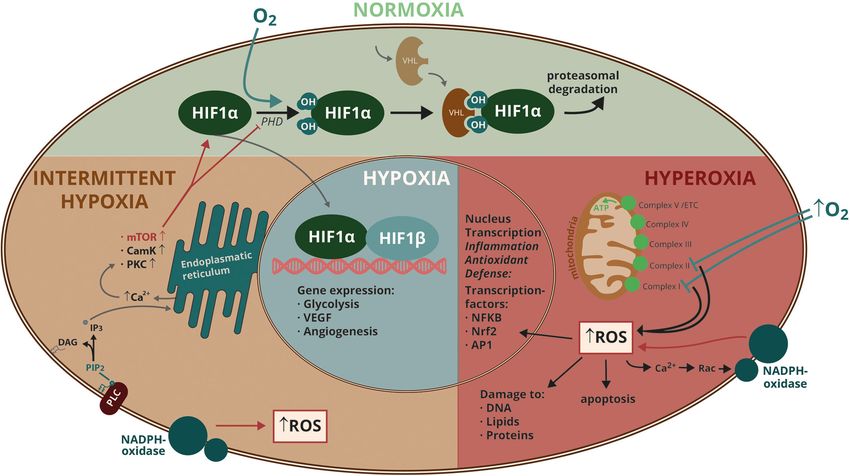
Tretter et al.                                                                                    Investigating Disturbances of Oxygen Homeostasis

apart from peaks of approximately 30% O2 in the atmosphere              boost in evolution was the invention of using the otherwise
during the Carboniferous period 300 million years ago and the           toxic O2 for respiration. Bacteria capable of using O2 for
Cretaceous period 100 million years ago (Berner et al., 2007).          respiration were taken up by other cells and developed into
Excess oxygen leads to accumulation of oxygen radicals, generally       specialized cell organelles, the mitochondria, which use oxidative
known as reactive oxygen species (ROS), that have the potential         phosphorylation to efficiently boost the development of higher
to detrimentally modify proteins, lipids and nucleic acids.             multicellular living entities. Oxygen is used to generate the
    In clinical practice, physicians routinely take all measures        universal cellular energy currency ATP in a tightly controlled
to prevent hypoxemia, accepting in many cases accidental                cascadic pathway. Energy is released in redox reactions, where
hyperoxemia as a side effect of the therapeutic intervention.           electrons flow from donors to acceptors and where O2 is the
The World Health Organization (WHO) guideline in 2016                   terminal electron acceptor, being reduced to water molecules.
recommended, based on a meta-analysis of then current                   This electron chain, however, is not ”perfect“, and some
literature, that any patient who becomes anesthesized, intubated        “escaping“ electrons are directly transfered to O2 , thereby
and mechanically ventilated for surgery should receive 80%              generating a certain amount (1–3%) of incompletely reduced
inspiratory O2 levels during and supplemental O2 up to 6 h              O2 radicals- i.e., superoxide, hydrogen peroxide and hydroxyl
after surgery (Allegranzi et al., 2016). The recommendation was         radical- commonly referred to as ROS.
based on evidence, that such high O2 concentrations may exert               Reactive O2 species are also by-products of many other cellular
important benefits by reducing surgical site infections (SSI), a        reactions, including oxidase/dehydrogenase enzymatic reactions
key problem occurring after surgery worldwide. This guideline           of membrane-bound and cytosolic enzymes. Small amounts of
has evoked considerable criticism, as it is not known which dose        ROS are used by cells as signaling molecules. Exaggerated ROS
of oxygen for what duration can be regarded as clinically safe          production, however, results in oxidative injury to proteins,
(Hedenstierna et al., 2017).                                            lipids and nucleic acids (Circu and Aw, 2010). Evolution has
    Several clinical trials demonstrated that hyperoxemia may be        equipped cells with antioxidative defense systems, comprised
associated with detrimental effects on mortality and morbidity          of either small molecules (vitamins, the tripeptide glutathione,
in different acute conditions, including stroke, myocardial             redox buffer proteins – i.e., thioredoxin and peroxiredoxin)
infarction (MI) and acute respiratory distress syndrome (ARDS).         or ROS dissipating enzymes such as superoxide dismutases or
Further efforts in translational science, in addition to more           peroxidases. When the anti-oxidative buffer capacity of these
clinical trials are needed to address this problem and to define        systems is exhausted, the cell experiences a condition of oxidative
the margins of safe oxygen application. We here discuss current         stress. It must be emphasized, that both excess as well as
experimental and clinical evidence regarding the mechanisms             under-supply of O2 induces oxidative stress. However, the exact
and consequences of dysregulated O2 homeostasis.                        threshold beyond which oxidative stress harms the cells remains
                                                                        to be determined. Cells in different tissues and organs experience
                                                                        a large range of different O2 tensions, and accordingly, the
MOLECULAR BIOLOGY OF OXYGEN                                             mitochondrial electron transport chain is functional ranging
SENSING IN NORMOXIA, HYPOXIA AND                                        from ambient O2 conditions (21% O2 ) down to near anoxia
HYPEROXIA                                                               (approximately 0.5% O2 ). Below this limit, cellular apoptosis
                                                                        is initiated. From an individual cell’s view, “hypoxia“ and
All life on earth has evolved most likely from a common                 “hyperoxia“ are relative terms and are context-dependent. In
ancestor around 4 billion years ago, facilitated by the chemical        this respect, “normoxia“ is defined as an O2 level that provides
properties of the elements carbon and water. Early primitive            optimal conditions for the cell-typical physiological processes.
organisms learned how to convert light energy into chemical             Any disturbance of this homeostatic balance (either up or down)
energy in a process termed photosynthesis, that initially used          results in pathological reactions.
hydrogen or hydrogen sulfide and only later, water as reducing              A master regulator of cellular responses to O2 is the
agent. Cyanobacteria thereby produced large quantities of O2            transcription factor hypoxia inducible factor (HIF)-1. In the
which was a starting point for the evolution of the more                1990s, Gregg L. Semenza (John Hopkins School of Medicine,
complex living organisms. The endosymbiotic theory provides a           Baltimore, United States), together with his fellows, identified this
possible explanation, how eukaryontic cells evolved: namely by          protein, which regulates approximately 5% of the whole genome
symbiosis of independent prokaryotes like bacteria and archaea.         (more than 2500 target genes) (Wang and Semenza, 1995). Joint
The eukaryotic organelles mitochondria and chloroblasts are             studies of the research groups of William G. Kaelin Jr. (from
believed to be phylogenetically derived from Rickettsiales and          Dana-Faber/Harvard Cancer Center) and Sir Peter J. Ratcliffe
Cyanobacteria, respectively. Cyanobacteria and multicellular            in Oxford finally contributed to uncode the full mechanism of
photosynthetic eukaryotes developed via algae into higher plants,       oxygen condition-dependent HIF-1 regulation, an achievement
that also colonized land around 470 million years ago. Oxygen           that was acknowledged with the shared 2019 Nobel Prize in
levels in the atmosphere rose and might have become a threat            Physiology and Medicine.
to other organisms, as O2 actively influences redox reactions,              Hypoxia-inducible factor-1 consists of a constitutively
which represented a main reaction type of early life. Primitive         expressed HIF-1ß subunit and an O2 -regulated HIF-1α subunit.
organisms produced their fuel energy via anaerobic respiration,         Oxygen is used as a substrate by prolyl hydroxylase domain
which maintained their life, but was rather inefficient. A major        (PHD) proteins, that hydroxylate HIF-1α on proline 402

Frontiers in Physiology | www.frontiersin.org                       2                                         August 2020 | Volume 11 | Article 947
Tretter et al. Investigating Disturbances of Oxygen Homeostasis
and/or 564. Proline hydroxylation allows binding to the “von protein-1 (AP-1) (Wright and Dennery, 2009). Nrf2 regulates
Hippel-Lindau“protein (VHL) that recruits an ubiquitin ligase the expression of ROS-detoxifying enzymes such as glutathione
and facilitates proteasomal degradation of HIF-1α (Kaelin and S-transferase, superoxide dismutase, catalase, NAD(P)H quinone
Ratcliffe, 2008). Lack of O2 inhibits this reaction and therefore oxidoreductase and the oxidative stress response protein heme
stabilizes the transcription factor (Figure 1). Regulation of the oxygenase (HO-1) via DNA sequences called antioxidant
HIF-1 activity is complex and partly O2 -independent via RACK- response elements (ARE). Nrf2 was shown to be an important
1 protein, a mechanism, that is important in cancer therapy and protector against hyperoxic lung injury in linkage analysis (Cho
in the effect of the immunosuppressive drug cyclosporine A et al., 2002). Detailed bioinfomatic and genetic studies on humans
(Semenza, 2009). HIF-1 essentially regulates adaptive measures of as well as transgenic animal models revealed the lung-protective
the organism to hypoxia. Orginally identified as the transcription effect of Nrf2 (Cho and Kleeberger, 2010). AP-1 is composed of
factor that regulates erythropoietin (EPO) transcription, HIF-1 fos and jun proteins and controls cell proliferation and death
has been shown to regulate many necessary steps involved in response to stimuli like hyperoxia. Hyperoxia-induced cell
in erythropoiesis and angiogenesis. A central role for HIF-1 death is enhanced when AP-1 activation is blocked (Li et al.,
in cellular metabolism is to seek the right balance between 2003). Activated AP-1 also targets the IL-8 promoter, providing
glycolysis and oxidative metabolism to achieve the maximal ATP a mechanistic link to the inflammatory response in hyperoxia
yield under a given O2 condition. In this respect it is remarkable (Joseph et al., 2008; Wu et al., 2016a). NF-KB is a dimeric
that HIF-1 can even initiate mitochondrial autophagy in order to transcription factor that targets genes involved in the regulation
prevent cells from death due to excess mitochondrial ROS, which of apoptosis, inflammation and oxidative stress. Activation of
is generated under both hypoxia and hyperoxia. Different levels NF-KB has a “canonical“ and an “atypical“ pathway, with the
of hypoxia are a hallmark in tumor biology. Solid tumors are latter being especially important in the response to hyperoxia
frequently fast-growing cell masses with abnormal angiogenesis, (Wright et al., 2009). Hyperoxic NF-KB activation is cell-type
where individual cells encounter a broad range of O2 tensions, specific, and so is the response of different cell types in the
depending on how close they are to blood vessels. In tumors lung to hyperoxia. From 40 different cell types in the lung,
HIF-1 is long-term stabilized not only by hypoxia, but frequently endothelial cells are especially sensitive to hyperoxic injury,
also over-expressed following gain-of-function mutations of while type II epithelial cells are resistant (Crapo et al., 1980).
oncogenes or loss-of-function mutations of tumor suppressors Endothelial dysfunction and destruction as well as inflammation
(Semenza, 2003). are hallmarks of phase II in pulmonary oxygen toxicity (as
Exposure of cells to increasing hyperoxic conditions results discussed later in this review). It has been discussed, that
in an exponential release of ROS. Studies in pulmonary endothelium’s susceptibility to hyperoxic lung injury is caused
capillary endothelial cells, using DCF (20 ,70 -dichlorofluorescein) by its high metabolic activity and its close contact to a large
fluorescence imaging and specific inhibitors of mitochondrial number of “active” substances from the circulation (Kallet and
complexes or nicotinamide adenine dinucleotide phosphate Matthay, 2013). Endothelial metabolism has been shown to
(NAD(P)H) oxidase, revealed an early phase, where ROS be affected by high oxygen concentrations with a subsequent
was predominately released from the mitochondrial electron dysregulation of vasoactive substances. Hyperoxic lung injury is
transport chain as a consequence of complex I and II inhibition, enhanced by an increased metabolism in these cells, probably
and a late phase with more ROS being released by NAD(P)H also by an elevated oxidative phosphorylation and concomitant
oxidase. Mitochondrial ROS initiates a calcium (Ca2+ ) signal, ROS generation. The endothelial nitric oxide synthase (eNOS)
that translocates Rac1 (a small GTPase) to the plasma membrane, generates large amounts of nitric oxide (NO) and both, the
where it activates NAD(P)H oxidase (Brueckl et al., 2006) enzyme and its reaction product are sources of aggressive ROS
(Figure 1). This cascadic release of ROS might indicate, and radical nitrogen species under hyperoxic conditions. Also,
that short-term hyperoxia is capable of activating endothelial the antioxidative defense is cell-type specific and might make the
cells, but is not necessarily toxic. The time-shift between endothelium especially vulnerable. Phase III in pulmonary O2
the two steps observed in above mentioned experiment was toxicity constitutes a pro-coagulative state and the formation of
30 min. Longer exposure times, however, lead to massive ROS microthrombi, which is supported by the activation of mediators
amplification, that overwhelms the cell’s defense systems and like plasminogen activator inhibitor (PAI)-1 in lung epithelial
lead to irreversible damage. Hyperoxia further disturbs the cells by inflammatory cytokines induced by NF-KB (Jacobson
necessary balance of mitochondrial fusion and fission and favors and Birukov, 2009). Further, NF-KB has a differentiated and
mitochondrial fragmentation, a phenomenon that is similarily sophisticated role in embryonal development, a major reason
observed in different pathological conditions such as diabetes, why it has not been developed as a general drug target for
artherosclerosis and pulmonary artery hypertension (Ma et al., treatments. However, in the case of patients with acute lung
2018). The increase of ROS during hyperoxia affects a large injury, that require mechanical ventilation with high levels
number of intracellular signal transduction proteins, including of O2 , a combination of the anticoagulant protein C and
protein kinases, transcription factors, channels, receptors and statins, which inhibit NF-KB and downregulate PAI-1 have been
members of the apoptosis pathway (Gore et al., 2010). Central proposed to be tested in clinical trials (Liu et al., 2009). Several
to the molecular responses to hyperoxia are the redox-activated other factors contribute to the complex response of the cell
transcription factors nuclear factor, erythroid 2 related factor to hyperoxia, i.e., STAT (signal transducers and activators of
2 (Nrf2), nuclear factor kappa B (NF-KB) and activator transcription protein)-subtypes, CEBP (ccat/enhancer binding
Frontiers in Physiology | www.frontiersin.org 3 August 2020 | Volume 11 | Article 947Tretter et al. Investigating Disturbances of Oxygen Homeostasis FIGURE 1 | Molecular mechanisms involved in cellular responses to different oxygen conditions. Abbreviations: AP1, activator protein 1; Ca2+ , calcium; CamK, Ca2+ /calmodulin dependent protein kinase; DAG, diacyl glycerol; ETC, electron transport chain; HIF, hypoxia-inducible factor; IP3, inositol 1,4,5-triphosphate; mTOR, mammalian target of rapamycin; NADPH, nicotine amide adenine dinucleotide phosphate; NFKB, nuclear factor ’kappa-light-chain-enhancer’ of activated B-cells; Nrf2, nuclear factor erythroid 2-related factor 2; O2 , oxygen; PHD, prolyl hydroxylase domain protein; PIP2, phosphatidyl inositol (4,5)-bisphosphate; PLC, phospholipase C; PKC, protein kinase C; Rac, small GTPase; ROS, reactive oxygen species; VHL, von Hippel-Lindau protein. protein), c-myc, Fra-1, junB and c-Fos. To better understand to remote tissues and organs, including the brain (Klein et al., the tissues’ responses to hyperoxia, several recent studies 2013) and the kidneys (Thomas et al., 2017). investigated proteomic alterations in response to various degrees Baumgardner, Klein, Markstaller and others have of hyperoxia (Konsavage et al., 2013; Hinkelbein et al., 2015; hypothesized that these O2 oscillations might represent an Hafner et al., 2017b). The complexity of the cellular proteome independent pathomechanism in ventilator-induced lung as well as the use of different experimental and analytical injury that might aggravate biotrauma (Boehme et al., 2019). protocols necessarily leads to different results and conclusions Cell culture studies confirmed that O2 oscillations in the in these studies. However, the analysis of the proteome adds hyperoxic/hypoxic range result in specific pathway activations significantly to the understanding of ongoing mechanisms, as that are different from constant hypoxic/hyperoxic conditions gene expression analysis not necessarily directly reflects steady- (Nanduri and Nanduri, 2007; Wu et al., 2016a,b; Hafner et al., state protein levels. Further proteomic investigations using 2017a; Wohlrab et al., 2018). HIF-1α has been shown to be specific cell types in culture and also further animal experiments activated faster by intermittent hypoxia than by continous are indicated to obtain a detailed understanding of hyperoxic hypoxia, though through different mechanisms (Nanduri and systems biology. Nanduri, 2007). Intermittent hypoxia induces ROS production Alternating O2 conditions, even with oscillating patterns, are by NADPH oxidase, activating phospholipase (PLC)γ, which observed in many clinical conditions and diseases - e.g., tumors, generates inositol 1,4,5-triphosphate (IP3) and diacylglycerol, asthma, sickle cell disease, smoking, obstructive sleep apnea thereby mobilizing intracellular Ca2+ . Calcium activates syndrome (OSAS) and cyclic recruitment and derecuitment of calcium-calmodulin kinase (CamK), protein kinase C and atelectasis during mechanical ventilation in patients with acute finally mTOR, which facilitates HIF-1α synthesis and inhibits respiratory distress syndrome (ARDS). These conditions differ PHD2-dependent degradation (Figure 1). Intermittent hypoxia, importantly in extent of frequencies of oscillations, O2 ranges and as occuring in OSAS, is believed to activate the carotid body, O2 amplitudes – most likely causing different cellular responses which normally detects hypoxia, leading to an increased (Almendros et al., 2014). For example, pulmonary O2 oscillations sympathetic tone and systemic hypertension. Besides differences caused by cyclic recruitment and derecruitment during ARDS are in HIF-1α activation, O2 oscillations also affect more signaling not only local phenomenons within the lungs on an alveolar level, pathways including other transcription factors, kinases and but have been shown to be transmitted as arterial O2 oscillations phosphatases. In particular, intermittent hypoxia has been shown Frontiers in Physiology | www.frontiersin.org 4 August 2020 | Volume 11 | Article 947
Tretter et al.                                                                                        Investigating Disturbances of Oxygen Homeostasis

to have a larger impact on the activation of the immediate early            is designated ptO2 . Tissue partial pressure of O2 is dependent
gene c-fos than hypoxia itself, resulting in an increased AP-1              on blood flow, oxygen availability and consumption. Intracellular
transcriptional activity (Yuan et al., 2004). NF-KB is another              partial pressure of O2 (ipO2 ) determines availability of oxygen to
transcriptional activator that is enhanced by intermittent hypoxia          mitochondria and energy production.
(Greenberg et al., 2006). Sustained and intermittent hypoxia also              Homeostasis with regard to O2 supply is maintained by
have divergent effects on activation of signaling pathways as               several O2 sensing systems (Weir et al., 2005). A decrease in the
exemplified by the differential activation of MAPkinases, PKC               arterial partial ressure of O2 is sensed by the glomus cells of
and CamKII (Yuan et al., 2005). Alterations in the activity of              the chemoreceptor organ “carotid body“ (CB). This peripheral
these pathways affect cellular differentiation, proliferation as well       O2 sensor is a small para-ganglion at the bifurcation of bilateral
as inflammation and apoptosis.                                              Arteriae carotis communis each into the Arteriae carotis interna
    Intermittent hyperoxia has been less investigated as compared           and externa and was discovered by Jean-Francois and Corneille
to intermittent hypoxia, although it frequently occurs in the               Heymans in the 1920s. A drop in the partial pressure of O2
clinical setting, mainly through supplemental O2 via facemask,              (hypoxemia) or pH (acidosis) as well as a raise of the partial
or even in neonates ventilated with room air. Combinations                  pressure of carbon dioxide (hypercapnia) lead to a depolarization
of intermittent hypoxia and intermittent hyperoxia have been                of the cell membrane, a block of potassium channels (TASK
shown to blunt inflammatory responses (Hafner et al., 2017b) as             and KV channels) and opening of voltage-dependent L-type
well as physiological reactions (Bavis et al., 2019). Whether this is       calcium channels. The elevated intracellular calcium initiates
attributable to physiologic antagonism or specific and divergent            the exocytosis of neurotransmitter-laden vesicles that activate
signaling mechanisms remains to be investigated in more detail.             via their cargo afferent nerve fibers (Renshaw and Nikinmaa,
The study of Hafner et al. directly compares cardiac cell responses         2007). Afferent axons from the CB send signals to the Nucleus
after different time points in response to normoxia, hypoxia,               tractus solidarius (NTS) in the caudal medulla. Projections
hyperoxia and intermittent hypoxia/hyperoxia. Comparing the                 then reach the respiratory neuronal network and brainstem
release of cytokines under these conditions reveals frequently              autonomic sympathetic nuclei (rostral ventrolateral medulla,
an intermediate response of intermittent hypoxia/hyperoxia.                 RVLM) From there, the respiratory rate is regulated and the
Interleukin-8 release is much increased under intermittent                  adrenal medulla is stimulated to increase catecholamine release
hypoxia/hyperoxia, which might be a result of a strong activation           (adrenaline, noradrenaline) leading to an increased heart rate
of NF-KB under chronic oscillations. Quantitative proteomics of             and blood pressure (Semenza and Prabhakar, 2018). Enhanced
cell extracts revealed frequently, but not in all cases an opposite         CB input can lead to an overactivation of the sympathetic
tendency in the level of some signaling or structural proteins,             nervous system resulting in pathological developments like
which implicates the complexity of alterations of responses.                hypertension, sleep apnea and the metabolic syndrome. In
                                                                            some cases, ablation of the CB is considered as a therapy
                                                                            to treat these diseases (Iturriaga et al., 2016). The brain lies
PHYSIOLOGY OF OXYGEN SENSING IN                                             downstream of the arterial respiratory chemoreceptors and
NORMOXIA, HYPOXIA AND HYPEROXIA                                             as animal experiments indicate, possesses its own central O2
                                                                            response system. Key players seem to be astrocytes, where
The concentration gradient of partial O2 pressure within                    hypoxia induces an inhibition of the mitochondrial respiratory
supplying blood vessels and surrounding tissues is the main                 chain resulting in an increase of ROS. Further signaling activates
determinant and the driving force of O2 supply by diffusion                 phospholipase C and calcium release from internal stores.
through membranes to the tissues. In addition to the pressure               The astroglial calcium signals are highly sophisticated and
gradient, effective levels of dissolved O2 are altered by barometric        induce multiple downstream effects. They can be transmitted to
pressure, temperature, humidity, physiological conditions and all           adjacent glia cells via connexins and can induce the exocytosis
factors acting on the dissociation curve of O2 from hemoglobin              of messenger-laden vesicles. An important messenger in the
(Ortiz-Prado et al., 2019). The inspired partial pressure of O2             response to hypoxia is ATP, which binds to purinergic receptors
in the upper airways is reduced from 159 mmHg present in                    also found on neurons, that generate the respiratory rhythm (pre-
room air to 100 mmHg in the alveolar compartment (pAO2 ),                   Bötzinger Complex neurons) and sympatoexcitatory neurons of
mostly due to the addition of water vapor, the effect of dead space         the ventrolateral medulla oblongata (Gourine and Funk, 2017;
ventilation and the mixing of inspired gas with expired gas. As             Ramirez et al., 2018).
the pAO2 cannot directly be measured, it needs to be calculated                The respiratory center consists of four respiratory groups:
from the alveolar gas equation. Rapid gas exchange at the                   dorsal and ventral respiratory group (medulla) and pneumotaxic
alveolar-capillary membrane results in an arterial partial pressure         and apneustic center (pons). From there the diaphragm is
(paO2 ) that is almost equal to the alveolar partial pressure of            activated to allow air moving in and out of the lungs to exchange
O2 (pAO2 = 75–100 mmHg) under healthy conditions. This                      O2 for the exhaled CO2 . Depression of the central respiratory
arterial partial pressure remains unchanged for the first decades           center (due to diseases or drugs) can lead to a failure in ventilation
of life and then slowly declines with age due to alterations in the         and respiratory acidosis due to an accumulation of the acidic
ventilation-perfusion matching within the lungs. In smokers or              CO2 . Respiratory acidosis is corrected to some extent by the
patients with lung diseases, this decline occurs more rapidly and           cellular and plasma buffers and secondary renal compensation. In
is more pronounced. The average partial pressure of O2 in tissues           1977, Lauweryns described in the airway mucosa neuroepithelial

Frontiers in Physiology | www.frontiersin.org                           5                                         August 2020 | Volume 11 | Article 947
Tretter et al. Investigating Disturbances of Oxygen Homeostasis
bodies (NEB), which are widely distributed in lungs, and are is a measureable reduction in vital capacity of lung function,
innervated clusters of amine (serotonin) and peptide producing followed by tachypnea and increasing hypoxemia leading to
cells containing O2 sensitive K+ channels (Lauweryns et al., 1977; respiratory failure. Horncastle and Lumb (2019) describe four
Cutz et al., 2013). As they are similar to CB and as they are stages in pulmonary toxicity: 1. increase in ROS and reduction
especially numerous in the fetal and neonatal lungs, when the CB of antioxidative defense; 2. inflammation, endothelial and
are not yet fully developed, it has been proposed, that they serve surfactant damage leading to hyperpermeability and edema 3.
a similar purpose early in development. activation of coagulation, formation of microthrombi; 4. collagen
Low O2 tensions in the alveolar space induce a lung-specfic deposition and fibrosis. Clinical diagnosis is difficult as other co-
phenomenon called “hypoxic pulmonary vasoconstriction“ (HPV; existing conditions might produce similar symptoms. Hyperbaric
Euler-Liljestrand mechanism), where smooth muscle cells in hyperoxia can result in central nervous system toxicity with
the pulmonary arteries contract in response to hypoxia in symptoms like nausea, headache, dizziness, visual and mental
order to redirect the blood-flow to well-ventilated lung areas, disturbances and is frequently encountered by oceanic divers.
and to improve the ventilation/perfusion ratio. Although the Apart from pulmonary function tests a novel approach to detect
exact mechanism leading to O2 sensing by the precapillary oxygen toxicity is the analysis of exhaled breath for volatile
smooth muscle cells remains to be elucidated, involved organic compounds (Wingelaar et al., 2019).
signaling pathways may include activation of voltage-gated
potassium channels, calcium channels and transient receptor
potential channels (Archer et al., 1998; Dunham-Snary et al., ASSESSING OXYGEN SENSING IN
2017). This hypoxia-induced pulmonary vasoconstriction causes VITRO AND IN VIVO
pulmonary hypertension when breathing in low inspired O2
tension situations as encountered in exposure to high altitudes. The most widely used experimental approach to investigate
Supplemental O2 under such circumstances leads to rapid and molecular responses of living cells to altered gas conditions are
efficient relief of both pulmonary hypertension and tissue edema in vitro cell cultures. The implementation of such experiments
(Aragones et al., 2009). initially proved very cumbersome due to low solubility and
Hypoxia also has an impact on the acid-base equilibrium slow diffusion of gases in culture media (Place et al., 2017).
in the body and depending on the condition might induce A major criticism arises from the fact that many cell culture
either alkalosis or acidosis (Swenson, 2016). Hyperventilation or experiments use ambient O2 levels of 18–21% O2 as control
exposure to high altitude can result in alkalosis with reduced conditions, a level of “normoxia“, that differs importantly from
sympathetic tone, blunted hypoxic pulmonary vasoconstriction the O2 levels encountered by cells under physiological conditions
and increased hemoglobin O2 affinity. Severe hypoxia or ischemia in their natural environment (1–13% O2 ; “physioxia” or tissue
frequently results in metabolic and hypercapnic acidosis (due normoxia). Cells are usually cultivated for days to weeks or
to lactate formation and CO2 accumulation). Hypoxia and the even longer under such “relative” hyperoxic conditions and
Warburg effect (aerobic glycolysis for massive proliferation) play might have adapted their metabolism to these new conditions.
a major role in the rapid growth of tumors, where HIF-1 signaling However, well-defined cell cultures are still the starting point,
regulates many genes involved in acid-base heomeostasis. when the activation of signal transduction pathways is of central
When an individual breaths hyperoxic gas mixtures, the interest in the investigation. Here, technical limitations with
partial pressure of arterial O2 will increase. The physiological regard to fast cellular delivery of gases have been overcome in the
limits can be expanded by using higher pressures (hyperbaric recent years (Pavlacky and Polak, 2020). Several systems using
hyperoxic O2 ), which is clinically used to treat conditions cell culture dishes with gas-permeable membranes have been
like traumas. The net effect is a steeper intracellular O2 introduced. These membrane-based systems offer the advantage
gradient promoting better diffusion from the capillaries to of direct delivery of gases to the cultured cell layer without the
the tissues. After an initial transient decrease in ventilation need for slow equilibration of the culture medium (Wu et al.,
a paradoxical increase of the respiratory response follows 2016b; Farre et al., 2018). Depending on the material of the gas-
(hyperoxic hyperventilation), that is centrally regulated. permeable membrane, very fast transition times can be achieved
High concentrations of oxygen further completely saturate (below 0.5 s for polydimethylsiloxane (PDMS) membranes on
hemoglobin, while at the same time the affinity for CO2 silicone basis). This is especially important when investigating
is decreased (Haldane effect). The resulting acidification high frequency oscillations (up to 300 cycles/h as sometimes
induces an enhancement of CO2 removal via hyperventilation used in mechanical ventilation). In addition, these gas-permeable
(Brugniaux et al., 2018). membrane-based systems omit the need for repetitive exchange
Harmful effects of O2 mainly depend on concentration of equilibrated medium to induce changes in O2 levels, thereby
and pressure. Oxygen toxicity under normobaric conditions avoiding exposure of the cells to shear stress (Tsapikouni
(“normobaric hyperoxia”) is mainly manifested as oxidative et al., 2012). Shear stress is also an issue in perfusion-based
damage to the respiratory epithelium of the tracheobronchial tree systems, but has been minimized in setups using microfluidic
with symptoms like painful breathing, chest pain and dyspnea chips (Pavlacky and Polak, 2020). To separate the differential
and depends on exposure time and concentration. A benchmark contribution of shear stress and strain on ventilator induced
is a more than 24h exposure to a fraction of inspiratory O2 lung injury (VILI) as compared to O2 toxicity, in vitro model
(FiO2 ) > 0.6. A clinical read-out for occurring O2 toxicity systems exist that are capable of simulating mechanical stress
Frontiers in Physiology | www.frontiersin.org 6 August 2020 | Volume 11 | Article 947Tretter et al. Investigating Disturbances of Oxygen Homeostasis
on cultured cells. Various systems have been described and further developed in the following years. A major disadvantage of
many of them were custom-built. Cell cultures are cultivated in Clark-type electrodes is the fact, that they consume O2 ; therefore,
special chambers and are then exposed to different types of cyclic measuring very low levels of O2 can be biased.
stretch and shear stress followed by analysis of gene expression, In the 1970s, the Japanese bioengineer Aoyagi developed a
visualization of cytoskeletal rearrangements or calcium (Ca)2+ method which is now known as pulse oximetry, measuring O2
fluxes. Using such an in vitro VILI model, it has been shown that saturation (SO2 ) of the blood (Aoyagi and Miyasaka, 2002).
VILI, in combination with hyperoxia, changes cell membrane In this method, two wavelengths of light are passed through a
properties (enhances stiffness) and leads to rearrangement of thin body part, and changes in the absorption by the pulsing
the cytoskeleton. The concomitant increase in resistance to arterial blood are measured. In the 1980s, Wilson et al. developed
deformation exposes cells to mechanical strain and stress- platinum and palladium-based probes that, when excited in the
induced injury (Roan et al., 2012). The premium class of in vitro UV range, exhibit an O2 sensitive decay of phosphorescence
cell culture systems are three-dimensional (3D) cell cultures, emission. Later ruthenium probes with fluorescence quenching
especially when multiple cell types are included. A special were used (Boehme et al., 2013).
challenge in these cultures is the formation of O2 gradients, which While under physiological conditions small alterations (e.g.,
need to be monitored with microelectrodes or other O2 reporters 10–20 mmHg) in paO2 can occur in patients with healthy lungs,
(Pavlacky and Polak, 2020). significant paO2 oscillations can be evident due to injured lungs
The majority of animal models for studies of hyperoxia and (e.g., in ARDS) as a consequence of cyclic changes in shunt
hypoxia have been performed in rodents, mainly rats or mice. fraction and cyclic opening and collapse of alveoli. Formenti et al.
Beside their ease in breeding, handling, storage, cost, etc, these developed ultrafast fiberoptic O2 sensors with a fast response
animal models offer the unique opportunity to manipulate the time that can be used to detect rapid variations in paO2 , and can
genetic background of the respective strain, which has provided potentially be used in humans (Formenti et al., 2015, 2017). These
the path to specific deletion or overexpression of single genes. probes can be used to visualize within-breath oscillations of paO2 .
In addition, the long history and the vast experience with these Current research proposes to use the information gained from
models provide a solid background for the experimental design dynamic monitoring to tailor ventilator settings according to the
of studies. For example, newborn rodents exposed to hyperoxia individual patients’ conditions (Formenti and Farmery, 2017).
provide a simple model for BPD, as their lung development is Positron emission tomography (PET), magnetic resonance
not finalized upon birth, but at a stage similar to an extremely imaging (MRI) and electron spin resonance (ESR/EPR) are more
prematurely born human (saccular stage of lung development). sophisticated imaging methods used to visualize hypoxic regions
A recent study characterized a rat model of BPD in detail in vivo. These methods are used for diagnosis in humans as well as
by exposing neonatal rats to a FiO2 of 0.6–1.0 from day with adaptations to research animals also in pre-clinical research
of life 1–19 (Greco et al., 2019). These hyperoxic challenged (Mason et al., 2010). PET uses positron-emitting isotopes (18 F,
neonatal rats developed severe growth restriction, rarefaction 64 Cu) for instance of imidazole derivates that diffuse into cells,
of pulmonary vessels, alveolar simplification, increased a-SMA where they are reduced and trapped. Under normoxia the
(smooth muscle actin) content in vessels (indicating lung fibrosis tracer is reoxidized and leaves the cells. For example, 15 O-
due to differentiation of lung fibroblasts into myofibroblasts) and oxygen labeled gas tracers are used to evaluate cerebral O2
right heart hypertrophy. metabolism in humans. The method has been also adapted to
A large number of experiments have been performed using research animals (rats) with some modifications to overcome
rat or mouse models of VILI and HALI, or combinations of technical challenges. For a better application of the tracer gas
both, indicating that high O2 concentrations aggravate VILI and intravenous administration method has been developed. Rat
symptoms. Effects are even worse in older animals, probably due blood is collected and red blood cells are labeled with 15 O-
to increasing pro-oxidant developments and loss of antioxidant oxygen gas using an artificial lung (Magata et al., 2003). The
defense mechanisms during aging (Andrade et al., 2014). As the labeled blood is then injected into the animal for the experiment.
basal metabolic rate is linearly inversely related to animal size, Small rodents are preferred disease models to study conditions
small animals live and exercise on an elevated level of aerobic like neurodegenerative diseases and cerebral ischemia. PET is
metabolism, being more sensitive to phenomenona like VILI used to assess parameters like cerebral metabolic rate of oxygen
and HALI. Therefore, translation of experimental results is not (CMRO2 ), cerebral blood flow (CBF), cerebral blood volume
necessarily straightforward. (CBV) and oxygen extraction fraction (OEF).
Magnetic resonance imaging (MRI) methods are frequently
Measuring Oxygen used to assess the degree of hypoxia in tumors in pre-clinical
One of the first available methods to measure blood oxygenation research and, if suitable for humans, also in clinical diagnostics.
was spectroscopy based on the observation that oxygenated MR signals arise from the transition of dipoles (hydrogen nuclei;
and de-oxygenated hemoglobin have different absorption curves 1 H) between different energy states. The subject is posed in a
over the spectrum of wavelengths. Initial inventions of oxygen static magnetic field (with field strength measured in Tesla),
sensitive electrodes in the early 1950s using a polarized platinum where the dipoles align according to the magnetic field. Radio
cathode and a nonpolarizable silver anode were advanced by frequency pulses are used for the transition of the dipoles to a
Leland Clark to measure partial pressures of O2 in blood gas higher energy level at the resonance frequency. Based on this
analysis (“Clarke-type Electrodes“) (Clark et al., 1953), and were principle individual body structures, perfusion or even functional
Frontiers in Physiology | www.frontiersin.org 7 August 2020 | Volume 11 | Article 947Tretter et al. Investigating Disturbances of Oxygen Homeostasis
parameters can be displayed. The 1 H signals can be modified with blood pressure in the lung with a concomitant exuberant stress on
the help of contrast agents, which are metal-based, 19 F or use the the heart. This hypoxic pulmonary hypertension is accompanied
BOLD contrast mechanism. by a remodeling of the vessels, especially the pulmonary
Blood-oxygenation level-dependent magnetic resonance artery (Tuder, 2018). This includes enhanced proliferation and
imaging (BOLD-MRI) can visualize stimulus-dependent dedifferention of vascular smooth muscle cells leading to a loss
activations of brain activity and relies on changes in the magnetic of contractility, hyperplasia and hypertrophy, extracellular matrix
properties of oxygenated and deoxygenated hemoglobin. Oxygen deposition, endothelial cell dysfunctions and muscularisation of
consumption increases due to increased brain activity will normally non-muscular arteries. Many of these alterations are
lead to a change in cerebral blood flow with a concomitant mediated by the cellular oxygen sensors of the hypoxia-inducible
reduction of paramagnetic deoxyhemoglobin. The method may factor (HIF) family.
yield procedure-related biased results under general anesthesia Intermittent hypoxia with regular reoxygenation events
(Logothetis and Wandell, 2004). BOLD signals are used to activates different mechanisms. A classical disease associated
provide insight into neuronal activity associated with different with intermittent hypoxia is Obstructive sleep apnea syndrome
tasks. The responses relate to changes in cerebral blood volume, (OSAS). OSAS is a breathing disorder occurring as a consequence
blood flow and O2 consumption with mutlifactorial influencing of cyclically collapsed upper airways during sleep with a high
factors (neuronal activity, metabolism, type of neuronal circuit, prevalence, especially among obese patients. The resulting cycles
cell type and many others). Therefore, data interpretation of hypoxia-reoxygenation (intermittent hypoxia) generate large
with respect to information processing in the brain is still amounts of ROS, activate HIF-1α and de-activate its antagonist
highly challenging. HIF-2a. Consequently, patients suffer from hypertension and are
In electron paramagnetic (spin) resonance (EPR/ESR) proned to develop secondary organ damage and diabetes.
oximetry, the absorption of energy by unpaired electrons in the Supra-normal paO2 levels (e.g., caused by applying high FiO2
sample at radio- or microwave frequencies is measured, and levels) have an impact upon pulmonary, vascular, metabolic and
provides information about the magnetic environment (Williams cerebral effects (Hafner et al., 2015). A rise in alveolar and mixed
et al., 2010). Free radicals containing unpaired electrons are venous pO2 inhibits hypoxic pulmonary vasoconstriction and
exposed to microwaves in a changing magnetic field until leads to “absorption atelectasis“ with increased right-to-left shunt
resonance occurs. In research ESR is used in vitro to understand due to low ventilation/perfusion ratios (VA /Q) (Lorrain-Smith
the role of free radicals such as ROS and nitric oxide (NO). Non- effect). This is not observed under hyperbaric conditions, and
invasive in vivo measurements are feasible, but technically more is partially prevented by continuous positive airway pressure
challenging due to sensitivity and influence of radical scavengers. breathing or a higher positive end-expiratory pressure (PEEP).
For these measurements low-frequency ESR spectroscopy has However, longer periods of high oxygen breathing are associated
been developed for measurements in whole research animals with the danger of oxygen toxicity, which presents itself as severe
(Ohya-Nishiguchi and Packer, 1995). A reporter probe is injected pulmonary inflammation and edema. It is worthy to note that
into the tissue before the measurement. This method is used to when periods of breathing 100% O2 are interspersed with short
monitor tissue oxygenation during disease and therapy. periods of normal air breathing, O2 toxicity may be attenuated
(Hendricks et al., 1977). We have evidence from in vitro
experiments on cardiomyocytes, vascular endothelial cells and
PATHOPHYSIOLOGY OF OXYGEN pulmonary endothelial cells that intermittent hyperoxia/hypoxia
(hyperoxia interspersed with hypoxic periods) over a longer time
SENSING DYSREGULATION: HYPOXIA, (72 h) results in significantly lower amounts of pro-inflammatory
HYPEROXIA AND INTERMITTENT cytokine release (Hafner et al., 2017b).
HYPOXIA
Hyperoxic Acute Lung Injury (HALI)
The essential role of oxygen for physiologic cell metabolism Patients with respiratory failure are mechanically ventilated using
at rest and under stress implicates that any disturbance in the supraphysiological concentrations of O2 . This intervention is
supply-demand ratio can lead to significant pathophysiologic frequently life-saving in the short-term, but prolonged exposure
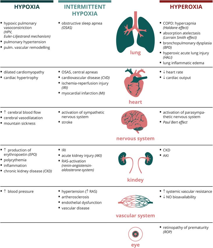
reactions. Figure 2 depicts examples; how different organ systems to high O2 breathing gas produces large amounts of ROS
respond to dysregulated O2 conditions. from mitochondria and NADPH oxidase NOX1. These radicals
are aggressive per se, thereby oxydizing proteins, lipids and
Lungs nucleic acids, but also engage in cellular signaling. Downstream
In order to provide efficient oxygenation of the blood the lung of NOX1 MAPkinases ERK1/2, JNK and p38 are activated
responds to hypoxic conditions with the physiological response (Dias-Freitas et al., 2016). Cell death is initiated by activation
of hypoxic pulmonary vasoconstriction with the purpose to of caspases 3 and 9, and finally NF-KB activation results in
redirect the blood flow from lesser to more oxygenated areas of release of pro-inflammatory cytokines such as interleukins IL-
the lung. After phases of acute hypoxia, arteries can reversibly 1ß, IL-6, IL-8, tumor necrosis factor (TNF)α and macrophage
dilate. Chronic general hypoxic conditions (as in high altitude inflammatory protein (MIP-)2. Extracellular HMGB1, a pro-
living or conditions like chronic obstructive pulmonary disease), inflammatory damage (danger) associated molecular pattern
however, lead to widespread vasoconstriction thereby increasing (DAMP), is increased, promoting ALI progression. The HMGB1
Frontiers in Physiology | www.frontiersin.org 8 August 2020 | Volume 11 | Article 947Tretter et al. Investigating Disturbances of Oxygen Homeostasis FIGURE 2 | Organ-specific physiological and pathological responses to different oxygen conditions. Abbreviations: AKI, acute kidney injury; BPD, bronchopulmonary dysplasia; COPD, chronic obstructive pulmonary disease; CKD, chronic kidney disease; CVD, cardiovascular disease; EPO, erythropoetin; HALI, hyperoxic acute lung injury; HF, heart failure; HPV, hypoxic pulmonary vasoconstriction; IRI, ischemia reperfusion injury; MI, myocardial infarction; NO, nitric oxide; OSAS, obstructive sleep apnea syndrome; RAS, renin-angiotensin-aldosterone-system. receptor RAGE (receptor for advanced glycation endproducts) lung inflammation. Calfee et al. showed that lung protective plays a major role in normal lung development and functionality. ventilation (VT = 6 ml/kg) can lower RAGE levels (Calfee During hyperoxia, RAGE levels are increased, mediating et al., 2008). Destruction of the alveolar-capillary barrier is Frontiers in Physiology | www.frontiersin.org 9 August 2020 | Volume 11 | Article 947
Tretter et al.                                                                                    Investigating Disturbances of Oxygen Homeostasis

due to endothelial and epithelial cell apoptosis, and changes             due to the generation of an abundance of ROS. This phenomenon
in the expression of Na+ channels and the Na/K ATPase                     was not well understood until it was determined that the
of alveolar type-II (ATII) cells disturb the fluid balance and            physiological oxidation of hypoxanthine to xanthine and uric
ultimately lead to edema. Chemokines attract neutrophils and              acid by xanthine dehydrogenase is altered during ischemia.
macrophages that accumulate in the interstitium and air spaces.           In ischemia-reperfusion, under reduced conditions, xanthine
Activation of phagocyte NOX2 further increases ROS levels and             dehydrogenase is converted to xanthine oxidase producing large
leads to cell death.                                                      amounts of harmful ROS during the conversion of hypoxanthine
                                                                          (Wu et al., 2018).
Broncho-Pulmonary Dysplasia (BPD)                                            In the cardiovascular system, hyperoxia induces a decreased
Low gestational age neonates frequently need mechanical                   heart rate and lowered cardiac output. This is due to an
ventilation for treatment of respiratory distress syndrome. BPD           increased parasympathetic tone and a rise in systemic vascular
is a form of chronic lung disease that occurs in these patients           resistance. The increase in vascular resistance is explained by
with high incidence and is characterized by abnormal lung                 decreased ATP release from red blood cells and reduced NO
compliance, increased pCO2 , lung inflammation and pulmonary              bioavailability. Prolonged hyperoxia has an impact on endothelial
hypertension following disrupted microvascular maturation                 NO synthase (eNOS) and increased ROS converts available NO
and poor alveolarization. Triggers are deficiency in vascular             to peroxynitrite (nitrosative stress). Also, release of NO from
endothelial growth factor (VEGF), matrix metalloproteases                 S-nitrosothiole is inhibited.
and lack of antioxidants. Oxygen partial levels in utero are
sufficiently low (20–30 mmHg) to ensure proper angiogenesis
and extracellular matrix (ECM) deposition. However, pre-term              Nervous System
birth at an early stage interrupts ongoing lung development               Reduced O2 supply to the brain (hypoxia) is counter-acted by
and instant hyperoxic conditions lead to massive oxidative stress         a regulation of cerebral blood flow and vasodilatation in order
(Valencia et al., 2018).                                                  to meet the metabolic demand. Further adaptations are changes
                                                                          in regulation of respiratory rate, metabolism, enzyme activities
                                                                          and gene expression, which provide a certain hypoxic-ischemic
Heart
                                                                          tolerance. This tolerance can be extended to some extent with
Increased pulmonary arterial pressure in response to chronic
                                                                          non-injurious hypoxic exposures at an appropriate time interval
hypoxia results in hypertrophy of cardiac myocytes and
                                                                          and dosage (“preconditioning”). However, severe hypoxia can
thickenening of the cardiac muscle wall. Cardiomyocyte cell
                                                                          lead to cognitive impairment and seizures. An example for an
death leads to a thinning of the remaining heart muscle as well
                                                                          associated pathology is acute mountain sickness, that goes along
as dilation of the chamber (dilated cardiomyopathy).
                                                                          with symptoms like headache, tiredness, dizziness and vomiting.
    Effects of intermittent hypoxia on the heart are encountered
                                                                          According to the alveolar gas equation the partial pressure of
in diseases related to sleep-disordered breathing such as OSAS as
                                                                          oxygen in the alveoli is reduced due to a reduced atmospheric
well as in central apnea (CA) independent of sleep or wakefulness.
                                                                          pressure at high altitudes.
OSAS is caused by cyclic upper airway obstruction during sleep
                                                                             Intermittent hypoxia has an enhancing effect on the
and can lead to several types of cardiovascular disorders, like
                                                                          sympathetic activity, carotid body activity, inflammation,
systemic hypertension, cardiac arrhythmias, infarction and heart
                                                                          oxidative stress and endothelial dysfunction. Further, the
failure. CA is caused by a temporary withdrawal of the respiratory
                                                                          bioavailability of NO is reduced, all of which, depending on the
drive from the brainstem and is observed in heart failure
                                                                          severity of intermittent hypoxia, can contribute to an increased
patients in form of the Cheyne-Stokes respiration (CSR) with
                                                                          risk of cognitive decline and stroke.
cycles of crescendodecrescendo ventilation and apnea/hypopnea
                                                                             Breathing gas with high O2 concentrations (hyperoxia) bears
(Costanzo et al., 2015). CSR is interpreted to be caused by an
                                                                          the risk of cerebral toxicity. The most dramatical manifestation
increased chemosensitivity to hypoxia and hypercapnia as well
                                                                          are generalized tonic-clonic grand mal seizures upon exposure
as a chemoreflex delay due to an increased circulatory time
                                                                          to pure oxygen under supra-atmospheric pressures (diving,
(Ponikowski et al., 2001; Giannoni et al., 2019). Production of
                                                                          hyperbaric chamber), named the “Paul-Bert effect”. Hyperbaric
pro-inflammatory cytokines, ROS and mediators of the renin-
                                                                          oxygenation (HBO) therapy is implicated in carbon monoxide
angiotensin system (RAS) in the carotid body underly the
                                                                          (CO)-poisoning, decompression injury and gas embolism.
increased chemosensitivity (Aimo et al., 2020).
    Intermittent hypoxic conditions are also present in ischemia-
reperfusion injury (IRI) and myocardial infarction (MI). IRI is           Kidneys
probably one of the best-studied examples of oxidative injury             The kidneys have a relatively low innate O2 tension also in
following O2 deprivation with subsequent O2 toxicity. It occurs           the healthy individual, that is explained by the O2 shunt
whenever O2 and nutrient supply is interrupted due to blood               between the arterial and venous vessels running parallel in
vessel occlusion or clamping. A paradoxical situation is observed         the kidney. Chronic kidney disease (CKD) frequently develops
upon reperfusion. Immediate reopening of the vessel is required           from clinical conditions like diabetes mellitus, hypertension
to restore oxygen and nutrient supply to the tissues. However,            or glomerulonephritis. Risk factors are sleep apnea syndrome
depending on the time and amount of oxygen deprivation,                   and chronic obstructive pulmonary disease and progression
reperfusion induces a profound and sustained oxidative stress             of CKD is accelerated by increase of hypoxia in the kidneys.

Frontiers in Physiology | www.frontiersin.org                        10                                       August 2020 | Volume 11 | Article 947
Tretter et al. Investigating Disturbances of Oxygen Homeostasis
Hypoxia induces activity of HIF, which upregulates down- with critical illness, stroke, trauma, sepsis, cardiac arrest and
stream genes like VEGF and erythropoietin (EPO). HIF is myocardial infarction. However, to approach guidelines, which
inactivated by prolyl-hydroxylases (PHD) under normal oxygen provide the best possible outcome, it is necessary to look into
conditions. However, loss of PHDs results in polycythemia, which detail of the individual conditions (Selected disease-related clinic
is caused by EPO overproduction. Renal ischemia-reperfusion trials are listed in Table 1).
injury (IRI) can be a complication of major surgery resulting
in acute kidney injury (AKI), that frequently further progresses Use of Oxygen During General Surgery
to CKD. Intermittent hypoxia as hallmark for instance of OSA Perioperatively, a FiO2 of 0.8 vs 0.3 during anesthesia and
can aggravate renal disease by vascular/endothelial dysfunction, for several hours after is mostly applied in order to prevent
oxidative stress, inflammation, increased sympathetic nervous SSI, nausea and vomiting, and to promote wound healing.
system activity and dysregulation of the RAS, especially activation Preoxygenation with 100% O2 is often used in order to provide
of angiotensin II. Supplemental O2 can alleviate hypoxic enough time for endotracheal intubation (“safe time” during
conditions also in the kidneys, but excess O2 also exerts toxicity in apnea); however, lower concentrations of O2 might be preferable
this organ which can ultimately lead to kidney disease or failure. in order to avoid formation of atelectasis.
In the 1980s, it was proposed that giving high fractions of
Vascular System O2 in the perioperative phase might have a positive side-effect:
Organ and cellular homeostasis are maintained by the vascular increased ROS has bactericidal properties like an antibiotic and
system, that responds to changes in O2 . In contrast to the might therefore reduce SSI, especially in abdominal surgeries
pulmonary circulation, the systemic circulation responds to (Knighton et al., 1984). Also, oxidative bursts of neutrophils
hypoxia with vascular dilation and only with more severe hypoxia require molecular O2 . In the following years, several further
with peripheral vasoconstriction to redistribute the oxygenated approaches to explain this beneficial effect were taken: a
blood to the organs. Chronic intermittent hypoxia (CIH) can reduction of inflammation and a boost of local macrophages, as
lead to hypertension due to activation of the sympathetic nervous well as a decreased release of TNFα by leukocytes was discussed.
system and the RAS, scavenging of NO, increased levels of However, a higher mortality and tumor recurrence was observed
endothelin and altered baroflex control. A hallmark of CIH is in cancer patients. Three trials reported statistically significant
endothelial dysfunction and oxidative stress and development of results (Greif et al., 2000; Belda et al., 2005; Myles et al., 2007).
artherosclerosis or vascular disease. Hyperoxia and the associated All other trials either did not reach statistical significance or
generation of ROS reduces the bioavailability of NO resulting in were for other reasons inconclusive (Meyhoff, 2019). A meta-
an increased systemic vascular resistance. analysis from two trials on surgical site infections in patients
undergoing elective colectomy did not reveal an increase in long-
Eye term mortality (Podolyak et al., 2016). The WHO guideline from
Prematurely born infants who receive neonatal intensive care 2016 advocating the use of 80% O2 in anesthesized, intubated
including high O2 therapy to support respiration are especially and ventilated patients during and up to 6h after surgery was
prone to develop retinopathy of prematurity (ROP). Retinal based on a subgroup analysis from precendent trials and initially
vascularization is a process that normally develops under low O2 ignored potential harmful effects, which were the main points
tension. Excess O2 leads to disorganized growth of retinal vessels, of subsequent criticism (Meyhoff et al., 2017). Potential hazards
scar formation and retinal detachment, in the worst case leading would be an increase in mortality and respiratory complications
to blindness (Hellstrom et al., 2013). (Meyhoff et al., 2012; Staehr-Rye et al., 2017). An earlier
Cochrane review from 2015 stated that evidence was insufficient
to support the use of increased perioperative O2 (Wetterslev et al.,
THERAPEUTIC STRATEGIES: WHERE 2015). Therefore, the WHO re-assessed the available evidence
DO WE STAND AND WHAT IS THE regarding the effectiveness of high FiO2 to reduce SSI, and
FUTURE? taking into consideration possible associated adverse events, the
strength of the recommendation was revised from strong to
Literature on outcome studies regarding the liberal use of conditional in 2018.
supplemental O2 and consecutive hyperoxemia is heterogenous. Another very recently published paper still advocates
A recent review of 37 published studies revealed that in half of the hyperoxia in noncritically ill intubated adult surgical patients
studies, statistical evaluation resulted in no detectable association (Weenink et al., 2020). The unclear risk-benefit ratio led Meyhoff
between hyperoxemia and detrimental clinical outcome. Other and others to stress the need of large trials to obtain the
studies, however, found higher mortality in the context of stroke, required statistical power to evaluate the effects on SSIs (Meyhoff
cardiac arrest and traumatic brain injury (TBI) (Stolmeijer et al., et al., 2011; Cohen et al., 2018; Meyhoff, 2019). Conflicting
2018). A recent meta-analysis of high-quality data from 25 conclusions exist from studies comparing longterm mortality
randomized controlled trials including more than 16,000 patients among colorectal cancer surgery patients who received 80% vs.
concluded that liberal oxygen therapy (SpO2 = 94–99%) is 30% O2 during surgery. The PROXI trial identified a slightly
very likely detrimental with regard to short-term and long-term earlier reoccurrence of cancer and increased mortality among
mortality when compared to conservative administration of O2 cancer patients in the 80% O2 group (Meyhoff et al., 2009).
(Chu et al., 2018). This overarching analysis included patients Fonnes et al. re-analyzed data from the PROXI trial and
Frontiers in Physiology | www.frontiersin.org 11 August 2020 | Volume 11 | Article 947You can also read