The Department of Biosciences and Nutrition - Scientific Report 2013-2015 - Karolinska ...
←
→
Page content transcription
If your browser does not render page correctly, please read the page content below
The Department of
Biosciences and Nutrition
Scientific Report 2013-2015
Contents

Introduction ........................................................................................................................................... 4
The department in brief ........................................................................................................................ 5
   Organisation ........................................................................................................................................ 5
   Finances 2013-2015 ............................................................................................................................ 5
Discoveries ............................................................................................................................................. 7
Renewals ................................................................................................................................................ 7
New groups and improved gender balance......................................................................................... 7
Research ................................................................................................................................................. 8
   Ageing ................................................................................................................................................. 9
   Bioinformatics ................................................................................................................................... 11
   Bioorganic Chemistry........................................................................................................................ 14
   Cancer Biology .................................................................................................................................. 16
   Developmental Biology..................................................................................................................... 20
   Developmental Neurobiology ........................................................................................................... 22
   Epigenetics ........................................................................................................................................ 24
   Functional Genomics......................................................................................................................... 28
   Molecular Endocrinology .................................................................................................................. 38
   Nutrition ............................................................................................................................................ 40
   Signal Transduction........................................................................................................................... 42
   Stem Cells ......................................................................................................................................... 44
   Structural Biology ............................................................................................................................. 46
   Toxicology ........................................................................................................................................ 52
   Virology ............................................................................................................................................ 53
Core Facilities ........................................................................................................................................55
   BEA - the core facility for Bioinformatics and Expression Analysis................................................ 55
   CryoEM – Cryo Electron Microscopy Facility ................................................................................. 56
   LCI - The Live Cell Imaging facility ................................................................................................ 57
   KHTC - Karolinska high throughput center ...................................................................................... 58
Dissertations 2013-2015 ........................................................................................................................ 59
Undergraduate teaching ..................................................................................................................... 60
Contact.................................................................................................................................................. 62
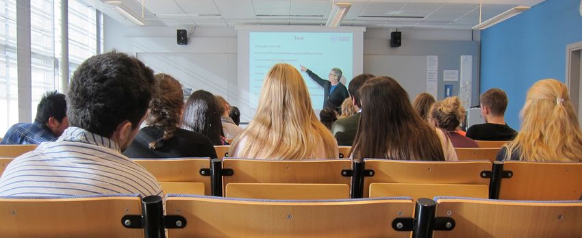
Introduction

Karl Ekwall, Head of Department

I am proud of having been appointed the new head of the Department of Biosciences and Nutrition
“BioNut” in August 2015 and I am very inspired and motivated to lead the department into an exciting
future within KI, that will be undergoing large structural improvements in the next few years.
Historically, BioNut was created in 2006 by merging two departments Biosciences and Medical
Nutrition and it has grown to become the largest biomedical department at the KI South Campus with
a strong focus on basic research. As you can see in this report of our activities 2013-2015, we cover a
wide range of topics in biomedical research ranging from Ageing to Virology, with many prominent
research groups. We also run high level educational activities including a master’s programme in
Nutrition in collaboration with SU and we contribute to the Biomedicine programme at KI. In the
future, we wish to strengthen the quality of research and be attractive for collaboration with the
healthcare sector and other partners at KI South Campus. We also aim to strengthen our international
environment in basic and applied experimental research and education.
My visions for the next three years (2016-2018)
At the end of 2017, our new research building NEO will be ready in Huddinge. The move to NEO will
be a major step for BioNut, since it will provide new conditions for our infrastructure, as well as
possibilities to change the contents of our activities, thereby creating better conditions for
collaboration across our research groups, with neighbouring departments and across research
disciplines. In this context I have three important goals for BioNut:
1. To create a more integrated coherent BioNut Department: We wish to develop a creative and safe
work environment, with a constructive collegial atmosphere.
2. To increase and improve our collaborations with our neighbouring Departments and Research
Centres in particular NVS, LabMed, MedH, KTH STH and CIMED (SLL) and the Karolinska
Hospital in Huddinge: The goal here is to obtain a very open environment for research and education
with common areas where all employees can meet and interact.
3. Finally we should not forget ‘Tredje uppgiften’ - our collaboration with Swedish society and
enterprise (näringsliv): I am determined to make BioNut a consistently attractive partner in this
context by inspiring and enabling higher standards of excellent quality research and education.

Karl Ekwall
The department in brief
Organisation

Finances 2013-2015

INCOME STATEMENT                     2015      2014      2013
Revenues from grants                49 668    51 739    47 504
Revenues from fees                   9 598    10 657    13 260
Revenues from allowances           145 677   175 438   169 801
Internal revenues                   21 300    17 235    20 075
Total revenues                     226 243   255 068   250 640
Key financial figures
External / total financing          72,7%     72,9%     68,4%
Research and doctoral education     93,9%     95,8%     95,4%
First and second level education     6,1%      4,2%      4,6%

                                                                 5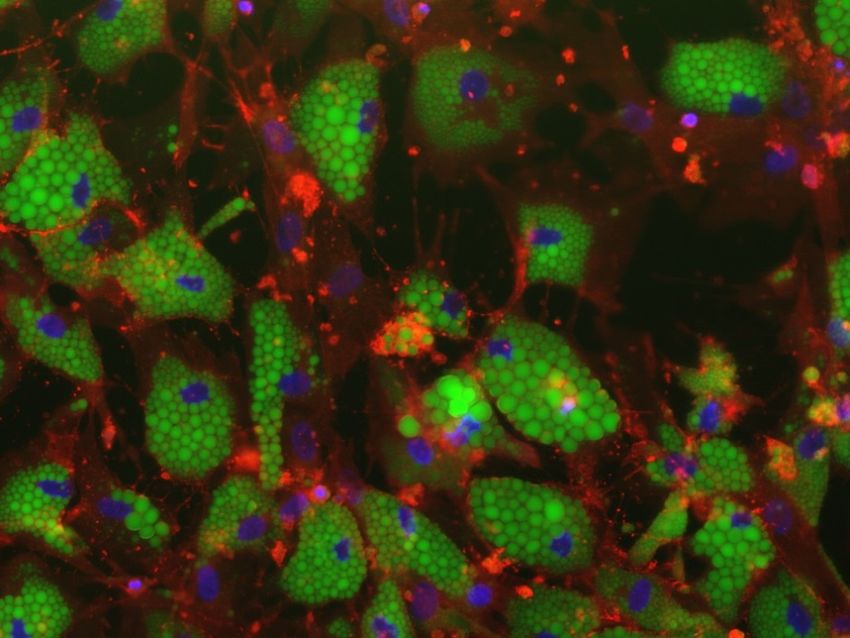
Revenues 2015

                            Costs 2015

External grants 2013-2015

                                     6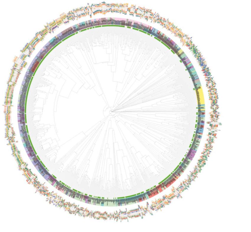
Discoveries
There are many examples of key scientific discoveries made at BioNut. This report highlights selected
papers from each of the research groups. Here follows some examples from two different research
areas i.e. functional genomics and nutrition. Jussi Taipale’s group have uncovered the DNA binding
specificities of human transcription factors and mapped the binding of these factors to cohesion sites
(Cell 2013; Cell 2015). Marie Löf’s group made an important contribution to childhood obesity
research with a mobile phone tool to improve dietary habits (BMC public health 2015; Int J Obesity in
press). Please note that our research covers as many as 14 different research areas resulting in many
important findings published in 2013-2015.

Renewals
New seminar series started during 2015 include “Chairman’s seminar series”, “Introduction seminars
by new BioNut Group Leaders” and “Group leaders journal club”. The chairman’s seminar series aims
to highlight selected top scientists at Swedish universities and is designed to specifically promote
female scientists. The introduction seminars aim to increase the visibility for new research groups at
our department. Group leaders’ journal club has been created to inspire our most junior scientists, the
newly registered PhD students, and to increase their academic networks.

                                     Group leaders’ Journal Club

New groups and improved gender balance
One of our top priorities is to obtain an improved gender balance in the department. At the beginning
of 2015 we had 4 female and 17 male group leaders. During 2015 and the beginning of 2016, five
newly established PI’s with research groups, one new Professor with a group, three Guest Professors,
and one foreign Adjunct Professor have been recruited. Of these 10 new group leaders 4 are female,
which is a significant improvement of the gender balance on the group leader level. The department
head is very pleased to see this positive development and to lead such a dynamic department
supporting newly established teams and international recruitments. Please note that the activities of
seven new PI’s, recruited during 2015 and 2016, will not be presented until the next scientific report
(2016-2018).

                                                                                                         7
Name of new group leader             Research area
Martin Bergö                         Biochemical and medical importance of CAAX protein
                                     processing and the role of reactive oxygen species and
                                     antioxidants in cancer
Piero Carninci                       Studies of mammalian transcriptomes
(Foreign adjunct professor)
Pekka Katajisto                      Tissue homeostasis loss and ageing
Andreas Lennartsson                  Epigenetic regulation of acute myeloid leukaemia
Linda Sofie Lindström                Molecular and genetic cancer epidemiology
Victoria Menendez Benito             Centrosomes in cell division
Cecilia Williams                     Hormone signalling and non-coding RNAs in cancer
(Guest professor)

Research
Three group leaders have left the Department during the period 2013-2015. Thomas Bürglin
(Regulation of cell specialization, Mauro D’Amato (Molecular genetics of gastrointestinal disease)
and Dan Segerbäck (UV radiation and DNA damage). Their research is not presented in this report.

BioNut administration

                                                                                                     8
Ageing
Maria Eriksson
+46-8-524 810 48, maria.eriksson.2@ki.se
http://ki.se/bionut/eriksson

Genetic mechanisms of premature and healthy ageing
Genetic mechanisms that affect ageing are of high interest to society, yet
not well understood. Although genetic variation between individuals has
been studied extensively, few studies have investigated genetic variation
within an individual and genetic variation acquired during ageing.
Hutchinson-Gilford progeria syndrome (HGPS, progeria) is a very rare
genetic disorder with several clinical features reminiscent of premature
ageing, including atherosclerosis, osteoporosis, loss of subcutaneous fat
and hair, and thinning of the skin.
                                           The overall aim of our research is to identify genetic
                                           mechanisms that contribute to the declined tissue
                                           homeostasis associated with ageing and disease mechanisms
                                           in progeria. In our studies we use next generation
                                           sequencing technologies to investigate the genome of
                                           human cells and cells from transgenic models of premature
                                           ageing to identify disease mechanisms and early targets for
                                           treatment. We expect our findings to contribute to the
                                           understanding of genetic mechanisms in ageing and age-
                                           associated disease, and ultimately the prevention and
                                           treatment of these processes.

                                           Murine skin stained for nuclear lamina (red), DNA (blue), and
                                           keratin (green).

Selected publications
1) Rodríguez SA, Grochová D, McKenna T, Borate B, Trivedi NS, Erdos MR, Eriksson M. Global
genome splicing analysis reveals an increased number of alternatively spliced genes with aging. Aging
Cell. 2015; Dec 21.
2) Strandgren C, Nasser HA, McKenna T, Koskela A, Tuukkanen J, Ohlsson C, Rozell B, Eriksson M.
Transgene silencing of the Hutchinson-Gilford progeria syndrome mutation results in a reversible
bone phenotype, whereas resveratrol treatment does not show overall beneficial effects. FASEB J.
2015; 29: 3193-3205.
3) Baek JH, Schmidt E, Viceconte N, Strandgren C, Pernold K, Richard TJ, Van Leeuwen FW,
Dantuma NP, Damberg P, Hultenby K, Ulfhake B, Mugnaini E, Rozell B, Eriksson M. Expression
of progerin in aging mouse brains reveals structural nuclear abnormalities without detectible
significant alterations in gene expression, hippocampal stem cells or behavior. Hum Mol Genet.
2015; 24: 1305-21.
4) McKenna T, Sola Carvajal A, Eriksson M. Skin Disease in Laminopathy-Associated Premature
Aging. J Invest Dermatol. 2015; 135:2577-2583.
5) McKenna T, Rosengardten Y, Viceconte N, Baek J-H, Grochová D, Eriksson M. Embryonic
expression of the common progeroid lamin A splice mutation arrests postnatal skin development.
Aging Cell. 2014; 13: 292-302.

Prizes/Awards to group members 2013-2015
Nikenza Viceconte: Fernström travel award 2014

                                                                                                           9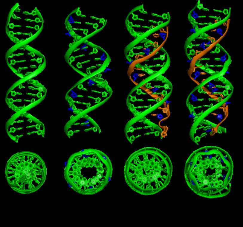
Group members
Agustin Sola Carvajal   Tomas McKenna           Emelie Wallén Arzt
Irene Franco            Sofia Rodriguez         Nikenza Viceconte
Hafdís Helgadóttir      Raquel Pala Rodriguez   Jean-Ha Baek
Gwaldys Revechon        Charlotte Strandgren

                                                                     10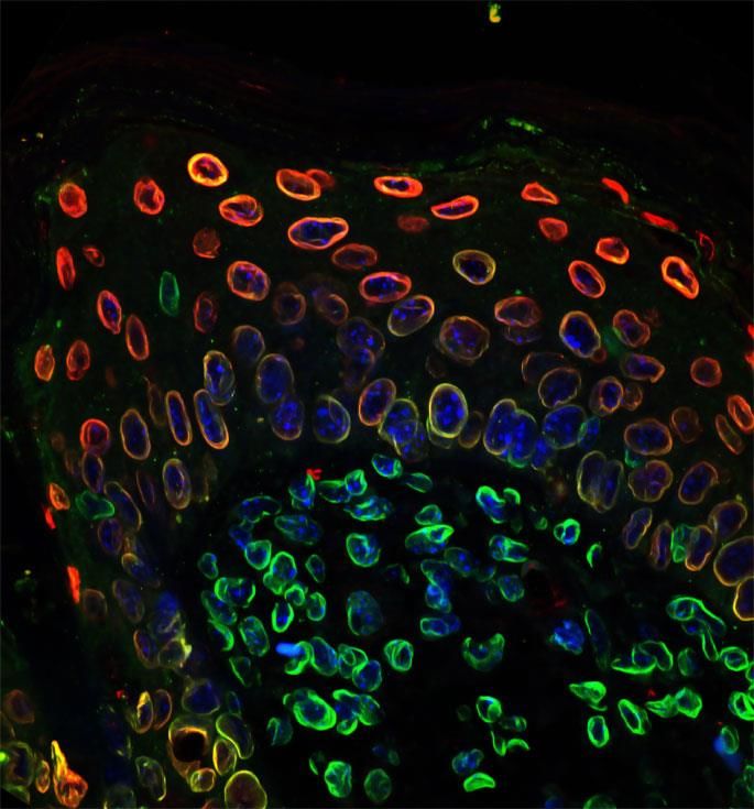
Bioinformatics
Carsten Daub
+46-8-524 812 24, carsten.daub@ki.se
http://ki.se/bionut/daub

Transcriptomics for gene regulation in development and disease
Our interests focus on the understanding of the molecular basis of gene
regulation of diseases through translational research. The key aspects of
our work include genome-wide gene expression analysis from human
patient samples employing technologies such as RNA-Seq, Cap Analysis
of Gene Expression (CAGE) or small RNA sequencing (miRNA).

Our analysis goes beyond differentially expressed genes and identifies a variety of candidate elements
responsible for the observed expression differences in the disease patients and the associated clinical
phenotypes. Application of sequencing technology to the transcriptome previously has been utilized to
uncover a range of regulatory elements and mechanisms, including regulation through transcription
factors (TFs), nearby but distinct alternative promoters resulting in the same protein but employing
different sets of regulatory TFs, expression of anti-sense RNA to modulate the sense-RNA and the
regulatory role of expressed repeat elements and miRNAs. Subsequent functional validation studies
confirm the suggested regulatory relationships.

Selected publications
1) Yu NY, Hallström B M, Fagerberg L, Ponten F, Kawaji H, Carninci P, Forrest A R; Fantom
Consortium, Hayashizaki Y, Uhlén M, Daub CO. Complementing tissue characterization by
integrating transcriptome profiling from the Human Protein Atlas and from the FANTOM5
consortium. Nucleic Acids Res. 2015; 43;14 6787-98.
2) Arner E et al. Transcribed enhancers lead waves of coordinated transcription in transitioning
mammalian cells. Science. 2015; 347;6225 1010-1014.
3) Persson H, Kwon A T, Ramilowski J A, Silberberg G, Söderhäll C, Orsmark-Pietras C, Nordlund
B, Konradsen J R, de Hoon M J, Melén E, Hayashizaki Y, Hedlin G, Kere J, Daub CO.
Transcriptome analysis of controlled and therapy-resistant childhood asthma reveals distinct gene
expression profiles. J Allergy Clin Immunol. 2015; Sep;136(3):638-48.
4) Andersson R et al. An atlas of active enhancers across human cell types and tissues. Nature. 2014;
Mar 27;507(7493):455-61.
5) FANTOM Consortium and the RIKEN PMI and CLST (DGT). A promoter-level mammalian
expression atlas. Nature. 2014; Mar 27;507(7493):462-70.

Research networks 2013-2015
FANTOM5 “Functional Annotation of the Mammalian Genome” coordinated by the Riken Institute,
Omics Science Center, Japan
DANIO-CODE Encyclopedia of DNA Elements in Zebrafish

Group members
Tahmina Akhter                     Abdul Kadir Mukarram               Niyaz Yoosuf
Olga Hrydziuszko                   Enrichetta Mileti                  Nancy Yu
Matthias Hörtenhuber

                                                                                                    11
Bioinformatics
Joseph Rafter
+46-8-524 835 45, joseph.rafter@ki.se
http://ki.se/bionut/rafter
Understanding the interplay between gut microbiota, gut function and
host genes in the generation of gastrointestinal symptoms and disease
The human gut is colonized by billions of microbes, which constitute a
complex community known as the gut microbiota. The microbiota exerts
positive physiological/nutritional effects, and alterations in its composition
are associated with conditions such as inflammatory bowel disease, colon
cancer and metabolic disorders.
Our work (collaboration with Mauro D’Amato, BioCruces Institute, Bilbao) attempts to understand
how host genes, gut microbiota and gastrointestinal function are interconnected and how they are
eventually related to GI disease (IBS, IBD). This involves correlating variation in microbiota
composition with variation in gut function and correlating human genetic variation with alterations in
microbiota and gut function.
We showed that, in humans, a correlation exists between microbiota and gut function, in that measures
of stool frequency and pattern that are associated with gut transit time show a negative correlation with
α-diversity indices. We showed that variation in the human genome contributes to shaping the
composition of the gut microbiota. We provide preliminary evidence that specific genes/associated
pathways may be relevant to the control of bowel movement frequency, and establish a set of
candidate targets for follow-up and replication in independent datasets.
Therapies can be considered, where
modifications in gut microbiota may
be introduced via pharmacological or
dietary changes, in order to restore
“normal” intestinal flora and human
wellbeing.
The interplay between Host Genes, Gut
Microbiota and Gut Function in the
generation of GastroIntestinal Disorders
Selected publications
1) Quince C, Lundin E, Andreasson A N, Greco D, Rafter J, Talley N J, Agreus L, Andersson A F,
Engstrand L, D’Amato M. The impact of Crohn’s disease genes on healthy human gut microbiota: a
pilot study. Gut. 2013; 62: 952-4.
2) Ek W E et al. Exploring the genetics of irritable bowel syndrome: a GWA study in the general
population and replication in multi-national case-control cohorts. Gut. 2015; 64:1774-82.
123) Westerlind H, Mellander M R, Bresso F, Munch A, Bonfiglio F, Assadi G, Rafter J, Hübenthal
M, Lieb W, Källberg H, Brynedal B, Padyukov L, Halfvarson J, Törkvist L, Bjork J, Andreasson A,
Agreus L, Almer S, Miehlke S, Madisch A, Ohlsson B, Löfberg R, Hultcrantz R, Franke A,
D'Amato M. Dense genotyping of immune-related loci identifies HLA variants associated with
increased risk of collagenous colitis. Gut. 2015; Nov 2. pii: gutjnl-2015-309934. doi:
10.1136/gutjnl-2015-309934. [Epub ahead of print]
Prizes/Awards to group members 2013-2015
Maria Henström: National Scholar Award (UEG) 2015.
Research network 2013-2015
FP7-KBBE-2007-2A-222720 (CP-IP - Large-scale integrating project)
TORNADO: Molecular Targets Open for Regulation by the gut flora – New Avenues for improved
Diet to Optimize European health” (2009 – 2014).
Group members
Fatemeh Hadizadeh
Maria Henström
13Bioorganic Chemistry
Roger Strömberg
+46-8-524 810 24, roger.stromberg@ki.se
http://ki.se/bionut/stromberg
Targeted oligonucleotides and other alternative approaches for
treatment of disease
The main research aims at chemically enabling novel treatments for
inherited, metabolic or infectious disease. This through development of
oligonucleotide (ON) therapeutics that target RNA molecules, and in
treatment of infections also by triggering our own innate defense molecules
and mechanisms.
ON therapeutics can be used to target proteins difficult to modulate with small molecule drugs and
also to affect regulatory non-coding RNAs. Advances in ON chemistry can increase potency and
provide new therapeutic molecules. The field of ON therapeutics encompass different modes of action,
including effects on mRNA, microRNAs, pre-mRNA as well as mRNA therapy.
A key feature is synthetic modified oligonucleotides and their conjugates with other biomolecules that
act as signals directing them to the site of action.
Globally, infections are responsible for two thirds all deaths among children of the age 1 month to 4
years. A possible treatment is induction of our own antimicrobial peptides.
Three main directions of the research are:
Oligonucleotide based artificial nucleases and PNAzymes for biomedical applications and also aiming
at treatment of Malaria.
Stabilized, cell penetrating and target seeking oligonucleotides for enhanced therapy, aiming at
treatment of inherited, metabolic and infectious diseases.
Treatment of infections through substances that induce our own innate defense (antimicrobial
peptides) against microbes as well as through enhancing autophagy by targeting of microRNA.
To the left: Confocal microscopy image of uptake of
our cell penetrating AECM oligonucleotides in U-2
OS cells treated with a fluorescein-labelled fully
AECM modified oligonucleotide (green colour).
To the right: Graph showing reduction of bacterial
count in Shigella infected rabbits after treatment with
different doses of an inducer of antimicrobial
peptides. Treated rabbits recovered clinically in four
days.
Selected publications
1) Ghidini, A, Ander, C, Winqvist, A, Strömberg, R. (2013) An RNA modification with remarkable
resistance to RNase A. Chem. Comm., 49, 9036.
2) Milton S, Honcharenko D, Moreno PMD, Rocha C, Smith CIE, Strömberg R. (2015) Nuclease
resistant oligonucleotides with cell-penetrating properties. Chem Comm. 51, 4044. (Patent application:
Strömberg R, Milton S, Honcharenko D. PCT Int. Appl. WO 2014131892 A1 20140904).
3) Honcharenko M, Zytek M, Bestas B, Moreno P, Jemielity J, Darzynkiewicz E, Smith C I E,
Strömberg R. (2013) Synthesis and evaluation of stability of m3G-CAP analogues in serum-
supplemented medium and cytosolic extract. Bioorg. Med. Chem., 21, 7921
4) Ghidini A, Steunenberg P, Murtola M, Strömberg R (2014) Synthesis of PNA Oligoether
Conjugates. Molecules, 19, 3135
5) Honcharenko D, Bose PP, Maity J, Kurudenkandy FR, Juneja A, Flöistrup E, Henrik Biverstål H,
Johansson J, Nilsson L, Fisahn A, Strömberg R. (2014) Synthesis and Evaluation of Antineurotoxicity
Properties of an Amyloid-β Peptide Targeting Ligand Containing a Triamino Acid. Org. Biomol.
Chem., 12, 6684
14Research networks 2013-2015
Swedish Research Council’s “framtidens behandlingar”
MMBIO, EU training network
Group members
Ghidini Alice Jezowska Herrera Martina Ottosson Håkan
Honcharenko Dmytro Maity Joytirmoy
Honcharenko Malgorzata Murtola Merita
15Cancer Biology
Staffan Strömblad
+46-8-524 811 22, staffan.stromblad@ki.se
http://ki.se/bionut/stromblad
Cell biology of cancer
Our research focuses on key cellular events in cancer progression, with
emphasis on cell migration and p21-activated kinase 4 (Pak4). Depending
on the properties of the surrounding extracellular matrix, cancer cells can
utilize different migration strategies for dissemination. This adaptive
behavior expands the range of tissue contexts under which cancer cells can
efficiently invade.
Expanding on this knowledge, we recently identified two distinct modes of mesenchymal migration
and that perturbing cell-ECM interactions or tensile forces caused switching between these modes. We
combine different quantitative microscopy techniques, including traction force microscopy and FRET
signalling biosensors aiming to reveal mechanisms of migration mode switching and how distinct
temporal phases are controlled and executed. These studies are expected to provide novel treatment
opportunities targeting the most malignant aspect of any cancer, the ability to metastasize.
Pak4 is overexpressed in several human cancers and we previously linked Pak4 to promotion of cancer
cell migration and to control of cell growth. To examine the role of Pak4 in cancer progression, our
laboratory has in place a number of techniques, including in vitro models, transgenic mice, cancer
mouse models, patient database bioinformatics and patient derived material, which will be combined
within our comprehensive yet molecularly detailed investigations, stretching also into testing Pak4
pharmacological targeting.
Simultaneous combination of Traction force
microscopy (TFM) and a signalling biosensor (RhoA
FRET) in a human colon cancer cell. Left: RhoA
FRET heat map; Right: Calculated TFM force fields
displayed as force heat map. Image: Jianjiang Hu
Selected publications
1) Lock J G, Jafari-Mamaghani M, Shafqat-Abbasi H, Gong X, Tyrcha J, Strömblad, S. Plasticity in
the Macromolecular-Scale Causal Networks of Cell Migration. PLoS One. 2014; 9: e90593.
2) Kiss A, Gong X, Kowalewski J M, Shafqat-Abbasi H, Strömblad S, Lock J G. Non-monotonic
cellular responses to heterogeneity in talin protein expression-level. Integrative Biol. 2015; 7, 1171 –
1185.
3) Zhuang T, Zhu J, Li Z, Lorent J, Zhao C, Dahlman‐Wright K, Strömblad S. P21‐activated kinase
group II small compound inhibitor GNE‐2861 perturbs estrogen receptor alpha signaling and
restores tamoxifen sensitivity in breast cancer cells. Oncotarget. 2015; 6, 43853-68.
4) Hernández-Varas P, Berge U, Lock, J.G, Strömblad S. A plastic relationship between vinculin-
transmitted tension and adhesion area defines adhesion complex size and lifetime. Nat Commun. 2015;
6, 7524.
5) Kowalewski JM, Shafqat-Abbasi H, Jafari-Mamaghani M, Endrias Ganebo B, Gong X,
Strömblad S, Lock JG. Disentangling Membrane Dynamics and Cell Migration; Differential
Influences of F-actin and Cell-Matrix Adhesions. PLoS One. 2015; Aug 6;10(8):e0135204.
16Research networks 2013-2015
FP7-HEALTH-2010-258068 (NoE)
Systems Microscopy Network of Excellence (2011-2015). Coordinated by prof. Strömblad.
The KI Breast Cancer theme Center (BRECT); Strömblad serves as vice director.
H2020-PHC-2014- 634107 (RIA)
Multimot: Capture, dissemination and analysis of multiscale cell migration data for biological and
clinical applications (2015- ) Strömblad is a partner.
Group members
Ulrich Berge Gabriela Imreh Helene Olofsson
Tânia Costa Alexa Kiss Parisa Rabieifar
Marianne van Dijk Jacob Kowalewski Hamdah Shafqat Abbasi
Xiaowei Gong Zhilun Li Matthias Spiess
Sara Göransson John Lock Miao Zhao
Pablo Hernández-Varas Miriam Masia-Balague Ting Zhuang
Jianjiang Hu
17Cancer Biology
Rune Toftgård
+46-8-524 810 53, rune.toftgard@ki.se
http://ki.se/bionut/toftgard
Hedgehog signalling and tissue stem cells in cancer development
The Hedgehog (Hh) signalling pathway plays a key role in directing
cellular growth and tissue patterning during embryonic development. In
normal adult physiology the pathway is implicated in stem cell
maintenance and tissue repair.
Inappropriate activation of the Hh-signalling pathway is increasingly implicated in human cancer.
Mutational inactivation or activation of core components of the Hh-pathway underlie cell autonomous
activation in basal cell carcinoma of the skin (BCC), medulloblastomas, meningiomas and
rhabdomyosarcomas. In other tumour types, such as colorectal and pancreatic cancer, tumour cells
upregulate expression of Hh-ligands that signal to the surrounding tumour stroma.
A major focus of our research is to understand the details of Hh signalling at the genetic, molecular
and structural level with emphasis on the key intracellular SUFU and GLI components. Moreover, to
elucidate how aberrant activation of this pathway influences cancer development in skin, mammary
gland and colon we combine studies of genetically modified models and patient samples.
To understand cancer biology and how to best
eradicate tumour cells it is necessary to know
also the biology of normal tissues, the nature of
tissue stem and progenitor cells and their ability
to serve as cancer cells of origin. With this aim
lineage tracing and cell fate mapping is used to
investigate the presence and functional
properties of tissue stem cells marked by
expression of Lgr5 and Lgr6 in the skin and
mammary gland.
Confocal 3D projection of mammary gland alveoli
from a pregnant mouse. A network of contractile
myoepithelial cells (green) encloses milk-producing
luminal cells (blue). Mammary progenitor cells
expressing Lgr6 were genetically labelled during
puberty and their progeny (red) traced into mid-
pregnancy.
Selected publications
1) Norum JH, Bergström Å, Andersson AB, Kuiper RV, Hölzl M, Sörlie T, Toftgård R. A conditional
transgenic mouse line for targeted expression of the stem cell marker LGR5. Dev Biol. 2015;
404(2):35-48.
2) Lauth M, Toftgård R. Think inside the BOCs: a mechanism underlying medulloblastoma
progression. Dev Cell. 2014; 31(1):1-2.
3) Villegas VE, Rahman MF, Fernandez-Barrena MG, Diaou Y, Liapi E, Sonkoly E, Ståhle M,
Pivarcsi A, Annaratone L, Sapino A, Ramirez Clavijo S, Bürglin TR, Shimokawa T, Ramachandran
S, Kapranov P, Fernandez-Zapico ME, Zaphiropoulos PG. Identification of novel non-coding RNA-
based negative feedback regulating the expression of the oncogenic transcription factor GLI1. Mol
Oncol. 2014; 8(5):912-926.
184) Cherry AL, Finta C, Karlström M, Jin Q, Schwend T, Astorga-Wells J, Zubarev RA, Del Campo M,
Criswell AR, de Sanctis D, Jovine L, Toftgård R. Structural basis of SUFU-GLI interaction in human
Hedgehog signalling regulation. Acta Crystallogr D Biol Crystallogr. 2013; 69(12):2563-2579.
5) Kumari S, Bonnet MC, Ulvmar MH, Wolk K, Karagianni N, Witte E, Uthogg-Hachenberg C,
Renauld JC, Kollias G, Toftgård R, Sabat R, Pasparakis M, Haase I. Tumor necrosis factor receptor
signaling in keratinocytes triggers interleukin-24-dependent psoriasis-like skin inflammation in
mice. Immunity. 2013; 39(5):899-911.
Prizes/Awards to group members 2013-2015
Marco Gerling: 3-year Postdoctoral Fellowship Cancerfonden 2015
Romina Croci : 2-year Postdoctoral Fellowship Barncancerfonden 2015
Research networks 2013-2015
Breast Cancer Theme Center (BRECT), Karolinska Institutet (Member PI)
Strategic Research Programme on Cancer (StratCan), Karolinska Institutet (Director)
Center for Innovative Medicine (CIMED), Karolinska Institutet (Director)
Group members
Agneta Andersson Mohammed Ferdous-Ur Rahman Fabian Schneider
Ani Azatyan Csaba Finta Stephan Teglund
Åsa Bergström Marco Gerling Elin Tüksammel
Leander Blaas Maria Hölzl Sandra Falck
Romina Croci Biljana Jovanovic Victoria Villegas
Yumei Diao Uta Rabenhorst Peter Zaphiropoulos
19Developmental Biology
Emma R Andersson
+46-8-524 873 60, emma.andersson@ki.se
http://ki.se/bionut/andersson
Genetic and environmental control of embryonic development
Our group studies the genetic underpinnings of disease, and how genes
interact with the environment to produce specific phenotypes.
Within this, our lab has two main focuses:
1. Alagille syndrome pathogenesis with a focus on biliary and vascular
development.
2. Development of ultrasound-guided in utero nanoinjection as a
powerful tool to manipulate gene expression during development.
Alagille syndrome is a pediatric disorder caused by mutations JAGGED1 or NOTCH2, which leads to
liver defects, heart defects, vertebral and ocular malformations and stereotypic facial features. We
investigate the role of Notch signaling in bile duct development, liver regeneration and liver
malignancy in a mouse model for Alagille syndrome and in human patient material using RNA
sequencing of liver and biliary organoids. We also investigate the role of Notch signaling in the
vasculature, since a large portion of Alagille patients in fact die from vascular accidents.
In order to rapidly manipulate gene expression in the developing embryo, to answer basic biological
questions in various organ systems, we have collaborated with Elaine Fuchs’s group and further
developed ultrasound-guided nanoinjection to target other organ systems than the skin. We use this
technology to screen gene libraries for roles in cancer or normal development of various organ
systems, with a focus on the nervous and hepatic system.
Vascular development is controlled by
Notch signaling. Our lab uses the retina as a
model for angiogenesis to study how blood
vessels grow, remodel and establish
functional arteries (vascular smooth muscle
cells labelled in green) and veins in the
nervous system (astrocytes labelled in red).
Selected publications
1) Andersson ER, Lendahl U. Therapeutic modulation of Notch signalling– are we there yet?
requested review. Nat Rev Drug Discov. 2014; May; 13(5):357-78.
2) Main H, Radenkovic J, Lendahl U, Andersson ER. Notch signaling maintains neural rosette
polarity. PLoS One. 2013; May 10;8(5):e62959.
3) Andersson ER, Saltó C, Villaescusa JC, Cajanek L, Yang S, Bryjova L, Nagy II, Vainio SJ,
Ramirez C, Bryja V, Arenas E. Wnt5a cooperates with canonical Wnts to generate midbrain
dopaminergic neurons in vivo and in stem cells.. Proc Natl Acad Sci USA. 2013; Feb
12;110(7):E602-10.
20Prizes/Awards to group members 2013-2015
Emma R Andersson: Sven and Ebba Christina Hagberg’s prize 2014
Simona Hankeova: Best Technique Presentation, From Basic to Clinic, March 2015, Sweden; title:
Imaging of 3D structures using resin casts and µC 2015
Best Poster Presentation, From Basic to Clinic, March 2014, Sweden ; title: Notch Signalling and
Alagille Syndrome 2014
Research networks 2013-2015
Center for Innovative Medicine (CIMED), Karolinska Institutet
21Developmental Neurobiology
Peter Swoboda
+46-8-524 810 70, peter.swoboda@ki.se
http://ki.se/bionut/swoboda
Understanding the role of cilia in human brain conditions
We work on the biology of cilia, crucial signal reception and transduction
organelles present on many different eukaryotic cell types. Cilia stick out
from the cell surface, akin to antennae. Thereby cells can communicate
with their immediate environment.
Many components of cilia are controlled by RFX transcription factors. With their unique DNA
binding domain RFX factors bind to the X-box promoter motif (RFX = Regulatory Factor binding to
the X-box). In this way they directly regulate their target genes. In animals RFX factors regulate genes
involved in the immune response and in cilia development and function.
By searching for X-boxes in several animal genomes (C. elegans, Drosophila, mouse and humans) we
have identified numerous direct RFX targets. We confirmed many of these targets to function in cilia
by using various assays in C. elegans worms and in human (neuronal) cell lines. Accordingly we
assigned a number of these cilia genes – upon malfunction – to being at the root of a human disease
class termed ciliopathies. We focus on the cell biological underpinnings of human brain-related,
suspected ciliopathies, like dyslexia (reading disorder). We attempt to tie together the different
biological functions (in ciliogenesis) of direct RFX targets by cross-comparing a large number of
candidate X-box regulated genes in various different genomes. With these approaches we will be able
to track RFX target gene modules from basic biological function to disease states in humans.
The head of the worm C.
elegans is shown. Two
bilaterally symmetrical “salt-
tasting” neurons are marked
with GFP. Through cell-specific
genetic rescue experiments the
neuron at the top has regained a
fully functional sensory cilium
(arrowhead) and thus is able to
“taste” salt (cf. calcium imaging
trace on the left). The neuron at
the bottom remains mutant for
cilia development and thus is not
able to “taste” salt (cf. calcium
imaging trace on the left).
Selected publications
1) Gonzalez-Barrios M, Fierro-Gonzalez JC, Krpelanova E, Mora-Lorca JA, Pedrajas JR, Peñate X,
Chavez S, Swoboda P, Jansen G, Miranda-Vizuete A. Cis- and trans-regulatory mechanisms of gene
expression in the ASJ sensory neuron of Caenorhabditis elegans. Genetics. 2015; May;200(1):123-
134.
2) Klang IM, Schilling B, Sorensen DJ, Sahu AK, Kapahi P, Andersen JK, Swoboda P, Killilea DW,
Gibson BW, Lithgow GJ. Iron promotes protein insolubility and aging in C. elegans. Aging. 2014;
Nov;6(11):975-991.
223) Arodin L, Miranda-Vizuete A, Swoboda P, Fernandes AP. Protective effects of the thioredoxin
and glutaredoxin systems in dopamine induced cell death. Free Radic Biol Med. 2014; Aug;73:328-
336.
4) Choksi SP, Lauter G, *Swoboda P, *Roy S. Switching on cilia: transcriptional networks regulating
ciliogenesis. Development. 2014; Apr;141(7):1427-1441. (*equal contribution)
5) Henriksson J, Piasecki BP, Lend K, Bürglin TR, Swoboda P. Finding ciliary genes: a
computational approach. Methods Enzymol. 2013; (Cilia, Part B, ch. 16, ed. W. Marshall);525:327-
351.
Prizes/Awards to group members 2013-2015
Gilbert Lauter: Fellowship award from Hjärnfonden (HF), Fellowship award from Svenska Sällskapet
för Medicinsk Forskning (SSMF) 2015.
Research networks 2013-2015
KI Neurosciences network
Nordic C. elegans researcher network (Nord-Forsk)
Nordic Cilia and Centrosome network (Nord-Forsk)
European C. elegans researcher network (EU COST Action)
Group members
Johan Dethlefsen Gilbert Lauter Flavie Soubigou
Karin Fürtenbach Prasad Phirke Debora Sugiaman-Trapman
Ida Klang
23Epigenetics
Karl Ekwall
+46-8-524 810 39, karl.ekwall@ki.se
http://ki.se/bionut/ekwall
Basic research on epigenetic mechanisms and cancer epigenetics
My group is carrying out both basic research in epigenetics and applied
research in cancer epigenetics. We are studying yeast cells (S. pombe) and
human cell lines for the basic research and we are using human blood cells
as a model to study cell differentiation and cancer.
Our recent work is focused on chromatin remodelling mechanisms, gene regulation and genome
stability.
See also http://ki.se/bionut/ekwall
Part of a chromosome with the DNA double
helix organized into a more compact
structure by formation of nucleosomes
(round spheres). Each nucleosome contains
histone proteins and 146 base-pairs of DNA.
Selected publications
1) Sadeghi L, Siggens L, Svensson J.P, Ekwall K. Centromeric histone H2B monoubiquitination
promotes noncoding transcription and chromatin integrity. Nature Struct. Mol. Biol. 2014;
Mar;21(3):236-43.
2) Prasad P#, Rönnerblad M#, Arner E, Itoh M, Kawaji H, Lassmann T, Daub C, Forrest A.R.R, the
FANTOM consortium, Lennartsson A# and Ekwall K#. High-throughput transcription profiling
identifies putative epigenetic regulators of hematopoiesis. Blood. Apr 24; 123(17):e46-57. Epub 2014
Mar 26. (#shared last authors).
3) Svensson J P, Shukla M, Menendez-Benito V, Norman-Axelsson U, Audergon P, Sinha I, Tanny
J C, Allshire R C, Ekwall K. A nucleosome turnover map reveals that the stability of histone H4
Lys20 methylation depends on histone recycling in transcribed chromatin. Genome Research. 2015;
Mar 16. pii: gr.188870.114.
4) Siggens L, Cordeddu L, Rönnerblad M, Lennartsson A and Ekwall K. Transcription-coupled
recruitment of human CHD1 and CHD2 influences chromatin accessibility and histone H3 and H3.3
occupancy at active chromatin regions. Epigenetics Chromatin. 2015; Jan 15;8(1):4.
5) Steglich B, Strålfors A, Khorosjutina O, Persson J, Smialowska A, Javerzat JP and Ekwall K. The
Fun30 chromatin remodeler Fft3 controls nuclear organization and chromatin structure of insulators
and subtelomeres in fission yeast. PLoS Genet. 2015; Mar 23;11(3):e1005101. eCollection 2015
Mar.
24Prizes/Awards to group members 2013-2015
Karl Ekwall : Distinguished Professorship at Karolinska institute (2010-2014)
Punit Prasad : Award from Åke Olssons foundation for hematology 2013
Research networks 2013-2015
Member of the NordForsk Network "Chromatin, Transcription, and Cancer" (2010-14)
Member of the NordForsk Network "Non-coding RNA" (2011-14)
The FANTOM5 project coordinated by the Riken Institute, Omics Science Center (2013-2015)
Principal investigator for the KAW project ‘Clinical epigenetics of acute leukemia’ involving three
research groups at KI (S Lehmann, R Ohlsson and K Ekwall (2012-17)
Group members
Ulrika Axelsson Olga Khorosjutina Michelle Rönnerblad
Galina Bartish Andreas Lennartsson Laia Sadeghi
Jiang Cheng Victoria Menendez-Benito Lee Siggens
Lina Cordeddu Jenna Persson Babett Steglich
Wenbo Dong Punit Prasad Peter Svensson
Alexander Julner
25Epigenetics
Eckardt Treuter
+46-8-524 810 60, eckardt.treuter@ki.se
http://ki.se/bionut/treuter
Coregulators, epigenomes and metaflammation
Our research attempts to better understand how alterations of the
epigenome control metaflammation, i.e. inflammation in the context of
metabolic diseases such as obesity, type-2 diabetes and atherosclerosis.
Thereby, we hope to identify novel epigenomic targets and chromatin-
based strategies for future prevention and treatment of these diseases.
Epigenome alterations linked to gene expression are fundamental reprogramming processes of the
chromatin landscape that are associated with diseases. However, the underlying regulatory
mechanisms, the critical components, and the causal relationship of these associations are currently
poorly defined. We address these issues with an emphasis on coregulators, proteins that modify
chromatin and cooperate with transcription factors. Our search for candidates involved in
metaflammation revealed a key role of a fundamental corepressor complex linked to histone
deacetylation and demethylation. We suspect that inappropriate function of the complex in adipose
tissue triggers epigenomic reprogramming and thereby enhances the susceptibility to develop
inflammatory disturbances, insulin resistance and type-2 diabetes. To dissect the underlying
mechanisms, we apply a multidisciplinary approach including conditional corepressor knockout mice,
genomic and epigenomic profiling, and translational studies.
Model of how obesity-associated
epigenome alterations caused by
inappropriate corepressor function
trigger insulin resistance. In
metabolically healthy adipose tissue, a
GPS2-containing complex represses
transcription of pro-inflammatory genes
encoding chemokines such as CCL2
(known as chemo-attractant protein
MCP-1), thereby preventing
macrophage infiltration. In
metabolically unhealthy adipose tissue
(e.g. in obese humans and mice), loss of
the subunit GPS2 causes inappropriate
function of the entire complex,
resulting in epigenome alterations (e.g.
histone H3K27 acetylation at
enhancers) and increased signal
responsiveness of transcription to
propagate an inflammatory disease
environment.
Selected publications
1) Giudici M, Goni S, Fan R, Treuter E. Nuclear Receptor Coregulators in Metabolism and Disease.
Handb Exp Pharmacol. 2015; 233, 95-135.
2) Jakobsson T, Vedin L.L, Hassan T, Venteclef N, Greco D, D'Amato M, Treuter E, Gustafsson J-Å,
Steffensen K.R. The oxysterol receptor LXRβ protects against DSS- and TNBS-induced colitis in
mice. Mucosal Immunol. 2014; 7:1416-28.
3) Zhu J, Zhao C, Kharman-Biz A, Zhuang T, Jonsson P, Liang N, Williams C, Lin CY, Qiao Y,
Zendehdel K, Strömblad S, Treuter E, Dahlman-Wright K. The atypical ubiquitin ligase RNF31
stabilizes estrogen receptor α and modulates estrogen-stimulated breast cancer cell proliferation.
Oncogene. 2014; 33:4340-51.
264) Toubal A, Clément K, Fan R, Ancel P, Pelloux V, Rouault C, Veyrie N, Hartemann A, Treuter E
(shared corresponding author), Venteclef N. SMRT-GPS2 corepressor pathway dysregulation
coincides with obesity-linked adipocyte inflammation. J Clin Invest. 2013; 123:362-79.
Research networks 2013-2015
FP7 HEALTH F5-2013-602757 (SME-targeted collaborative project)
HUMAN: Health and the understanding of metabolism, aging and nutrition (2013-2018)
FP7 PEOPLE ITN-2013-606806 (Marie Curie Initial Training Network)
NR-NET: Control of metabolic and inflammatory networks by nuclear receptors (2013-2017)
Group members
Serena Barilla Marco Giudici Ning Liang
Anastasios Damdimopoulos Saioa Goñi Huang Zhiqiang
Rongrong Fan
27Functional Genomics
Lauri Aaltonen
+46-8-524 811 27, lauri.aaltonen@ki.se
http://ki.se/bionut/aaltonen
Tumor genomics
Since June 2015 I have been a visiting Professor at the Department of
Biosciences and Nutrition, KI. My research revolves around genomics of
benign and malignant tumors. The work scrutinizes both hereditary and
acquired genetic mutations and variations that can cause uncontrolled cell
growth.
The genomics of colorectal cancer and uterine leiomyoma are long-term interests of mine with the
focus on the role of the non-coding regions of the DNA in susceptibility and somatic genesis of the
disease. Whole genome sequencing and genome wide association studies have made this poorly
characterized part of the genome visible to researchers, but how variation in this region can lead to
uncontrolled growth remains difficult to predict. In collaboration with Professor Jussi Taipale, we are
exploring its role in these two tumor types, in order to provide a more profound understanding on the
underlying mechanisms.
Sample materials are important in this type of research. A collection of fresh leiomyoma samples is
planned to start in May 2016. The collection will be performed at Danderyds hospital in collaboration
with doctor Helena Kopp-Kallner. My group has recently revealed that different leiomyoma subtypes
have distinct driver pathways and biomarkers (Mehine et al., 2016). To build on this finding we will
use the prospective sample collection to investigate possible associations of the leiomyoma subclasses
to treatment responses.
Clustering of sequencing data from 94 leiomyomas from 60 patients. The clustering revealed that most
leiomyomas grouped together according to the mutation status of MED12 (green), HMGA2 (blue), FH (red),
and COL4A5-COL4A6 (purple).
28Selected publications
1) Mehine M, Kaasinen E, Mäkinen N, Katainen R, Kämpjärvi K, Pitkänen E, Heinonen H-R, Bützow
R, Kilpivaara O, Kuosmanen A, Ristolainen H, Gentile M, Sjöberg J, Vahteristo P, Aaltonen LA.
Characterization of Uterine Leiomyomas by Whole Genome Sequencing. N Engl J Med. 2013; 369,
453-463.
2) Gylfe AE, Kondelin J, Turunen M, Ristolainen H, Katainen R, Pitkänen E, Kaasinen E, Rantanen
V, Tanskanen T, Varjosalo M, Lehtonen H, Palin K, Taipale M, Taipale J, Renkonen-Sinisalo L,
Järvinen H, Böhm J, Mecklin J-P, Ristimäki A, Kilpivaara O, Tuupanen S, Karhu A, Vahteristo P,
Aaltonen LA. Identification of candidate oncogenes discovered in human colorectal cancers with
microsatellite instability. Gastroenterology. 2013; 145, 540-543.
3) Heinonen H-R, Sarvilinna NS, Sjöberg J, Kämpjärvi K, Pitkänen E, Vahteristo P, Mäkinen N,
Aaltonen LA. MED12 mutation frequency in unselected sporadic uterine leiomyomas. Fertil Steril.
2014; 102, 1137-1142.
4) Katainen R, Dave K, Pitkänen E, Palin K, Kivioja T, Välimäki N, Gylfe A, Ristolainen H,
Hänninen UA, Cajuso T, Kondelin J, Tanskanen T, Mecklin J-P, Järvinen H, Renkonen-Sinisalo L,
Lepistö A, Kaasinen E, Kilpivaara O, Tuupanen S, Enge M, Taipale J, Aaltonen LA CTCF/cohesin
binding sites are frequently mutated in cancer. Nat Genet. 2015; 47, 818-821.
Research networks 2013-2015
Academy of Finland project "Finnish Center of Excellence in Cancer Genetics Research" (2012-2014).
This is a research consortium where Lauri Aaltonen serves as the director, funded by the Academy of
Finland (12M€). Major goal: To unravel the genetic components of human cancer susceptibility using
systems biology approaches and to translate the molecular findings into clinical benefits.
FP7-HEALTH-2010-258236 (CP-IP)
SYSCOL: Systems Biology of Colorectal Cancer (2011-2015).
29Functional Genomics
Patrick Cramer
+46-8-524 81127, patrick.cramer@ki.se
http://ki.se/bionut/cramer
Genome regulation
The goal of our research is to understand the molecular mechanisms of
gene transcription and the principles of genomic regulation in eukaryotic
cells. To this end we develop functional genomics techniques and
computational approaches. Eventually we wish to understand the
functional genome as a regulatory network based on the underlying
sequence determinants and molecular mechanisms.
We maintain a guest professor team at the Department whereas our main laboratory is located at the
Max Planck Institute for Biophysical Chemistry in Goettingen, Germany
(https://www.mpibpc.mpg.de/cramer). There we also use integrated structural biology (electron
microscopy, X-ray crystallography, mass spectrometry) to investigate the molecular basis of gene
transcription.
Recent highlights from the laboratory include the three-dimensional structure of the RNA polymerase
II transcription initiation complex and its coactivator Mediator (Plaschka et al., Nature 2015 and
upublished data) and the development of transient transcriptome sequencing (TT-Seq), a method that
uses metabolic RNA labeling to map the entire range of RNA species in cells, including very short-
lived non-coding RNAs (Schwalb, Michel, Zacher, et al., Science 2016). In the future we wish to
collaborate with various research groups in Stockholm and aim at using TT-Seq to address several
biological questions, including the mechanisms of gene activation during hedgehog signalling and the
deregulation of gene transcription in cancer cell lines.
TT-Seq maps the human transient transcriptome.
30Selected publications
1) Plaschka C, Larivière L, Wenzeck L, Seizl M, Hermann M, Tegunov D, Petrotchenko EV, Borchers
CH, Baumeister W, Herzog F, Villa E, Cramer P. Architecture of the RNA polymerase II-Mediator
core initiation complex. Nature. 2015; 518, 376-380.
2) Cramer, P. A tale of chromatin and transcription in 100 structures. (Review). Cell. 2014; 159, 985–
994.
3) Schulz D, Schwalb B, Kiesel A, Baejen C, Torkler P, Gagneur J, Soeding J, Cramer P.
Transcriptome Surveillance by Selective Termination of Noncoding RNA Synthesis. Cell. 2013;
155, 1075-1087.
4) Engel C, Sainsbury S, Cheung AC, Kostrewa D, Cramer P. RNA polymerase I structure and
transcription regulation. Nature. 2013; 502, 650-655.
5) Sainsbury S, Niesser J, Cramer P. Structure and function of the initially transcribing RNA
polymerase II-TFIIB complex. Nature. 2013, 493, 437-440.
Prizes/Awards to group members 2013-2015
Patrick Cramer: James B. Sumner Lectureship, Cornell University (2015), Arthur Burkhardt Prize
(2015)
Research networks 2013-2015
German Research Council (DFG) SFB 860 “Integrative structural biology of dynamic macromolecular
complexes”
German Research Council (DFG) SPP 1935 “Deciphering the mRNP code”
Group members
Katja Frühauf
Michael Lidschreiber
31Functional Genomics
Karin Dahlman-Wright
karin.dahlman-wright@ki.se
http://ki.se/bionut/karindahlmanwright
Functional genomics of breast cancer
Patients with estrogen receptor (ER)-positive breast cancer are usually
treated with anti-hormone therapies such as tamoxifen or aromatase
inhibitors. However, many of these patients are resistant to these drugs at
diagnosis or develop resistance during treatment resulting in treatment
failure. In addition, patients with triple-negative breast cancer (TNBC)
have limited treatment options.
Our group is using functional genomics approaches towards unravelling mechanisms of drug
resistance in ER positive breast cancer and identifying molecular determinants of malignant cell
behaviors in TNBC. The ultimate goal is to develop novel and improved prognostic tools and therapies
for patients with these breast tumors.
Recent published results from the group showed the first evidence that the AP-1 transcription factor
Fra-1 is overexpressed in TNBC and has prognostic value. This work provided novel insights into the
mechanisms through which TNBC cells acquire invasive and proliferative properties. Currently there
are three main projects in focus 1) Characterization of the role of AP-1 in regulating the invasive
phenotype of TNBC and in a breast cancer mouse model. 2) Identification of the ER cistrome
associated proteome in response to different ligands in ER-positive breast cancer cells. With the term
“the ER cistrome associated proteome” we refer to the global identification of proteins associated with
primarily the DNA bound ER. 3) Identification of the ER cistrome associated proteome in tamoxifen
resistant compared to tamoxifen sensitive breast cancer.
The group is approaching the genomic alterations responsible for drug resistance and malignant cell behaviors
in breast cancer combining phenotypic and functional genomics data with the ultimate goal to identify novel
diagnostic criteria and drug targets.
32Selected publications
1) Qiao Y, Shiue C, Zhu J, Zhuang T, Jonsson P, Wright A.P, Zhao C, Dahlman-Wright, K AP-1-
mediated chromatin looping regulates ZEB2 transcription: new insights into TNFalpha-induced
epithelial-mesenchymal transition in triple-negative breast cancer. Oncotarget. 2015, Apr
10;6(10):7804-14.
2) Zhu J, Zhao C, Zhuang T Jonsson P, Williams C, Sinha I, Strömblad S, Dahlman-Wright K. RING
finger protein 31 (RNF31) promotes p53 degradation in breast cancer cells. Oncogene. 2015; Jul 6.
doi: 10.1038/onc.2015.260.
3) Borbely G, Haldosén L.A, Dahlman-Wright K, Zhao, C. Induction of USP17 by combining BET
and HDAC inhibitors in breast cancer cells. Oncotarget. 2015; Oct 20;6(32):33623-35.
4) Zhu J, Zhao C, Kharman-Biz A, Zhuang T, Jonsson P, Williams C, Qiao Y, Zendehdel K,
Strömblad S, Treuter E, Dahlman-Wright K. The Atypical Ubiquitin Ligase RNF31 Stabilizes
Estrogen Receptor α and Facilitates Estrogen-dependent Breast Cancer Cell Poliferation. Oncogene.
2014; Aug 21;33(34):4340-51. doi: 10.1038/onc.2013.573.
5) Zhao C, Qiao Y, Jonsson P, Wang J, Xu L, Rouhi P, Sinha I, Cao Y, Williams C, Dahlman-
Wright K. Genome-wide profiling of AP-1-regulated transcription provides insights into the
invasiveness of triple-negative breast cancer. Cancer Res. 2014; Jul 15;74(14):3983-94.
Group members
Lucia Bialešová Huan He Yichun Qiao
Gábor Borbély Malin Hedengran-Faulds Indranil Sinha
Peik Brundin Min Jia Li Xu
Hui Gao Amirhossein Kharman Biz Chunyan Zhao
Marcela González-Granillo Ju Luan Jian Zhu
Lars-Arne Haldosén
33Functional Genomics
Juha Kere
+46-8-524 810 57, juha.kere@ki.se
http://ki.se/bionut/kere
The first week of human development
The earliest stages of human development before embryo implantation at 5-
7 days after fertilization remain poorly charted. The development starts
with individual transcriptome activation (Embryo Genome Activation,
EGA) accompanied by the degradation of mRNA brought along by the egg
cell, to be followed with new waves of transcriptional activation.
These steps can be approached by transcriptomic analysis, but they pose also challenges such as ≈30-
fold changes in cellular mRNA content. In order to understand these steps, we performed single-cell
transcriptome sequencing of over 340 cells, including oocytes, zyogtes and single blastomeres from 4-
cell and 8-cell embryos, obtained by informed consent as donations after in vitro fertilization
treatments. Comparison of the transcriptomes of oocytes and 4-cell stage blastomeres identified the
first 32 embryonally transcribed genes, including previously uncharacterized transcripts and
promoters, as well as the significant reduction of thousands of maternal transcripts. At the 8-cell stage,
129 additional genes were upregulated compared to the 4-cell stage. Our transcription start site
targeted data allowed also the identification of critical regulators of EGA as 36 bp and 35 bp
conserved promoter elements at the two stages of EGA, respectively. These data constitute a resource
for understanding the earliest steps of human embryonal development and provide new genes of
interest for study of pluripotency and stem cell technologies.
Analysis of RNA changes during the first 3 days after fertilisation. The embryo cells, blastomeres, become
successively smaller with each round of cell divisions, with a corresponding reduction in total mRNA content.
Massive mRNA degradation takes place in the zygote to 4-cell stage transition, when there is a fourfold
degradation effect on top of a fourfold cell division effect.
Selected publications
1) Töhönen V, Katayama S, Vesterlund L, Jouhilahti E-M, Sheikhi M, Madissoon E, Filippini-
Cattaneo G, Jaconi M, Johnsson A, Bürglin TR, Linnarsson S, Hovatta O, Kere J. Novel PRD-like
34You can also read