Overview of Brain-to-Gut Axis Exposed to Chronic CNS Bacterial Infection(s) and a Predictive Urinary Metabolic Profile of a Brain Infected by ...
←
→
Page content transcription
If your browser does not render page correctly, please read the page content below
REVIEW
                                                                                                                                              published: 21 April 2020
                                                                                                                                       doi: 10.3389/fnins.2020.00296

                                            Overview of Brain-to-Gut Axis
                                            Exposed to Chronic CNS Bacterial
                                            Infection(s) and a Predictive Urinary
                                            Metabolic Profile of a Brain Infected
                                            by Mycobacterium tuberculosis
                                            Simon Isaiah 1 , Du Toit Loots 1 , Regan Solomons 2 , Martijn van der Kuip 3 ,
                                            A. Marceline Tutu Van Furth 3 and Shayne Mason 1* †
                                            1
                                             Human Metabolomics, Faculty of Natural and Agricultural Sciences, North-West University, Potchefstroom, South Africa,
                                            2
                                             Department of Pediatrics and Child Health, Faculty of Medicine and Health Sciences, Stellenbosch University, Tygerberg,
                                            South Africa, 3 Pediatric Infectious Diseases and Immunology, Amsterdam University Medical Center, Academic Medical
                                            Center, Emma Children’s Hospital, Amsterdam, Netherlands
                           Edited by:
          Andreas Martin Grabrucker,
         University of Limerick, Ireland    A new paradigm in neuroscience has recently emerged – the brain–gut axis (BGA).
                        Reviewed by:        The contemporary focus in this paradigm has been gut → brain (“bottom-up”),
                    Tatiana Barichello,
                                            in which the gut-microbiome, and its perturbations, affects one’s psychological
   University of Texas Health Science
    Center at Houston, United States        state-of-mind and behavior, and is pivotal in neurodegenerative disorders. The
               Michelle Ann Erickson,       emerging brain → gut (“top-down”) concept, the subject of this review, proposes
            University of Washington,
                         United States      that dysfunctional brain health can alter the gut-microbiome. Feedback of this
                   *Correspondence:         alternative bidirectional highway subsequently aggravates the neurological pathology.
                       Shayne Mason         This paradigm shift, however, focuses upon non-communicable neurological diseases
                  nmr.nwu@gmail.com
                                            (progressive neuroinflammation). What of infectious diseases, in which pathogenic
                              † ORCID:

                      Shayne Mason
                                            bacteria penetrate the blood–brain barrier and interact with the brain, and what is
     orcid.org/0000-0002-2945-5768          this effect on the BGA in bacterial infection(s) that cause chronic neuroinflammation?
                                            Persistent immune activity in the CNS due to chronic neuroinflammation can lead to
                   Specialty section:
         This article was submitted to
                                            irreversible neurodegeneration and neuronal death. The properties of cerebrospinal fluid
            Neuroendocrine Science,         (CSF), such as immunological markers, are used to diagnose brain disorders. But
               a section of the journal
                                            what of metabolic markers for such purposes? If a BGA exists, then chronic CNS
            Frontiers in Neuroscience
                                            bacterial infection(s) should theoretically be reflected in the urine. The premise here is
       Received: 25 November 2019
          Accepted: 16 March 2020           that chronic CNS bacterial infection(s) will affect the gut-microbiome and that perturbed
           Published: 21 April 2020         metabolism in both the CNS and gut will release metabolites into the blood that are
                                Citation:   filtered (kidneys) and excreted in the urine. Here we assess the literature on the effects
      Isaiah S, Loots DT, Solomons R,
   van der Kuip M, Tutu Van Furth AM
                                            of chronic neuroinflammatory diseases on the gut-microbiome caused by bacterial
         and Mason S (2020) Overview        infection(s) of the CNS, in the context of information attained via metabolomics-based
          of Brain-to-Gut Axis Exposed
                                            studies of urine. Furthermore, we take a severe chronic neuroinflammatory infectious
 to Chronic CNS Bacterial Infection(s)
    and a Predictive Urinary Metabolic      disease – tuberculous meningitis (TBM), caused by Mycobacterium tuberculosis, and
           Profile of a Brain Infected by   examine three previously validated CSF immunological biomarkers – vascular endothelial
          Mycobacterium tuberculosis.
               Front. Neurosci. 14:296.
                                            growth factor, interferon-gamma and myeloperoxidase – in terms of the expected
        doi: 10.3389/fnins.2020.00296       changes in normal brain metabolism. We then model the downstream metabolic

Frontiers in Neuroscience | www.frontiersin.org                                   1                                                April 2020 | Volume 14 | Article 296
Isaiah et al. CSF Metabolic Response to Mycobacterium
effects expected, predicting pivotal altered metabolic pathways that would be reflected
in the urinary profiles of TBM subjects. Our cascading metabolic model should be
adjustable to account for other types of CNS bacterial infection(s) associated with
chronic neuroinflammation, typically prevalent, and difficult to distinguish from TBM, in
the resource-constrained settings of poor communities.
Keywords: gut-brain axis, tuberculous meningitis, immunological biomarker, metabolism, urinary profiling,
chronic neuroinflammation, bacterial infectious diseases
INTRODUCTION of a perturbed BGA, most of the relevant research centers on non-
communicable neurological diseases, synonymous with a slow,
A new paradigm in neuroscience has emerged in recent gradual progression of neuroinflammation. However, the link
years – the brain–gut axis (BGA) – involving bidirectional between the brain–gut concept and CNS bacterial infection(s)
communication between the brain and gut. This implicates is less prevalent in the literature, and hence the focus of this
a variety of pathways, including the enteric nervous system review. The most recent and comprehensive review of the
(ENS), central nervous system (CNS), gastrointestinal tract BGA was by Cryan et al. (2019). However, only a very small
(GIT), endocrine system/GI hormones, and immune response, section, amounting to half a page, discusses infections and the
all integrated to orchestrate the bidirectional feedback loop brain, even though bacterial penetration of the blood–brain
of the BGA. As averred by Hippocrates, the Greek physician barrier (BBB), and subsequent infection, leads to a cascade of
acknowledged by many as the father of modern medicine, events within the brain, modulating a feedback effect on the
“All disease starts in the gut.” The gut-microbiome is made host gut-microbiome (Dando et al., 2014; Bauer et al., 2016;
up of innumerable microbes, which function in a mutualistic Martin et al., 2018). Bacterial infection(s) of the CNS induce an
relationship with the human host (Collins et al., 2012; Zhu inflammatory response via glia mediators, pivotal to establishing
et al., 2017). Currently, scientific evidence supports the notion communication between the host’s immune system and the brain
that homeostatic imbalance is initiated in the gut-microbiome, (DiSabato et al., 2016) and, ultimately, generating sustained
mediated by several microbe-derived molecules, in the gut– feedback on the BGA (Geyer et al., 2019).
brain (“bottom-up”) direction of communication (Foster and As a proof of a novel concept for the BGA, we use three
Neufeld, 2013; Martin et al., 2018). Stable gut microbiota are previously validated immunological CSF markers of tuberculous
essential for normal gut physiology and contribute to appropriate meningitis (TBM) – vascular endothelial growth factor (VEGF),
signaling along the BGA (Forsythe et al., 2010; Cryan and interferon-gamma (IFN-γ), and myeloperoxidase (MPO) – to
Dinan, 2012; Schroeder and Bäckhed, 2016). Over the past model/predict the metabolic changes, and are the basis for
decade, however, neuroscience research on the BGA has focused postulating a metabolic cascade, expected within the brain of
on how perturbations in the gut-microbiome affect the brain a TBM patient. It is well known that important diagnostic
in a feedback loop, centered on the premise of “you are and prognostic information related to alterations in metabolic
what you eat” and “gut feelings” (Moos et al., 2016; Sherwin cascades and disruption of homeostasis can be characterized
et al., 2016; Zmora et al., 2019). Considering the bottom- through metabolite profiling of urine (An and Gao, 2015; Emwas
up motif, particularly its perturbations in the gut-microbiome, et al., 2015). Hence, logic dictates that if the BGA exists then
can have a clear and direct effect on the host’s psychological the impact of chronic CNS bacterial infection(s) (such as TBM)
state-of-mind (depression, anxiety, bipolar disorder), behavior should be reflected in the host’s urine.
(autism) and also in the pathogenesis and/or progression of
various neurodegenerative diseases (Alzheimer’s, Parkinson’s,
and multiple sclerosis). These disorders associated with the BRAIN–GUT CONCEPT
bottom-up direction of communication have been succinctly and
meticulously detailed in many topical research reviews (Mayer According to the brain–gut (“top-down”) concept, the brain
et al., 2014; Konturek et al., 2015; Powell et al., 2017; Zhu et al., can alter the community structure and function of the
2017; Martin et al., 2018; Ambrosini et al., 2019). Perturbations gut-microbiome in a bidirectional interaction feedback loop,
of the BGA associated with non-communicable neurological characterized by continuous communication between the CNS
diseases – to what degree, the precise mechanism involved, and the GIT (Zhu et al., 2017; Karol and Agata, 2019).
and their appropriate therapy – are not yet well understood. The GIT is a highly complex organ involved in multiple
Many studies on the role of microbiota in the pathogenesis of dynamic physiological processes, while interacting with the gut-
neurodegenerative/psychiatric diseases exist, however, and their microbiome – an extensive and diverse community of bacteria
main findings are summarized in Table 1. (Parker et al., 2018). The brain nerves (e.g., vagus nerve), which
The focus of this review is on the brain–gut (“top- control unconscious tasks, run from the brainstem to the gut,
down”) direction of the BGA. In particular, perturbations maintaining the physical bidirectional communication between
of brain metabolism induced by invading bacteria and, as a the CNS and intestinal wall. The brain-to-gut signaling pathway
consequence, gut dysbiosis. Within the contemporary paradigm affects host–bacteria interactions in the GIT by influencing
Frontiers in Neuroscience | www.frontiersin.org 2 April 2020 | Volume 14 | Article 296Isaiah et al. CSF Metabolic Response to Mycobacterium
TABLE 1 | Main findings from studies describing the role of microbiota in the pathogenesis of neurodegenerative/psychiatric diseases.
Disorders Main findings References
Neurodegenerative
Parkinson’s (i) Gut microbiota influence the activity of enteric neurons, affecting cellular α-synuclein (α-syn) secretion, Braak et al., 2003
disease (PD) characterized by the accumulation and aggregation of α-syn in the substantia nigra (SN).
(ii) Gastrointestinal dysfunction is present in ∼80% of PD patients. Mulak and Bonaz, 2015
(iii) α-Synucleinopathy is suggested to be an early indicator of PD pathology. Nair et al., 2018
(iv) The vagal nerve, which serves as channel for α-syn from the ENS to the CNS, is crucial for the Ulusoy et al., 2013;
communication between gut microbiota and the brain. Scheperjans et al., 2015;
Fitzgerald et al., 2019
(v) Pathological hallmarks of PD are a loss of dopaminergic neurons in the SN and the presence of Lebouvier et al., 2009
cytoplasmic eosinophilic inclusions termed Lewy bodies (LBs).
(vi) Immunolabeling with α-syn antibodies have become the reference standard in the assessment of LBs Lebouvier et al., 2009
and Lewy neurites in both the CNS and peripheral nervous system. Hence, α-synucleinopathy affects all
levels of the BGA.
Alzheimer’s (i) AD is characterized by a deposition of amyloid beta (Aβ) followed by the formation of plaques, Wang et al., 2014; Jouanne
disease (AD) characterized by a progressive decline in cognitive function. et al., 2017
(ii) Gut microbiota produce amyloids which aid bacterial cell binding, and form part of the biofilm protecting Friedland and Chapman, 2017
these from destruction by host immune factors.
(iii) Bacterial amyloid proteins exposure to the host, from the gut, may be detrimental since they prime of the Kowalski and Mulak, 2019
host’s immune system against endogenous production of neuronal amyloids in the brain.
(iv) Bacterial lipopolysaccharides are increased in the neocortex and the hippocampus in AD. Zhao et al., 2017
(v) Calprotectin is indicative of inflammation and has be detected in elevated amounts in the CSF, brain and Kowalski and Mulak, 2019
fecal matter of AD patients.
Multiple (i) MS is a demyelinating disease, clinically associated with autoimmune disease. Progressive degradation of Ochoa-Repáraz and Kasper,
sclerosis (MS) the integrity of the epithelia that comprise cellular barriers essential to maintaining the integrity of both 2014; Dendrou et al., 2015;
intestine and CNS, have been associated in MS patients suffering from autoimmunity, resulting in paralysis Ochoa-Repáraz et al., 2018
and other related symptoms of MS.
(ii) Clinical signs of MS are relapse of sensory, motor and cerebellar complications; while an acute disease Johnston and Joy, 2001;
stage is a characteristic feature of the relapsing-remitting MS (the latter of which are often diagnosed with D’Amico et al., 2016; Connick
neuronal dysfunction). et al., 2018
(iii) Secondary-progressive MS develops and transcends into progressive neurological impairment. D’Amico et al., 2016
(iv) Dysbiosis affects the immunological responses of the host to the microbiota, as described in an Mazmanian et al., 2005; Kirby
experiment where germ-free mice with an immune dysfunction, were characterized by an imbalance and Ochoa-Repáraz, 2018;
between pro- and anti-inflammatory immune cells in the gut, where after colonization of the gut with Ochoa-Repáraz et al., 2018
commensal microbes restored immune function.
Neuropsychiatric
Autism (i) Dysbiosis in children with ASD has been show to contribute to both gastrointestinal and CNS Wang et al., 2011; Santocchi
spectrum abnormalities. et al., 2016
disorders (ASD) (ii) Short-chain fatty acid producing bacteria, and their metabolites, especially propionic acid, has been De Angelis et al., 2015
indicated to adversely affect the CNS and contribute to autism behavior by modulating the BGA.
(iii) Behavioral abnormalities are accompanied by imaging abnormalities in the sensory and emotion Green et al., 2013
regulation regions of the brain.
(iv) Abnormally elevated levels of lipopolysaccharides have also been associated with the pathogenesis of Fattorusso et al., 2019
autism.
(v) 40% of ASD patients complain of GI symptoms; abnormalities such as chronic diarrhea, constipation, Mayer et al., 2014; Fattorusso
vomiting, feeding problems, reflux and abdominal pain, as well as anxiety. et al., 2019
(vi) Patients with ASD also have high fecal and urinary levels of bacterially derived p-cresol, and further Altieri et al., 2011; Persico and
exposure to p-cresol has been shown to contribute to the severity of behavioral symptoms and cognitive Napolioni, 2013; Gabriele et al.,
impairment in ASD. 2014
(vii) Optimized remedies that are practiced include rehabilitation, educational therapy and Fattorusso et al., 2019
psycho-pharmacological approaches.
Depression, (i) Pre-clinical studies of depression, anxiety and MDD indicate that the altered brain function associated Bercik et al., 2011; Park et al.,
anxiety, and with these, can partly be attributed to disturbances in the gut microbiota composition. 2013; Jiang et al., 2015; Kelly
major et al., 2016
depressive (ii) Studies have shown that the microbiome has the capacity to influence on emotional behavior, and is Bercik et al., 2011; Clemente
disorder (MDD) associated with various parameters relating to depression pathogenesis and severity. et al., 2012; Cryan and Dinan,
2012
(iii) Hippurate, dimethylamine and dimethylglycine, all by-products of gut microbiota, have been detected in Zheng et al., 2013, 2016
abnormal concentrations in MDD patients which further substantiates the aforementioned observations.
(iv) Increased severity in depression and anxiety have been noted following bacterial infection in patients. Naseribafrouei et al., 2014
Frontiers in Neuroscience | www.frontiersin.org 3 April 2020 | Volume 14 | Article 296Isaiah et al. CSF Metabolic Response to Mycobacterium
the enteric microbiota indirectly via an altered intestinal to the infectious agent. However, when the presence of an
permeability, or directly via signaling molecules released into infectious agent persists, a chronic state of inflammation within
the gut lumen from immune and enterochromaffin cells, thereby the brain results (Sochocka et al., 2017a) and the activated
increasing motor, sensory and secretory modalities of the glial cells are altered beyond “normal” proportions, which
GIT (Rhee et al., 2009; Grenham et al., 2011; Eisenstein, results in progressive neurodegeneration (Kempuraj et al., 2017;
2016). Those signaling systems that allow the brain, in this Sochocka et al., 2017a). Pattern recognition receptor (Newton
crosstalk communication, to influence gut-microbiome functions and Dixit, 2012; Suresh and Mosser, 2013) activation initiates
in the GIT, are: (1) the endocrine-immune system, (2) the the release of pro-inflammatory cytokines and chemokines, in
hypothalamus–pituitary–adrenal (HPA) axis, (3) the sympathetic order to modulate the immune response, leading to pleocytosis
and parasympathetic arms of the autonomic nervous system of white blood cells (Janowski and Newland, 2017). This in
(ANS), and (4) enteric nervous system (ENS) (Rhee et al., turn triggers an increased BBB permeability and the influx
2009; Grenham et al., 2011; Cong et al., 2015). These signaling of leukocytes from the blood into the CNS at the site(s)
systems are interlinked systematically to form a complex reflex of infection (Waisman et al., 2015; Kempuraj et al., 2017).
network, with afferent and efferent fibers (O’Mahony et al., Although this is the mechanism by which the brain attempts
2011). Hence, activation of any of these signaling systems, to restore homeostasis and protect itself against the invading
either alone or in combination, might influence the composition pathogen (More et al., 2013), the chronic production of immune
and functionality of enteric microbiota (Rhee et al., 2009). For cells induces neurodegeneration. Since activated microglia have
instance, under conditions of chronic stress the brain recruits both neuroprotective and neurotoxic functions (Kim, 2003;
these same mechanisms, by activation of the HPA axis in the Nimmerjahn et al., 2005; Dando et al., 2014; Liechti et al.,
brain, to regulate cortisol secretion. Cortisol in turn affects 2015; Doran et al., 2016), various toxic molecules released by
various immune cells (including cytokine secretion) locally in the the microglia during the immune response may also inflict
gut, subsequently inducing changes to microbiota composition, neuronal injury.
and increasing the gastrointestinal permeability (de Punder and
Pruimboom, 2015; Kelly et al., 2015; Farzi et al., 2018). Hence, an
exceedingly complex array of signaling systems, all interlinked, BACTERIAL INFECTIONS OF THE CNS
lies between the brain and gut in the “top-down” concept (Aziz AND THEIR EFFECT ON THE
and Thompson, 1998; Collins and Bercik, 2009; O’Mahony et al.,
2009; Forsythe et al., 2014; Khlevner et al., 2018; Weltens et al.,
BRAIN–GUT AXIS
2018; Zhao et al., 2018). Most bacterial CNS infections present acutely, including subacute
The CNS is well shielded by the BBB, the major site of blood– and chronic forms. Common acute bacterial CNS infections
CNS exchange. The barrier comprises microvascular endothelial involve Streptococcus agalactiae, Gram-negative bacilli including
cells, astrocytes and pericytes, and is tasked with the regulated Escherichia coli, Klebsiella pneumoniae, Listeria monocytogenes,
passage of molecules into and out of the brain (Abbott et al., Neisseria meningitidis, and Streptococcus pneumoniae (Durand
2010; Sochocka et al., 2017b). Neurotropic bacteria are capable et al., 1993; Gray, 1997; Grandgirard et al., 2013; Zhou,
of evading host defenses, gaining access to the CNS (Dando 2019), while subacute and chronic bacterial CNS infections,
et al., 2014), with >95% of brain abscesses caused by bacterial besides Mycobacterium tuberculosis, involve Borrelia burgdorferi,
infection(s) (Sonneville et al., 2017). Furthermore, the brain may Leptospira interrogans, Treponema pallidum, Mycobacterium
become particularly susceptible to bacterial infection(s), if the leprae. Microbial pathogens can gain entry into CNS by
BBB is chronically compromised by an initial infection (Mendes penetrating the BBB or via the olfactory (Kristensson, 2011). The
et al., 1980; Cantiera et al., 2019). Various brain cells – microglia nasopharynx is the usual portal of entry for major meningeal
(resident macrophages), endothelial, ependymal, neuronal and pathogens. Pathogens penetrate the olfactory epithelium, and
glial (astrocytes and oligodendrocytes) – convey innate immune could potentially cross epithelial barriers into the subarachnoid
molecules that prompt the recruitment of leukocytes into the space; compromising the epithelial tissue by exposure to bacterial
infected CNS compartments, in order to combat invading virulence factors, directly infecting the olfactory sensory neurons
neurotropic bacteria (Klein et al., 2017). This process results in (Dando et al., 2014; Rey et al., 2018). Meningeal invasion
a series of initial neuroinflammatory events within the brain, subsequently follows via penetration of the cellular barriers of
as well as phagocytosis of the infecting bacteria, in an attempt the CNS. The putative cascade of events caused by bacterial
to control disease progression. Neuroinflammation in the CNS infection(s) of the brain that alter permeability of the gut –
is mediated by the production of cytokines and chemokines, discussed in detail below, ultimately leads to dysbiosis.
that are pivotal in the coordinated communication between
the immune system and the brain (DiSabato et al., 2016). The (1) Within the cascade, the first step of bacterial invasion
host’s inflammatory reaction in the CNS is initiated by the involves transitioning across the compromised BBB into
recognition of the invading pathogens, which in turn leads to the subarachnoid space. Pathogens can cause disruption
the local production of mediators by the glial cells comprising of the BBB, which enables their passage into the brain.
microglia and astrocytes (Grandgirard et al., 2013). Thus, acute The various host defenses are usually inadequate to
inflammatory feedback is triggered by rapid and early activation control the infection. Leukocytes traverse the BBB and
of mediators released by activated glial cells in the CNS due patrol the brain parenchyma under normal conditions.
Frontiers in Neuroscience | www.frontiersin.org 4 April 2020 | Volume 14 | Article 296Isaiah et al. CSF Metabolic Response to Mycobacterium
During inflammation, as result of infection, the BBB (4) Bacterial pathogens may target neurons and glial cells,
junctions (adherens and tight) that regulate the flux of ions, inducing inflammation and exerting direct cytopathic
polar molecules, and macromolecules from the systemic effect due to the release of their products. Thereafter, brain
circulation can be compromised, thus traffic is greatly cell apoptosis begins to occur. For example, Pneumolysin
increased at these junctions. Bacteria may cross the BBB and hydrogen peroxide (H2 O2 ) are direct triggers of
by transcellular penetration after bacterial adhesion to Streptococcus pneumoniae. H2 O2 rapidly diffuses through
endothelial cells or via infected leukocytes. Pinocytosis, eukaryotic cell membranes to damage intracellular
increased by leukocytes combating bacteria that might targets thus increasing intracellular Ca2+ , damaging
have invaded following disruption of tight junctions mitochondria, and causing the release and translocation
or via the “Trojan horse” mechanism – phagocytes of mitochondrial apoptosis-inducing factor. Increased
infected with the pathogen transverse the BBB (Kim, intracellular ROS and Ca2+ precedes morphologic changes
2003; Pulzova et al., 2009). Leukocytes, activated by that lead to brain cell apoptosis (Mitchell and Andrew,
inflammatory molecules released during infection, cross 1997; Lipton and Nicotera, 1998; Braun et al., 2002;
the BBB by a multistep process that involves attachment to, Janowski and Newland, 2017). Brain cell apoptosis leads
and invasion through, the post-capillary venule wall and to neuronal injury in the form of brain manifestations,
the surrounding endothelial and parenchymal basement such as: basal ganglia and thalami communication that
membranes which differ in their laminin composition and become obstructive, cranial nerve dysfunction, minor
permeability (Owens et al., 2008; Kristensson, 2011; Dando focal neurological signs, infiltrates of inflammatory cells,
et al., 2014). During infection of the CNS various acute exudation of protein-rich fluid, and edema (Gray, 1997;
pathological events may occur which further compromise Hussein and Shafran, 2000; Van de Beek et al., 2004;
the CNS. The brain parenchyma is populated by resident Østergaard et al., 2005; Al Khorasani and Banajeh, 2006;
immune cells, the microglia, which are highly specialized Hähnel and Bendszus, 2009; Abdulrab et al., 2010).
tissue macrophages. (5) Pathogenic bacteria that causes meningitis exhibit
(2) Microglia cells, the primary immune effector cells in antiphagocytic capsular polysaccharide ability which
the brain, continuously survey the brain parenchyma enables survival within the blood. Hence, changes in the
and respond to very subtle alterations in their gut involves hematogenous dissemination of bacteria,
microenvironment and in the brain’s structural integrity initiating meningitis via mucosal adhesion of the organism
(Nimmerjahn et al., 2005). Microglia are highly motile and subsequent systemic invasion (Seib et al., 2009; Harvey
immune effector cells in the brain that respond to neuronal et al., 2011; Dando et al., 2014). The intestinal immune
infection and damage. The role of microglia in a healthy system is tasked to maintain homeostasis within the
brain, along with immediate reaction to brain damage, is gut-microbiome via the processes of minimizing direct
paramount in response to the prevention of any kind of contact between intestinal bacteria and the epithelial cell
major brain damage. Microglia are considered essential surface (stratification), and confining penetrant bacteria to
for communication in the intrinsic immune system intestinal sites and limiting their exposure to the systemic
of the CNS, as well for intercellular crosstalk between immune compartment (compartmentalization) (Hooper
astrocytes and neurons (Kreutzberg, 1996; Stollg and et al., 2012; Macpherson and McCoy, 2013). Mucosal
Jander, 1999; Streit, 2002; Streit et al., 2004; Akiyoshi surfaces represent the major interface and constitute the
et al., 2018). Microglia maintain CNS health via mediators point of entry of most infectious pathogens, and are in
involved in the function of neurogenesis, modeling of contact with potentially injurious antigens (Janeway et al.,
synapses, excitotoxicity prevention and regulation of 2001; Kaetzel, 2005).
neuroinflammation. Short-chain fatty acids derived from (6) Stratification of intestinal bacteria on the luminal
the gut-microbiome play a pivotal role in the function side of the epithelial barrier also depend on secreted
and maturation of microglia. Hence, microglia are crucial immunoglobulin A (IgA). IgA specific for intestinal
mediators in the interaction between the CNS and the gut bacteria is produced with the help of intestinal dendritic
microbiota (Wang et al., 2018; Abdel-Haq et al., 2019). cells that sample the small numbers of bacteria penetrating
(3) Bacterial cell wall material, enzymes, and toxins cause the overlying epithelium. Some meningeal pathogens
direct injury to neurons and indirect damage by increasing produce proteases that cleave to human immunoglobulin
vascular permeability that causes edema and further subclasses (e.g., IgA1), allowing adherence of bacterial
injury. Microglial cells respond to bacterial pathogens strains to mucosal surfaces and crossing the mucosal
and neuronal injury by the production of reactive oxygen barrier (Lorenzen et al., 1999; Hooper et al., 2012; Brooks
species (ROS), nitrous oxide, and peroxynitrite. Immune and Mias, 2018). IgA1 proteases separate the pathogen-
response also contribute to neurotoxicity via release of recognition (Fab) and host signaling (Fc) components
proteases and excitatory amino acids. Several signaling of the antibody, thereby severing communication with
molecules, such as catecholamines, serotonin, dynorphin host defense cells. This also leaves pathogens coated
and cytokines, used by the host for neuronal and with cleaved Fab fragments and camouflaged from the
neuroendocrine signaling, are also likely to be secreted into immune system. IgA1 proteases disable this important
the gut lumen (Rhee et al., 2009). defense immune molecule allowing for direct escape of the
Frontiers in Neuroscience | www.frontiersin.org 5 April 2020 | Volume 14 | Article 296Isaiah et al. CSF Metabolic Response to Mycobacterium
invading pathogen from host immunity (Woof and Russell, bacterial infection(s) of the CNS should, in principle, result
2011; Marshall et al., 2017). This communication/crosstalk in persistent feedback on the gut via the BGA, communicated
involving the gut microbiota from the CNS encompasses via the CSF and blood, leading to dysbiosis and an altered
several channels along various neural, enteric and immune urinary metabolome.
systems. Sensory and motor fibers from the vagus nerve In research on infectious diseases, urinary profiling has
connect the gut and the brainstem, and serve as a conduit received much attention, in particular regarding pulmonary
for neural signals involving the microglia. Increased tuberculosis (TB) – a disease caused by Mycobacterium
CNS inflammation signals vagal efferent nerves to relay tuberculosis (Mtb) – about which several studies have been
information about the immune status of the brain to conducted using urine for the detection of clinically relevant
the gut and the gut microbes. In the same manner, vagal biomarkers (Banday et al., 2011; Bonkat, 2012; Das et al., 2015;
afferents transduce and relay information from the GIT Luies and Loots, 2016; Luies et al., 2017; Preez et al., 2017;
to the CNS, signaling microglia via increased production Isa et al., 2018). The detection of lipoarabinomannan (LAM),
of various pro-inflammatory cytokines that modulate for instance, a Mycobacterium-specific liposaccharide from the
neuroinflammation (Goehler et al., 1999, 2005; Borovikova Mtb cell wall, is an example of the basis of a well-studied
et al., 2000; Forsythe et al., 2014; Abdel-Haq et al., 2019). commercial ELISA assay that shows promise for its diagnostic
use in urine with a reported sensitivity of 74% and specificity
of 86.9% in a study performed on 148 confirmed TB patients
URINE REFLECTS DYSBIOSIS WITHIN (Tessema et al., 2001); a sensitivity of 80.3% and specificity of
BACTERIAL CNS INFECTION(S) 99% in a study conducted on 132 confirmed TB patients (Boehme
et al., 2005); and a sensitivity of 44% and specificity of 89% in
The CNS can communicate with the gut via signaling molecules a study conducted on 195 TB-positive patients in a high-HIV
carried by the CSF and blood, which in turn may alter gut prevalence setting (Mutetwa et al., 2009). Within TBM cases
composition and physiology. Evidence for this communication (see Box 1), the direct LAM-ELISA assay of CSF has similarly
between the gut and the brain includes the following: (1) it shown a sensitivity of 64% and specificity of 86.9% in a study
is well known that toxins or abnormal metabolites that enter including 50 TBM cases in a high-HIV-prevalence setting (Patel
the bloodstream are ultimately removed from the blood, in et al., 2009); and a sensitivity of 43% and specificity of 91% for
an attempt to maintain a state of cellular homeostasis, and definite TBM cases in a study performed on CSF collected from
excreted via the urine (Li, 2015; Wu and Gao, 2015); (2) the 4th ventricle, post-mortem (Cox et al., 2015). However, Bahr
biomarkers for various neurological diseases are detected using et al. (2015) determined that this LAM-based TB antigen test
body fluids including CSF, blood and urine (An and Gao, yielded negative results for all the CSF samples (∼100) analyzed
2015). The CSF transfers waste products to the blood, which in their study, of whom 18 had a confirmed diagnosis of TBM.
is filtered by the kidneys, whereby blood-borne waste products In a short communication the following year, Bahr et al. (2016)
accumulate in the urine and are then excreted (Wu and Gao, voiced their concern about the reliability of the LAM assay for
2015). It is also well known that various perturbations or other
physiological changes in the human body – such as an altered
microbiome, for instance – may change what is considered
BOX 1 | Tuberculous meningitis (TBM).
a normal urinary metabolome fingerprint into a new disease- TBM, a severe infectious disease caused by Mtb, is a chronic form of bacterial
specific fingerprint (Want et al., 2010; Emwas et al., 2015; meningitis (BM), resulting in chronic neuroinflammation often associated with
Wu and Gao, 2015). There exists well-described examples in irreversible neurological damage/dysfunction. TBM develops in severity in
the literature of metabolites found in urine that are associated progressive stages (TBM stages I, II and III), and a uniform case definition
(definite, probable and possible TBM) for diagnosis has been standardized
with microbial metabolism or microbial–host co-metabolism and
(Marais et al., 2010). TBM is the most common form of CNS-tuberculosis (TB)
found to change in response to diseases where gut dysbiosis is the (Van Well et al., 2009) and is considered severe due to its high associated
predominant perturbation (Holmes et al., 2011; Vernocchi et al., prevalence of mortality and morbidity (Rohlwink et al., 2019). Transmitted via
2016; Dumas et al., 2017; Malatji et al., 2019). Furthermore, urine infectious aerosols into the lung, Mtb may enter the circulatory system,
is considered the preferred sample matrix for the detection of traverse the BBB and then enter the brain meninges (Rock et al., 2008;
Nicholas et al., 2012). Microglia, the resident macrophages of the brain, are
certain metabolites, which are otherwise difficult to detect from
the cells preferentially infected by the Mtb bacilli (Rock et al., 2005). The Rich
a blood sample due to their low concentrations. Moreover, urine foci (Rich and McCordock, 1933), lesions that form in the meninges,
collection is considered relatively non-invasive (Bouatra et al., eventually rupture, spilling the Mtb microbes, cytokines and chemokines into
2013; Li, 2015). For these reasons, the metabolomics of urine the subarachnoid space, resulting in infection and extensive inflammation of
has been successfully exploited for new biomarker discovery in the meninges (Dastur et al., 1995; Donald et al., 2005; Rock et al., 2008). The
pathogenesis of TBM is dynamic and Mtb bacteria exhibit a resilience that
various diseases, including neuropsychiatric disorders, such as allows them to survive hostile environments, which results in a persistent
schizophrenia, major depressive disorder, bipolar disorder, and neuroinflammatory response if not treated correctly and swiftly (de Carvalho
autism spectrum disorder (Yap et al., 2010; Cai et al., 2012; Zheng et al., 2010; Beste et al., 2011, 2013; Warner, 2015). Despite all efforts
et al., 2013; Chen et al., 2014), and various neurodegenerative toward improved solutions to curbing TB since the discovery of Mtb as the
causative agent in 1882, there is still a very limited understanding of Mtb
diseases, such as PD, AD, and MS (Luan et al., 2014). Based
infection within the host, especially so for TBM, and hence the need for new
on the premise that the urine contains the accumulation of all biomarkers better describing this.
end-product metabolites of the body, logic dictates that chronic
Frontiers in Neuroscience | www.frontiersin.org 6 April 2020 | Volume 14 | Article 296Isaiah et al. CSF Metabolic Response to Mycobacterium
use on CSF for diagnosis of TBM, and also discussed the study by sensitive metabolomics analytical platform (GC × GC–TOFMS),
Cox et al. (2015). Ultimately, the LAM-ELISA, like many other Luies and Loots (2016) independently compared urine collected
TB diagnostic tests, is not sufficient as a stand-alone assay for a from 46 confirmed TB adults to 30 TB-negative healthy controls,
definitive diagnosis of TB. and identified similar urinary markers indicative of the same
Of particular interest, as it pertains to our review, is that alterations for the host’s tryptophan metabolism. They attributed
bacterial antigen-specific assays perform particularly poorly these to the result of an inflammatory response due to releases of
when used for diagnosing bacterial CNS infection from urine cytokines, specifically IFN-γ. Hence, an inflammatory response
collected from patients, even in documented septicemia cases induced by Mtb-infection, whether in the lungs or brain, results
(Barnes et al., 1998). Barnes et al. postulated that the reason for in the release of IFN-γ, which stimulates the upregulation
this is that these complex polysaccharide antigens break down of tryptophan catabolism (Yoshida et al., 1981; Taylor and
before excretion in urine. Using the well-tested LAM-ELISA Feng, 1991; Blumenthal et al., 2012; Hashioka et al., 2017; Lu
assay, Blok et al. (2014) analyzed urine collected from 21 TBM et al., 2017). The presence of increased urinary tryptophan
cases and obtained a sensitivity of only 4.8% and specificity of catabolites therefore contributes to a differential diagnosis
93.1%, and hence concluded that urinary LAM detection offers of Mtb-based infection, but they do not serve as uniquely
little value for the diagnosis of TBM. Although LAM is detectable distinctive biomarkers.
in the urine of TB cases and the CSF of TBM patients, it is Second, Mtb–host related metabolites were identified. In
almost undetectable in urine collected from patients with TBM. particular, significantly elevated concentrations of methylcitric
A postulated reason for this inconsistency is the inability of LAM acid were speculated to be likely to have originated from
to transgress the BBB. This hypothesis can likely be extended the well-characterized methylcitrate cycle of Mtb (Muñoz-
to complex bacterial antigens in general, as supported by the Elías et al., 2006; Savvi et al., 2008). Interestingly, a positive
results of Barnes et al. (1998). We therefore conclude from these correlation between urinary quinolinic acid and methylcitric acid
Mtb-antigen-specific assay studies that the diagnosis of bacterial concentrations was observed by Mason et al. (2016) in all the
infection(s) of the CNS, based on the detection of bacterial TBM patients’ urine samples collected both before and after Mtb-
antigens in urine, is not a viable option. specific treatment commenced. Hence, the roles of quinolinic
For this reason, we believe that the detection of the catabolic acid and methylcitric acid in the host are intertwined during Mtb
components (metabolites) of complex signaling pathways is a infection, and its treatment.
better option for the accurate and sensitive differential diagnosis Lastly, urinary metabolite markers associated with alterations
of bacterial CNS infection(s), using urine collected from patients. to the gut-microbiome were identified as a major consequence of
Mason et al. (2016) provided proof-of-concept by using an perturbed metabolism associated with TBM. Of the significant
untargeted gas chromatography–mass spectrometry (GC-MS) urinary metabolites, those that are linked to gut microbiota
metabolomics approach to analyze the urine of 12 confirmed were identified as uracil, hippuric acid, 4-hydroxyhippuric acid,
TBM cases, 19 non-TBM cases (sick controls proven negative phenylacetylglutamine and 4-cresol (Mason et al., 2016). Luies
for both TB and meningitis) and 29 controls. This explorative and Loots (2016) also identified elevated urinary concentrations
study identified urinary metabolite markers that showed two of oxalic acid and rhamnulose, as evidence for an altered
important changes in the TBM cases: (1) a dysfunctional host gut-microbiome in pulmonary TB. In a follow-up study
metabolism, and (2) indicators of an altered host–microbe by Luies et al. (2017), the failure of treatment of TB via
response in TBM (Mason et al., 2016). The indicators of standard anti-TB combination therapy was characterized
dysfunctional host metabolism included: lipolysis and ketosis by an imbalanced gut-microbiome, with the two largest
(elevated 2-hydroxybutyric acid, 3-hydroxybutyric acid, 2- predictors for a poor treatment outcome being two altered
methyl-3-hydroxybutyric acid, and acetoacetic acid); perturbed micobiome urinary markers [3,5 dihydroxybenzoic acid and
energy metabolism (elevated branched-chain amino acid 3-(4-hydroxy-3-methoxyphenyl)propionic acid]. Additionally,
derivatives, citric acid cycle intermediates and vanillylmandelic another independent GC-MS metabolomics longitudinal
acid); liver damage (from the presence of 4-hydroxyphenyllactic treatment study conducted on TB patient urine (Das et al.,
acid and 4-hydroxyphenylacetic acid, and highly elevated 2015) showed a treatment-dependent trend of a deregulated
4-hydroxyphenylpyruvic acid). Of greater importance to this tyrosine–phenylalanine axis, also associated with an abnormal
review was the discovery of those markers serving as indicators microbiome. Considering these urinary TB metabolomics
of an altered host–microbe response in TBM, as is discussed in studies, although not yet fully understood, strong evidence exists
greater detail below. for the association of TB disease and an altered microbiome,
First, Mtb-induced changes to tryptophan metabolism was detectable via altered metabolite markers present in urine
evident, due to the presence of elevated urinary concentrations collected from TB patients.
of indole-3-acetic acid, 5-hydroxyindole acetic acid, tryptophan, Independent urinary metabolomics studies on pulmonary TB,
kynurenic acid and quinolinic acid, accompanied by significantly therefore, although not related to the CNS but still involving
elevated levels of N-acetylanthranilic acid (the N-acetylated an infectious disease distinguished by chronic inflammatory
product of anthranilic acid; Paul and Ratledge, 1970, 1971, 1973), response(s), support the findings of Mason et al. (2016) in
the latter of which is a novel microbial metabolite indicative characterizing chronic neuroinflammation from TBM through
of gut microbiota involved in the perturbed host’s tryptophan urinary profiling. Herein lies the strength of untargeted
metabolism (Mason et al., 2016). Using a similar but more metabolomics studies – the complementary evidence of three
Frontiers in Neuroscience | www.frontiersin.org 7 April 2020 | Volume 14 | Article 296Isaiah et al. CSF Metabolic Response to Mycobacterium
independent, open-minded analyses of metabolomics data et al., 1996; Wang et al., 2001). Classically associated with chronic
obtained from urine on a similar analytical platform with a inflammatory diseases, such as rheumatoid arthritis (Fava et al.,
common, general hypothesis of the importance of the gut 1994), VEGF is also associated with the increased permeability,
microbiota. For the remainder of this review, we focus on TBM and subsequent dysfunction, of the BBB (Dobrogowska et al.,
and take a validated 3-marker CSF immunological signature of 1998; Proescholdt et al., 1999; Harrigan et al., 2002) and in
TBM and discuss it in conjunction with previously identified, the pathogenesis of brain edema related to ischemia, trauma,
altered urinary metabolomics markers of TBM. vasculitis and tumors (Van Bruggen et al., 1999; Viac et al., 1999).
VEGF exhibits direct neuroprotective effects during in vitro
ischemia (Jin et al., 2000). Another study showed that topical
VALIDATED 3-MARKER CSF application of VEGF on the cerebral cortex induces a reduction
IMMUNOLOGICAL SIGNATURE OF TBM of infarct size in a rat model of transient cerebral ischemia
(Hayashi et al., 1998).
Bacteriological confirmation of TBM from CSF is not always In 2001, Van der Flier et al. showed no detectable CSF
possible, especially in children, so that diagnosis is mostly VEGF concentrations in patients with viral meningitis (VM),
based on a combination of clinical findings, CSF analysis and whereas 30% (11/37) of those patients with bacterial meningitis
radiological results (Marais et al., 2010). Since various biomarker- (BM) displayed detectably elevated concentrations of CSF VEGF
based tests of the host have shown promise in extrapulmonary (ranging from 9.4, >99.5, and >25,823 patients was 58% (15/26) (at 98 ± 31 pg/mL) with a calculated
pg/mL, respectively (Manyelo et al., 2019). Hence, VEGF, IFN- VEGF index of 486 ± 976, the latter once again indicative of
γ, and MPO in combinaton was validated by Manyelo et al.
(2019) as a 3-marker CSF immunological signature of TBM.
The background behind these three markers is now described, BOX 2 | Matrix metalloproteinases (Kolb et al., 1998; Leib et al., 2000;
in order to provide insights into how they led to our predictive Shapiro et al., 2003; Lee et al., 2004).
metabolic model. MMPs are a large family of zinc-dependent proteolytic enzymes. Their main
function involves remodeling of the connective tissues by degrading
extracellular matrix molecules and are regulated by tissue inhibitors of
metalloproteinases. These many compounds are subdivided according to
VASCULAR ENDOTHELIAL GROWTH their main substrates:
FACTOR (VEGF)
• Gelatinases: MMP-2, MMP-9.
• Collagenases: MMP-1, MMP-8, MMP-13.
VEGF, a 46 kDa glycosylated homodimeric cytokine protein, • Stromelysins: MMP-3, MMP-10, MMP-11.
is expressed intracellularly in several cell types, including
microglia (Cohen et al., 1996). It is a potent growth factor MMP-2 and MMP-9 digest type IV collagen and are subsequently implicated
in the breakdown of the BBB via dissolution of the basement membrane
inducer of vascular endothelial cell proliferation, vascular underlying the endothelial cells. MMP-2 and MMP-9 production is strongly
permeability (Soker et al., 1997) and angiogenesis (Connolly, correlated with the development of neurological sequelae and induced by
1991; Yancopoulos et al., 2000). Endothelial changes associated pro-inflammatory cytokines (IFN-γ) and other mediators (such as MPO). The
with VEGF include: (1) separation of intercellular tight amount of MMP present in CSF varies, depending on the severity of
inflammation. MMP-2 and MMP-9 are detected in elevated amounts in the
junction, (2) increased vesicle transport, and (3) formation of
CSF of meningitis cases (TBM, VM and BM), with MMP- 9 correlating strongly
vesico-vacuolar organelles, all of which results in increased with the number of neutrophils in VM.
macromolecular transport over the endothelial barrier (Feng
Frontiers in Neuroscience | www.frontiersin.org 8 April 2020 | Volume 14 | Article 296Isaiah et al. CSF Metabolic Response to Mycobacterium
TABLE 2 | Summary of CSF VEGF concentrations in different types of meningitis. an index of inflammation (Liechti et al., 2014) and leukocyte
influx (Grandgirard et al., 2012). In a review by Ray and
TBM BM VM
Katyal (2016), MPO was clearly associated with the etiology of
CSF VEGF 142.8 pg/mL 14.5 pg/mL 27.9 pg/mL neurodegenerative disorders.
[28.1–225.7]a [8.7–86.5]a [7.9–48.7]a MPO is synthesized in reaction to infection (Pohanka, 2013),
144.4 ± 75.1 pg/mLd 47 ± 9 pg/mL 27.6 ± 26.3 pg/mLd
resulting in elevated ROS. The occurrence of oxidative stress in
106 ± 50 pg/mL [Isaiah et al. CSF Metabolic Response to Mycobacterium
also proved that 3NO2 -Tyr can be used as a biomarker for responsiveness to antigens, and (3) host–pathogen interactions
peroxynitrite formation and is associated with an unfavorable (Lu et al., 2017).
outcome of BM. In a study of 59 children with confirmed Consolidating from the literature, the CSF studies on IGRAs
BM (Mirić et al., 2010), CSF MPO activity, although relatively as a diagnostic tool for TBM (Table 3), a weighted average of the
low, was significantly increased at baseline compared to diagnostic performance of IGRAs (pooled from 326 TBM cases)
controls (n = 23), increasing even further by day 5 of was calculated to give an overall average sensitivity and specificity
treatment. It was concluded that MPO may be involved in of 65 and 87%, respectively – insufficient for application as a
the oxidative stress associated with BM, as well as potentially stand-alone diagnostic tool. On similar data, a meta-analysis of
contributing to BBB disruption. Marais et al. (2016) indicated 6 studies from the literature, all using IGRAs conducted on CSF,
a significant increase in neutrophil-dependent inflammatory showed a pooled (156 cases) sensitivity of 77% (69–84%) and
response biomarkers, including MPO, in adult TBM and HIV specificity of 88% (74–95%) for TBM diagnostic applications
co-infection patients with paradoxical immune reconstitution (Yu et al., 2016). Furthermore, IGRAs require 3–7 mL of
inflammatory syndrome. Lastly, Üllen et al. (2013) indicated CSF, a volume often unobtainable, especially from children and
that BBB dysfunction associated with neuroinflammation caused infants. Moreover, the measure of sensitivity and specificity is
by MPO can be partially reversed by using para-aminobenzoic dependent upon a pre-defined cut-off point which is currently
acid (PABA) hydrazide, first shown by Forghani et al. (2012) not yet standardized.
to effectively treat multiple sclerosis in mice. PABA (or vitamin The use of IGRAs for the differential diagnosis of meningitis
Bx) is non-essential for humans, but exhibits anti-fibrotic has, however, yielded a practical outcome. Chonmaitree and
properties. Fibrosis in the brain occurs via the proliferation Baron (1991) analyzed CSF from 16 VM and 41 BM cases
or hypertrophy of glial cells, such as microglia – microgliosis, and determined that elevated concentrations of IFN-γ were
during neurotrauma caused by infection. Subsequently, PABA present in 75 and 24% of these patient groups, respectively.
may later be considered for its use as a possible adjunctive A review of the literature (1964–1991) by Chonmaitree and Baron
therapeutic agent in TBM, since the inhibition of MPO has (1991) revealed a similar trend, showing elevated concentrations
been posited to be a valuable therapeutic approach to reduce of IFN-γ in 68% (133/196) of all VM patients (based on
oxidative-stress-mediated damage in neurodegenerative diseases 11 studies), whereas in patients with BM, only 28% (59/189)
(Green et al., 2004). showed elevated IFN-γ in the pooled population (8 studies
used). Hence, patients with VM exhibit higher IFN-γ levels
than those with BM. Based upon quantified data in 50 patients
INTERFERON-γ with VM, using a radioimmunoassay, Minamishima et al. (1991)
determined CSF IFN- γ to be on average 9.8 ± 7.5 UI/mL.
Interferon-γ (IFN-γ) is predominantly produced by CD4+ T Minamishima et al. additionally suggested that IFN-γ produced
cells and functions by activating microglia, thereby stimulating in the inflamed intrathecal space may be associated with the
lymphocyte Th1 differentiation (Farrar and Schreiber, 1993) pathogenesis of the disease, and associated the elevated CSF
and antimicrobial activity of the microglia (Mastroianni et al., IFN-γ levels with (1) CSF protein concentrations, (2) total cell
1997), after infection. A plethora of literature studies report the counts, and (3) number of febrile episodes. San Juan et al.
performance of IFN-γ release assays (IGRAs) for diagnosing TB (2006), also using a radioimmunoassay on CSF collected from
under different conditions. These studies are comprehensively patients, calculated a mean IFN-γ for definite (n = 12) and
covered by systematic reviews and meta-analyses and include probable (n = 8) TBM patients to be 28.7 ± 8.2 and 10.6 ± 2.8
applications to diagnosing: (1) latent Mtb infection (53 studies: UI/L, respectively. However, Ohga et al. (1994) showed only 3
Diel et al., 2011); (2) latent Mtb infection in rheumatic patients out of the 13 BM patients investigated, and Kornelisse et al.
(11 studies: Ruan et al., 2016); (3) latent TB in patients with (1997) only 20 of 35 BM patients investigated, to have CSF
autoimmune diseases under immunosuppressive therapy (17 IFN-γ elevated to concentrations above the detection limit
studies: Wong et al., 2016); (4) active TB (27 studies: Sester et al., of 10 pg/mL. In an analysis of 30 TBM patients, Lu et al.
2011); (5) active TB among HIV-seropositive individuals (11 (2016) determined, via ELISA, a mean CSF IFN-γ value for
studies: Huo and Peng, 2016); (6) active TB in immunocompetent patients with TBM to be 350.97 ± 372.94 pg/mL. Lu et al. also
children (15 studies: Laurenti et al., 2016), immunodiagnosis determined that in 10 of these TBM patients the average CSF
of TB (75 studies: Pai et al., 2004); (7) active and latent IFN-γ levels were 500.48 pg/mL before treatment and 103.62
TB in HIV-positive populations (32 studies; Overton et al., pg/mL following 4 weeks of treatment, indicating that while
2018); and (8) extra-pulmonary TB (22 studies: Zhou et al., IFN-γ decreased significantly (5-fold), it still remained elevated
2015). Similarly, several studies (Table 3) using IGRAs have compared to the norm, after 4 weeks of treatment (that is,
also been performed using CSF as a possible sample matrix inflammation in the brain persisted). Mansour et al. (2005)
for diagnosing TBM, with the two main commercially used reported a highly elevated mean concentration of CSF IFN-
IGRAs tested being T-SPOT.TB and QuantiFERON-TB. IGRAs γ (794 ± 530 pg/mL) in 39 patients with TBM (all of whom
function by measuring the release of IFN-γ from T cells, after were HIV negative) prior to receiving medication, which was
in vitro stimulation with Mtb antigens, such as early secreted correlated with markers of neuroinflamation in these individuals.
antigenic target 6 (ESAT-6) and culture filtrate protein 10 (CFP- Mansour et al. (2005) also showed that the CSF IFN-γ remained
10); they are influenced by (1) the antigenic load, (2) host elevated for many weeks after treatment was begun in patients
Frontiers in Neuroscience | www.frontiersin.org 10 April 2020 | Volume 14 | Article 296Isaiah et al. CSF Metabolic Response to Mycobacterium
TABLE 3 | Performance of IGRAs on CSF from TBM cases as a stand-alone diagnostic tool.
References IGRA TBM cases (n) Sensitivity % (range) Specificity % (range)
Pan et al. (2017) T–SPOT.TB 53 61 (40–92) 97 (75–100)
Lu et al. (2017) T–SPOT.TB 61 62 (49–74) 73 (62–82)
Qin et al. (2015) T–SPOT.TB 12 92 (62–100) 93 (76–99)
Park et al. (2012) T–SPOT.TB 25 72 (51–88) 79 (66–89)
Kim et al. (2010) T–SPOT.TB 31 71 (51–86) 89 (72–98)
Patel et al. (2010) T–SPOT.TB 38 58 (41–74) 94 (83–99)
Thomas et al. (2008) T–SPOT.TB 10 90 (56–100) 100 (59–100)
Caliman-Sturdza et al. (2015) QuantiFERON-TB 63 84 98
Vidhate et al. (2011) QuantiFERON-TB 36 13 (2–40) 63 (35–85)
Weighted average diagnostic performance of IGRAs 329 65 87
with TBM, whereas in those cases diagnosed with VM and compounds, we propose a predictive metabolic model for TBM
BM the CSF IFN-γ returned to undetectable concentrations in the brain based upon previously published biochemistry
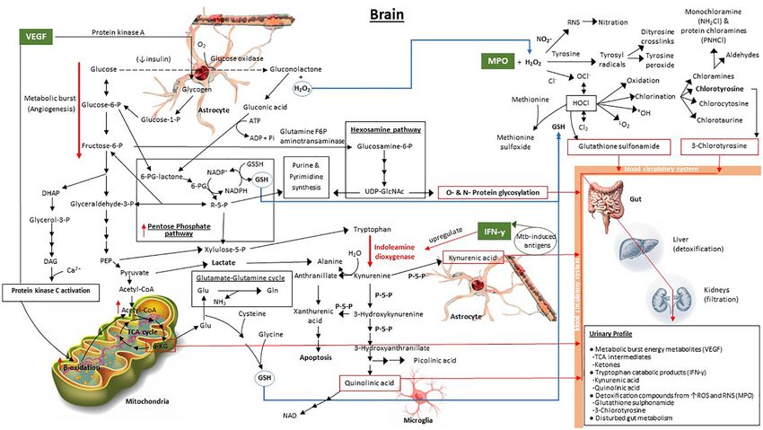
within a couple of days post-treatment. Considering all of fundamentals. This model, illustrated in Figure 1, shows the
the above, patients with VM and TBM exhibit a similar interaction of the overlapping metabolic cascades initiated by
increase in CSF IFN-γ levels, both far greater than in patients TBM, and its associated 3-marker CSF immunological signature.
with BM. This suggests that CSF IFN-γ could potentially be Our predictive metabolic model shows how increased levels
used as a differential diagnostic marker for the exclusion of of VEGF result in a persistent metabolic burst caused by the
BM. Furthermore, CSF IFN-γ levels in TBM cases remain induction of angiogenesis (Stapor et al., 2014; Treps et al.,
elevated for weeks following treatment, differentiating TBM 2016), whereby glycolysis, and the release of glycogen from
from VM. However, as described previously, in order to astrocyte stores to fuel glycolysis, is increased significantly.
acquire a definitive TBM diagnosis additional measures of CSF Secondary pathways that are subsequently upregulated include:
parameters are needed. (1) the pentose phosphate pathway, that contributes to an
In summary, the overall trend across all CSF VEGF studies elevated synthesis of glutathione (Ben-Yoseph et al., 1996),
is a significantly higher concentration of VEGF in TBM patients elevated xylulose-5-phosphate (also via phosphoenolpyruvate
than in other cases of meningitis. Of further note, Van der Flier in the glycolysis pathway) to fuel tryptophan catabolism
et al. (2004) reports significantly increased CSF VEGF (178 ± 52 (Stephanopoulos and Simpson, 1997; Simpson et al., 1999; Maria
pg/mL) in TBM patients with nausea and vomiting, indicating et al., 2018), and elevated purine and pyrimidine synthesis
that elevated CSF VEGF has a potential direct impact on the (Zimmer, 1988, 1996); (2) the hexosamine pathway, which
BGA, leading to a perturbed gut. CSF IFN-γ levels show a similar contributes to increased O- and N-protein glycosylation,
increase in TBM and VM but less so in BM. Hence, CSF IFN-γ imperative for the host’s immune response since glycosylation
levels could potentially be used for the exclusion of the diagnosis controls cell migration, host defense, and antigenicity (Varki,
of BM. The HOCl produced by the MPO–H2 O2 –Cl2 system 1993); (3) increased β-oxidation providing substrate in
yields similar oxidative markers in both TBM and BM. the form of diacylglycerol from downstream catabolism of
The addition of VEGF and MPO with IFN-γ, as part of a 3- dihydroxyacetone phosphate and activation of protein kinase C
marker immunological biosignature of TBM in CSF (Manyelo from VEGF (Takahashi et al., 1999; Harhaj et al., 2006), ultimately
et al., 2019), has yielded a diagnostic measure with an AUC of yielding increased acetyl-CoA; and (4) the boosted mitochondrial
0.97, and a sensitivity and specificity of up to 91.3% and up citric acid (TCA) cycle, due to the increased acetyl-CoA. The
to 100%, respectively. Hence, this 3-marker biosignature yields elevated TCA intermediate α-ketoglutarate (α-KG), previously
excellent results for diagnosis of TBM from a CSF sample. indicated to be a urinary marker of TBM (Mason et al., 2016),
But, what of the urinary metabolomics profile? If these three contributes to glutamate synthesis and downstream glutathione
immunological markers are present in the CSF of a TBM patient, (GSH) production, the latter being a needed antioxidant,
then a downstream metabolic effect, based upon the BGA, should synthesized in response to the elevated MPO.
be reflected in the urine. This concept is explored in our proposed Increased levels of IFN-γ, stimulated by Mtb-induced antigens
predictive metabolic model that follows. (Blumenthal et al., 2012; Lu et al., 2017), specifically upregulate
indoleamine dioxygenase (Yoshida et al., 1981; Taylor and Feng,
1991; Hashioka et al., 2017), the initial enzyme in the tryptophan
PROPOSED PREDICTIVE METABOLIC catabolic pathway. A massive burst in tryptophan catabolism
MODEL OF TBM IN THE BRAIN BASED results in astrocyte-based kynurenic acid and microglia-based
UPON IFN-γ, MPO AND VEGF quinolinic acid synthesis – also previously identified urinary
markers of TBM (Mason et al., 2016). Several enzymes
Given the background of IFN-γ, MPO, and VEGF described within the tryptophan metabolic pathway require pyridoxal-5-
above, and the associated metabolic pathways of these signaling phosphate (P-5-P), an active form of vitamin B6 , as a cofactor.
Frontiers in Neuroscience | www.frontiersin.org 11 April 2020 | Volume 14 | Article 296You can also read