Predominant fecal microbiota of captive adult cheetahs housed in European zoos: a cross-sectional study
←
→
Page content transcription
If your browser does not render page correctly, please read the page content below
Acknowledgments

                                                                   FACULTY OF SCIENCES

            Predominant fecal microbiota of captive adult
                   cheetahs housed in European
                    zoos: a cross-sectional study

                                 Niloufar MAJDZADEH

                                   Master Dissertation
                        Master of Biochemistry and Biotechnology
                             Major Microbial Biotechnology
                                 Academic year 2013-2014

Promoter: Prof. Dr. Geert Huys
Mentor: drs. Anne Becker
Department of Biochemistry and Microbiology (WE10)
Laboratory of Microbiology

                                                                                         1
Acknowledgments

ACKNOWLEDGMENTS

This Master project was only possible because of the involvement of few people to whom I
wish to dedicate the following lines. Hereby I want to thank them for their inspiration,
motivation and help.
My special thanks go to Professor Dr. Huys (Laboratory of Microbiology, University of Gent)
for making this research possible. I would like to appreciate and show my gratefulness to his
guidance, support and advice throughout this research project.
Hereby I would like to express my deepest gratitude to my scientific supervisor, Drs. Anne
Becker (Laboratory of Microbiology, University of Gent) for her thorough support,
encouragement and guidance throughout this project. She helped me warmheartedly and
genuinely despite her busy agenda. Her enthusiasm, open mind, perseverance and brilliance
enlightened my scientific world. Dear Anne, I sincerely wish you the best personal and
professional life ever!
I am also grateful to all the people in BCCM/LMG bacteria collection and Laboratory of
Microbiology for providing a friendly working atmosphere with their kindness and
helpfulness during my master thesis.

I would like to bring this acknowledgment to an end by showing my appreciation to my
beloved family for believing in me all the way through my education. My deepest gratitude
goes to my husband for providing the best possible atmosphere to study and work in. Your
patience, love and support were the key to my success.

                                                                                           2
Table of Contents

TABLE OF CONTENTS

Acknowledgments...................................................................................................................... 2

Abbreviation list ......................................................................................................................... 6

Chapter I. Summary ................................................................................................................... 7

Chapter II. Introduction ............................................................................................................. 8

   II.1. Gut microbiota ................................................................................................................ 8

       II.1.1. The intestinal tract as microbial ecosystem ............................................................ 8

       II.1.2. Function of gut microbiota ...................................................................................... 9

       II.1.3. Role of diet on gut microbiota ............................................................................... 12

   II.2. Techniques for studying diversity of gut microbiota .................................................... 14

   II.3. Cheetah (Acinonyx jubatus) .......................................................................................... 17

       II.3.1. Classes based on dietary habits ............................................................................. 17

       II.3.2. Vulnerable status of cheetahs ............................................................................... 18

       II.3.3. Cheetah’s diet and its role in cheetah’s metabolism ............................................ 19

       II.3.4. Domestic cat as a model for studying cheetah gut microbiota ............................. 20

Chapter III. Goals ...................................................................................................................... 23

Chapter IV. Results ................................................................................................................... 24

   IV.1. DGGE data .................................................................................................................... 24

       IV.1.1. Analysis of all DGGE fingerprints .......................................................................... 26

       IV.1.2. Band richness of the DGGE fingerprints per zoo .................................................. 32

   IV.2. Linking the DGGE data with the clone library ............................................................. 35

       IV.2.1. Common band-classes in the fingerprints ............................................................ 35

       IV.2.2. Linking band-classes with fingerprint clusters ..................................................... 36

   IV.3. quantitative-PCR results .............................................................................................. 40

       VI.3.1. The quantity of Clostridium cluster XIVa per zoo ................................................. 40

                                                                                                                                           3
Table of Contents

       VI.3.2. The quantity of Clostridium cluster XIVa per animal ............................................ 41

       VI.3.3. Clostridium cluster XIVa versus the DGGE band intensity.................................... 41

Chapter V. Discussion .............................................................................................................. 43

Chapter VI. Conclusion ............................................................................................................. 46

Chapter VII. Materials and methods ........................................................................................ 47

   VII.1. Fecal sample collection ............................................................................................... 47

   VII.2. Homogenization of fecal samples .............................................................................. 47

   VII.3. DNA extraction ........................................................................................................... 47

   VII.4. DNA quantity and quality check ................................................................................. 47

       VII.4.1. Agarose gel electrophoresis ................................................................................ 47

       VII.4.2. Optical density measurement by spectrophotometer ........................................ 48

   VII.5. V3-16s rRNA PCR ........................................................................................................ 48

   VII.6. DGGE analysis and gel processing .............................................................................. 49

   VII.7. Data analysis ............................................................................................................... 49

   VII.8. quantitative PCR ......................................................................................................... 50

       VII.8.1. Calibration of a standard curve ........................................................................... 50

       VII.8.2. Quantifying Clostridium cluster XIVa in the fecal samples .................................. 51

Chapter VIII. References .......................................................................................................... 52

Chapter IX. Addendum ............................................................................................................. 63

   IX.1. Fecal samples collection .............................................................................................. 64

   IX.2. Homogenization of the fecal samples ......................................................................... 64

   IX.3. DNA extraction ............................................................................................................. 64

   IX.4. DNA quantity and quality check .................................................................................. 65

       IX.4.1. Agarose gel electrophoresis ................................................................................. 65

       IX.4.2. Optical density measurement by spectrophotometer ......................................... 65

                                                                                                                                        4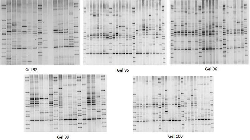
Table of Contents

  IX.5. V3-16s rRNA PCR .......................................................................................................... 65

  IX.6. DGGE ............................................................................................................................ 66

  IX.7. Band extraction and sequencing ................................................................................. 67

  IX.8. q-PCR ............................................................................................................................ 67

     IX.8.1. Building a standard curve ..................................................................................... 67

     IX.8.2. Quantification of Clostridium cluster XIVa ........................................................... 68

                                                                                                                                           5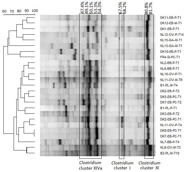
Abbreviations

ABBREVIATION LIST

CFU----------------------------------------------Colony-Forming Unit

MAMP-------------------------------------------Microbe-Associated Molecular Pattern

TLR-----------------------------------------------Toll-Like Receptor

SCFA --------------------------------------------Short-Chain Fatty Acid

EAZA-------------------------------------------- European Association of Zoos and Aquaria

DGGE------------------------------------------- Denaturing Gradient Gel Electrophoresis

GALT --------------------------------------------Gut-Associated Lymphoid Tissue

TH17 --------------------------------------------T Helper 17 cell

TTGE --------------------------------------------Temporal Temperature Gradient Gel Electrophoresis

TGGE --------------------------------------------Temperature Gradient Gel Electrophoresis

RFLP ---------------------------------------------Restriction Fragment Length Polymorphism

ARISA -------------------------------------------Automated Ribosomal Intergenic Spacer Analysis

SSCP ---------------------------------------------Single-Strand Conformation Polymorphism

qPCR---------------------------------------------quantitative PCR

OTU----------------------------------------------Operational Taxonomic Units

PL------------------------------------------------- Planckendael

GA------------------------------------------------Gaia Zoo

OV------------------------------------------------Zoo Parc Overloon

BB------------------------------------------------Safaripark Beekse Bergen

EB------------------------------------------------Ree Park Ebeltoft Safari

SI-------------------------------------------------Réserve Africaine de Sigean

                                                                                                  6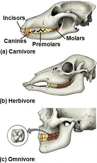
Summary

CHAPTER I. SUMMARY

The gastrointestinal tract of mammals is associated with a diverse, dynamic and complex
microbial community, in which the biggest group is represented by bacteria. The gut
microbiota is responsible for production of essential vitamins, differentiation of the immune
system, tissue homeostasis, energy harvest and short-chain fatty acid production. The
cheetah (Acinonyx jubatus) is an endangered animal with limited metabolic ability to digest
anything except proteins. This carnivore hunts gazelle, rabbit or antelope in the wild.
However, they are mostly fed either meat or prey in captivity. One of the biggest problems
that endanger cheetahs is the feeding mismanagement in the zoos that increases
suboptimal health, gastrointestinal and metabolic diseases. Diet, microbiota and health are
strongly correlated and several studies have been conducted on the diet and health of these
carnivores. Yet, there is not sufficient information about their gut microbiota, which form
the main go-between of diet and health. Since the current information about the gut
microbiota of captive cheetahs has been obtained from 2 animals in Belgium, we aim to
collect more fecal samples from animals in different zoos with various conditions. The aim is
to investigate the diversity of their gut microbiota and whether this analysis reveals the
presence of a core microbiota.

Using population fingerprinting by denaturing gradient gel electrophoresis (DGGE) of V3-16S
rRNA amplicons in a cross-sectional study, we studied the fecal microbial diversity of 55
fecal samples from 36 captive cheetahs in 6 members of EAZA zoos. After homogenization
of the fecal samples, community DNA extraction was carried out followed by V3-16S rRNA
PCR. Afterwards, DGGE profiling was performed followed by data analysis. The fingerprints
were compared with the available clone library and phylogenetic tree, previously built
based on two captive adult cheetahs in Belgium. Afterwards, Quantitative PCR (qPCR) was
performed in order to quantify Clostridium cluster XIVa.

As a result of DGGE analysis, there was no zoo-specific or diet type specific clustering in the
DGGE fingerprints, which suggested that the fingerprints are affected by a combination of
factors such as zoo, diet, genetic background and housing. Clostridium cluster XIVa, XI and I
were present in almost all the DGGE profiles, which indicate that these clusters might be
members of the core microbiota of these 55 fecal samples. Band-classes 28.3% and 41.3%
did not correlate with any clone in the clone library, which belonged to Lactobacillus spp.
after sequencing. Furthermore, the boxplot analysis of the fingerprints revealed the
variation of the predominant bacterial diversity between the fingerprints per zoo, per
animal and per diet type. The quantification of Clostridium cluster XIVa depicted that all the
DNA samples consisted of this cluster except for one animal that suffered from a renal
disease, which may probably be one of the reasons that this cluster was not detectable.
In the future studies, more fecal samples could be collected and analyzed by DGGE to
confirm the diversity and the gut microbiota in captive adult cheetahs. Moreover, more
bands, which belong to potentially discriminating band-classes, can be sequenced to identify
the unknown bacterial genera and species in the clone library. Since multiple taxa can
occupy the same position in the DGGE gel, as it is one of the shortcomings of DGGE, more
zoomed DGGE can be performed.

                                                                                             7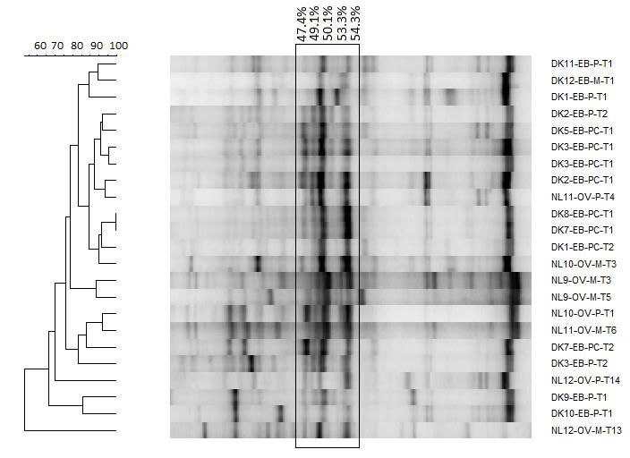
Introduction

CHAPTER II. INTRODUCTION

II.1. GUT MICROBIOTA

II.1.1. THE INTESTINAL TRACT AS MICROBIAL ECOSYSTEM

The gastrointestinal tract of mammals, including humans, is an organ system that starts
from the mouth and ends with the anal canal (Martin, 2010) (Figure 1). The intestine, one
essential organ in this system in which the mucosal immune system operates, has several
fundamental tasks in humans. One is nutrient absorption made possible by its 200m2 total
surface area in humans (Hooper et al, 2012). The other is to provide an environment in
which bacteria can grow and survive, which is done through the few micrometers of mucus
layer between the bacterial community and the layer underneath (O’Hara & Shanahan,
2006).

The gastrointestinal tract is not only associated with eukaryotic tissues and cells as in other
organs, but also with a diverse, dynamic and complex microbial community, in which the
biggest group is represented by bacteria. The gastrointestinal tract of human and mammals
also includes archaea, viruses, fungi and protozoa (Hooper et al, 2002). These bacteria are
spread in almost all parts of the gastrointestinal tract of most mammals, with a relatively
low intensity in the proximal and middle small intestine and high intensity in the large
intestine. The number of the bacteria reaches to 1011–1012 CFU/g in the human colon
(Mackie et al, 1999) (Figure 1).

The human gastrointestinal tract (Matamoros et al, 2013) is sterile at birth (Matamoros et
al, 2013). The bacteria start colonization of the host gastrointestinal tract during birth from
the mother, the surrounding environment, infant feeding, gestational age, hospitalization,
and antibiotic use by the infant (Greiner & Bäckhed, 2011; Penders et al, 2006). Therefore,
the microbial composition of gut microbiota changes after birth both in diversity and
richness throughout development. The diversity and richness of the gut microbiota in each
individual reaches its highest complexity and homeostatic climax composition in adulthood
(Sommer & Bäckhed, 2013; Scholtens et al, 2012). The gut microbiota remain relatively
stable during most of adult life and is expected to become less sensitive to modification
(Jalanka-Tuovinen et al, 2011).

Both anaerobic and aerobic bacteria exist in the gastrointestinal tract of humans in which
those that are anaerobic, such as Bacteroides and Clostridium (Vedantam & Hecht, 2003),
are dominant by 100- to 1,000-fold over the aerobic and facultative anaerobic bacteria in
the human gastrointestinal tract (Clemente et al, 2012). In total, the human intestinal
microbiota consists of approximately 500–1,000 species that belong to only a few of the
known bacterial phyla (Qin et al, 2010) (Figure 1). Amongst them, Bacteroidetes and
Firmicutes account for more than 90% of all the phyla of human intestinal bacteria (Tehrani
et al, 2012).

                                                                                             8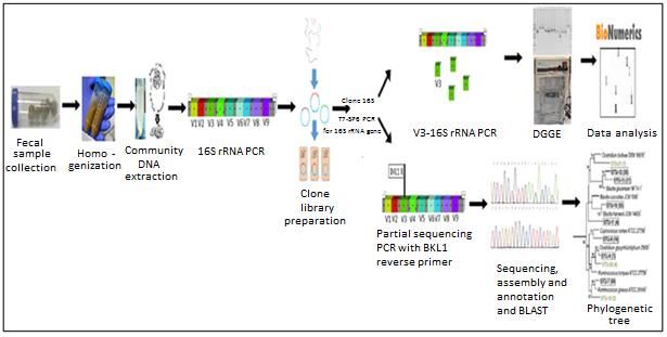
Introduction

                                                                          Figure 1.The bacterial
                                                                                    composition,
                                                                          concentration      and
                                                                          spatial distribution in
                                                                          the human intestinal
                                                                          tract. As depicted, the
                                                                          highest concentration
                                                                          of bacteria among the
                                                                          human gut microbiota
                                                                          belongs to the colon
                                                                          with    1012    CFU/g.
                                                                          Figure obtained from:

                                                                           http://mpicel.myweb.
                                                                          uga.edu/

Human gastrointestinal tract harbors eukaryotes, viruses, archaea and bacteria. Culture-
based techniques have illustrated that all healthy human adults share most of the same gut
bacterial species referred to as a core microbiota. For instance, Escherichia coli can be
isolated from most people. However, culture-independent techniques have demonstrated a
vast microbial diversity with high variability over time and across populations (Knight et al,
2012). Studies have shown that Bacteroidetes and Firmicutes are the phyla in human adults
that dominate their microbiota, whereas Actinobacteria, Proteobacteria and
Verrucomicrobia are generally minor phyla (Eckburg et al, 2005). Additionally, the diversity
of gut microbiota is hugely influenced by age, genetics, environment, diet, human health
and medicine (Knight et al, 2012).

II.1.2. FUNCTION OF GUT MICROBIOTA

Microbiota have coevolved over millions of years with mammalian hosts (Ley et al, 2008). A
balanced gut microbiota are important for host health due to its important role in numerous
developmental, nutritional, physiological and immunological processes (Mackie et al, 1999;
Hooper et al, 2001). The most important functions are:

Differentiation of the immune system: The gut microbiota is essential for differentiation of
the immune system in the host through providing signals. These signals promote the
maturation of immune cells and the normal development of immune functions in order to
eventually protect the host against invasion by opportunistic pathogens and to suppress
inflammatory responses (Chow et al, 2010; Smith et al, 2007; Round et al, 2011). It has been
shown that both the innate and adaptive immune systems in mammals have evolved to
require microbial interactions through their development (Chow et al, 2010; O’Hara &
Shanahan, 2006). The human innate immune system recognizes general microbe-associated
molecular patterns (MAMPs) that are parts of the bacteria such as their flagella or their cell
walls. These antigens are recognized by the host Toll-like receptors (TLRs). In case of

                                                                                               9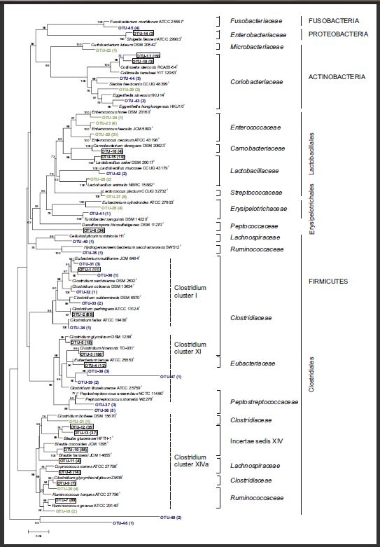
Introduction

absence or mutation of TLRs, the gut and mucosal immune systems do not develop normally
in human (O’Hara & Shanahan, 2006). The human gut microbiota also suppress
inflammatory responses and promote immunological tolerance through TLRs (O’Hara &
Shanahan, 2006). The human gut microbiota also help educating the T cells in the adaptive
immune system to discriminate the self from non-self-cells to protect the body from killing
its own cells, which leads to autoimmunity diseases (Kuhn & Stappenbeck, 2013) (Figure 2).

Production of essential vitamins: Production of vitamins is one of the many functions of gut
microbiota in mammals (Smith et al, 2007; LeBlanc et al, 2013). Yet, some vitamins are not
synthesized by the host. Therefore, these vitamins should be obtained from external
sources with the help of the gut microbiota (Hill, 1997). Biotin (B7) is one of the many water-
soluble vitamins, in which our body absorbs through the help of gut microbiota. Vitamin B12
is another important water-soluble vitamin that is crucial for DNA synthesis and is also
produced by the gut microbiota by consumption of proteins since B12 is bound to proteins.
Vitamin K, a fat-soluble vitamin, is of high importance for blood clotting and if absent the
host may die due to bleeding. The daily requirement of this vitamin comes from the gut
microbiota in mammals (Hill, 1997). These vitamins are synthesized by several intestinal
bacterial genera, including Bacteroides, Eubacterium, Propionibacterium, and Fusobacterium
(Hooper et al, 2002; Wilson & Nicholson, 2009).

Tissue homeostasis: This function involves a tightly regulated cell cycle and requires a
balance between cell renewal and cell death, which is mediated by the gut microbiota in
mammals (Sommer & Bäckhed, 2013). Studies conducted on mice have shown that TLR
signals derived from the gut microbiota are required for regaining tissue homeostasis
following injury in the intestine (Rakoff-Nahoum et al, 2004). Moreover, gut microbiota can
influence tissue homeostasis in bones by decreasing the bone mass when more Ca2+ is
needed in the body through recruiting specific cells called “osteoclasts”. These cells that are
present in bones, are responsible for resorption of boney tissues when the body needs
calcium ion (Sommer & Bäckhed, 2013) (Figure 2). The gut microbiota modulate bone
homeostasis through modulation of T cell function, serotonin levels and cytokine (Sjogren et
al, 2012).

Energy harvest and short-chain fatty acid production: Carbohydrates are important energy
sources for human and microbial cells, in which the human enzymes cannot degrade most
complex carbohydrates and plant polysaccharides (Tremaroli & Bäckhed, 2012). Therefore,
the undigested and unabsorbed dietary carbohydrates, such as fibers, reach the colon and
are fermented by the microbiota, resulting in the production of CO2, H2 and most
importantly “short-chain fatty acids” (SCFA), which are organic fatty acids with 1 to 6 carbon
atoms. Acetate, propionate and butyrate are the main short-chain fatty acids with certain
advantages for the human and mammal’s body (Willing & Van Kessel, 2007; Hijova &
Chmelarova, 2007). The production rate of short-chain fatty acids in several mammal’s
body, is the highest for butyrate, while is the lowest for acetate, with the ratio of 3 to 1
(Topping & Clifton, 2001). Short-chain fatty acids are rapidly absorbed in the cecum and
colon and only 5-10% is eventually excreted in the feces (Macfarlane & Macfarlane, 2003). It
has been shown that SCFAs are associated with reduced risk of inflammatory bowel disease,

                                                                                            10
Introduction
cardiovascular diseases and different sorts of cancers such as colon cancer (Floch & Hong-
Curtiss, 2001; Radulian et al, 2009).
Human enterocytes and colonocytes use butyrate as the major substrate to maintain energy
producing pathways and 70-90% of this metabolite is metabolized by the colonocytes
(Hijova & Chmelarova, 2007). Butyrate is regarded as a metabolite that is beneficial for the
host health due to its positive influences on cell growth, differentiation and anti-
inflammatory effects (Hamer et al, 2008) . Inhibition of the transcription factor NF-kB, which
is implicated in innate immunity, cell cycle control and apoptosis, is amongst the anti-
inflammatory effects of butyrate (Luhrs et al, 2001; Segain et al, 2000). Butyrate also
regulates cell proliferation in mammals through the release of growth factors or
gastrointestinal peptides or by modulation of mucosal blood flow. Moreover, butyrate is the
main short-chain fatty acid that can act directly on genes that regulate cell proliferation
(Blottiere et al, 2003).
Acetate and propionate are taken up by the portal circulation and have an impact on lipid
metabolism in an opposite manner. While acetate contributes to lipid and cholesterol
synthesis in liver, propionate inhibits the effects of acetate (Laparra & Sanz, 2010). In
addition, liver cells metabolize residual butyrate and propionate in order to use them in
glycogenesis. While 50-70% of acetate is taken up by the liver, residual acetate is used by
the muscles in order to generate energy (Hijova & Chmelarova, 2007). Moreover, several
studies have shown that acetate is considered to be the predominant short-chain fatty acid,
in concentration in the human colon. Yet, butyrate has the most beneficial effects on human
health (Tedelind et al, 2007).
Many factors determine the production of short-chain fatty acids in the gut such as the
transit time (Hijova & Chmelarova, 2007), substrate source (Cook & Sellin, 1998) and
bacterial composition in the colon (Roberfroid, 2005). The fermentation takes place in the
proximal colon since the substrate availability is the greatest (Hijova & Chmelarova, 2007).
By the increase in the amount of short-chain fatty acids in the human gut, several beneficial
bacterial species, such as Lactobacillus and Bifidobacterium, which are associated with
improved health, increase as well (Roy et al, 2006). In contrast to most clostridial species in
the mammal’s gastrointestinal tract, Lactobacillus and Bifidobacterium are generally
regarded as beneficial microbes due to their ability to eliminate harmful bacteria and
pathogens by producing various antimicrobial agents such as antibiotics (Rastall, 2004). In
addition to antimicrobial production, these microbes are highly capable of fermentation of
dietary fibers. Therefore, Lactobacillus and Bifidobacterium are considered to ferment
dietary fibers much more effectively than the pathogenic species in the gut (Hijova &
Chmelarova, 2007). Increase in the amount of all short-chain fatty acids results in a decrease
in the pH that indirectly affects the composition of the colonic microbiota, increases mineral
absorption, decreases solubility of bile acids and reduces ammonia absorption (Radulian et
al, 2009; Cook & Sellin, 1998) (Figure 2).
Inhibition of pathogen from causing infection: Another important function of the gut
microbiota in the mammals is protection of gut from infection by pathogenic bacteria.
During pathogen infection, three main functions may be seen:
11Introduction
i) The gut microbiota might block the pathogen growth and thus interfere with the infection
(Stecher & Hardt, 2011). ii) The microbiota might prime the host innate and adaptive
immune system to prevent the development of infection and diseases, which is caused by
the virulence factors of pathogen bacteria (Sekirov et al, 2010). iii) The gut microbiota
eliminate the pathogens from the host gut at the end of the infection (Endt et al, 2010).
The effects of human gut microbiota on host physiological processes. Arrows represent either
Figure 2.
stimulatory or inhibitory effects of gut microbiota on host physiological processes. Figure obtained from:
http://www.nature.com/nrmicro/journal/v11/n4/full/nrmicro2974.html (Sommer & Bäckhed, 2013)
II.1.3. ROLE OF DIET ON GUT MICROBIOTA
Diet and different food habits are important factors in microbiota composition and
development of mammals including the human being, either in the early life or in the late
life. For instance, the diversity and composition of microbiota vary significantly between
breast-fed and formula-fed infants in the early life. The intestinal microbiota of the breast-
fed infants is more heterogeneous and more taxonomically diverse than the formula-fed
infants (Schwiertz et al, 2010) with bifidobacteria as the key player in the gut microbial
community of neonates (Turroni et al, 2012). The gut microbiota composition in the human
and mammals is also under the influence of food habits. For instance malnutrition results in
the lower abundance of Bacteroidetes in human gut microbiota, which are specialized for
breaking down the carbohydrates in energy rich diet types (Ottman et al, 2012).
Carbohydrates form the bulk of most human and animal diets and are important nutrients
for both host and microbiota. Mammals easily absorb simple sugars such as glucose and
12Introduction
galactose, as well as disaccharides and starch, in the proximal part of their small intestine
(Ferraris, 2001). Yet, they are limited to hydrolyze and utilize more complex polysaccharides
(Hooper et al, 2002). Therefore, a large quantity of undigested dietary carbohydrates
including polysaccharides from plant cell wall components such as cellulose, xylan, pectin
and fibrous substrates as well as undigested starch reaches the distal part of the
gastrointestinal tract. This is where the gut microbiota play an important role by degrading
these biomolecules (Hooper et al, 2002). Gut microbiota of most mammals and humans
ferment these carbohydrate polymers that reach the distal gut and help extract nutrient
value from otherwise poorly utilized dietary substrates. Therefore, the distal regions of the
gut of several mammals are highly colonized with bacteria (Zoetendal et al, 2002). Gut
microbiota hydrolyze these carbohydrates and consequently the hosts avoid the need to
evolve the complex repertoire of glycosylhydrolases that would be required to break down
the wide variety of linkages in dietary polysaccharides. The gut microbiota, in turn, gain
access to a nutrient-rich environment, including complex dietary plant polysaccharides and
host-derived glycans secreted on the surface of the epithelium and on the mucus layer,
which is rich in carbohydrates (Corfield et al, 2001). Among these bacteria, bifidobacteria
play the biggest role for carbohydrate fermentation in several mammals and humans
(Ottman et al, 2012).
Proteins form a portion of diet of humans, carnivores and omnivores. Several bacteria use
proteins as a carbon and nitrogen source and it has been demonstrated that protein-rich
diets increase Clostridium populations (Zentek et al, 2004). The amount of protein that
reaches the lower bowel may be consumed by gut microbiota, in which this amount
depends greatly on the protein quality. Lower quality proteins, which are poorly digested,
will provide more protein to microbes in the lower bowel. Therefore, either lower quality
proteins or higher amounts of proteins can increase the occurrence of proteolytic bacteria
such as Bacteroides (Wu et al, 2011). These bacteria can be pathogenic and/or produce
putrefactive compounds such as ammonia, indoles, phenols, and sulphur-containing
compounds. These compounds, which are regarded as detrimental for gut health in either
humans or several animals, are toxic at high levels and are associated with several disease
states (Backus et al, 2002) such as Inflammatory bowel disease (Pedersen et al, 2002; Tuohy
et al, 2006) and chronic renal failure (Niwa, 2010).
According to the study conducted by Wu et al., short-term diet does not change the
microbial composition in the human gut, while long-term diet has a tremendous role on
modulating gut microbiome composition and enterotype partitioning (Wu et al, 2011). For
instance, long term diet rich in animal protein, sugar, starch and fat and low in fiber, will
result in Firmicutes and Proteobacteria dominance in the human gut microbiota. On the
other hand, a diet type rich in vegetables and thus plant fibers, will result in the dominance
of Actinobacteria and Bacteroidetes phyla (Wu et al, 2011; De Filippo et al, 2010).
Recent studies have shown that obese and lean people have different composition in their
gut microbiota, specifically in the proportion of Bacteriodetes and Firmicutes. In obese
people the Firmicutes are dominant while the Bacteriodetes are dominant in lean people
(Ley et al, 2006). The Bacteroidetes degrades complex and otherwise indigestible dietary
polysaccharides in the large intestine, resulting in the production of short-chain fatty acids,
13Introduction
which are the energy source for the host (Tremaroli & Bäckhed, 2012; Thomas et al, 2011).
In conclusion, the studies conducted to date about the role of diet on gut microbiota,
endorse the fact that the intestinal microbiota in several mammals thrives on using
polysaccharides and peptides, which are indigestible to mammals (Guarner & Malagelada,
2003).
II.2. TECHNIQUES FOR STUDYING DIVERSITY OF GUT MICROBIOTA
Several techniques are available for studying the diversity of gut microbiota in either
humans or animals. Early experiments in this field used culture-based techniques, with a
certain level of discriminatory power for identification of microorganisms (Temmerman et
al, 2004). Most bacteria in the gastrointestinal tract are fastidious and hard to culture since
they are anaerobic. Besides there is no uniform substrate preference for all of them (Inglis
et al, 2012). Consequently only a fraction of the microorganisms in the gastrointestinal tract
can be cultured and studied by culture-dependent techniques. Therefore, these techniques
are limited in scope and lack precision for studying the diversity or functionality of
microbiota, and the progress in this field was greatly hindered due to this fact (Kerr et al,
2013a).
The fact that 99% of the bacteria cannot be cultured or has not yet been cultured
(Handelsman, 2004), leads the scientists to use culture-independent or molecular
techniques, which is one of the most important developments of environmental
microbiology since the 1980s (Keller & Zengler, 2004). These culture-independent
techniques have several advantages over the culture-dependent techniques. Recent
developments and rapid advances in molecular biology and sequencing have resulted in a
more accurate understanding of the gut microbiota composition, the genetic elements of all
the microorganisms in the gastrointestinal tract (microbiome), their dynamics, their
interaction with the other gastrointestinal organs and the functionality of the intestinal
ecosystem in human and many mammalian species (Ritchie et al, 2010). There are many
molecular techniques of which phylogenetic clone libraries, phylogenetic microarray,
community DNA profiling and metagenomics are most commonly used to study gut
microbiota diversity (Lau & Liu, 2007).
Phylogenetic clone library is a sequencing-based technique that targets the 16S-rRNA gene,
which is present and conserved among all members of a bacterial community, to study
phylogenetic and bacterial community structures. In this technique, primers are used to
amplify a portion of the 16S-rRNA gene, followed by ligation of the amplified region of the
universal gene in bacterial plasmids. Afterwards, the plasmids are transformed into
competent cells, such as E. coli, to generate a clone library (Inglis et al, 2012). The
advantages are relative simplicity, representation of a single individual from the bacterial
community by possessing a single insert in each clone; and the ability to near complete
sequencing of 16S-rRNA gene (Inglis et al, 2012). On the other hand, the disadvantages are
the underrepresentation of low abundance bacteria in the community, efficiency
dependency on the quality of the inserts (Lovell et al, 2008) and the high expenses of
sequencing as well. Despite all these aforementioned limitations, this method has still merit
14Introduction
and has provided invaluable information about the bacterial community and composition of
gut microbiota either in humans or mammals (Inglis et al, 2012) (Figure 3).
Phylogenetic microarray is a glass surface covered by thousands of covalently linked DNA
probes, in which they will be hybridized by DNA or RNA. Subsets of probe sequences, which
are specific to the ecological environment of interest, are required. The probes for studying
the gastrointestinal microbial community are targeting only one or several parts of 16S-
rRNA gene such as the V3 variable region (Claesson et al, 2009). This technique is widely
used for monitoring gene expression, mutation in genomic DNA and DNA sequence
polymorphism. Yet, it is not capable of detecting the unknowns, hybridization conditions are
not optimal for all the hybrids and many probes are required for exploring the diversity
(Gentry et al, 2006) (Figure 3).
Community DNA profiling is either sequence-dependent, such as Denaturing Gradient Gel
Electrophoresis (DGGE) and Temperature Gradient Gel Electrophoresis (TGGE), or size-
dependent electrophoresis such as Restriction Fragment Length Polymorphism (RFLP),
Automated Ribosomal Intergenic Spacer Analysis (ARISA) and Single-Strand Conformation
Polymorphism (SSCP).
In DGGE, a set of amplicons equal in size but different in sequence obtained from the 16S
rRNA gene (Inglis et al, 2012), is applied to an polyacrylamide electrophoresis gel. The
polyacrylamide gel has a denaturing agent including urea and formamide, so that the
amplicons melts at various stages through the gel based on their GC content (Muyzer et al,
1993). The ability to excise and clone individual bands, is a significant advantage of DGGE
analysis. Yet, visual identification of the bands in samples with complex banding patterns
and high diversity can be difficult (Inglis et al, 2012) (Figure 3). TTGE is a form of
electrophoresis that uses temperature to denature the sample as it moves across an
acrylamide gel. Therefore, the amplicons are separated based on differences in melting
temperature, sequence length, and GC content (Inglis et al, 2012). RFLP is a technique that
uses fluorescently labeled PCR primers to amplify the gene of interest, followed by
electrophoresis (Inglis et al, 2012). In order to study different environmental samples, ARISA
can be used that uses PCR on the specific region of rRNA gene operon between 16S and 23S
subunits (Ranjard et al, 2001). In SSCP electrophoresis is carried out to distinguish
sequences fragments according to their different conformations (Hori et al, 2006).
Metagenomics is the study of genetic material recovered directly from environmental
samples such as soil or water samples obtained from the environment. This technique
enables studies of organisms that are not easily cultured in a laboratory, as well as studies of
organisms in their natural environment (Sleator et al, 2008; Handelsman, 2004). This
technique has been made possible by advances in next generation DNA sequencing
technologies and bioinformatics, including sequencing the 16S-rRNA gene (Inglis et al, 2012)
(Figure 3). Therefore, this technique has provided us with invaluable insight about the
lifestyle and metabolic capabilities of the bacteria from environmental samples (Tringe et al,
2005). For instance, metagenomics allows to study the physiology and ecology of
environmental microorganisms, novel genes and gene products, novel proteins and novel
molecules with an antimicrobial activity (Handelsman, 2004). It also allows simultaneous
15Introduction
monitoring of a diversity of microorganisms and does not have the biases of DNA
amplification. Yet, it is not possible to obtain finished genomes from all the microorganisms,
present in the sample, after performing assembly and annotation due to several reasons
such as incompleteness of reference databases. This results in several gaps and
misannotations in the chromosomes (Handelsman, 2004; Sleator et al, 2008). Particularly
from less abundant members of the microbiota or in situations where a community contains
many closely related species. Therefore, it is difficult to assign a function to specific species
within one community (Sleator et al, 2008).
Techniques used for studying the diversity of gut microbiota. A) Phylogenetic clone library is a library
Figure 3.
in which the nucleotide sequences of interest are preserved as inserts to a plasmid that has been used to infect
bacterial cells. B) Phylogenetic microarray is a high-throughput analytical method for microbial community
analysis and consists of a glass covered with thousands of covalently linked DNA probes to which the DNA from
the samples is added. C) Community DNA profiling is either “sequence dependent electrophoresis”, such as
DGGE, TGGE and TGGE or “size dependent electrophoresis” such as RFLP, ARISA and SSCP. D) Metagenomics is
the study of genetic material recovered directly from environmental samples, in which all community DNA is
sequenced. This technique provides us with not only diversity but also the functionality of gut microbiota.
Figures derived from: http://metagenomicsrevealed.yolasite.com/process-1.php
http://www.envirologek.com/products_detail.php?indid=3&compid=15&cid=6&appid=&prodid=344&langid=1
http://biointelligence.wordpress.com/microarray_research/
16Introduction
II.3. CHEETAH (ACINONYX JUBATUS)
II.3.1. CLASSES BASED ON DIETARY HABITS
Mammals can be divided into three general classes based on dietary habits, namely
carnivores, herbivores and omnivores. Carnivores are animals that derive their energy and
nutrients from mainly or exclusively animal tissues. They can either hunt other animals or
scavenge animal bodies. Therefore, they have organs for capturing prey such as sharp teeth,
strong jaws and claws (Gittleman et al, 2001). The cheetah (Acinonyx jubatus) is one of the
274 species in the mammalian order of Carnivora (Britannica, 2011), which is the only
member of the genus Acinonyx (Treves & Karanth, 2003). This fast and relatively large
animal has a deep chest and a relatively small head. It is also well-known by the tear marks
running down its eyes in order to protect its eyes from sunlight and help this animal hunt
(Hayward et al, 2006) (Figure 4).
Herbivores, such as deer and goat, consume plant material and the structure of their body
and the inner organs is well adapted to eating only plants (Karban & Agrawal, 2002).
They typically have wide flat teeth adapted to grinding the plant material like grass, tree
bark and other tough plant material (Hummel et al, 2011) (Figure 4).
Omnivores, such as most bears as well as the human being, derive their energy and
nutrients from sources that may include animals, plants, algae and fungi that possess teeth
proper for consumption of various sorts of tissues such as plant and animal tissues
(Deweerdt, 2011) (Figure 4).
The scull and teeth structure in a) carnivores,
Figure 4.
b) herbivores and c) omnivores. a) The carnivores
have sharp teeth appropriate for eating and tearing
meat, tendons, etc. apart as well as strong jaws. b)
The herbivores, on the other hand, have flat teeth
suitable for eating plant materials, in which their
teeth are stronger in the back side of their jaws. c)
Omnivores have the kind of teeth that potentially can
consume both animal and plant materials.
Figure obtained from: http://avonapbiology2011-
2012.wikispaces.com/Maya+Regalado
17Introduction
II.3.2. VULNERABLE STATUS OF CHEETAHS
Cheetah, the world’s fastest mammal, is a predator carnivore. As a result these animals have
limited metabolic ability to digest anything except proteins (Bradshaw et al, 1996). Cheetahs
inhabit various places such as grassland savannahs and woodland habitats. They are listed
as an endangered animal in the last decades and their distribution has declined dramatically
in the wild (Nowell K., 1996). This is mostly due to the human actions such as destroying
their natural habitat like much of Africa, Middle East and South Asia. Other human actions
that further endanger this animal is hunting cheetahs and their prey such as gazelle and
especially antelope, which is the most important prey for the cheetahs in the wild (O’Brien
et al, 1985). Occasionally road accidents can cause danger to this animal as well (Williams,
2007). Based on an investigation in Zimbabwe, it was also found that several cheetahs have
been killed in the last twenty years due to ingestion of anthrax infected meat, pneumonia,
nephritis, asphyxiation, flea infection, accidental poisoning and fractures in their bones
(Williams, 2007).
Cheetahs are mid-sized predators. Therefore, if there is not enough prey in the
environment, they cannot compete for their food requirements with the larger predators
such as lions, leopards, tigers and the African wild dogs. Obviously this unfortunate event
can cause their starvation (Marker et al, 2003; Durant, 2004; Radloff & Du Toit, 2004;
Hayward et al, 2006). It has also been seen and reported in the wild and also in the
preserved areas that the cheetah cubs are preyed by other large predators such as lions and
jaguars (Gros, 1998).
Lack of genetic variation, polymorphism and heterozygosity are other problems that may
cause susceptibility and vulnerability of these endangered animals to specific diseases such
as renal failure. Additionally, the level of variation in the aforementioned factors is higher in
other mammals. Therefore, the fact that they compete with the other animals and survive in
the wild, as well as adaptation to the ecological niche, is of great significance (O’brien et al,
1983).
The increase in the habitat destruction of wild animals has led to the prospect of protecting
these animals in captive breeding programs (O’Brien et al, 1985). In wildlife preservation,
with the aim of protecting animals from extinction, conservation of threatened and
endangered species is quite challenging. Captive breeding of cheetahs is very difficult to
perform since a large number of the cubs die at birth due to disease susceptibility and
maternal neglect (O’Brien et al, 1985; van Gelder, 1973).
Another problem that endangers cheetahs is the feeding mismanagement in the zoos that
increases suboptimal health, gastrointestinal and metabolic diseases in these vulnerable
animals. Additionally, improper feeding habits in captivity can lower the breeding
performance in the cheetahs (Kotsch et al, 2002; Munson et al, 2005). Therefore, well-
balanced diets can be a therapeutic intervention with representing an important route to
prevent these feeding and breeding problems (Garcia-Mazcorro et al, 2011; Gaggìa et al,
2010).
18Introduction
II.3.3. CHEETAH’S DIET AND ITS ROLE IN CHEETAH’S METABOLISM
Cheetahs are specialized in predation and have a protein-rich diet due to consuming mostly
meat (Treves & Karanth, 2003). In the wild, these carnivores, generally prefer to capture
and kill the most available medium-sized prey within a body mass range of 23-56 kg, such as
antelope and gazelle (Nowak, 1999; Hayward et al, 2006). Though they are also able to
utilize both larger and smaller prey (Mills et al, 2004). They mostly feed from the flesh of
their prey, due to their relatively fragile teeth, skull and jaw (Van Valkenburgh et al, 1990).
They also consume indigestible animal tissues such as small bones, tendons, skin, hair, and
feathers derived from the prey (Depauw et al, 2011). These indigestible materials, which
cannot be enzymatically digested in the body, have the potential to act as a substrate for
microbial fermentation in the intestine of carnivores with the ability to produce high
amounts of short-chain fatty acids. These short-chain fatty acids have numerous benefits on
colonic function and metabolism such as energy source for colonocytes, stimulation of
colonic blood flow and motility as well as decrease of the growth of pathogenic microbiota
(Rondeau, 2003; Wong et al, 2006).
Wild cheetahs consume tendons, small bones and cartilage on a regularly basis. On the
other hand, cheetahs in captivity are fed either raw meat diet supplemented with a vitamin
and mineral premix or by a prey-based diet containing the whole carcasses, which has high
amounts of indigestible animal tissues (Depauw et al, 2011). Therefore, a big difference
between the diet of captive cheetahs and wild cheetahs is the nutrition composition of their
diet. According to recent studies about the role of animal fibers on the cheetah gut
microbiota and their health, the undigested part of an animal-based diet can be a source of
short-chain fatty acids in the captive cheetahs and potentially other carnivores (Depauw et
al, 2011). Moreover, the presence of animal fibers in the diet of captive cheetahs has a
modifying effect on the bacterial fermentation in the intestine (Depauw et al, 2011).
Therefore, it is of high importance to investigate whether the presence or absence of these
indigestible animal tissues causes changes in intestinal fermentation. Moreover, the effect
of prey-based diet type on the gut microbiota of cheetahs is promising due to the beneficial
actions of the gut microbiota.
In general, the fermentation of protein sources is regarded as detrimental for gut health in
either human or mammals. This process results in many by-products, such as ammonia,
indolic and phenolic compounds. These compounds are toxic and have been linked to
intestinal diseases e.g. inflammatory bowel disease (Pedersen et al, 2002; Tuohy et al, 2006)
and chronic renal failure (Niwa, 2010), which is one of the causes of death in the captive
cheetahs (Depauw et al, 2011). These putrefactive compounds have the potential to
damage the gut ecosystem and influence the general metabolism of carnivores as they can
be converted into toxic metabolites. To this end, several studies have been carried out, in
which it has been observed that putrefactive compounds, namely indole, phenol and p-
cresol, are higher when cheetahs are fed supplemented beef in comparison with cheetahs
fed whole prey such as rabbit (Vester et al, 2008, 2009).
19Introduction
II.3.4. DOMESTIC CAT AS A MODEL FOR STUDYING CHEETAH GUT MICROBIOTA
Domestication is a continuous transition that differs by species, genes and environment, in
which some characters may vary with circumstances (Price, 1984). In addition, it is reported
that all domesticates manifest a considerable tolerance of proximity to people and also
adaptations to a new diet. Domestication is not a single trait but a group of traits that
consists of elements affecting emotion, mood and social communication, in which all have
been modified in one way or another (Driscoll et al, 2009). The metabolic and morphological
changes that result in behavioral adaptation to the human environment can lead to a
remarkable dependence on humans for food and shelter, such as for domestic cats (Driscoll
et al, 2009).
Patterns of sequence variation have been seen in the genome of domestic cats, which
reflect a history of domestication and breed development (Driscoll et al, 2007). It is
probable that the cat began its association with humans as a commensal, feeding on the
rodent pests that infested the grain stores of the first farmers (Flood, 2001). It is generally
considered that domestic cats have descended from the old world wildcats. Yet, they differ
from wildcats in behavior, tameness, and coat color diversity (Peters et al, 2009).
The domestic cat has been proposed as a model for basic nutrients and energy
requirements and feeding methods for captive felid populations such as the cheetah (Vester
et al, 2008; Kerr et al, 2013b). It has been proposed by Dierenfeld et al. that nutritional
requirements of domestic cats should form the basis of comparison in managed feeding
programs for captive cheetahs (Dierenfeld, 1993). They both share several nutrition-related
aberrations including food allergies, obesity and hyperlipidemia. Additionally, nutritionists
and veterinarians can take blood samples from domestic cats, which is easier in comparison
with the cheetahs. The blood samples are used to explore the blood metabolite
concentrations, digestibility of food materials and intrinsic feline metabolism with specific
nutritional requirements. Therefore, an extrapolation of dietary recommendations for
domestic cats as well as their dietary profile, has been applied to other exotic felines like
cheetahs (Lubbs et al, 2009; Vester et al, 2009).
It is not thoroughly scientifically applicable to extrapolate the data obtained from the
domestic cats, including the data from dietary requirements and dietary profile, to the
cheetahs. The behavioral, anatomical and nutritional characteristics of these two animals
vary tremendously. For instance, domestic cats are frequently fed carbohydrate-rich
extruded kibble diets (MacDonald et al, 1984; Zoran, 2002) while captive cheetahs are fed a
meat-based or prey-based diet (Vester et al, 2009). Different sources of dietary fiber, such
as pectin and cellulose, are present in domestic cats’ diet. Taxonomic and functional studies
of the intestinal microbial communities have shown that domestic cats possess a well-
developed complex microbial community which can also change depending upon their diet
type (Lubbs et al, 2009; Vester et al, 2009). For instance, a higher protein level present in
their diet has been associated with an increase in Escherichia coli, Lactobacillus spp.,
Clostridium perfringens and Fusobacterium spp. and a decrease in Bifidobacterium
population in the fecal samples of cats (Lubbs et al, 2009; Vester et al, 2009). Moreover, C.
perfringens, which has been identified as an intestinal pathogen in cheetahs (Citino, 1995),
20Introduction
E. coli and Lactobacillus spp., were shown to increase when the cat’s diet was supplemented
with pectin, whereas Bifidobacterium increased upon fructooligosaccharide
supplementation (Barry et al, 2012). Bermingham et al. showed that the diversity of the
bacterial population in cats fed dry diets were lower compared with cats fed wet diets
(Bermingham et al, 2013). Moreover, it has been reported that cats fed a diet containing 4%
pectin have a higher percentage of Firmicutes and Spirochaetes in comparison with cats that
were fed a diet comprising 4% cellulose (Barry et al, 2010).
Recently, it was reported by Tun et al. that Bacteroidetes is the most predominant (68%)
bacterial phylum in the feline intestinal microbiome followed by Firmicutes and
Actinobacteria, as it was found in the fecal microbiota of domestic cats (Khin et al, 2012).
The current information about the gut microbiota of adult captive cheetahs has been
obtained from only two captive cheetahs housed in Belgium. This information is collected in
a clone library based on these two captive animals (Becker et al, 2014). In order to build a
clone library, fecal samples are collected and homogenization and community DNA
extraction is carried out. Afterwards, 16S rRNA PCR is performed to prepare the clone
library followed by two distinct steps. The first one is to perform V3-16S rRNA PCR, DGGE
and data analysis. The second step is partial sequencing PCR followed by sequencing,
assembly and annotation in order to build a phylogenetic tree by comparison against The
Ribosomal Database Project II (RDP) (Figure 5). The group of sequences with ≤3% sequence
divergence was regarded as an Operational Taxonomic Unit (OTU) or phylotypes. This
phylogenetic tree works as a reference to identify the bacterial groups on the DGGE
fingerprints of fecal samples under study.
Unexpectedly, Bacteroidetes and Bifidobacteriaceae, which can contribute in intestinal
homeostasis, were not present in the 16S rRNA clone libraries of two Belgian captive
cheetahs studied recently. Yet, a hardly detectable concentration of Bacteroidetes phylum
was seen by qPCR. On the other hand, Firmicutes were in high number as the dominant
group while the minority were Actinobacteria, Proteobacteria and Fusobacteria. Amongst
the Firmicutes, Clostridium cluster XIVa, XI and I were in majority with 43%, 38% and 13% of
all the bacteria respectively (Becker et al, 2014).
21Introduction
Constructing a clone library based on 16S rRNA gene. In order to build a clone library, fecal samples
Figure 5.
are collected followed by their processing to prepare the clone library. On one hand, sequencing, assembly and
annotation are performed to eventually build a phylogenetic tree. On the other hand, V3-16S rRNA PCR is
performed followed by DGGE to have the clones on the gel. The results obtained from both steps are used as a
reference when analyzing the fecal samples under study.
To sum up, the current information about the gut microbiota of captive adult cheetahs is
scarce. Consequently, more fecal samples should be collected from cheetahs housed in
various zoos with different backgrounds and diet in order to eventually bring the correlation
between diet, microbiota and health to light. By analyzing multiple fecal samples from these
animals, more knowledge will be obtained about the diversity, composition and core
microbiota of captive cheetahs.
22You can also read