Defining the role of CAAX protein proteolysis and methylation in the pathogenesis and treatment of progeria - Xue Chen
←
→
Page content transcription
If your browser does not render page correctly, please read the page content below
The Department of Biosciences and Nutrition
       Karolinska Institutet, Stockholm, Sweden

Defining the role of CAAX protein proteolysis
  and methylation in the pathogenesis and
            treatment of progeria

                     Xue Chen

                  Stockholm 2021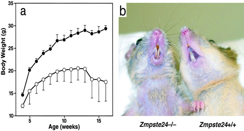
All previously published papers were reproduced with permission from the publisher.
Published by Karolinska Institutet.
Printed by US-AB 2021
Xue Chen, 2021
ISBN 978-91-8016-355-2
Defining the role of CAAX protein proteolysis and
     methylation in the pathogenesis and treatment of
                           progeria

Thesis for Doctoral Degree (Ph.D.)

By

Xue Chen

Principal Supervisor:                     Opponent:
Professor Martin Bergö                    Professor Vicente Andrés
Karolinska Institutet                     Centro Nacional de Investigaciones
Dept of Biosciences                       Cardiovasculares
and Nutrition                             Madrid

Co-supervisors:                           Examination Board:
Professor Maria Eriksson                  Professor Elias Arnér
Karolinska Institutet                     Karolinska Institutet
Dept of Biosciences                       Dept of Medical Biochemistry and
and Nutrition                             Biophysics

Dr Mohamed Ibrahim                        Docent Giedre Grigelioniene
Gothenburg University                     Karolinska Institutet
Sahlgrenska Center for Cancer Research    Dept of Clinical Genetics
Dept of Molecular and Clinical Medicine
                                          Professor Lena Kjellén
                                          Uppsala University
                                          Dept of Medical Biochemistry and Microbiology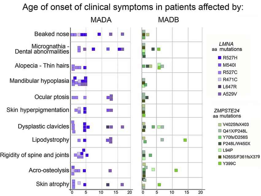
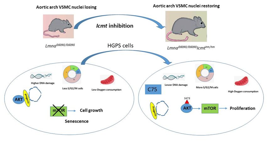
ABSTRACT

Hutchinson-Gilford progeria syndrome (HGPS) is a rare childhood disease characterized by
failure to thrive, bone abnormalities, hair loss, and a shortened life span due to cardiovascular
disease, and nuclear shape abnormalities in cultured cells. HGPS is caused by de-novo mutations
in LMNA, the gene encoding prelamin A. Lamin A is produced from prelamin A following three
modifications at a carboxyl-terminal CAAX motif: farnesylation of the cysteine by
farnesyltransferase (FTase); release of the –AAX by RAS-converting enzyme 1 (RCE1); and
methylation of the farnesylcysteine residue by isoprenylcysteine carboxyl methyltransferase
(ICMT); mature lamin A is subsequently produced by proteolytic removal of the last 15 amino
acids (including the farnesylcysteine residue) by Zinc metalloproteinase Ste24 homologue
(ZMPSTE24). The HGPS mutation produces a truncated prelamin A—progerin—which lacks the
ZMPSTE24 cleavage site; thus, farnesylated and methylated progerin accumulates at the nuclear
lamina in HGPS patient’s cells and causes all disease phenotypes. Other rare forms of progeria
result from inactivating mutations in ZMPSTE24 which leads to accumulation of full-length
prelamin A.
Targeting FTase with small-molecule drugs prevents prelamin A and progerin farnesylation and
corrects the nuclear shape abnormalities observed in cultured progeria cells and reduces
symptoms in progeria mouse models. Today, FTase inhibitors (FTIs) constitute the only approved
therapy for HGPS. However, FTIs have anti-proliferative properties and their effects in HGPS
patients including the survival benefit are modest. Thus, new therapeutic strategies are needed.
In this thesis, I have explored the biochemical and medical consequences of targeting ICMT and
RCE1 on the function of prelamin A and progerin and the therapeutic benefits of these
interventions.
In paper I, we followed up on our group’s earlier genetic studies showing that targeting Icmt
overcomes senescence of Zmpste24-deficient cells and human HGPS cells and increases
survival of Zmpste24-deficient mice with progeria. We found that knockout of Icmt improves
survival of progerin knock-in mice—a more relevant model of HGPS. Moreover, we synthesized
a small-molecule ICMT inhibitor, C75, and showed that it inhibits ICMT, stimulates AKT signaling,
reduces senescence, and increases proliferation of cultured progeria cells but has no effect on
cells lacking the Icmt gene, indicating drug specificity.
In paper II, we tested the hypothesis that targeting RCE1-mediated cleavage of prelamin A would
reduce its toxicity and be useful for therapeutic purposes. It was already known that in the absence
of RCE1, ZMPSTE24 can perform the same –AAX cleavage; thus, this strategy would only work
on progeria caused by ZMPSTE24 deficiency. We found that knockout of Rce1 in Zmpste24-
deficient mice prevented –AAX proteolysis, reduced progeria phenotypes, and improved survival,
but to a lesser extent than targeting Icmt. Inhibiting RCE1 expression in cells from a ZMPSTE24-
deficient patient stimulated AKT signaling, reduced senescence, and improved proliferation.

We conclude that targeting RCE1 and ICMT reduces the toxicity of prelamin A and progerin and
that an ICMT inhibitor can overcome senescence and stimulate proliferation of HGPS cells. The
results suggest RCE1 could be a potential drug target in future therapy of ZMPSTE24 deficiency
and that ICMT inhibitors should be further developed for in-depth preclinical evaluation in HGPS
therapy.
摘要
Hutchinson-Gilford 早衰综合征 (HGPS) 是一种罕见的儿童疾病,伴有发育迟缓、骨骼异
常、脱发和心血管疾病导致的寿命缩短。 HGPS 是由 LMNA 中的点突变引起的,LMNA
是编码前核蛋白 A 的基因。核蛋白 A 是由前核蛋白 A 在羧基末端 CAAX 基序进行三个修
饰后产生的:法尼基转移酶 (FTase) 对半胱氨酸进行法尼基化;通过 RAS 转换酶 1
(RCE1) 释放 –AAX;通过异戊二烯基半胱氨酸羧基甲基转移酶(ICMT)甲基化法尼基半
胱氨酸残基;随后通过锌金属蛋白酶 Ste24 同源物 (ZMPSTE24) 蛋白水解去除最后 15
个氨基酸(包括法尼基半胱氨酸残基)产生成熟的核纤层蛋白 A。 HGPS 突变产生截短
的 prelamin A-progerin,它缺少 ZMPSTE24 切割位点;因此,法尼基化和甲基化早老素
在核层 HGPS 患者的细胞中积累并导致所有疾病表型。其他罕见形式的早衰症是由
ZMPSTE24 失活突变导致的,该突变导致全长 prelamin A 的累积。

用小分子药物靶向 FTase 可防止 prelamin A 和早衰蛋白法尼基化,并纠正在培养的早衰
细胞中观察到的核形状异常,并减少早衰小鼠模型中的症状。 直至今日,FTase 抑制剂
(FTI) 是唯一被批准的 HGPS 疗法。 然而,FTI 具有抗增殖特性,它们对 HGPS 患者的
影响(包括生存益处)不大。 因此,需要新的治疗策略。 在本论文中,我探讨了靶向
ICMT 和 RCE1 对 prelamin A 和 progerin 功能的生化和医学影响以及这些干预措施的治
疗益处。

在论文 I 中,我们跟进了我们小组早期的遗传研究,表明靶向 Icmt 挽救了 Zmpste24 缺
陷细胞和人类 HGPS 细胞的衰老,并增加了 Zmpste24 缺陷小鼠早衰症的存活率。 我们
发现 Icmt 的敲除提高了 progerin-knock-in 小鼠的存活率,这是一种更相关的 HGPS 模
型。 此外,我们合成了一种小分子 ICMT 抑制剂 C75,并表明它可以抑制 ICMT,刺激
AKT 信号传导,减少衰老,并增加培养的早衰细胞的增殖,但对敲除 Icmt 基因的细胞没
有影响,表明药物特异性。

在论文 II 中,我们测试了以下假设,即靶向 RCE1 介导的 prelamin A 裂解会降低其毒性
并有助于治疗目的。 众所周知,在没有 RCE1 的情况下,ZMPSTE24 可以进行相同的 –
AAX 切割; 因此,该策略仅适用于由 ZMPSTE24 缺乏引起的早衰。 我们发现在
Zmpste24 缺陷小鼠中敲除 Rce1 可防止 -AAX 蛋白水解,减少早衰症表型并提高存活
率,但程度低于靶向 Icmt。 抑制 ZMPSTE24 缺陷患者细胞中 RCE1 的表达可刺激 AKT
信号传导、减少衰老并改善增殖。

我们得出结论,靶向 RCE1 和 ICMT 降低了 prelamin A 和 progerin 的毒性,并且 ICMT
抑制剂可以克服衰老并刺激 HGPS 细胞的增殖。 结果表明,RCE1 可能是未来
ZMPSTE24 缺乏症治疗中的潜在药物靶点,应进一步开发 ICMT 抑制剂,以在 HGPS 治
疗中进行深入的临床前评估。
LIST OF SCIENTIFIC PAPERS

I. A small-molecule ICMT inhibitor delays senescence
of Hutchinson-Gilford progeria syndrome cells
Xue Chen, Haidong Yao, Muhammad Kashif, Gwladys Revêchon, Maria Eriksson,
Jianjiang Hu, Ting Wang, Yiran Liu, Elin Tüksammel, Staffan Strömblad, Ian M Ahearn,
Mark R Philips, Clotilde Wiel, Mohamed X Ibrahim*, Martin O Bergo*

eLife 2021; 10: e63284   DOI: 10.7554/eLife.63284

II. Targeting RAS‐converting enzyme 1 overcomes
senescence and improves progeria‐like phenotypes
of ZMPSTE24 deficiency

Haidong Yao*, Xue Chen*, Muhammad Kashif, Ting Wang, Mohamed X. Ibrahim, Elin
Tüksammel, Gwladys Revêchon, Maria Eriksson, Clotilde Wiel*, Martin O. Bergo*

Aging Cell. 2020; e13200 DOI: 10.1111/acel.13200

*Authors contributed equally
CONTENTS
1. INTRODUCTION ................................................................................................................................... 1
1.1 Aging ................................................................................................................................................ 1
1.2 Premature aging ............................................................................................................................ 2
1.3 Hutchinson-Gilford progeria syndrome .................................................................................. 4
1.3.1 History ....................................................................................................................................... 4
1.3.2 HGPS clinical phenotypes ................................................................................................... 4
1.3.3 HGPS cellular and molecular phenotypes..................................................................... 12
1.3.4 Molecular background of HGPS and CAAX protein processing ............................. 16
1.3.5 Mouse model of HGPS ........................................................................................................ 20
1.3.6 Possible treatment of HGPS.............................................................................................. 28
1.3.7 Conclusion ............................................................................................................................. 36
1.4 Restrictive dermopathy (RD) .................................................................................................... 36
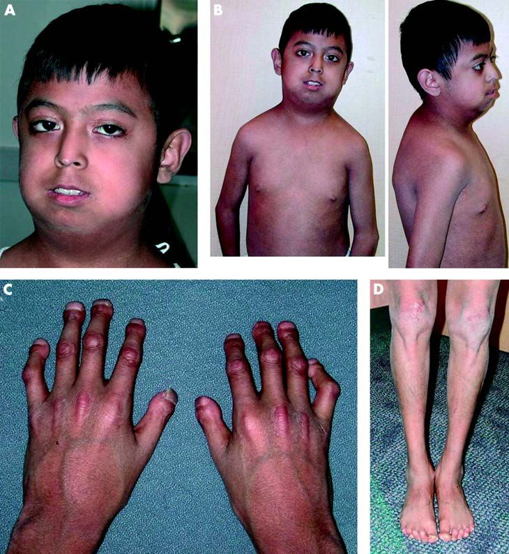
1.5 Mandibuloacral Dysplasia (MAD) ............................................................................................ 37
2. OBJECTIVES AND AIM..................................................................................................................... 40
3. RESEARCH APPROACH .................................................................................................................. 41
3.1 ICMT inhibitor – Structure and development ....................................................................... 41
3.2 Cell lines and In vitro experiments ......................................................................................... 41
3.3 Mouse models and in vivo experiments................................................................................ 42
3.4 Methods to evaluate the treatment effect of inhibiting Rce1 and Icmt ......................... 45
4. RESULTS AND DISCUSSION .......................................................................................................... 45
4.1 A small-molecule ICMT inhibitor delays senescence of Hutchinson-Gilford progeria
syndrome cells ................................................................................................................................... 45
4.2 Targeting RAS converting enzyme 1 overcomes senescence and improves progeria-
like phenotypes of ZMPSTE24 deficiency ................................................................................... 50
5. CONCLUSION AND FUTURE PERSPECTIVES........................................................................... 56
6. ACKNOWLEDGEMENTS .................................................................................................................. 59
7. REFERENCES ..................................................................................................................................... 65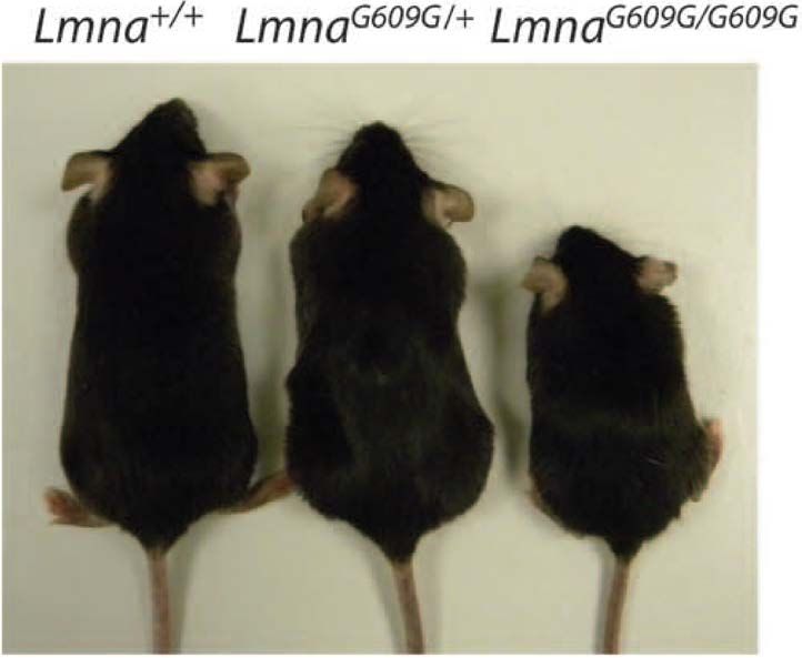
LIST OF ABBREVIATIONS

ABI                     Ankle-Brachial Indices
ATM                     Ataxia-Telangiectasia Mutated
CTSU                    Clinical and Translational Study Unit
CVD                     Cardiovascular disease
DCM                     Dilated cardiomyopathy
DNA-PKcs                DNA-PK catalytic subunit
DSBs                    Double-strand breaks
Ecs                     Endothelial cells
ER                      Endoplasmic reticulum
FADS                    Fetal akinesia deformation sequence
FTase                   Farnesyltransferase
FTIs                    Farnesyltransferase inhibitors
GGTase-I                Geranylgeranyltransferase-I
HGPS                    Hutchinson-Gilford progeria syndrome
ICMT                    Isoprenylcysteine carboxyl methyltransferase
LV                      Left ventricular
MAD                     Mandibuloacral dysplasia
MMP                     Mitochondrial membrane potential
MSC                     Mesenchymal stem cell
NAC                     N-acetylcysteine
PML-NB                  Promyelocytic leukemia nuclear bodies
PPi                     Inorganic pyrophosphate
PWVcf                   Carotid-Femoral Pulse Wave Velocity
RCE1                    RAS-converting enzyme 1
RD                      Restrictive dermopathy
SAHF                    Senescence-associated heterochromatic foci
SASP                    Senescence-associated secretory phenotype
SFN                     Sulforaphane
SOD                     Superoxide dismutase
UPR                     Unfolded protein response
VDR                     Vitamin D receptor
VSMC                    Vascular smooth muscle cell
WAT                     White adipose tissue
γH2AX                   Phosphorylated histone H2AX
1. INTRODUCTION

1.1 Aging

Aging starts already at conception and can be defined as the time-dependent progressive
loss of function which ultimately results in increased probability of disease and death 1.
Different people exhibit different symptoms of aging and these symptoms can vary
significantly, even among monozygotic twins 2. Multiple physiological changes occur in
organs during aging: the skin loses its vigor; the hair its color and regenerative ability;
blood pressure increases; plaques form in arteries; and the heart reduces its output and
contractile abilities. Many research groups around the world are trying to figure out the
mechanisms underlying aging and time-dependent decline in bodily functions and many
theories have been proposed, but no unifying theory exists.

Animals age and die because of senescence phenotypes where cells stop regenerating
and functioning   3–5.   However, animals also die of extrinsic factors including accidents,
starvation, disease, and war. Whether or not aging is programmed has been the subject
of debate for centuries across many research areas. In the worm C. elegans, hundreds
of genes were found to contribute to extended longevity 6. The identification of the
insulin/insulin-like     growth   factor-1   signaling   pathway   was   a   breakthrough   in
understanding mechanisms of lifespan extension in C. elegans. Even though many genes
and pathways have now been documented to contribute to accelerated or decelerated
aging, there is no gene or combination of genes that can abolish aging. Thus, there is no
organism immortality strategy.

Several molecular theories have been proposed to explain aging: The somatic mutation
theory emphasizes longevity in relation to DNA repair capabilities 7. A theory around
telomeres pointed out that these chromosomal end-structures act as intrinsic “cell division
counters,” perhaps to reduce the likelihood of tumor initiation but with the price of
contributing to aging 8. In a mitochondrial theory, when mitochondrial DNA (mtDNA)
mutations reach a high level, it reduces tissue bioenergetics and speeds up aging 9.

                                                 1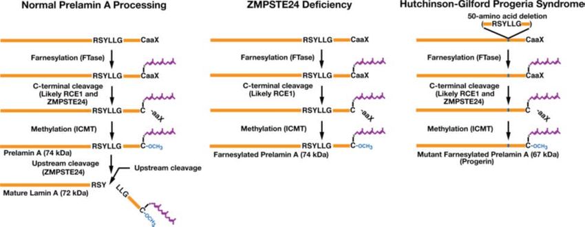
However, although these mechanisms clearly exist in cells and organs, it will be very
difficult to determine to what degree they interact and contribute to physiologic aging.

There has been a substantial increase in our understanding of proteins, microRNAs, and
signaling pathways which contribute to aging and lots of interesting research is underway,
for example to isolate compounds which can manipulate these factors and pathways and
thereby increase longevity. Equally important is research on prevention—i.e., identifying
lifestyle factors such as diet, exercise regiments, and environmental exposures—which
may lead to both healthier and longer life. It would be very interesting to know if the first
person who will reach a 150- or 200-year lifespan is already born,given the remarkable
increase in lifespan over the past two centuries, the emergence of super-centenarians is
within the realm of possibilities.

Nevertheless, understanding the intrinsic and extrinsic mechanisms which underlie
physiologic aging, may also provide tools to understand accelerated aging syndromes
such as Hutchinson-Gilford progeria syndrome and Werner syndrome. And vice versa,
knowledge about progeria can provide information on physiologic aging. The latter
concept has generated an enormous interest in progeria and for this reason there are
today many more progeria researchers than there are patients.

1.2 Premature aging

Premature aging syndromes are also known as progeria, recapitulate many phenotypes
of normal aging and they occur at an accelerated rate and appear in children and
teenagers rather than in later decades of life. They are classified into laminopathies, a
group of genetic diseases which are caused by different mutations in genes encoding
proteins of the inner nuclear rim; and another group of diseases caused by mutations in
genes encoding proteins involved in DNA repair (Table 1).

                                             2
Table 1 Different progeria syndromes. Reprinted from Premature aging syndromes: from patients to
mechanism 10. Creative Commons Attribution-NonCommercial-No Derivatives License (CC BY NC ND).
Disease Gene Phenotype Cellular Reference
name phenotype
Hutchinson- LMNA prominent from 1 year, Nuclear envelope Eriksson 2003;
Gilford short stature, deformities, de Sandre
progeria early loss of hair, increase in DNA Giovannoli
syndrome lipodystrophy, and telomere 2003; Bridger
cardiovascular problems, live damage, and Kill 2004;
into their mid-teens to early senescence Liu 2005;
twenties Decker 2009;
Chojnowski
2015
Restrictive ZMPSTE24 perinatal lethal, skin defects, Nuclear envelope Navarro 2004
dermopathy obvious superficial blood deformities
vessels, micrognathism, bone
problems, and
joint contractures
Mandibuloacral LMNA or Slow growth, craniofacial PrelaminA Novelli 2002,
dysplasia ZMPSTE24 abnormalities, mandibular increase, nuclear Filesi 2005,
hypoplasia, progressive bone enlargement, Cenni 2018,
defects , and skin atrophy , heterochromatin
pigmentation, and loss
lipodystrophy affecting face
and the extremities
Werner LMNA growth retardation, short Nuclear shape Hisama 2011,
syndrome stature, alopecia , abnormalities and Motegi 2014
skin atrophy , lipodystrophy , heterochromatin
abnormal fat deposition, and organization
severe ulcerations around deformation
the Achilles
tendon and malleoli
Néstor- BANF1 severe osteolysis, lipoatrophy Nuclear envelope Puente 2011,
Guillermo and absence of cardiovascular deformities, Cabanillas 2011
progeria disease emerin
syndrome mislocalization.
31.3 Hutchinson-Gilford progeria syndrome
Hutchinson-Gilford Progeria Syndrome (HGPS) is a rare fatal disorder which occurs in 1
per 4 to 8 million newborn babies 11. The lifespan of these children is between ten and
twenty (median ~14 yrs.). HGPS patients typically cannot be distinguished from normal
children at birth, and the phenotypes instead become gradually apparent from 1 year on.
1.3.1 History
In 1886, Jonathan Hutchinson identified a boy with stunted growth, hair loss, and skin
atrophy 12. Similar phenotypes were also described by Hastings Gilford in 1897 who
followed his first patient until the patient died. In recognition of their identification of this
rare disease, these two men’s names were used to describe the disease: Hutchinson-
Gilford Progeria Syndrome. The term progeria comes from the Greek word "pro", which
means "before" or "premature", and "gēras", which means “old age”; Geras was also the
god of old age in Greek mythology. In 1972, a review of 60 cases of HGPS by DeBusk
was published and contributed to a better understanding of the scope and diagnosis of
the disease 13.
1.3.2 HGPS clinical phenotypes
As outlined later, Hutchinson-Gilford Progeria Syndrome (HGPS) arises from a de-novo
mutation in the LMNA gene and is therefore typically not inherited. Unsuspecting parents
are blessed with a new child who appears completely normal at birth. Symptoms do not
start to become apparent until 1 year of age when parents will note a failure to thrive,
abnormal growth, and absence of hair. The diagnosis of HGPS can today be made with
a simple genetic test combined with the clinical phenotypes. Figure 1 on the next page
shows photographs of a child with HGPS at multiple stages of life until 12 years of age.
4Figure 1: Representative photographs of a patient at different ages. Reprinted from Hutchinson-Gilford
progeria syndrome: review of the phenotype 14. JOHN WILEY AND SONS LICENSE (License Number
5152060139044).
Clinical signs of HGPS include slow growth, prominent beak-like nose, small mandible
(micrognathia), alopecia of scalp, eyebrows, face and body, prominent scalp veins, paper-
thin sclerodermatic skin, and nail problems; other bone defects such as joint contractures
and pain, skeletal hypoplasia, delayed fontanelle closure, and fractures; endocrine
abnormalities; and metabolic disorders such as lipodystrophy, hyperlipidemia, and
atherosclerosis. The latter lead to the main cause of death manifested by myocardial
infarction, and stroke. Table 2 on the next page and Figure 2 on the following page show
the name and frequency of common and rare HGPS symptoms.
5Table 2: Major phenotypes in 142 HGPS patients. Reprinted from Hutchinson-Gilford progeria syndrome:
review of the phenotype 14. JOHN WILEY AND SONS LICENSE (License Number 5152060139044).
6Figure 2. HGPS clinical phenotypes.
Growth retardation
HGPS patients show only slightly reduced growth in the first year of life but a sharp
decrease thereafter. Children with HGPS are always much shorter than unaffected
children and they rarely weigh more than 30 kg, even as teenagers 15. Surprisingly, they
consume enough food to be able to grow but still they don’t grow. There is no effect on
puberty.
Malformation of face
Following the second year of life, HGPS patients usually develop hypoplasia of facial
bones and lower jaw. This results in the formation of a small face and retrognathia or
micrognathia. Frontal and temporal bones are more prominent, also the nose is sharp
and small, and the ears are small and sometimes have the lobes missing. The children
7also exhibit crowded teeth, delayed loss of the primary teeth, and eruption of secondary
teeth. Parotid glands of some patients are prominent without intrinsic lesions 15.
Head and skin
Normally, HGPS patients develop alopecia, i.e., they lose their hair and become bald on
the scalp and eyebrows, although in some cases, immature hairs remain. Because of
alopecia and the lack of fat on the skin of the scalp, blood vessels on the head appear
prominent. There is little or no osteolysis of calvaria but a few patients have had skull
fractures 16. Skin problems include a thin epidermis and loss of hair follicles and stem
cells 17. The skin over the abdomen and thigh is tighter compared with unaffected children.
A reduction in skin keratinocyte populations including those with stem and progenitor cell
characteristics has been observed in mouse models of progeria (tetop-LAG608G+;
K5tTA+) compared to wildtype mice 18.
Lipodystrophy
Lipodystrophy appears in nearly all cases of HGPS, with the degree varying in different
individuals. Some children show lipodystrophy across the whole body and some only on
extremities. A prominent feature of lipodystrophy is lack of subcutaneous fat in the skin.
Because of the disappearance of subcutaneous fat, the surface veins become more
prominent. Sometimes, the vein of the nasal bridge is visible, which in some children was
one of the first clinical phenotypes. Later, scalp veins (Figure 3) and other veins of the
body, including legs are easy to visualize. The eyes of children with HGPS are often
protruding because of the loss of fat around the eye and orbit.
8Figure 3. Prominent scalp veins. Reprinted from Hutchinson-Gilford progeria syndrome: review of the
phenotype 14. JOHN WILEY AND SONS LICENSE (License Number 5152060139044).
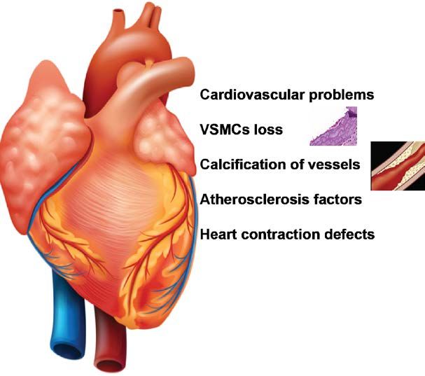
Cardiovascular problems
The endocrine and metabolic abnormalities associated with HGPS include dyslipidemia
and cellular defects in the artery wall which inevitably lead to cardiovascular problems in
older HGPS patients—all due to the toxic progerin protein (as discussed below). The most
common cause of death is myocardial infarction and stroke in the early teenage years.
Indeed, there is evidence from patients and mouse models of progressive loss of smooth
muscle cells in arteries and of fibrosis and dysfunction in the heart. Many factors likely
converge on the cardiovascular problems in HGPS and attempts to classify them lead to
four different aspects: Vascular smooth muscle cell (VSMC) loss; blood vessel
calcification; atherosclerosis; and heart contraction defects (Figure 4).
A clinical trial which included 27 HGPS patients examined heart and arterial function
through echocardiography 19. Although no left ventricular (LV) systolic dysfunction was
discovered, a high LV diastolic dysfunction was evident and could potentially be an early
marker for HGPS disease progression. Moreover, bradycardia and prolongation of the
PR, QRS, and QTc intervals on electrocardiograms have been reported in Zmpste24–/–
mice, one of the early mouse models of progeria 20. Importantly, however, in most HGPS
patients it seems the electrocardiographic intervals are normal with only an elevation of
the heart rate.
Another research group investigated the vascular problems in HGPS patients with
ultrasound measurements 21. The intima-media, near adventitia, and deep adventitia of
9the artery wall were greater in HGPS patients compared to control. The mean Carotid-
Femoral Pulse Wave Velocity (PWVcf) of HGPS patients was like that of 60-year-old
adults. Also, Ankle-Brachial Indices (ABI) was abnormal in most progeria patients.
In progerin knock-in LmnaG609G mice—an authentic mouse models of HGPS discussed
further below—the calcification of the aortic arch and thoracic aorta was much more
severe than in wildtype mice 22. These vascular abnormalities were accompanied with
high expression of osteogenic markers (as judged by immunohistochemical analysis) and
with defects in mitochondrial function and ATP synthesis.
Figure 4. Heart problems in HGPS.
Endocrine problems
Leptin
Leptin is a hormone secreted by adipose tissue in the small intestine and mainly functions
as a regulator of energy consumption. Consistent with lipodystrophy and subsequent lack
of subcutaneous adipose tissue, the leptin levels of HGPS patients is lower than in
unaffected people 23. Low leptin levels may be associated with cardiovascular disease,
as indicated in some studies 24.
10Insulin
Hyperinsulinemia means abnormally high circulating insulin levels and can contribute to
obesity and heart disease25. Hyperinsulinemia is often associated with (but doesn’t
require) diabetes and hyperglycemia. But hyperinsulinemic patients can develop insulin
resistance and finally progress to insulin-dependent type II diabetes 26. The insulin levels
of HGPS patients are more than 3-fold higher than in unaffected people, which could
potentially increase the risk of diabetes and cardiovascular disease, although diabetes is
not a uniform part of the HGPS disease spectrum 23.
Sex hormones
Since HGPS patients normally di during their 10-20s, they have been considered infertile.
Some of the girls had menarche but others did not 27. Very few studies have been done
regarding the sexual development with estrogen in female HGPS patients. More need to
be done related to the hormone levels and whether they differ from unaffected people.
There have also been numerous reports from studies of hormone levels in HGPS mouse
models. The Lamin A knock-in (LAKI) mice, harboring the genotype LmnaG609G develop
hypoglycemia and fat loss during the progeria disease progression, and they died
prematurely. They also showed higher energy consumption and mitochondrial
dysfunction 28. More research is needed to fully understand hormonal and metabolic
alterations of HGPS and to identify functional links between them and the fatal
cardiovascular disease.
Limited range of motion and ostolysis
HGPS patients usually exhibit reduced mobility in joints including elbows, wrists, fingers,
hips, knees, and ankles which leads some children to develop kyphosis of the spine or
develop the abnormal posture of “horse riding stance” 14. Similarly, mouse models of
progeria, i.e., Zmpste24-deficient mice and LmnaG609G develop bone abnormalities,
osteoporosis, and spontaneous fractures 29,30.
111.3.3 HGPS cellular and molecular phenotypes
Cellular senescence
Reduced Lamin B1 expression
Low levels of Lamin B1 has been observed in senescent HGPS cells and in cells
undergoing replicative senescence 31,32. Consistent with those observation, disruption of
the Lamin B1 gene reduces DNA replication ability and pol II activity 33–36. LaminB1 binds
to and regulates Oct1, and reduced LaminB1 results in non-sequestered active Oct1 and
overproduction of the microRNA mir-31 37. Mir-31, in turn, interacts with and regulates the
levels of p16(Ink4a)/p14(Arf) and thereby contributes to senescence; it can also increase
p53 levels consistent with that notion 38. Now, low Lamin B1 expression is used as a
marker for senescence in HGPS cells and the phenotype is readily detectable in cells
from HGPS patients and cells from the progeria mouse models.
SASP (Senescence-associated secretory phenotype)
Different kind of cells secrete a packet of proteins when they undergo senescence, amply
called the senescence-associated secretory phenotype (SASP). These proteins interact
with the local environment and result in inflammation and continued interleukin secretion.
Mainly inflammatory cytokines and chemokines are included in the SASP, for example
interleukins (IL)-6, 7, and 8; but the SASP also includes growth factors (e.g., GRO, HGF),
cell surface molecules (e.g, ICAM, uPAR, and TNF receptors), and other survival factors
39. In Zmpste24–/– mice, which exhibit progeria phenotypes, NF-κB and inflammatory
cytokines, such as IL-6, CXCL1, and tumor necrosis factor α (TNF-α), and adhesion
molecules, such as ICAM1, all SASP components, were upregulated. Inhibiting NF-κB
extends life span and reduces progeria-related phenotypes in this mouse model
according to one study 40. Although SASP was originally identified in senescent cancer
cells, many studies of progeria include the SASP as a marker of senescence. The mTOR
inhibitor rapamycin increases NF-κB transcriptional activity and reduces the SASP and
senescence-associated inflammation 41.
12SAHF (Senescence-associated heterochromatin foci)
When mice and humans age, the expression of p16INK4a in cells increases 42–45 and can
stimulate the formation of senescence-associated heterochromatic foci (SAHF) by
activating the pRB tumor suppressor, which inhibits cell cycle progression and reduced
proliferation (Figure 5). In addition to SAHF, there are several other phenotypes which
occur in both HGPS cells and in cells from physiologically aged individuals (Figure 6).
Figure 5 Hallmarks of senescent cells. Reprinted from Four faces of cellular senescence 46. Copyright
Clearance Center, Inc. ("CCC") (License ID1148668-1)
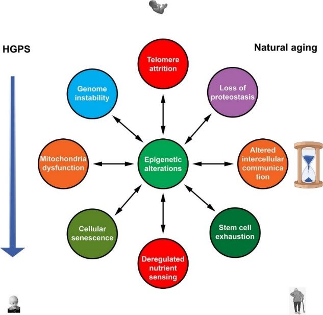
Figure 6 Nine hallmarks of aging. Reprinted from Are there common mechanisms between the
Hutchinson–Gilford progeria syndrome and natural aging 47. Copyright © 2019 Ashapkin, Kutueva,
Kurchashova and Kireev. Creative Commons Attribution License (CC BY).
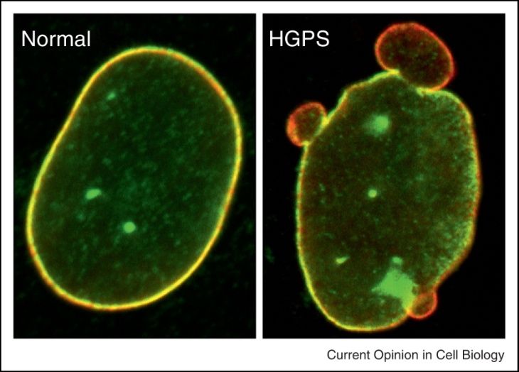
13Nuclear blebbing
The genetic mutation in exon 11 of the LMNA gene (outlined further below) activates a
cryptic splice site, which leads to a 50-amino acid deletion and production of the toxic
protein progerin. Because of this internal deletion, the farnesylcysteine residue is retained
and provides a strong affinity to the inner membrane of nuclear 48. Progerin inserts itself
into the nuclear lamina and causes a characteristic nuclear blebbing phenotype (Figure
7). Extensive manual and automatic quantification of nuclear shape abnormalities
revealed that the frequency of such abnormal nuclei is up to 73% in HGPS cells, whereas
only 24% of wildtype cell nuclei are abnormal 48. The abnormalities range from
invaginations, blebs, and herniations, to frank nuclear disruptions.
Figure 7 Immunostaining of primary fibroblasts from control and HGPS patient. Note irregular shape
and blebs on the nuclear lamina of an HGPS cell on the right. Reprinted from Hutchinson-Gilford progeria
syndrome 49. © 2013 Creative Commons Attribution 3.0 License
Nuclear architecture changes
Heterochromatin changes
A gradual loss of peripheral heterochromatin has been observed in HGPS late passage
fibroblasts 50. At early passages, a loss of the methylated histone marker H3K27me3
occurred before obvious changes in nuclear shape 51,52. Moreover, the EZH2 gene, which
plays an important role in the trimethylation of H3K27 53, is dramatically downregulated
in HGPS cells which may or may not be causally linked to the abnormal nuclei. At later
14passages, downregulated H3K9me3 has been detected accompanied by up-regulation
of pericentric satellite III repeat transcripts. One theory behind these findings is that the
mutant HGPS protein, progerin, influences histone methylation and heterochromatin loss.
The precise mechanisms however are unknown 54,55.
Thickened lamina, genome instability, and DNA damage
The lamin proteins are quite mobile in normal cells but more immobilized in cells from
HGPS patients which tends to make the lamina thicker 50,56. Many studies have found
high levels of phosphorylated histone H2AX (γ-H2AX)—a molecular biomarker of DNA
double-strand breaks (DSBs) 57—in HGPS cells compared to control 58,59. The high levels
of DNA damage likely contribute to the premature senescence.
DNA damage checkpoints are also activated in HGPS cells, most probably G1/S and/or
intra-S checkpoints 58. The checkpoint kinases Ataxia-Telangiectasia Mutated (ATM) and
ATR (ATM and RAD3-Related) are also indicators of DNA damage and are associated
with phosphorylated Chk1 and Chk2; all of these can be found upregulated in HGPS cells.
Interestingly, inactivation of ATM and ATR could reprogram the cells to enter S phase
and overcome their replication arrest (Figure 8, next page).
Farnesyltransferase inhibitors (FTIs) inhibit the farnesylation of progerin or prelamin A
and readily restore nuclear shape abnormalities. However, FTIs fail in reducing DNA
damage and the levels of γ-H2AX and SAHF levels remain high in FTI-treated cells 60,61.
15Figure 8 Major DNA damage in human cells. Reprinted from Genomic instability and DNA damage
responses in progeria arising from defective maturation of prelamin A 62. Creative Commons Attribution 3.0
License (CC BY 3.0).
1.3.4 Molecular background of HGPS and CAAX protein processing
The LMNA gene and etiology
HGPS is a rare genetic and fatal disease most often caused by a point mutation in the
gene LMNA 63,64. LMNA is located on chromosome 1 (1q22) and encodes two major lamin
proteins, lamin A and lamin C. Lamin A (72 kDa) is transcribed from exons 1–12, while
Lamin C (65 kDa) transcripts comes from two fewer exons (exon 1–10) at the 5′ splice
site (Figure 9, next page). There are also B type lamins (Lamin B1 and lamin B2)
encoded by LMNB1 and LMNB2. Lamins are intermediate filament proteins that make up
the inner layer of nuclear envelope, and the lamina interacts with many proteins and with
DNA in the form of chromatin.
Mutations in the LMNA gene can result in a series of disorders called laminopathies.
Distinct mutations lead to patients developing muscular dystrophy, lipodystrophy,
neuropathy, cardiomyopathy, and premature aging. The most talked-about laminopathy
is HGPS but it is far from the most frequent.
The point mutation that causes HGPS is a cytosine-to-thymine transition in the LMNA
coding region at nucleotide 1824 (c.1824C>T) 64,65. This site is located in exon 11 63. The
16mutation doesn’t change an amino acid and the site encodes glycine both before and
after the mutation. Instead, the Gly608 → Gly608 mutation (C to T transversion in the
codon) activates a cryptic splice site which leads to the production of prelamin A with an
internal deletion of 50 amino acids in the carboxyl-terminal domain which also contains
the cutting site for ZMPSTE24. This results in the accumulation at the nuclear envelope
of a farnesylated and methylated form of prelamin A that can’t be processed to mature
lamin A, and this truncated protein is called progerin. Retention of the farnesylated and
methylated lipid anchor in prelamin A and progerin causes all disease phenotypes. To
fully understand and appreciate the HGPS mutation, it is essential to understand the
processing of so-called CAAX proteins.
Figure 9 Representation of the A-type lamin encoding lamin A and lamin C. Reprinted from Do lamin
A and lamin C have unique roles 66. SPRINGER NATURE LICENSE. (License Number 5152001181566)
17CAAX proteins undergo three posttranslational modifications
Prelamin A is a so-called CAAX protein which means it possesses a CAAX motif at the
carboxyl-terminal end. The “C” represents a cysteine residue; the AAs are typically two
aliphatic residues, and X represents different C-terminal amino acid 67–69. In prelamin A,
these residues are Cys-Ser-Iso-Met. Prelamin A and most other CAAX proteins undergo
three enzymatic processing steps at this carboxyl-terminal CAAX motif (Figure 10, next
page):
Farnesylation: Farnesylation means the attachment of a 15-carbon farnesyl lipid to the
thiol group of the cysteine residue by farnesyltransferase (FTase). Beside prelamin A,
RAS and RHEB proteins and lamin B1 are also farnesylated. Other CAAX proteins
including RHOA, RAC1, and CDC42 are lipidated by a 20-carbon geranylgeranyl lipid by
geranylgeranyltransferase type I (GGTase-I). In mice, knockout of Fntb (encoding the
unique beta subunit of FTase) is an early embryonic lethal, and inactivation of FTase in
both normal and transformed cells results in proliferation arrest 70.
Initial –AAX proteolysis: Following farnesylation, the last three amino acids of prelamin
A are clipped off by RAS-converting enzyme 1 (RCE1) or alternatively by ZMPSTE24 71,72.
Rce1-deficient mice die late during embryonic development (E15.5), and RCE1 is
essential for RAS processing 73. Bergo et al also developed a Rce1 conditional knockout
mouse and found that the absence of Rce1 leads to mislocalization of all RAS proteins
and inhibits proliferation and oncogenic RAS transformation of cells 74.
Methylation: Isoprenylcysteine carboxyl methyltransferase (ICMT) is responsible for the
methylation of the newly exposed farnesylcysteine residue. The three processing steps
make the protein more hydrophobic and are believed to enhance interactions with
membranes and with other proteins, and to regulate protein stability and turnover 75,76.
Icmt knockout mice die by embryonic day 12.5 77. Icmt conditional knockout mice have a
normal life span and were previously used as a model for CAAX-protein methylation
research, including in progeria 78.
18Prelamin A undergoes a fourth posttranslational processing step: Prelamin A is the
only known CAAX protein that undergoes a fourth processing step following methylation.
The entire 15 amino acids of the carboxyl terminus, including the farnesyl-methyl-cysteine
residue is cleaved off and leaving mature lamin A to be formed from the amino terminus.
Around 20 years ago, Bergo et al and Pendas et al found that the protease ZMPSTE24
is responsible for this step 29,79. Another group developed a humanized yeast system to
evaluate the function of ZMPSTE24-dependent cleavage of prelamin A and helped to
increase our understanding of the structure and function of ZMPSTE24 80.
Figure 10 Posttranslational processing of prelamin A in normal cells, in ZMPSTE24 deficient cells,
and in HGPS. Reprinted from Prelamin A farnesylation and progeroid syndromes 81. Copyright © 1969,
Elsevier. Creative Commons CC-BY license.
Biochemical importance of CAAX protein processing: The posttranslational
processing increases the hydrophobicity of one end of the CAAX proteins which increases
interactions with membrane surfaces within cells. Indeed, cells lacking either Fntb, Rce1,
or Icmt show mislocalization of all the RAS proteins away from the plasma membrane
into the cytosol and internal membranes. The CAAX processing steps are also important
for controlling the interaction between the CAAX protein and its protein binding partners
and for protein stability, where some proteins accumulate and others are degraded faster
if either of these modifications are blocked 82–84.
191.3.5 Mouse model of HGPS
Mouse models have proved highly useful for studying mechanisms and treatment of
premature aging disorders. But it is important to be aware of the strengths and
weaknesses of mice as models for humans 85. Mice can live for 3 years and humans for
more than 100; thus aging, particularly physiologic aging, is difficult to study in a
comparative fashion. In contrast, disorders caused by the accumulation of a specific toxic
protein like progerin or prelamin A, or by the absence of a specific enzyme like
ZMPSTE24, are more likely to give valuable results.
1.3.5.1 Lmna-deficient mice
To address the issue of whether A-type lamins are essential, Sullivan et al in 1999 created
Lmna-deficient mice which lack both lamin C and lamin A (Figure 11). Homozygous
embryos developed normally in utero, but the mice developed severe muscular dystrophy
and growth retardation, and died by 8 weeks of age 86.
Figure 11. Schematic representation of the Lmna locus and allele for modification. Reprinted from
Loss of a-type lamin expression compromises nuclear envelope integrity leading to muscular dystrophy 86.
Springer Nature Customer Service Centre GmbH (the Licensor) (License ID1148668-1).
1.3.5.2 Zmpste24-deficient mice
In 2002, Zmpste24-deficient mice were developed by two groups and was found,
serendipitously, to mimic features of accelerated aging (Figure 12). Homozygous mice
were normal at birth and up to weaning, but then grew slowly and developed alopecia,
bone fractures, osteoporosis, reduced grip strength, dental problems, dilated
cardiomyopathy, and small amounts of subcutaneous fat 87. Their lifespan was 6–8
months.
20Figure 12. Body weight curves and photograph of Zmpste24-deficient mice (open circles) compared
to littermate wildtype siblings. Reprinted from Zmpste24 deficiency in mice causes spontaneous bone
fractures, muscle weakness, and a prelamin A processing defect 87. Copyright (2002) National Academy of
Sciences.
Zmpste24−/− mice showed reduced levels of subcutaneous fat which is an important
phenotype in HGPS patients 88. Adipocytes of the Zmpste24-deficient homozygotes were
less than half the size of adipocytes from wild-type mice. There was oxidative stress due
to high mitochondrial activity and dysregulation of lipid metabolism.
Cardiac problems are prominent with age in HGPS patients and this also was detected in
Zmpste24−/− mice 20. Older mice developed ventricular defects, bradycardia, and
prolongation of PR, PQ and QRS intervals on electrocardiograms.
There is another similarity between Zmpste24−/− mice and HGPS patients: Nearly all
children with HGPS have problems with their teeth and micrognathia and two-three-
month-old Zmpste24−/− mice have obvious micrognathia and fragile teeth (Figure 13) 29.
21Figure 13 μCT scans illustrating bone lesions in Zmpste24−/− mice. Reprinted from Zmpste24 deficiency
in mice causes spontaneous bone fractures, muscle weakness, and a prelamin A processing defect 87.
Copyright (2002) National Academy of Sciences.
Although they have many common phenotypes, the Zmpste24 mutation is not the same
as the HGPS LMNA point mutation. Prelamin A, which accumulates in Zmpste24
deficiency, is not the same as progerin which is shorter. Several groups have tried to and
succeeded in generating mice which express progerin, either from transgenic switchable
constructs or by introducing the HGPS point mutation into the mouse Lmna gene.
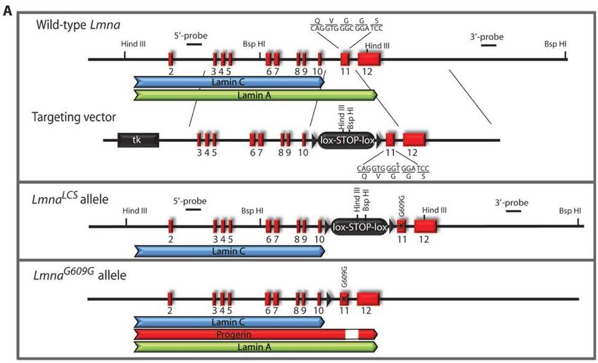
1.3.5.3 LmnaG609G knock-in mice
Thus, additional HGPS mouse models were subsequently developed by manipulating the
Lmna locus directly 89. In 2011, LmnaG609G knock-in mice were produced with a mutation
in the Lmna gene (1827C > T) 30. The researchers created a point mutation at
c.1827C>T;p.Gly609Gly, which is equivalent to the human HGPS
c.1824C>T;p.Gly608Gly mutation in the LMNA gene (Figure 14, next page).
LmnaG609G/G609G mice (Figure 15, following page) exhibit progeria phenotypes from the
age of 3–4 weeks. They developed reduced grip strength, subcutaneous fat, and bone
density, and shorter survival. More importantly and contrary to the Zmpste24 deficiency,
the mice developed a cardiovascular disease phenotype: loss of vascular smooth muscle
22cells in the aortic arch and a prolonged QRS-wave on electrocardiogram. The mice also
exhibited changes in plasma hormone levels 30.
Interestingly, LmnaG609G/+ mice, i.e., the heterozygotes, also developed progeria
phenotypes, but at a much later stage, around 8 months after birth. Then, they started to
lose weight, and developed most of the phenotypes of homozygotes, and they died
prematurely (Zmpste24+/– mice have no discernible phenotypes and a normal life span).
Figure 14. Schematic representation of the wild-type Lmna locus, and the method to edit the point
mutation. Positions of cleavage sites and probes are shown. Reprinted from Splicing-directed therapy in a
new mouse model of human accelerated aging 30. Copyright Clearance Center, Inc. ("CCC") (License
ID1148698-1).
23Figure 15. Representative photographs of 3-month-old Wild type, heterozygous, and homozygous
mice. Reprinted from Splicing-directed therapy in a new mouse model of human accelerated aging 30.
Copyright Clearance Center, Inc. ("CCC") (License ID1148698-1).
In our experiments, LmnaG609G/G609G transgenic mice bred poorly, likely because of early
disease symptom appearing at the time of breeding (~6 weeks) 90. To reduce the early
mortality, we also extended the weaning time from 21 to 30 days.
Interestingly, it was reported that survival of female and male LmnaG609G/G609G mice differs
in a statistically significant fashion (Figure 16, next page) 90. Female mice had a maximal
lifespan of 101 days whereas male mice lived almost one month longer.
Survival of LmnaG609G/+ female and male mice also differed. Here, female mice lived for
266 days while males lived for 311 days 90. We haven’t found any evidence to support
these finding. Accordingly, it was suggested that maybe later when we did animal
experiments in Lmna mice, we should consider the sexual difference.
24Figure 16. Life span. A, Kaplan-Meier plots showing survival of wildtype (green), LmnaG609G/+ (black), and
LmnaG609G/G609G (red) mice. B, LmnaG609G/G609G and C, LmnaG609G/+ mice: females (pink), and males (blue in
B; aqua in C). Reprinted from Long term breeding of the LmnaG609G progeric mouse: characterization of
homozygous and heterozygous models 90. Creative Commons Attribution-NonCommercial-No Derivatives
License (CC BY NC ND).
The LmnaG609G mice has become a widely-used model for HGPS. We use these mice but
complement the experiments with Zmpste24-deficient mice as they are easier to breed
and have more penetrant and robust phenotypes; and results can be compared with
previous studies. There is a scoring system for LmnaG609G transgenic mice (Table 3) 90,
and this table can be used to evaluate their condition.
Table 3. Grading system to evaluate severity of clinical phenotypes in mouse models. Total 3 = best
condition; total 10 = worst condition. Reprinted from Long term breeding of the LmnaG609G progeric mouse:
characterization of homozygous and heterozygous models 90. Creative Commons Attribution-
NonCommercial-No Derivatives License (CC BY NC ND).
25In this model design, LmnaG609G mice were created by Cre-excision of a segment from
LmnaLCS (Lamin C–Stop) mice, which have a deletion of Lamin A but with Lamin C still
expressed. These “lamin C-only” mice are indistinguishable from wild-type control mice
until 50 weeks old, which is also consistent with previous knowledge that Lamin A is
dispensable in lamina formation and mouse development 91.
1.3.5.4 Tissue specific HGPS mouse models
There are many other mice models that express progerin, sometimes with innovative
switching on and off systems, in specific cell types and tissues to help understand
mechanisms of progerin-induced toxicity and tissue-specific pathology and to test the
efficacy of potential treatment strategies—and to do so without having to worry about
systemic wide-spread disease. Moreover, the LmnaG609G and LmnaLCS models have been
used to evaluate tissue-specific effects of progerin expression.
Heart and vessel tissue specific models
One study analyzed atherosclerosis development and progression in LmnaG609G/G609G
mice bred on a background of apolipoprotein E deficiency (Apoe–/–), which are prone to
develop vascular lesions if they are on a high-fat diet 92. One of the goals was to study
mechanisms underlying progerin-induced atherosclerosis. The mice expressing progerin
developed a more severe cardiovascular phenotype compared with controls, with more
severe lipid deposition, adventitial fibrosis, and overall atherosclerosis. When Nevado et
al obtained Ldlr–/–LmnaG609G/G609G mice, it was used as another mouse model of
atherosclerosis of progeria 93. It showed pathology in the intima of the artery wall and
resulted in VSMCs death.
In additional experiments, Apoe–/–LmnaLCS/LCS mice were bred with
with LysMCre and SM22αCre mice 92. The latter two models express Cre in
macrophages and vascular smooth muscle cells (VSMCs), respectively. Apoe–/–
LmnaLCS/LCSSM22αCre mice did not exhibit premature aging phenotypes, as expected,
but died earlier due to atherosclerosis-related disease. This study provided useful
information that only expressing progerin in VSMCs could induce atherosclerosis and
shorten the lifespan.
26Another group created a G608G bacterial artificial chromosome (BAC)-transgenic
progeria mouse model, in which the “human” BAC harbored the HGPS mutation 94. The
most prominent phenotype of these mice was vascular defects characterized by loss of
VSMCs in the arterial vessel wall.
There is also an HGPS mouse model which selectively expresses progerin in endothelial
cells 95. Here, the researchers used an endothelial cell–specific Cdh5 promoter and
expressed a tetracycline-responsive transcriptional activator. These mice exhibited a
shorter survival compared with controls and expressed progerin in PECAM1-positive
microvasculature, but not in cardiomyocytes. They developed myocardium dysfunction
and hypertrophy, but no loss of vascular smooth muscle cells. Total eNOS and NO level
were decreased in the mice and pro-fibrotic pathways were activated.
Another group, Campo et al crossbred LmnaLCS/LCS mice with SM22
Cre+/–mice and Tie2-Cre+/–mice to express progerin in VSMCs and ECs (Endothelial cells),
respectively96. They found that it is the vascular smooth cells which play an essential role
in vessel impairment in HGPS mice and suggested that the combination of progerin
expression of ECs and VSMCs may lead to endothelium dysfunction.
Fat-cell specific mouse model
Another mouse model expressed progerin in a small portion of preadipocytes and
adipocytes of mouse subcutaneous white adipose tissue (sWAT) 97. These mice showed
decreased volume of sWAT and developed fibrosis and lipoatrophy with increasing age.
They also exhibited increased senescence, DNA damage, and inflammatory responses.
To mimic and study skin and teeth problems of HGPS patients, Sagelius et al 98 created
a tetracycline-inducible transgenic mouse model and found fibrosis, loss of subcutaneous
adipocytes, hair follicle and sebaceous glands defects, and abnormal incisors. Moreover,
the extent of the phenotypes was found to correlate with the level of progerin expression.
This model provided important new insight into skin abnormalities of HGPS.
All these mouse models have helped us understand more about the mechanism
underlying the toxic effects of progerin in a whole animal and in specific cell types.
Although it has been suggested that progerin plays a role in physiological aging—it has
27been found expressed at increasing levels with age in senescent cells 47—the evidence
for this is weak and needs further work which is difficult to address in mice.
1.3.6 Possible treatment of HGPS
Primary skin fibroblasts from HGPS patients proliferate slowly and exhibit premature
senescence phenotypes 99. Another prominent cellular phenotype is the abnormal nuclear
shape and impaired DNA repair mechanisms 100. All these phenotypes are caused by
progerin accumulation around the nuclear rim and lots of effort have gone into identifying
therapies which reduce progerin toxicity using these cellular biomarkers as a read-out.
Current therapy
In the years following the identification of the HGPS mutation in LMNA and the role of
ZMPSTE24 in prelamin A maturation, several groups hypothesized that the farnesyl lipid
attached to progerin could contribute to the toxic effects of this mutant protein.
Consequently, inhibiting farnesylation with farnesyltransferase inhibitors (FTIs) previously
developed to block RAS-driven cancer was tested. These efforts used as a read-out the
ability of compounds to reduce the frequency of nuclear shape abnormalities in HGPS
cells. FTIs were thus tested in cells like HeLa, HEK293, and NIH3T3 cells engineered to
express progerin/prelamin A and nuclear blebbing was significantly reduced 101. Few of
these studies addressed the issue of whether FTIs could influence the senescence
phenotype of cells and improve their proliferation.
The first in vivo test of FTIs used ABT-100 mixed in drinking water which was given to
Zmpste24-deficient mice and then to LmnaHG/+ mice (an early progerin-expressing mouse
model). Most of the premature aging phenotypes were attenuated by FTI treatment.
Indeed, the FTI increased body weight and subcutaneous fat pad size and reduced
kyphosis and bone fractures, and increased the bone mineralization102. However, all
treated mice eventually died from progeria suggesting that an FTI can delay but not cure
symptoms. Considering these findings, it is worthwhile to note that FTIs are anti-
proliferative drugs designed for cancer therapy.
In other studies, the FTI tipifarnib (R115777, Zarnestra) was given to LmnaG608G mice to
also test the ability of an FTI to influence the development of cardiovascular phenotypes.
28The FTI significantly delayed the onset of cardiovascular problems and attenuated the
existing cardiovascular disease 103 .
All these findings suggested that FTIs might be an effective drug for helping children with
HGPS. Clinical trials started with FTIs in these children. These clinical trials are difficult
to perform considering how few patients there are, how fragile they are, and how far some
of them must travel to participate. It is also difficult or even impossible to use a placebo-
controlled regimen. Nevertheless, clinical trials with FTIs were performed and achieved
some effects on key phenotypes although it is clear that these drugs cannot cure the
disease.
Twenty-six HGPS patients from 16 countries took part in a phase II clinical trial of the FTI
lonafarnib for at least two years 104. Overall, lonafarnib was well tolerated by the children
and 9 of 25 achieved a 50% increase in body weight, mainly due to increased bone and
muscle mass (but not fat). Other results from this trial showed the carotid-femoral pulse
wave velocity (PWVcf)—which is higher in HGPS patients than controls—decreased at
the end of the treatment. In addition, the FTI increased skeletal rigidity, sensorineural
hearing, and bone mineral density. The results indicate that FTIs reverse some
phenotypes, ameliorate others, but have no impact on most phenotypes—and altogether
this led to an increase in mean survival by 1.6 years 105. Although 1.6-years extra life
means the world to HGPS patients and their families, the FTIs did not meet the high
expectations placed on them following the preclinical studies.
Progerin and prelamin A are farnesylated by FTase, and like KRAS and NRAS it has been
proposed that they can undergo alternate prenylation by GGTase-I when FTase is
inhibited by the FTIs. That could conceivably explain why the FTIs had such modest
effects. To address this issue some studies have evaluated the impact of combining
statins and aminobisphosphonates—which may inhibit both farnesylation and
geranylgeranylation of progerin/prelamin A—and found that they inhibited the loss of bone
density and increased survival in Zmpste24–/– mice 106. Nuclear abnormalities and DNA
damage levels were reduced in HGPS cells after this combination treatment. The
preclinical studies with statins and bisphosphonates prompted clinical trials.
29A single-arm triple therapy trial (ClinicalTrials.gov, NCT00879034) was designed to
evaluate the impact of adding pravastatin and zoledronate to lonafarnib therapy and
compare the effect with lonafarnib monotherapy 107. Thirty-seven HGPS patients from 23
countries participated in the trial. Compared with lonafarnib monotherapy, the addition of
pravastatin and zoledronate did not lead to tolerance problems and side effects. The triple
therapy improved some bone parameters, as might be expected from a bisphosphonate,
but the heart phenotypes and survival did not improve compared to lonafarnib
monotherapy. The idea that statins and bisphosphonates inhibit prenylation are based on
conjecture and effects of high doses of drugs on cultured cells. To this day, there is no
robust in vivo evidence that these classes of drugs inhibit either farnesylation or
geranylgeranylation in tissues in vivo.
Potential future therapies
Based on what the field has learned about mechanisms of progerin-induced toxicity and
disease development, a list of different potential future therapeutic targets can be
compiled: 1. Change or repair the HGPS mutation in LMNA, or targeting lamin A
transcription; 2. Increase progerin degradation; 3. Reduce the toxicity of progerin
downstream 4. Inhibiting progerin methylation with ICMT inhibitors 108.
Change or repair the mutation or targeting lamin A transcription
The point mutation, i.e., the cytosine-to-thymine transition in the LMNA exon 11 coding
region at nucleotide 1824 (c.1824C>T) 62, does not alter the protein coding sequence but
the new splicing event created by the mutation leads to progerin production 63. Earlier
studies revealed that mice with a knockout of mature Lamin A, are completely normal due
to Lamin C expression 91. Thus, disease symptoms are caused by toxic progerin and not
any potential disturbance in Lamin A expression.
Ergin et al used two guide RNAs (gRNAs: gLmna-1 and gLmna-2) targeting Lamin A and
reduced Lamin A/progerin expression without perturbing Lamin C in LmnaG609G/G609G mice
which were also engineered to express CAS9 109. The treatment extended survival by
25%, reduced fat loss, attenuated aortic arch medium layer degeneration, and increased
grip strength. But the treated mice still died earlier comparing to wild type mice due to
30You can also read