Gene Editing and Gene Therapy: Entering Uncharted Territory in Veterinary Oncology
←
→
Page content transcription
If your browser does not render page correctly, please read the page content below

Preprints (www.preprints.org) | NOT PEER-REVIEWED | Posted: 17 May 2021                   doi:10.20944/preprints202105.0376.v1

                 Gene Editing and Gene Therapy: Entering Uncharted Territory in
                                                   Veterinary Oncology

             Wesley A. Wierson1, Alex M. Abel1, Elizabeth L. Siegler2, Stephen C Ekker1,3, Chad
                          M Johannes1,4, Saad S Kenderian1,2,5,6, and Jonathan P Mochel1,7

             1LifEngine Animal Health Laboratories, Rochester, MN 55902, USA.
             2Divisionof Hematology, Mayo Clinic, Rochester, MN 55905, USA.
             3Mayo Clinic Cancer Center Department of Biochemistry and Molecular Biology, Rochester, MN 55905,

             USA.
             4Iowa State University, Department of Veterinary Clinical Sciences, Ames, IA 50011, USA.
             5Department of Immunology, Mayo Clinic, Rochester, MN 55905.
             6Department of Molecular Medicine, Mayo Clinic, Rochester, MN 55905.
             7SMART Pharmacology, Department of Biomedical Sciences, Iowa State University, Ames, IA 50011,

             USA.

             Abstract: With rapid advances in gene editing and gene therapy technologies, the development
             of genetic, cell, or protein-based cures to disease are no longer the realm of science fiction but
             that of today’s practice. The impact of these technologies are rapidly bringing them to the
             veterinary market as both enhanced therapeutics and towards modeling their outcomes for
             translational application. Simply put, gene editing enables scientists to modify an organism’s
             DNA a priori through the use of site-specific DNA targeting tools like clustered regularly
             interspaced short palindromic repeats and CRISPR-associated protein 9 (CRISPR/Cas9). Gene
             therapy is a broader definition that encompasses the addition of exogenous genetic materials
             into specific cells to correct a genetic defect. More precisely, the U.S Food and Drug
             Administration (FDA) defines gene therapy as “a technique that modifies a person’s genes to
             treat or cure disease” by either (i) replacing a disease-causing gene with a healthy copy of the
             gene; (ii) inactivating a disease-causing gene that was not functioning properly; or (iii)
             introducing a new or modified gene into the body to help treat a disease. In some instances,
             this can be accomplished through direct transfer of DNA or RNA into target cells of interest or
             more broadly through gene editing. While gene therapy is possible through the simple addition
             of genetic information into cells of interest, gene editing allows the genome to be reprogrammed
             intentionally through the deletion of diseased alleles, reconstitution of wild type sequence, or
             targeted integration of exogenous DNA to impart new function. Cells can be removed from the
             body, altered, and reinfused, or edited in vivo. Indeed, manufacturing and production
             efficiencies in gene editing and gene therapy in the 21st century has brought the therapeutic
             potential of in vitro and in vivo reprogrammed cells, to the front lines of therapeutic intervention
             (Brooks et al., 2016). For example, CAR-T cell therapy is revolutionizing hematologic cancer
             care in humans and is being translated to canines by us and others, and gene therapy trials are
             ongoing for mitral valve disease in dogs.

             Keywords: Gene Editing; Gene Therapy; Oncology; Comparative Medicine; One Health

                                                                   1

           © 2021 by the author(s). Distributed under a Creative Commons CC BY license.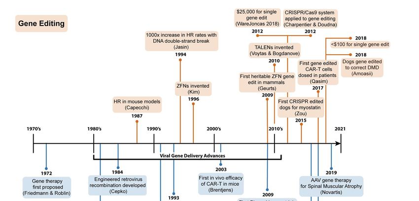
Preprints (www.preprints.org) | NOT PEER-REVIEWED | Posted: 17 May 2021           doi:10.20944/preprints202105.0376.v1

             Introduction
             Gene editing and gene therapy are increasingly used in the human clinic today and their
             application in companion animals is beginning to emerge. About 700 monogenic diseases have
             been reported in various dog breeds, with at least 200 of these diseases harboring known
             causative mutations (Switonski, 2020). About 400 of these spontaneous diseases in dogs are
             considered potential models for human disorders (https://omia.org/home). This makes the dog a
             particularly attractive animal model for accelerated commercial deployment of gene editing and
             gene therapy candidates with applications in cancer, hemophilia, lysosomal storage diseases,
             ophthalmology, immune-mediated disorders, muscular dystrophy, and others (Acland et al., 2001,
             Acland et al., 2005)., These indications may share genetic drivers, physiology, and presentation
             with their human counterparts. This creates bidirectional value, where on one-hand human
             experience and research may derisk similar innovations in veterinary patients. On the other-hand
             accelerated application of the underlying therapeutic in veterinary medicine can answer
             translational questions not effectively answered in pre-clinical studies or even human trials.
             Ultimately, regulatory incentives to develop parallel (veterinary and human) drug development
             programs would present an opportunity to streamline, accelerate, and improve pharmaceutical
             research and development in veterinary and human oncology.

             In cancer research, gene editing and gene therapy applications in canines have garnered
             considerable interest since, in contrast to mice, cancers develop spontaneously in dogs (i.e.,
             without genetic manipulation) and in the context of intact immune system with a syngeneic host
             and tumor microenvironment (Gordon et al., 2009). As more information on the dog genome is
             being released, multiple studies have demonstrated significant homologies between canine and
             human cancer-associated genes, including MET, mTOR, KIT and TRAF3 (Paoloni and Khanna,
             2008). As such, biological and genomic similarities between canine and human cancer provides
             an impetus for parallel development of novel drug candidates (including gene therapy and gene
             editing) in canine and human clinical trials (Schneider et al., 2018). Specifically, ample literature
             has established similarities between the pathologic, biologic, immunophenotypic, and genetic
             components of Diffuse Large B-cell Lymphoma (DBCL) in dogs and humans (Richards et al.,
             2013; (Mochel et al., 2019). Based on these similarities, preliminary proof-of-concept studies from
             Dr. Nicola Mason’s group are the first public reports of CAR-T cell therapy being used in
             companion animals (Panjwani et al., 2016). Even more recently, Sakai et al. have generated
             second and third-generation canine CAR-T cells using retroviral gene transduction with
             RetroNectin and showed positive cytotoxic responses against CD20-positive cells in vitro (Sakai
             et al., 2020).

             Besides applications in companion dogs, descriptions of gene editing in veterinary medicine have
             been reported in various species, including horses and cats. In horses, CRISPR/Cas9 was used
             to correct a deleterious point mutation associated with Glycogen Branching Enzyme Deficiency
             in primary fibroblasts (Pinzon-Arteaga et al., 2020). Likewise, lentivirus-delivered CRISPR/Cas9
             directed gene editing was used in a series of in vitro experiments to modulate the proviral load
             and production of virions of the Feline Immunodeficiency Virus (FIV) (Murphy et al., 2020). In this
             proof-of-concept study, the authors reported a reduction of cell-free viral RNA in gene-edited cells

                                                              2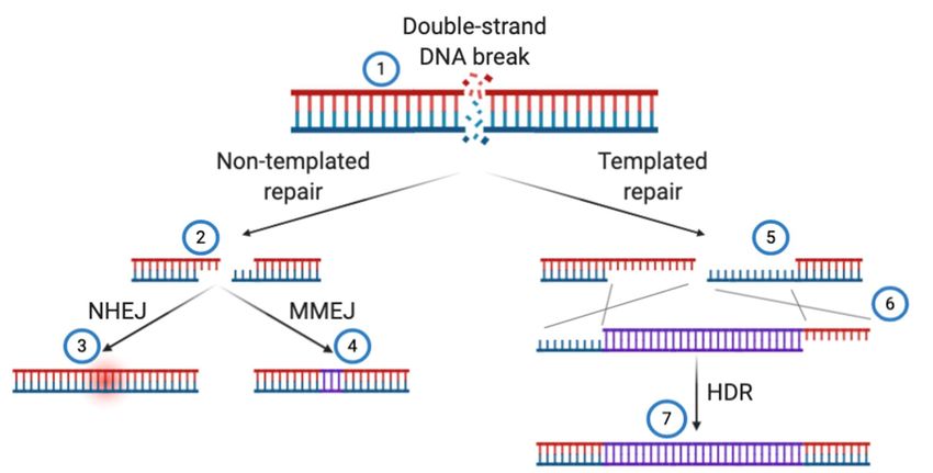
Preprints (www.preprints.org) | NOT PEER-REVIEWED | Posted: 17 May 2021          doi:10.20944/preprints202105.0376.v1

             relative to control. The reduced infectious potential of this new construct was later confirmed by
             infecting feline naïve T-cells with cell-free FIV harvested from FIV-infected and CRISPR/Cas9
             lentivirus-treated cells.

             Our knowledge on the role of genetic variation in disease continues to evolve and developments
             in the Dog Genome Annotation Project (DoGA) are rapidly aiding in comparative medicine efforts.
             Advances in DoGA combined with faster, cheaper, and more efficient gene editing and gene
             therapy methods will lead to even greater emphasis on translational modeling and parallel drug
             development efforts using spontaneous dog diseases. In this chapter, through a translational lens
             based on successes and failures in human medicine, we will present imminent application of gene
             therapy and gene editing in companion animals; how we got to the use of gene editing and gene
             therapy in companion animals; how these technologies may bring curative outcomes to canines
             with cancer and other diseases; and how the future of companion animal medicine as a whole
             will undoubtedly include genetic cures to disease.

             How we got here: a journey from past to present
             In considering how gene therapy and gene editing are now mentioned alongside routine
             veterinary medicine applications, one must understand how abrupt and revolutionary the
             advances in these enabling technologies have been (Figure 1). Advances in the design, safety,
             and transduction efficiencies of gamma retroviral- and lentiviral-based gene delivery in the mid-
             90’s and early 2000’s were paramount to the pioneering study of CD19 targeting CAR-T cells in
             Phase I human trials in 2009 (Hucks and Rheingold, 2019) and the plethora of ongoing gene and
             cell therapy FDA approvals and trials using viral vectors (Shahryari et al., 2019). Moreover, recent
             analyses demonstrated the cost for a single pair of the first readily re-engineered, modular gene
             editor, Zinc-Finger Nucleases, was $25,000 as recently as 2012 (WareJoncas et al., 2018).
             Today, generating gene editing reagents like CRISPR/Cas9 in academic labs costs well under
             $100 per experiment. This substantial reduction in operating cost, democratizing access to
             enabling technologies and enhancing the efficiency of desired outcomes, has led to over 22,000
             peer-reviewed publications mentioning CRISPR/Cas9 and over 29,000 peer-reviewed
             publications mentioning gene therapy in the last decade (PubMed search, January 2021). Here,
             we summarize seminal studies that have led to modern day gene therapy and gene editing, and
             their current applications in basic research and translational science, emphasizing dog models
             and/or applications where appropriate.

                                                              3
Preprints (www.preprints.org) | NOT PEER-REVIEWED | Posted: 17 May 2021         doi:10.20944/preprints202105.0376.v1

               Figure 1: Seminal discoveries in gene editing and gene therapy leading to the proposal and
               application of these technologies in veterinary medicine.

             Modern gene therapy
             Gene therapy was introduced in biomedical sciences as early as 1972 by Theodore Friedmann
             and Richard Roblin after Stanfield Rogers came up with the idea to replace defective DNA for the
             treatment of inherited diseases (Friedmann and Roblin, 1972). While eukaryotic viral vectors were
             developed from DNA viruses in animals, none of these viruses provide solutions to recombine the
             viral genome into the host genome. Thus, the idea for gene therapy was not realized for decades
             due to limiting technologies for the introduction of foreign DNA into cells. In 1981, Shimotohno
             and Temin reported the first viral recombination of foreign DNA into mammalian cells using
             retrovirus; however the production of these retroviruses was inefficient, limiting wide adoption of
             the technology (Shimotohno and Temin, 1981). A pioneering study in 1984 demonstrated the first
             efficient technology for production of infectious virions and subsequent transfer of foreign DNA
             into mammalian cells using engineered retrovirus vector systems (Cepko et al., 1984), though the
             cost and scale of this approach did not allow broader use of the technology.

             As research into using viruses as DNA delivery vectors continued to progress, warranted
             apprehension arose regarding the use of these infectious agents in the clinic. Therefore, modern
             gene therapy approaches that rely on viral vectors for DNA delivery utilize highly engineered viral

                                                              4
Preprints (www.preprints.org) | NOT PEER-REVIEWED | Posted: 17 May 2021             doi:10.20944/preprints202105.0376.v1

             particles wherein only a minimal amount of viral genome is retained. Non-replicative and tissue-
             specific viruses have been developed using such genetic engineering approaches which continue
             to drive innovations in safety, quality control, and efficacy of modern gene therapy.

             Within the past 50 years, technological innovations in recombinant DNA technology have rapidly
             propelled advancements in gene therapy. Along with continuous gains in our understanding of
             molecular genetics and gene regulation brought about by the genetic revolution, methods to safely
             and efficiently deliver foreign genes into cells remains an active area of research. In particular,
             the development of viral vectors as gene delivery systems capable of stably integrating genetic
             cargo into the genome has shown clinical efficacy as years of incremental advances in the
             production and efficiency of retroviral recombination led to the first NIH-sanctioned gene therapy
             clinical research study in 1991 (Ferrari et al., 1991). In this pioneering study, two children afflicted
             with adenosine deaminase (ADA) deficiency were given retroviral transduced T cells containing
             a functional copy of the adenosine deaminase gene to rescue a severe combined
             immunodeficiency (SCID) phenotype.

             Gene therapy-based interventions utilizing both viral and non-viral vectors in humans have
             continued to progress over the past 30 years with important implications for veterinary medicine.
             We will here discuss the foundational biology of retroviruses, lentiviruses, adenoviruses, and
             adeno-associated viruses with special emphasis on the technological innovations and genetic
             interventions that have allowed these infectious agents to become some of the most widely
             utilized gene delivery systems for gene therapy. Examples of non-viral methods of gene delivery
             will be briefly discussed in the latter portion of this section. Figure 2 summarizes these main
             mechanisms for introduction of genetic, protein, or small-molecule based material into living cells.

                                                                5
Preprints (www.preprints.org) | NOT PEER-REVIEWED | Posted: 17 May 2021             doi:10.20944/preprints202105.0376.v1

               Figure 2: Shown are examples of viral (left) and nonviral (right) transgene delivery methods. Left:
               (1) Virus-based delivery using +ssRNA retrovirus (RV) or lentivirus (LV), dsDNA Adenovirus (AdV),
               or ssDNA Adeno-associated virus (AAV) mediate cellular entry through specific cell surface ligands
               or ubiquitously expressed proteoglycans. (2) Genomic integration of the transgene is mediated by
               processes exclusive to the viral vector utilized for delivery. Though these integration events mostly
               occur at random locations throughout the genome, in the case of AdV, cargo DNA is replicated and
               transcribed but not stably integrated. (3) Transgenes are expressed as functional proteins via
               mRNA translation by ribosomes. Right: (1) Nonviral delivery can be accomplished via temporary
               disruption of the cell membrane by physical methods, such as electroporation for the introduction
               of free nucleic acids, or with lipid nanoparticle complexes encompassing nucleic acids that enter
               cells indiscriminately through electrostatic interactions with glycoproteins and proteoglycans. (2)
               Transgene integration can be accomplished using enzyme-mediated processes that cleave
               genomic DNA and may include transposases or nucleases, such as CRISPR/Cas9. (3) Transgene
               expression via mRNA translation by ribosomes can occur transiently through direct inoculation of
               mRNA into cells or as a result of transcribed DNA which has been integrated into the genome or
               from an episomal source.

             Viral gene delivery
             RNA virus vectors: retroviruses and lentiviruses
             RNA virus vectors derived from retroviruses (RVs) were one of the first viral delivery systems
             used for gene therapy and remain among the most used today. They are represented by a large
             family of enveloped viruses encompassing 7-12 kb plus-sense RNA genomes. Lentiviruses (LVs),
             including human immunodeficiency virus (HIV), represent a class of retrovirus often used for

                                                                6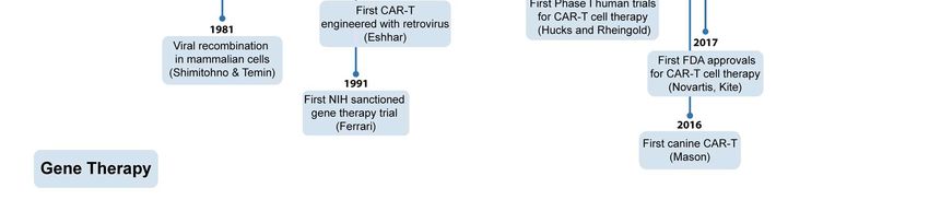
Preprints (www.preprints.org) | NOT PEER-REVIEWED | Posted: 17 May 2021           doi:10.20944/preprints202105.0376.v1

             biomedical research and require the same basic components as other RVs for successful cellular
             entry and viral genome integration (Verma and Weitzman, 2005).

             To employ the natural ability of RVs to facilitate gene transfer for either ex vivo or in vivo gene
             therapy applications, notable modifications to the viral genome were required to maximize safety
             and ensure efficacy before clinical benefit could be evaluated. RVs bring natural tissue tropism,
             like HIV’s propensity to infect T cells, and are replicative in nature. Accordingly, first-generation
             lentiviral vectors were modified to include the envelope protein of vesicular stomatitis virus, VSV-
             G, which functions to broaden cellular tropism by targeting the low-density lipoprotein (LDL)
             receptor which is ubiquitously expressed (Finkelshtein et al., 2013). However, these first-
             generation vectors retained much of the original HIV genome and warranted broad safety
             concerns. Thus, known virulence factors were removed from the viral genome to produce second-
             generation lentiviral vectors (Vannucci et al., 2013). Third-generation lentiviral vectors were then
             developed to further improve safety wherein multiple modifications were incorporated to eliminate
             the possibility of producing replication-competent lentiviruses (Dull et al., 1998).

             DNA virus vectors: adenoviruses and adeno-associated virus
             Adenoviruses (AdVs) and adeno-associated virus (AAV) are the most prominent DNA viruses
             used for modern gene therapy applications. Although both viruses encapsulate a DNA genome
             within and nonenveloped icosahedral protein capsid, the AdV genome consists of ~36 kb dsDNA
             while AAV possesses a much smaller genome, ~4.7 kb, and is comprised of ssDNA. Similar to
             RVs and LVs, plasmids encoding essential viral components can be introduced into packaging
             cell lines to generate AdV and AAV vectors for transgene delivery; however, critical differences in
             virus replicative cycles necessitate unique modifications for their production and implementation
             as gene therapy agents (Verma and Weitzman, 2005).

             The human AdV family contains over 50 unique serotypes with a wide tissue tropism. The majority
             of these viruses utilize the Coxsackie-adenovirus receptor for cellular attachment (Philipson and
             Pettersson, 2004) and internalization is facilitated through an interaction with cellular integrin αv
             receptors (Wickham et al., 1993). AdV DNA is not normally integrated into the host genome
             (Pombo et al., 1994) so, in terms of gene therapy applications, only transient transgene
             expression can be achieved with AdV vector systems. To ensure safety, essential viral replication
             genes were deleted in the first-generation AdV vectors which could incorporate transgenes
             ranging from 4.7-4.9 kb (Bett et al., 1993), and transgenes up to 8.3 kb where later cloned into
             second-generation AdV vectors wherein additional viral genes had been deleted. Although, it is
             noteworthy that recent developments towards third-generation AdV vectors have further
             enhanced the potential for efficient gene delivery and long-term transgene expression, high
             immunogenicity remains a hurdle that must be overcome before AdV vectors are to be widely
             used for gene therapy in vivo (Crystal, 2014).

             AAV serotype 2, a nonpathogenic human parvovirus, is the best characterized AAV vector to date.
             AAV2 utilizes ubiquitously expressed heparan sulfate proteoglycan for cell attachment
             (Summerford and Samulski, 1998) and membrane internalization is facilitated by either the
             fibroblast growth factor receptor or integrin αvβ5 (Summerford et al., 1999) (Qing et al., 1999).

                                                              7
Preprints (www.preprints.org) | NOT PEER-REVIEWED | Posted: 17 May 2021 doi:10.20944/preprints202105.0376.v1
In gene therapy studies, AAV vectors have demonstrated transduction in the muscle, retina, brain,
liver, and lungs where a slow rise in transgene expression plateaued after a few weeks in vivo
(Xiao et al., 1996). Although AAV vectors have been successfully used for gene therapy
applications in vivo in animal models, a relatively small transgene capacity ~4 kb and a high
prevalence of neutralizing antibodies directed against the viral capsid proteins represent
significant limitations (Verma and Weitzman, 2005).
Non-viral methods of gene delivery
It is important to note that non-viral methods indeed exist for delivery of proteins and nucleic acids
into cells, however they are not typically considered part of the “gene therapy” class, as they are
all currently utilized ex vivo for cell programming. Although viral vectors are currently the most
efficient approach used to deliver DNA cargo into host cells in vivo, non-viral approaches are
becoming much more common due to a better potential safety profile and technical advantages
in use and production. From a biosafety perspective, viral vectors can be highly immunogenic and
lead to adverse inflammatory reactions in patients such that, in 1999, the first gene therapy-
related fatality was reported in a clinical trial due to an inflammatory reaction in response to an
adenovirus (AdV) vector (Lehrman, 1999). A phenomenon known as ‘insertional mutagenesis’,
wherein the chromosomal insertion of viral DNA unintentionally results in cellular transformation
by either disrupting the expression of a tumor suppressor gene or activating an oncogene,
represents another potential safety concern regarding the use of viral vectors in gene therapy.
Additionally, as viral vector-mediated integration into the genome can occur at random loci, each
individual engineered cell is theoretically different. Ultimately, this leads to heterogeneity of the
resulting therapy with potential consequences resulting from differential expression of the cargo
gene. For these reasons, non-viral vectors are widely considered to be a safer alternative
(Ramamoorth and Narvekar, 2015) and, due to advances in nanotechnology, in vivo applications
for non-viral delivery systems have recently been realized in the context of gene therapy (El-
Sayed and Kamel, 2020).
The flexibility of non-viral delivery systems is a notable advantage as they can be used to
introduce various types of nucleic acids into cells including chemically synthesized small DNA
molecules (Oligodeoxynucleotides), large DNA molecules (plasmid DNA), various RNA
molecules such as ribozymes, small interfering RNAs (siRNA), and messenger RNAs (mRNA),
and even proteins directly into cells. Although many nucleic acids can be passively delivered to
cells via endocytosis, physical methods of gene delivery can help facilitate the introduction of
genetic material into cells by temporarily disrupting the cell membrane using physical forces.
Common physical methods of gene delivery include microinjection, electroporation, sonoporation,
particle bombardment, and magnetofection (Ramamoorth and Narvekar, 2015). Chemical carriers
can also be used to deliver nucleic acids into cells. These delivery systems are most commonly
made of a nucleic acid complexed with either cationic lipids (Lipoplexes), cationic polymers
(Polyplexes) or a combination of cationic lipids and polymers (Lipopolyplexes) (Midoux et al.,
2009). These chemical complexes function to protect encapsulated nucleic acids from
degradation and enhance their intracellular uptake via electrostatic interactions with glycoproteins
and proteoglycans on the cell membrane. Nucleic acid complexes may also influence intracellular
trafficking of nucleic acids. For instance, the cationic polymer polyethylenimine (PEI), disrupts
8Preprints (www.preprints.org) | NOT PEER-REVIEWED | Posted: 17 May 2021 doi:10.20944/preprints202105.0376.v1
endosomal membranes resulting in the translocation of the complexed nucleic acid into the
cytosol and cationic peptides comprised of basic residues, such as lysine and/or arginine, can be
employed to target specific cell surface receptors or provide nuclear localization signals to
facilitate the nuclear entry of cargo DNA (Al-Dosari and Gao, 2009).
Modern gene editing
Gene editing allows the mutation or alteration of DNA at specific locations in the genome a priori
(Yeh et al., 2019). Gene editing can be used as a basic scientific tool to heighten understanding
of disease pathophysiology using cell systems and animal models, including complex disease like
cancer, as well as monogeneic disorders such as cystic fibrosis, hemophilia, sickle cell disease,
heart disease, and human immunodeficiency virus (HIV) infection (Doudna, 2020). Multiple
approaches to gene editing have been developed in the 21st century, but the crux of the
technology requires the ability to direct enzymatic activity to a chosen locus in the genome. To
achieve this, scientists use “programmable nucleases” that induce DNA damage in the form of a
double-strand break or nick at specific loci in the genome. Whereas gene editing is the broader
concept, programmable nucleases are the critical tool for the modern development of cell-based
therapies and gene therapies. Programmable nuclease-induced DNA double-strand breaks can
result in random mutations around the break, specific alterations of endogenous base pairs using
homology templates, or the targeted integration of exogenous DNA via an engineered donor
template.
What makes programmable nucleases so powerful is their ability to induce DNA repair at their
target sites, allowing scientists to rewrite the genetic code at these genomic loci. Though
oversimplified, an effective analogy converts the genome to a word document, the nucleases to
a mouse cursor, and donor DNA templates to a keyboard. Scientists can “click” the genome, and
“delete” or “rewrite” the paragraph surrounding the cursor. Sometimes referred to as “genome
writing”, this technology gives scientists endless possibility towards inducing targeted mutations,
correcting genetic defects, or giving cells an ability to perform functions that nature did not evolve
(Doudna, 2020).
Brief history of gene editing tools
Modern gene editing ideals can be traced back to seminal works by Dr. Mario Capecchi and Dr.
Maria Jasin. Dr. Capecchi, who later won the 2007 Nobel Prize of Physiology and Medicine for
his work, pioneered strategies using the DNA repair pathway homologous recombination to
specifically engineer mouse embryonic stem cells and mouse models using targeted DNA
integration (Thomas and Capecchi, 1987). Dr. Jasin’s pioneering discovery demonstrated that
targeted DNA integration, that is the addition of exogenous DNA to a locus of interest through
homology-directed repair, can be stimulated 100-1000 times over Capecchi’s work by causing a
double-strand DNA break at the locus of interest in the presence of a repair template (Rouet et
al., 1994). This finding has served as a seminal discovery in the field of gene editing. However,
the nuclease used in this case, I-SceI, is a large restriction enzyme whose endogenous DNA
binding activity cannot be reprogrammed to target DNA outside of its restriction site, as possible
with modern programmable nucleases like Transcription Activator Like Effector Nucleases
(TALENs) (Joung and Sander, 2013), and CRISPR systems (Jinek et al., 2012). Zinc-Finger
9Preprints (www.preprints.org) | NOT PEER-REVIEWED | Posted: 17 May 2021 doi:10.20944/preprints202105.0376.v1
Nucleases, as well as some homing endonucleases like I-CreI (MacLeod et al., 2017), pre-date
TALENs and are used today in clinical trials (Tebas et al., 2014), but limitations in their genomic
targeting range and expense to engineer generally relegate them to use in industry settings and
they will not be discussed here.
TALENs were invented in 2010 by Bogdanove, Voytas, and colleagues (Christian et al., 2010),
after fusing the FokI nuclease to a bacterial transcription factor, called a TAL effector. In nature,
TAL effectors evolved to bind to promoters and activate host genes in plant:pathogen interactions
(Moscou and Bogdanove, 2009). TAL effectors rely on highly conserved, modular repeats
containing 34-35 amino acid loops repeated 12-20 times, with 2 amino acid residues in each loop
specifying near 1:1 nucleotide binding. Thus, through the addition of the FokI nuclease, individual
TALENs can be engineered to bind short stretches of DNA to induce a double-strand DNA break.
The FokI used is an obligate heterodimer that requires another TALEN to be supplied to the target
site spaced 12-20 base pairs opposite of the first. TALENs require relatively complex, multi-day
cloning reactions to engineer a single TALEN pair for gene editing. Even still, TALENs have been
used to target DNA breaks, inducing mutations, or used for exogenous gene integration in basic
research, translational science, plant biotechnology, and clinical settings for a decade.
The most widely adopted programmable DNA binding agent, and 2020 Nobel Prize of
Biochemistry winning technology, is called CRISPR/Cas9, short for clustered regularly
interspaced short palindromic repeats and CRISPR-associated protein 9. Evolved as a bacterial
immune system to catalog and resist bacteriophage, CRISPR systems are now utilized to target
DNA double-strand breaks, DNA nicks, targeted nucleotide edits, or exogenous gene integrations
to precise loci in genes of interest on a routine basis. While TALENs and ZFNs require the
engineering of entirely new proteins to target different genes, the power of CRISPR systems is
that the genomic target site is specified by a short, ~20 base pair RNA molecule (called a guide
RNA or gRNA) which, with the help of Cas enzyme DNA melting, is able to bind 1:1 to a
homologous match through simple RNA:DNA base pairing. Engineering CRISPR to bind new
genomic target sites is thus as simple as in vitro transcription of gRNAs specifying a new target
site or ordering an RNA primer from a nucleic acid synthesis company. While CRISPR/Cas9
technology specifically won a 2020 Nobel Prize, studies have shown that up to 40% of bacteria
and 90% of archaea contain at least 1 functional CRISPR system (Westra and Levin, 2020).
These systems differ in their CRISPR-associated proteins, guide RNAs, nucleic acid binding
kinetics, and nucleolytic activities, but all accomplish the same feat; targeting enzymatic activity
to a genomic locus of interest to induce gene editing.
Outcomes of gene editing
For simplicity, we will narrow our focus of gene editing to the use of CRISPR tools, but the ideas
that follow generally hold true for other programmable nucleases. In most applications, these tools
are combined with gene delivery as discussed above:
After CRISPR induces a double-strand DNA break, cells work to repair it using one of two major
pathways generally called “non-templated repair” or “templated repair” (Figure 3). These
pathways differ in their outcome pending the availability of a repair template either supplied by a
10Preprints (www.preprints.org) | NOT PEER-REVIEWED | Posted: 17 May 2021 doi:10.20944/preprints202105.0376.v1
sister chromatid for endogenous repair or a donor DNA template for genome writing applications.
Two specific pathways for non-templated repair are non-homologous end joining (NHEJ), which
results in random insertions or deletions (indels) and microhomology mediated end joining
(MMEJ), which results in short deletions but can be predicted (Bae et al., 2014) (Ata et al., 2018).
While viral gene therapy brings the ability to randomly supply diseased cells with donor DNA
carrying a corrected version of defective proteins to treat diseases, CRISPR mutations are rarely
therapeutic in and of themselves and require templated repair. Indeed, gene editing technologies
rely on precision control of the DNA repair outcomes after CRISPR targeting to alter single or few
nucleotides surrounding the target site or to integrate a cargo DNA for reverse genetics and
functional genomic applications and for therapeutic benefit in the clinic.
When programmable nucleases cut their target DNA site, enzymes specific to sub-pathways of
DNA repair compete to heal the double-strand DNA break (Figure 3) These pathways hinge upon
the initial recognition of the double-strand DNA break and subsequent processing of the DNA
ends for no, short-range, or long-range end resection.
Figure 3: Shown are examples of how DNA double-strand breaks are corrected in gene editing
applications. (1) A DNA double-strand DNA break is induced using a programmable nuclease, like
TALENs or CRISPR/Cas9. (2) In non-templated DNA repair, little to no end-resection takes place. (3)
Non-homologous end joining (NHEJ) resolves DNA breaks by short insertions or deletions (indels)
represented by the error cloud. (4) microhomology meditated end joining is used when 2-5 bp
homologies are present in DNA ends, resulting in a short deletion of the intervening sequence and
one of the homologous sequences (displayed in purple). (5) In templated DNA repair, long range end
resection results in >100bp ssDNA ends that are used as substrates for homology directed repair
(HDR), either with a sister chromatid or, in gene editing, an exogenously supplied template. (6)
Exogenously supplied templates, like double-stranded DNA, plasmids, AAV vectors, or ssDNA are
built with homology to match the DNA break site. (7) Pending the donor template and changes to be
introduced (SNPs or entire transgenes), HDR will proceed through either homologous recombination
(HR), single-strand annealing (SSA), or MMEJ-like mechanisms to recombine exogenous DNA
precisely into the genome.
11Preprints (www.preprints.org) | NOT PEER-REVIEWED | Posted: 17 May 2021 doi:10.20944/preprints202105.0376.v1
Non-Homologous End Joining (NHEJ)
The most common non-templated DNA repair outcome is called non-homologous end joining
(NHEJ), which results in short insertions/deletions (indels) at the target site. In research settings,
CRISPR is most often used in reverse genetics applications to generate indel mutations in the
genome in order to abrogate gene function and study resulting phenotypes, though CRISPR is
also particularly powerful for forward genetic screens. Indeed, CRISPR gene knockouts have
been used in canine reverse genetics to study cardiovascular disease (Feng et al., 2018),
Duchenne’s muscular dystrophy (Mata Lopez et al., 2020), and even to generate germline edits
of the myostatin gene (Zou et al., 2015). NHEJ activity requires KU70/80 to stabilize the DNA
ends, preventing end resection, and DNA-PKcs (Chang et al., 2017). These proteins recruit other
components, importantly DNA ligase IV which seals the broken DNA ends. Competition with end
resection enzymes and more complicated DNA break structures (like 5’ and 3’ overhangs) result
in the random indels associated with NHEJ.
Microhomology-Mediated End Joining (MMEJ)
Microhomology-mediated end joining (MMEJ) is another non-templated outcome of CRISPR-
induced DNA breaks and competes directly with NHEJ (Black et al., 2019). The core difference
between them is that MMEJ requires end resection to reveal short 3’ overhangs. Short stretches
(commonly 2-5 nucleotides) of homology are found by Polymerase Theta and LigIII is used to
seal the DNA break, thus resulting in small deletions of the intervening sequence and one copy
of the microhomology. Interestingly, CRISPR target sites can be chosen a priori to induce MMEJ,
thus resulting in predictable DNA repair outcomes, increasing the likelihood of an outcome of
interest (Martinez-Galvez et al., 2021). The ability to select CRISPR target sites that will result in
predictable outcomes is invaluable in research settings to ensure that a resulting mutant will carry
an out of frame deletion and early translational stop codon to abrogate gene function. More
importantly, in therapeutic settings, the ability to select MMEJ sites in somatic tissue editing brings
predictable outcomes to clinical applications. For example, 1 in ~1000 cases of human Limb-
Girdle Muscular Dystorphy 2G (LGMD2G) contain an 8bp duplication in exon 1 of TCAP (Nigro
and Savarese, 2014). Using CRISPR/Cas9, this mutation has been successfully reprogrammed
to WT in up to 57% of alleles in patient derived iPSCs using MMEJ repair (Iyer et al., 2019). Not
all diseases, however, carry such defined genomic signatures to be exploited for reversion to wild
type. In these instances, scientists could use diseased cell- and tissue-based genomics to identify
MMEJ-prone CRISPR target sites that may abrogate gene circuits involved in pathologies.
Homology-Directed repair (HDR)
Homology-directed repair, as the name suggests, is the general set of pathways that uses
homologous templates to direct DNA break repair (Jasin and Rothstein, 2013). After endogenous
DNA breaks e.g., from DNA replication stress or radiation, the cell uses a sister chromatid as
template. However, in gene editing applications, scientists supply an exogenous repair template
embedding the changes desired. Pending the desired edit, multiple sub-pathways of HDR are
utilized:
Templated repair to induce single nucleotide polymorphism (SNP) changes
● Most often, templates for SNP changes arePreprints (www.preprints.org) | NOT PEER-REVIEWED | Posted: 17 May 2021 doi:10.20944/preprints202105.0376.v1
SNP. The first demonstrated use of ssODNs in vivo was developed using TALENs
(Bedell et al., 2012), and subsequent deployment with CRISPR systems has
shown this is a universal HDR strategy to achieve single or few nucleotide changes
desirable to rescue a mutant phenotype, like the A to T SNP in sickle-cell anemia
(Frangoul et al., 2021). The molecular mechanisms of this type of targeted DNA
repair change are currently the subject of debate, but most likely involves
combinations of the Fanconi Anemia pathway important to stabilization of stalled
replication forks, and the synthesis dependent strand annealing (SDSA) pathway
(Yeh et al., 2019). In this model, it is speculated that the homology supplied in the
ssODN anneals with a matched, resected 3’ DNA end, and subsequent DNA
polymerase activity incorporates the desired changes before the template
dissociates. Whether or not the template itself is incorporated into the chromosome
is currently unknown.
● It is also possible to utilize longer homology templates to incorporate single or
multiple nucleotide changes into genes of interest. In general, the mechanisms
governing this type of templated gene editing fall under homologous recombination
(HR). HR is well known for its role in meiosis but can also be induced by supplying
donors with >500 bp homology arms flanking an insert of interest (Yeh et al., 2019)
(Jasin and Rothstein, 2013). HR proceeds after MRN/CtIP complex-induced long
range end resection involving many components, importantly the 5’-3’
exonuclease ExoI. After DNA ends are resected, the RAD family controls the
precision of repair through RAD51 DNA end stabilization, homologous template
search and sampling, RAD52 mediated strand invasion, annealing, and
subsequent template polymerization and end capture (Yeh et al., 2019). Many
types of donors have been used to induce this type of repair, including adeno-
associated viral templates, ssDNA, linear dsDNA, and plasmid DNA. The use of
these types of donors and the requirement for homologous recombination is
arguably less efficient than the use of ssODN for SNP incorporation.
Templated repair to incorporate new genes
● For many gene-edited cell therapy applications, scientists do not wish to fix single
nucleotide polymorphisms, but need to add exogenous DNA to educate cells with
new functions that nature did not evolve. In research applications, this could be the
addition of DNA encoding fluorescent proteins to tag spatiotemporal gene
expression patterns in model organisms and cell lines (Wierson et al., 2020). In
the clinic, the best example is the addition of DNA encoding chimeric antigen
receptors to impart tumor killing activity onto T cells. Whatever the application, the
incorporation of multi-kilobase gene cassettes into CRISPR induced double-strand
DNA breaks generally requires a repair template carrying homology arms that flank
the DNA break. Just like in templated SNP alterations, the homologous
recombination pathway of DNA repair is a commonly employed mechanism for
these alterations. There are publications demonstrating the efficacy of many types
of templates to accomplish this feat; single stranded or double stranded linear
DNAs (Roth et al., 2018) (Shin et al., 2014), plasmid DNA (Wierson et al., 2020),
or rAAV donors (MacLeod et al., 2017) to mediate site-specific integration of this
13Preprints (www.preprints.org) | NOT PEER-REVIEWED | Posted: 17 May 2021 doi:10.20944/preprints202105.0376.v1
DNA. More recently, scientists have discovered that delivering a donor template
with CRISPR sites flanking homology arms and cargo DNA is an efficient way of
inducing a sub-pathway of HDR, dubbed homology-mediated end joining, to create
engineered model organisms and cell lines (Wierson et al., 2020, Wierson et al.,
2019) (Yao et al., 2017) (Hisano et al., 2015).
Base editing for SNP changes
An alternative form of CRISPR-based single nucleotide alterations is accomplished through the
fusion of “base editors” to Cas enzymes. Base editors can be used to alter single nucleotides in
DNA or RNA. The first base editor, engineered by David Liu’s lab in 2016, is a fusion of a Cas9
nickase enzyme (so that only a single-strand DNA break is induced) to the cytidine deaminase
enzyme APOBEC1 (Komor et al., 2016). Nicking the non-base edited DNA strand using a Cas9-
nickase induces a mismatch repair pathway that effectively “tricks” the cell to use the uracil as a
template for DNA replication, thus converting the target cytosine/guanine base pair to a
thymine/adenine base pair. Similarly, the Liu lab used directed evolution to engineer an adenosine
deaminase that can effectively target adenine/tyrosine base pairs for conversion to
guanine/cytosine (Gaudelli et al., 2017). With these two tools and their enhanced versions, 63%
of known pathogenic single nucleotide variants could theoretically be corrected. Additionally, an
advantage of base editing is that no double-strand DNA break has to be generated, as emerging
concerns with DNA breaks have been noted (Cullot et al., 2019) (Kosicki et al., 2018) (Ihry et al.,
2018). An elegant review was recently published on the advances and opportunities for furthering
base editing technology (Porto et al., 2020).
Armed with the toolboxes of viral integration, gene editing, and non-viral gene delivery, scientists
now have control over the genome at scale and resolution never before imagined to engineer
living therapies.
Revitalizing cells, tissues, and organs in vivo
Human clinical gene therapy
Numerous experimental gene therapy trials have been initiated with varying degrees of success,
leading to the first FDA-approved gene therapy in humans in 1998, Vitravene (Fomivirsen®)
(Stein and Castanotto, 2017). This antisense oligodeoxyncleotide was indicated for the local
treatment of cytomegalovirus (CMV) retinitis in immunocompromised patients but was later
removed from the market in 2002 and 2006 in the EU and U.S, respectively (Stein and Castanotto,
2017). In Europe, the very first gene therapy ever approved was based on a recombinant adeno-
associated virus (rAAV) vector to treat familial lipoprotein lipase deficiency (Alipogene tiparvovec,
marketed under the trade name Glybera®) (Yla-Herttuala, 2012). Since 1998, 22 gene therapy
products, including naked nucleic acids, non-viral and viral vectors, as well as cell-mediated
therapy have been approved for commercialization and are elegantly described in a 2020 review
by Ma and colleagues (Ma et al., 2020).
Advantages of canine models for gene therapy
Animal models serve a critical role in biomedical research both as basic science tools for
elucidating molecular mechanisms as well as for the preclinical evaluation of novel therapies.
14Preprints (www.preprints.org) | NOT PEER-REVIEWED | Posted: 17 May 2021 doi:10.20944/preprints202105.0376.v1
Dogs play an integral role in modern society and enhance the lives of countless individuals by
providing both mental and physical assistance, security, and companionship. The domestic dog
has also been recognized as a valuable model of monogenic diseases in humans and possesses
key advantages over inbred rodent models classically used for preclinical studies regarding gene
therapy. Not only do causative mutations spontaneously occur in dogs as they do in humans,
detailed pedigrees and opportunities to evaluate treatments over a long-term, often years, provide
crucial advantages in a preclinical setting (Switonski, 2020).
Specifically, immunotherapies present unique opportunities to be tested in vivo in dogs before
formal evaluation into human clinical trials, since an intact immune system and tumor
microenvironment can be adequately modeled in the dog (as opposed to mice). This is particularly
relevant for future accelerated development of many novel treatment options, including, among
others, immune checkpoint inhibitors, CAR-T cell, and adoptive T-cell transfer therapies in both
veterinary and human oncology.
Also, it has been well-established that spontaneous tumorigenesis occurs in dogs through similar
mechanisms as what is known to occur in humans. Although preclinical studies in dogs have
contributed to the development of cancer therapeutics for human medicine, approaches utilizing
gene therapy remain limited in this regard, as gene therapy is not an explicit class of therapeutic
options for oncology.
One could ponder how spontaneous disease in dogs could again be of use to test novel
hypotheses for gene therapy-based solutions in oncology that correct genomic risks in somatic
tissues. One such hypothesis involves hijacking a cancer cell’s reliance MMEJ. As MMEJ is
inherently mutagenic, cancers carry many genomic hallmarks of MMEJ-based re-arrangements
(Alexandrov et al., 2020), as well as overexpression of protein components involved in MMEJ
repair (Lemee et al., 2010). Drugs are currently under development that can induce lethality,
synthetic or otherwise, at the protein level. Theoretically, as tissue specific in vivo gene delivery
continues to advance, one could imagine a scenario where gene therapy is combined with gene
editing to exploit MMEJ to correct driver mutations or to induce synthetic lethality in cancer cell
populations. As discussed at length in the cell therapy section below, we are most interested in
the use of gene edited cell therapies (not explicitly “gene therapies”) in oncology. However, we
are interested in testing these hypotheses in spontaneous cancers via collaboration with in vivo
gene delivery and gene therapy experts. Given the few formal descriptions of gene therapy that
have been described in veterinary oncology (Thamm, 2019), examples of translational medicine
regarding gene therapy outlined herein apply specifically to monogenic diseases.
A special issue of Human Genetics published in 2019 highlights many recent discoveries
regarding monogenic diseases in dogs that have significant implications for human health
(Shaffer, 2019). For instance, variants in the TTN gene are known to contribute to
cardiomyopathies in humans and a missense mutation in TTN was discovered in Doberman
pinscher dogs with diagnosed dilated cardiomyopathy (Meurs et al., 2019). Another group
studying amelogenesis imperfecta (AI) in Parson Russell terriers and Akita dogs identified
variants in the ENAM and ACP4 genes which are implicated in the pathophysiology of human AI,
15Preprints (www.preprints.org) | NOT PEER-REVIEWED | Posted: 17 May 2021 doi:10.20944/preprints202105.0376.v1
further highlighting relevant physiological similarities between humans and dogs (Hytonen et al.,
2019).
There have been numerous successful proof-of-concept gene therapy studies in dogs, both as a
model for human translational application and to solve pressing needs in canine health. As of
2017, hundreds of gene therapy trials have been undertaken to deal with monogenic diseases
and researchers have recently been able to shed light on the molecular mechanisms underpinning
many disorders to pave the way for novel interventions utilizing gene therapy. Beginning in 1993,
the First International DogMap Meeting in Oslo, Norway sought to gain a more in-depth
understanding of the genetics driving approximately 700 monogenic diseases known to afflict dog
Table 1: Description of monogeneic diseases in dogs, their counterpart in humans, genetic
drivers, and any dog or human gene therapies developed for the disorders.
Dog Disease Human Known genetics Breeds Gene therapy
counterpart affected
Congenital CSNB/Type-2 Recessive RPE65 Briard AAV delivery of WT RPE65
stationary night Leber’s congenital (Aguirre et al., 1998) (Acland et al., 2005), Human
blindness (CSNB) amaurosis (LCA2) trials (Testa et al., 2013)
(Schimmer and Breazzano,
2015) led to FDA approvals
for humans in 2017
Muscular Duchenne’s DMD mutations (Yiu Retrievers, CRISPR/Cas9 to restore
dystrophy muscular and Kornberg, 2015). Pointers, DMD function (Amoasii et
dystrophy Rottweilers, al., 2018)
Spaniels,
Corgis, Terrier
Hemophilia A and Hemophilia A and Coagulation factor VIII Setters, Viral delivery of FVIII and
B B and Coagulation Schnauzers, FIX in dogs (Nichols, et al.,
factor IX Pointers, 2016), AAV gene therapy in
Lhasa Apso, humans (Peyvandi and
Retriever, Garagiola, 2019)
Terrier/Beagle
Retrovirus gene
Severe combined SCID Mutations in DNA-PK, Terriers, replacement in dogs (Ting-
immunodeficiency RAG1, IL2RG Frisian Water De Ravin et al., 2006)
(SCID) (Switonski, 2020) Dogs, Hounds,
Corgis
16Preprints (www.preprints.org) | NOT PEER-REVIEWED | Posted: 17 May 2021 doi:10.20944/preprints202105.0376.v1
Leukocyte LAD I, LAD Mutation in ITGB2 Setters, Retrovirus gene
adhesion III/CLAD I, (Zimmerman et al., German replacement of ITGB2 in
deficiency (LAD) CLADIII 2013), FERMT3 Shepherds dogs (Bauer et al., 2013)
(Hugo and Heading,
2014)
Lysosomal MPS I, MPS IIIB, Mutations in GUSB German Retrovirus gene
storage diseases MPS VI, and MPS shepherds, replacement of MPS VII
VII mixed (Xing et al., 2013)
breeds (Switonski, 2020). Although analysis of genome sequence variations between different
dog breeds is currently underway (Ostrander et al., 2019), known causative mutations are
continuously being identified and approximately 430 monogenic diseases in dogs have the
potential to serve as preclinical models for homologous human diseases (Switonski, 2020). In
fact, gene therapy has shown efficacy in treating many monogenic diseases that afflict both
canines and humans and positive results in preclinical canine studies have resulted in the initiation
of multiphase clinical trials. These diseases are represented among multiple organ systems and
include ocular diseases, muscular dystrophy, hemophilia, severe combined immunodeficiencies,
leukocyte adhesion deficiencies, and lysosomal storage diseases (Table 1).
Engineering living cells ex vivo for therapeutic use
Mainstream application of viral transduction, gene editing technologies, and most recently non-
viral gene delivery into cells ex vivo has led to a massive shift in the way scientists think about
disease treatment. It is now routine to “educate” cells ex vivo with genetic information to impart
therapeutic benefits with the intention of transplanting them back into diseased patients. However,
only in a handful of research groups are developing these strategies for dogs, with only two clinical
reports to date (Panjwani et al., 2016) (Panjwani et al., 2020). Here, we will focus on the enabling
technologies and their use in humans, knowing that translation of this technology to canines is
sure to follow.
In the context of oncology, the most commonly engineered cell used to date is the T cell. Though
there are many subtypes of T cells, CD8+ “cytotoxic” T cells are naturally equipped to rid the body
of virally infected cells and even some malignant cells through MHC Class I peptide recognition
with the T cell receptor (Kumar et al., 2018). Armed with this knowledge, scientists long sought to
reprogram the T cell receptor to engineer T cell specificity and killing. Indeed, the idea to use
genetically reprogrammed T cells as therapy began in 1992 with a pioneering study by Michel
Sadelain reporting the use of retroviral transduction to engineer T cells with exogenous DNA.
Later, in 1993, Zelig Eshhar generated the first chimeric antigen receptor (CAR) by fusing an
antibody domain to the CD3ζ domain of a T cell receptor in what has become known as a “1st-
generation CAR”. Studies in the late 1990’s and early 2000’s set the field ablaze by generating
“2nd-generation CAR-T cells” containing a costimulatory domain in addition to the antibody and
17Preprints (www.preprints.org) | NOT PEER-REVIEWED | Posted: 17 May 2021 doi:10.20944/preprints202105.0376.v1
CD3ζ signaling domain (Maher et al., 2002), which culminated in the first report of in vivo efficacy
of CAR-T cells by targeting CD19 on leukemic B cells in a mouse model (Brentjens et al., 2003).
Numerous subsequent reports and clinical trials utilized CD19-targeting, autologous CAR-T cells
generated from T cells isolated from the patient’s peripheral blood mononuclear cells (PBMCs)
(Hollyman et al., 2009). These efforts culminated in two FDA approvals in 2017 for relapsed and
refractory acute lymphoblastic leukemia (ALL) and diffuse large B cell lymphoma (DLBCL) and
one FDA approval for mantle cell lymphoma (MCL) in 2020 (Neelapu et al., 2017) (Maude et al.,
2018) (Wang et al., 2020). To date, the FDA approved CAR-T cell therapies are engineered via
lentiviral transduction of nucleic acid encoding the CAR and are all autologous, using no further
gene editing. However, there are numerous FDA-sanctioned clinical trials ongoing with gene-
edited CAR-T cells, both autologous and allogeneic, with various alterations to the manufacturing
process and resultant gene edited cells. As of January 2020, over 900 potential Investigative new
Drug (IND) applications are in the queue for cellular and/or gene therapy applications
https://www.fda.gov/news-events/press-announcements/fda-continues-strong-support-
innovation-development-gene-therapy-products.
Proof of concept CAR-T cell therapy in dogs
As noted in this chapter and described at length in [Nicola Mason’s chapter], the only peer
reviewed literature for CAR-T cell therapy in canines is via work from the University of
Pennsylvania with two publications by Panjwani et al., (Panjwani et al., 2016, Panjwani et al.,
2020). In these seminal studies, the authors used either mRNA or retroviral transduction of canine
T cells to generate doses of autologous CAR-T cell therapy designed to target CD20 on B cell
lymphomas. The first proof-of-concept experiment showed limited effect, and while the CAR-T
cells did traffic to the lymph nodes and reduce tumor burden, this effect was only transient,
primarily due to the nature of mRNA-based CAR expression, which are not stably integrated.
In the second publication by Panjwani et al., 5 dogs were treated with stably transduced
autologous CAR-T cells, though the doses of CAR-T cells varied in every dog and were all far
lower than the target 1-3x10⁶ CAR-T cells/kg used in humans. Canine anti-mouse antibodies were
detected, likely in response to the murine-derive CAR itself, which the authors postulated
triggered rejection of the CAR-T cells and subsequently limited clinical benefit. In any case, the
authors concluded that CAR-T cells were detectable in dogs, had modest anti-tumor activity, and,
in some instances, selectively forced CD20 antigen loss on malignant B cells, indicating that the
dog as a model can faithfully recapitulate pitfalls previously described in human CAR-T cell
therapy studies (Majzner and Mackall, 2018) (Maus et al., 2013) (Enblad et al., 2015).
In addition to canine CAR-T cell therapy being developed for B cell lymphoma, there is a report
of this modality being applied to the treatment of glioma in dogs (Yin et al., 2018). In this study,
scientists aimed to develop a novel antigen binding protein to develop CAR-T cells that target IL-
13Rα2, commonly found on both human and canine gliomas, but not on healthy tissues. Solid
tumors like glioma are historically harder to treat with CAR-T cell therapy than liquid cancers, such
as leukemia and lymphoma (O'Rourke et al., 2017). Yin et al., engineered human cells expressing
a cross-reactive CAR for both human and canine IL-13Rα2 and showed it effectively targeted IL-
18Preprints (www.preprints.org) | NOT PEER-REVIEWED | Posted: 17 May 2021 doi:10.20944/preprints202105.0376.v1
13Rα2 in both species in vitro and was effective in vivo towards an orthotopic mouse model of
canine glioma. Interestingly, the authors engineered both canine and human cells with IL-13Rα2
CARs, and showed that both elicit killing of IL-13Rα2 positive cells, indicating that human T cells
can kill canine cells when armed with a CAR specific for a canine antigen. The authors concluded
that they are moving forward to enroll a pilot trial to use this technology towards fighting canine
gliomas as a preclinical model of the approach in humans (Herranz et al., 2016).
It is notable to discuss here that there are currently multiple industry groups in the United States
offering autologous tumor vaccinations or “adoptive T cell transfer” to fight differing cancers, which
is quite different than CAR-T cell therapy. Respectively, dogs are either vaccinated with their own
tumors to induce a cellular immunity response to the cancer, or T cells are isolated from dogs,
grown in the presence of tumor neo-antigens, and reinfused in the hopes that the cells naturally
acquire tumor antigen specific killing properties (O'Connor and Wilson-Robles, 2014, O'Connor
et al., 2012). Data on these approaches in dogs is currently scant with various stages of regulatory
approvals ongoing, however it is well established in humans that genetically reprogramming cells
with anti-cancer properties (i.e., CAR-T) is absolutely a more effective approach to induce durable
disease remissions.
Gene-edited cell therapies in dogs
With Dr. Mason’s pioneering work, the translation of CAR-T cell therapy from humans to
companion dogs emerging. While the work in the Mason lab is so far the only peer-reviewed
literature for in vivo trials of CAR-T in dogs, publicly available knowledge for awarded federal and
foundation grants gives us a glimpse of what is to come in this area. These include federal grant
awards to Dr. Nicola Mason (3U24CA224122-02S1), Dr. Carl June and Dr. Gerald Linette
(1U54CA244711-01) and other colleagues at the University of Pennsylvania (5R01AR075337-
03), foundation awards to Dr. Heather Wilson-Robles and colleagues at Texas A&M University
(AKC-CHF #1418), and a Small Business Innovation Research award to the biotechnology
company LifEngine Animal Health Laboratories (LEAH Labs, NSF #2006130). In all instances,
the scientists note the need for better canine therapies and seek to use the high-impact potential
of spontaneously occurring disease in a large animal model system to study the effects of novel
cell therapies. As mentioned above, the opportunity to support parallel development of novel drug
candidates in veterinary and human oncology through a One Health approach could increase
availability of revolutionary treatment options in animal health, while reducing attrition rates in
Phase II clinical programs. The public summary statements of these grants indicate that gene
editing is being used in the development of novel cell-based
therapies; however, to date, there are no peer-reviewed reports of gene-edited cell therapies for
dogs.
One of the biggest challenges to overcome in regards to successful translation of CAR-T cell
therapy into canines is the cost to deliver the therapy to patients. Notably, the current cost of FDA-
approved CAR-T cells in human medicine ranges from $373,000 to $475,000 represents a large
barrier to effective translation of this technology to canines. This cost is largely due to the need
for individualized manufacturing, lentiviral preparations, FDA regulation, and GMP manufacturing
standards. In considering autologous cell therapy in dogs, apheresis to obtain PBMC populations
19You can also read