STUDIES OF INFLAMMATORY RESPONSES IN HANTAVIRUS INFECTION - From the Department of Medicine, Huddinge Karolinska Institutet, Stockholm, Sweden
←
→
Page content transcription
If your browser does not render page correctly, please read the page content below
From the Department of Medicine, Huddinge
       Karolinska Institutet, Stockholm, Sweden

    STUDIES OF INFLAMMATORY
RESPONSES IN HANTAVIRUS INFECTION

                    Kimia Maleki

                   Stockholm 2021
All previously published papers were reproduced with permission from the publisher.
Published by Karolinska Institutet.
Printed by Universitetsservice US-AB, 2021
© Kimia Maleki, 2021
ISBN 978-91-8016-191-6
Cover illustration: Created by Kimia Maleki, using BioRender.
Studies of inflammatory responses in hantavirus
infection

THESIS FOR DOCTORAL DEGREE (Ph.D.)

By

Kimia Maleki

The thesis will be defended in public at Karolinska Institutet, room 9Q Månen, Alfred Nobels
Allé 8 level 9, 22nd of June 2021 at 12.00.

Principal Supervisor:                          Opponent:
Docent Jonas Klingström                        Dr. Nicole Tischler
Karolinska Institutet                          Fundación Ciencia & Vida
Department of Medicine, Huddinge               Laboratorio de Virología Molecular
Center for Infectious Medicine

Co-supervisor(s):                              Examination Board:
Docent Niklas Björkström                       Docent Helen Kaipe
Karolinska Institutet                          Karolinska Institutet
Department of Medicine, Huddinge               Department of Laboratory Medicine
Center for Infectious Medicine                 Division of Biomolecular and Cellular Medicine

Professor Hans-Gustaf Ljunggren                Docent Peter Bergman
Karolinska Institutet                          Karolinska Institutet
Department of Medicine, Huddinge               Department of Laboratory Medicine
Center for Infectious Medicine                 Division of Clinical Microbiology

Docent Anna Smed-Sörensen                      Professor Marie Larsson
Karolinska Institutet                          Linköping University
Department of Medicine, Solna                  Department of Biomedical and Clinical Sciences
Division of Immunology and Allergy             Division of Molecular Medicine and Virology
"Seven human-mediated factors are most likely driving the emergence of zoonotic diseases:
1) increasing human demand for animal protein; 2) unsustainable agricultural intensification;
3) increased use and exploitation of wildlife; 4) unsustainable utilization of natural resources
    accelerated by urbanization, land use and extractive industries; 5) increased travel and
              transportation; 6) changes in food supply; and 7) climate change."
Preventing the next pandemic - Zoonotic diseases and how to break the chain of transmission
                      United Nations Environment Programme (2020)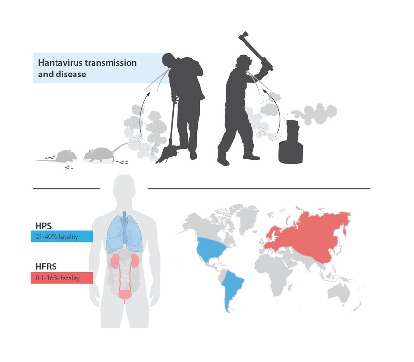
POPULAR SCIENCE SUMMARY
Hantaviruses cause acute infections in humans world-wide. In nature, hantaviruses that cause
disease in humans are carried by different rodent species that do not get sick themselves. The
viruses are transmitted to humans through air containing dust contaminated with droppings or
urine from infected rodents. Yearly, hantaviruses cause disease in around 100 000 individuals.
In contrast to the coronavirus that has caught the world's attention lately, most hantaviruses do
not transmit between humans. The hantavirus species' that circulate in Europe and Asia cause
a disease called hemorrhagic fever with renal syndrome, HFRS. This disease is in some
respects similar to the seasonal flu, with fever, headache, muscle ache, back pain, and
stomachache as common symptoms. Approximately one third of the diagnosed patients require
hospitalization and of these, some need dialysis due to kidney dysfunction. Hantaviruses in the
Americas cause a more severe form of disease, referred to as hantavirus pulmonary syndrome,
HPS. This disease often gives rise to severe lung symptoms that quickly can become life-
threatening. Among individuals with HPS, up to 40% succumb to the disease.

Generally, hantavirus infection is characterized by a strong inflammatory response and leakage
of fluid from the blood vessels out into the surrounding tissue. These hallmarks are not specific
for diseases caused by hantaviruses, but are also seen in several other viral infections, including
COVID-19. Despite the morbidity and mortality associated with hantavirus diseases, there is
no treatment or approved vaccine available. Moreover, the mechanisms behind how
hantaviruses cause disease in humans remain unknown.

In the work included in this doctoral thesis, my colleagues and I have studied the inflammatory
response in hantavirus-infected patients, with the aim to identify specific immunological
factors that can help us understand why some individuals get more ill than others. By studying
blood samples from Argentinian patients infected with hantavirus, we were able to identify that
increased levels of the inflammatory protein IL-6 were associated with severe HPS. Moreover,
we found that fatal HPS was associated with increased levels of a protein that is released upon
intestinal injury. In our subsequent experiments we wanted to investigate how IL-6 possibly
could be involved in causing disease during hantavirus infection. Using blood vessel cells that
we infected in the laboratory, we found that IL-6 in some contexts could drive an inflammatory
response and cause a separation between blood vessel cells. These findings suggest a possible
mechanism behind the leakage of blood fluid seen in patients. This thesis also includes a study
of a subset of T cells named MAIT cells. We found a prominent decline in MAIT cells in the
blood of Swedish hantavirus-infected patients. Further, we found that the MAIT cells were
strongly activated, and that this activation was associated with increased levels of IL-6 in the
patients. In the laboratory, we discovered that a subset of inflammatory mediators called type
I interferons are responsible for causing hantavirus-mediated activation of MAIT cells. We also
observed that these activated MAIT cells released protein-cleaving proteins called granzymes.
Altogether, this study shows that type I interferons have a more important role in MAIT cell
activation than what was previously known. It also suggests that MAIT cells may be important
producers of inflammatory proteins such as granzymes during hantavirus infection.
POPULÄRVETENSKAPLIG SAMMANFATTNING
Hantavirus orsakar akuta infektioner hos människor över hela världen. I naturen bärs hantavirus
av olika gnagararter som själva inte blir sjuka. Viruset sprids till människor via inandning av
damm som kontaminerats med avföring eller urin från infekterade gnagare. Årligen insjuknar
ungefär 100 000 individer i världen med hantavirusinfektion. Till skillnad från det coronavirus
som senaste året har fångat världens uppmärksamhet, smittar de flesta hantavirus inte mellan
människor. De hantavirusarter som cirkulerar i Europa och Asien orsakar en sjukdom som
kallas för "hemorrhagic fever with renal syndrome", HFRS, eller sorkfeber som vi kallar den
variant av sjukdomen som finns i Sverige. Sorkfeber påminner i vissa avseenden om
säsongsinfluensan och ger upphov till symptom så som feber, huvudvärk, muskelvärk,
ryggsmärta och magont. Ungefär en tredjedel av alla patienter som diagnosticeras med HFRS
behöver bli inlagda på sjukhus. Av dessa patienter behöver en mindre andel dialys, till följd av
njursvikt orsakat av virusinfektionen. I Amerika finns en allvarligare form av
hantavirussjukdom som kallas för "hantavirus pulmonary syndrome", HPS. Denna sjukdom
ger ofta upphov till allvarliga lungsymptom hos patienten, som i många fall är livshotande.
Upp till 40% av de individer som insjuknar i HPS avlider till följd av infektionen. Generellt
karaktäriseras hantavirusinfektion av en stark inflammatorisk respons samt ett läckage av
blodplasma från blodkärlen ut i vävnaden. Dessa kännetecken är inte specifika för de
sjukdomar som orsakas av hantavirus, utan ses även vid flertalet andra virussjukdomar,
inklusive COVID-19. Trots den sjuklighet och dödlighet som är kopplad till
hantavirussjukdomar finns det idag varken behandling eller godkänt vaccin mot
hantavirusinfektion. Vidare är mekanismerna bakom hur hantavirus orsakar sjukdom okända.

I arbetet som ingår i denna doktorsavhandling har jag och mina kollegor studerat
immunförsvaret vid hantavirusinfektion med syftet att försöka förstå varför vissa individer blir
mer sjuka än andra. Genom att studera blodprover från argentinska HPS-patienter kunde vi se
att förhöjda nivåer av ett inflammatoriskt protein som kallas för IL-6 är kopplat till svår
sjukdom. Dessutom fann vi att dödlig hantavirusinfektion är associerad med ökade nivåer av
en markör för tarmskada. I våra efterföljande experiment undersökte vi hur IL-6 skulle kunna
bidra till symptomen vid hantavirusinfektion. Genom att på laboratoriet infektera
blodkärlsceller fann vi att IL-6 i vissa sammanhang kan driva en inflammationsrespons samt
orsaka en separation mellan blodkärlsceller. Dessa resultat tyder på en möjlig mekanism bakom
det läckage av blodplasma som ses hos hantavirusinfekterade patienter. Avhandlingen
innehåller även en studie där vi har undersökt en grupp av T-celler som kallas för MAIT-celler.
Vi fann en nedgång i antalet MAIT-celler i blod hos svenska sorkfeberpatienter. Vidare
observerade vi en stark aktivering hos MAIT-cellerna som var kopplad till ökade nivåer av
IL-6. På laboratoriet fann vi att en typ av inflammatoriska proteiner som kallas för typ I
interferoner orsakar hantavirusmedierad aktivering av MAIT-celler. Vi såg också att dessa
MAIT-celler frisläppte protein-klyvande proteiner som kallas för granzymer. Sammantaget
visar denna studie att typ I interferoner har en viktigare roll vid MAIT-cellsaktivering än vad
som tidigare varit känt. Studien antyder också att MAIT-celler kan vara en viktig källa till
inflammatoriska proteiner så som granzymer under hantavirusinfektion.
ABSTRACT
Throughout the world, orthohantaviruses cause severe, acute infections in humans.
Orthohantaviruses, commonly referred to as hantaviruses, are zoonotic viruses with a single
stranded RNA genome of negative sense. Hantavirus strains endemic to Europe and Asia cause
a systemic infection with renal involvement referred to as hemorrhagic fever with renal
syndrome (HFRS). In the Americas, hantaviruses cause hantavirus pulmonary syndrome (HPS)
- a severe and highly fatal infection characterized by severe pulmonary compromise.
Individuals infected with hantaviruses typically display increased levels of cytokines,
decreased platelet counts, and vascular leakage. As specific treatments are lacking, the long-
term goal of the studies within this thesis was to provide leads that will aid in the development
of such. Specifically, this thesis aimed to characterize inflammatory responses and MAIT cell
responses during hantavirus infection, as a step to increase the understanding of protective
versus detrimental immune responses. Moreover, this thesis aimed to investigate the role of the
cytokine interleukin-6 (IL-6) in the pathogenesis of hantavirus infection.

In both HFRS patients and HPS patients, we observed increased systemic levels of many pro-
inflammatory cytokines and other inflammatory markers. In HPS patients, serum levels of IL-6
were found to be associated with increased odds of developing severe disease. On the contrary,
serum levels of complement factor (C) 5/C5a and B cell activating factor were associated with
decreased odds of developing severe disease. Intestinal fatty acid-binding protein (I-FABP), a
systemic marker of intestinal damage, was increased during HPS and associated with increased
odds of a fatal outcome. Next, we demonstrated that IL-6 trans-signaling in hantavirus-infected
endothelial cells led to increased pro-inflammatory responses and increased monolayer
permeability. In HFRS patients, we observed an altered balance of soluble IL-6 receptors in
plasma, which may increase the likelihood of IL-6 trans-signaling in patients. The imbalance
in these markers was associated with an increased need for supplemental oxygen treatment.

When investigating the phenotype of peripheral blood MAIT cells in HFRS patients, we
observed a strong decline in MAIT cell numbers during the acute disease. MAIT cells
remaining in the circulation were highly activated and exhibited decreased expression of
mucosal tissue homing markers. In vitro, we were able to recapitulate these findings, and show
that MAIT cell activation mediated by hantavirus was dependent on type I interferons (IFNs)
produced by antigen-presenting cells or endothelial cells.

In conclusion, this thesis adds to the view that HFRS and HPS are diseases characterized by
strong inflammatory responses. The identification of IL-6 and I-FABP as markers of disease
severity and fatality, respectively, may help in the understanding of hantavirus pathogenesis
and the development of treatment options. The demonstration of the effects of IL-6 on
hantavirus-infected endothelial cells suggest a potential mechanism behind IL-6-driven
pathogenesis. Finally, this thesis provides further evidence on the involvement of MAIT cells
during acute viral infection, and highlights type I IFNs as important mediators in MAIT cell
activation.
LIST OF SCIENTIFIC PAPERS

 I. Maleki KT, García M, Iglesias A, Alonso D, Ciancaglini M, Hammar U,
    Ljunggren H-G, Schierloh P, Martínez VP, and Klingström J. Serum markers
    associated with severity and outcome of hantavirus pulmonary syndrome.
    2019. J Infect Dis. 219: 1832–1840.

 II. Maleki KT*, Niemetz L*, Wigren Byström J, Ahlm C, and Klingström J.
     IL-6 trans-signaling causes increased cytokine secretion and barrier
     dysfunction in hantavirus-infected endothelial cells. Manuscript.
     *Contributed equally.

III. Maleki KT, Tauriainen J, García M, Kerkman PF, Christ W, Dias J, Wigren
     Byström J, Leeansyah E, Forsell MN, Ljunggren H-G, Ahlm C, Björkström
     NK, Sandberg JK, and Klingström J. MAIT cell activation is associated with
     disease severity markers in acute hantavirus infection. 2021. Cell Rep. Med.
     2. 100220.
SCIENTIFIC PAPERS NOT INCLUDED IN THE THESIS
I. Maleki, KT, Cornillet M, and Björkström NK. Soluble SEMA4D/CD100: A novel
immunoregulator in infectious and inflammatory diseases. 2016. Clin. Immunol. 163:
52–59.
II. Scholz S#, Baharom F#, Rankin G*, Maleki KT*, Gupta S, Vangeti S, Pourazar J,
Discacciati A, Höijer J, Bottai M, Björkström NK, Rasmuson J, Evander M,
Blomberg A, Ljunggren H-G, Klingström J, Ahlm C, and Smed-Sörensen A. Human
hantavirus infection elicits pronounced redistribution of mononuclear phagocytes in
peripheral blood and airways. 2017. PLoS Pathog. 13: e1006462. #,*Contributed
equally
III. Klingström J, Smed-Sörensen A, Maleki KT, Solà-Riera C, Ahlm C, Björkström
NK, and Ljunggren H-G. Innate and adaptive immune responses against human
Puumala virus infection: immunopathogenesis and suggestions for novel treatment
strategies for severe hantavirus-associated syndromes. 2019. J. Intern. Med. 285:
510–523.
IV. Solà-Riera C, Gupta S*, Maleki KT*, González-Rodriguez P*, Saidi D*, Zimmer
CL, Vangeti S, Rivino L, Leo YS, Lye DC, MacAry PA, Ahlm C, Smed-Sörensen A,
Joseph B, Björkström NK, Ljunggren H-G, and Klingström J. Hantavirus inhibits
TRAIL-mediated killing of infected cells by downregulating death receptor 5. 2019.
Cell Rep. 28: 2124-2139.e6. *Contributed equally.
V. Varnaitė R, García M, Glans H*, Maleki KT*, Sandberg JT*, Tynell J, Christ W,
Lagerqvist N, Asgeirsson H, Ljunggren H-G, Ahlén H, Frelin L, Sällberg M, Blom
K, Klingström J, and Gredmark-Russ S. Expansion of SARS-CoV-2-specific
antibody-secreting cells and generation of neutralizing antibodies in hospitalized
COVID-19 patients. 2020. J. Immunol. 205: 2437–2446. *Contributed equally
VI. Parrot T*, Gorin J-B*, Ponzetta A, Maleki KT, Kammann T, Emgård J, Perez-Potti
A, Sekine T, Rivera-Ballesteros O, Karolinska KI/K COVID‐19 Study Group,
Gredmark-Russ S, Rooyackers O, Folkesson E, Eriksson LI, Norrby-Teglund A,
Ljunggren H-G, Björkström NK, Aleman S, Buggert M, Klingström J, Strålin K, and
Sandberg JK. MAIT cell activation and dynamics associated with COVID-19 disease
severity. 2020. Sci. Immunol. 5. *Contributed equally.
VII. García M*, Kokkinou E*, Carrasco García A, Parrot T, Palma Medina LM, Maleki
KT, Christ W, Varnaitė R, Filipovic I, Ljunggren H-G, Björkström NK, Folkesson
E, Rooyackers O, Eriksson LI, Sönnerborg A, Aleman S, Strålin K, Gredmark-Russ
S, Klingström J, Mjösberg J, and Karolinska KI/K COVID‐19 Study Group. Innate
lymphoid cell composition associates with COVID-19 disease severity. 2020. Clin.
Transl. Immunol. 9: e1224. *Contributed equally.
VIII. Jiang X, Bergquist A, Löscher BS, Venkatesh G, Mold JE, Holm K, Laerdahl JK,
Skånland SS, Maleki KT, Cornillet M, Taskén K, Franke A, Karlsen TH,
Björkström NK, and Melum E. A heterozygous germline CD100 mutation in a
family with primary sclerosing cholangitis. 2021. Sci. Transl. Med. 13.
IX. Lagerqvist N, Maleki KT, Verner-Carlsson J, Olausson M, Dillner J, Wigren
Byström J, Monsen T, Forsell M, Eriksson J, Bogdanovic G, Muschiol S, Ljunggren
J, Repo J, Kjerstadius T, Muradrasoli S, Brytting M, Szekely Björndal Å, Åkerlund
T, Nilsson C, and Klingström J. Evaluation of 11 SARS-CoV-2 antibody tests by
using samples from patients with defined IgG antibody titers. 2021. Sci. Rep. 11:
7614.CONTENTS
1 INTRODUCTION ......................................................................................................... 1
1.1 HANTAVIRUS ................................................................................................... 1
1.1.1 Brief hantavirus history ........................................................................... 1
1.1.2 Hantavirus structure ................................................................................. 2
1.1.3 Hantavirus replication .............................................................................. 2
1.1.4 Natural hosts and transmission ................................................................ 3
1.1.5 Hantavirus-caused diseases ..................................................................... 4
1.2 THE IMMUNE SYSTEM ................................................................................... 7
1.2.1 Brief overview of the human immune system ........................................ 7
1.2.2 Viral recognition ...................................................................................... 8
1.2.3 Endothelial cells ....................................................................................... 9
1.2.4 Inflammation .......................................................................................... 10
1.2.5 MAIT cells ............................................................................................. 14
1.3 HANTAVIRUS PATHOGENESIS .................................................................. 16
1.3.1 Sensing of hantaviruses and inhibition of antiviral responses .............. 16
1.3.2 Virus-induced direct responses.............................................................. 16
1.3.3 Cytokine responses upon hantavirus infection ...................................... 17
1.3.4 Immune cell responses upon hantavirus infection ................................ 17
2 RESEARCH AIMS ..................................................................................................... 19
3 METHODS .................................................................................................................. 21
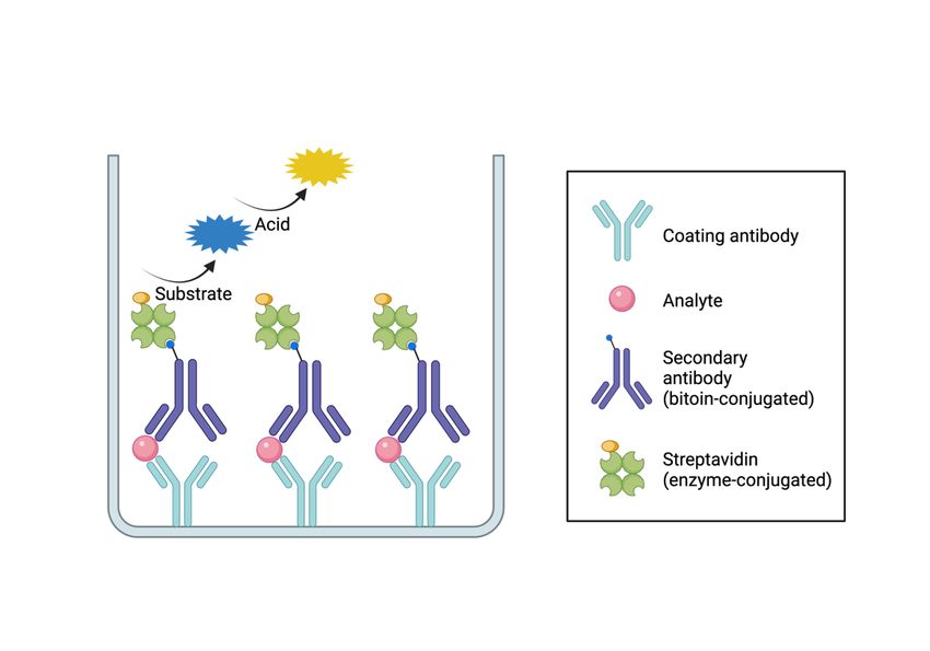
3.1 Analysis of soluble markers............................................................................... 21
3.1.1 Sandwich ELISA ................................................................................... 21
3.1.2 Multiplex immunoassay ........................................................................ 22
3.2 Analyses of MAIT cell phenotypes by flow cytometry .................................... 22
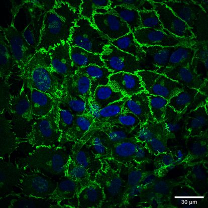
3.3 Analysis of endothelial cells using immunofluorescence microscopy ............. 22
3.4 Permeability assessment using transendothelial electrical resistance ............... 22
3.5 Ethical considerations ........................................................................................ 23
4 RESULTS AND DISCUSSION ................................................................................. 25
4.1 INFLAMMATORY RESPONSES IN HANTAVIRUS-INFECTED
PATIENTS ......................................................................................................... 25
4.1.1 The inflammatory response in HPS patients ......................................... 25
4.1.2 Serum markers associated with severity of HPS................................... 26
4.1.3 Serum markers associated with fatality of HPS .................................... 26
4.1.4 Conclusions and future directions on inflammatory responses in
HPS patients ........................................................................................... 27
4.2 ROLE OF IL-6 IN HANTAVIRUS PATHOGENESIS ................................... 28
4.2.1 Sources of IL-6 during hantavirus infection ......................................... 28
4.2.2 Pro-inflammatory effects of IL-6 trans-signaling during hantavirus
infection ................................................................................................. 294.2.3 Effects of IL-6 trans-signaling on the barrier integrity of infected
cells......................................................................................................... 29
4.2.4 Levels of soluble IL-6 receptors in hantavirus-infected patients .......... 30
4.2.5 Conclusions and future directions on the role of IL-6 in hantavirus
pathogenesis ........................................................................................... 31
4.3 MAIT CELL RESPONSES IN HANTAVIRUS INFECTION........................ 32
4.3.1 MAIT cell responses in HFRS patients ................................................. 32
4.3.2 PUUV-mediated MAIT cell activation in vitro..................................... 33
4.3.3 Function of PUUV-activated MAIT cells ............................................. 34
4.3.4 Conclusions and future directions on MAIT cells in hantavirus
infection.................................................................................................. 35
5 CONCLUDING REMARKS ...................................................................................... 37
6 ACKNOWLEDGEMENTS ........................................................................................ 39
7 REFERENCES ............................................................................................................ 41LIST OF ABBREVIATIONS 5-OP-RU 5-(2-oxopropylideneamino)-6-d-ribitylaminouracil ANDV Andes orthohantavirus ARDS acute respiratory distress syndrome BAFF B cell-activating factor C complement component CCL C-C motif chemokine ligand CRP C-reactive protein CCR C-C motif chemokine receptor CRS cytokine release syndrome DOBV Dobrava-Belgrade orthohantavirus ECMO extracorporeal membrane oxygenation ELISA enzyme-linked immunosorbent assay FDA The United States Food and Drug Administration gp130 glycoprotein 130 HBV hepatitis B virus HCV hepatitis C virus HDV hepatitis D virus HFRS hemorrhagic fever with renal syndrome HPS hantavirus pulmonary syndrome HTLV-1 human T-lymphotropic virus 1 HTNV Hantaan orthohantavirus HUVECs human umbilical vein endothelial cells ICAM-1 intercellular adhesion molecule 1 I-FABP intestinal fatty acid-binding protein IFN interferon IL interleukin IL-6R IL-6 receptor ISG interferon-stimulated genes JAK Janus kinase KHF Korean hemorrhagic fever
LBP        LPS-binding protein
LPS        lipopolysaccharide
MAdCAM-1   mucosal vascular addressin cell adhesion molecule 1
MAIT       mucosal-associated invariant T
MDA-5      melanoma differentiation-associated protein 5
MHC        major histocompatibility complex
MR1        MHC class I-related gene protein 1
MxA        myxovirus resistance protein 1
N          nucleocapsid
NE         nephropathia epidemica
NETs       neutrophil extracellular traps
NK         natural killer
NSs        nonstructural protein
PAMPs      pathogen-associated molecular patterns
PCDH-1     protocadherin 1
PHV        Prospect Hill orthohantaviurs
PRRs       pattern-recognition receptors
PUUV       Puumala orthohantavirus
RIG-I      retinoic acid-inducible gene 1
sCD14      soluble CD14
sCD25      soluble CD25
SEOV       Seoul orthohantavirus
sgp130     soluble gp130
sIL-6R     soluble IL-6R
SNV        Sin Nombre orthohantavirus
STAT       signal transducer and activator of transcription
sTRAIL     soluble TRAIL
TCR        T cell receptor
TEER       transendothelial electrical resistance
TLRs       Toll-like receptors
TNF        tumor necrosis factor
TRAIL    TNF-related apoptosis inducing ligand
TULV     Tula orthohantavirus
VCAM-1   vascular cell adhesion protein 1
VE       vascular endothelial
VEGF     vascular endothelial growth factor
ZO       zonula occludens
1 INTRODUCTION
1.1 HANTAVIRUS
1.1.1 Brief hantavirus history
Hantaviruses are zoonotic viruses with world-wide distribution. In humans, hantaviruses cause
two severe diseases; hemorrhagic fever with renal syndrome (HFRS), caused by "Old World"
hantaviruses, and hantavirus pulmonary syndrome (HPS), caused by "New World"
hantaviruses (1). Diseases resembling HFRS were reported in clinical records in China already
960 AD (1) and in Russia in 1913 (2). Similar syndromes were also described during World
War I (3) and the Korean War in 1951, under the names "war nephritis" and "Korean
hemorrhagic fever" (KHF), respectively (4).
In Sweden, HFRS-like illness was first reported in 1934 by the physicians Zetterholm and
Myhrman, independent of each other (5,6). The cases were characterized by an acute onset of
chills, abdominal pain, back pain, proteinuria, and kidney dysfunction (5,6). In 1945, Myhrman
proposed the name nephropathia epidemica (NE) for the disease (7). Myhrman noted that many
of his NE patients reported contact with mice and speculated that the agent was transmitted to
humans from animals (8). In 1976, NE was found to be related to the disease KHF (9).
However, the causative agents of these illnesses were unknown until 1978, when Lee et al.
reported isolation of the causative agent of KHF - Hantaan orthohantavirus (HTNV) - from a
striped field mouse captured close to the Hantaan river in South Korea (10). In the early 1980s,
the causative agent of NE, Puumala orthohantavirus (PUUV), was isolated from bank voles
(Myodes glareolus) captured in Puumala, Finland (11). Shortly after, KHF and NE were
collected under the name HFRS (12). Since then, additional HFRS-causing hantaviruses, such
as for example Seoul orthohantavirus (SEOV), carried by rats (Rattus rattus, R. norvegicus)
(13) and Dobrava-Belgrade orthohantavirus (DOBV), carried by the yellow-necked mouse
(Apodemus flavicollis) (14), have been identified in Europe and Asia.
In 1993, a cluster of cases of a highly fatal respiratory disease appeared in the Four Corner
region in the United States (15,16). The index cases were two young individuals with acute
onset of fever that after a couple of days rapidly progressed into severe respiratory distress with
fatal outcome (17). In just over a month, the causative agent was found to be a new hantavirus,
later given the name Sin Nombre orthohantavirus (SNV), carried by deer mouse (Peromyscus
maniculatus) (18–20). The disease was named hantavirus pulmonary syndrome (HPS) (also
known as hantavirus cardiopulmonary syndrome) (21). In 1996, another HPS-causing
hantavirus species was identified upon an outbreak in Argentina in 1995 (22). This virus was
named Andes orthohantavirus (ANDV) and is carried by the long-tailed pygmy rice rat
(Oligoryzomys longicaudatus) (23). Several other hantaviruses related to SNV and ANDV
have been reported to cause HPS in the Americas. These include Bayou orthohantavirus, Black
Creek Canal orthohantavirus and Laguna Negra orthohantavirus, among several others (24).
New World hantaviruses sporadically cause outbreaks in the Americas, with the SNV-outbreak
in Yosemite national park in 2012 and the ANDV-outbreak in Argentina in 2018-2019 being
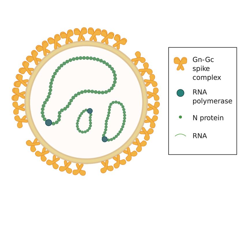
1some of the most recent (25,26). Also non-pathogenic or low-pathogenic hantavirus species exist, the most well-studied being Prospect Hill orthohantavirus (PHV) and Tuula orthohantavirus (TULV) (27–30). 1.1.2 Hantavirus structure Orthohantavirus comprises a genus within the Hantaviridae family of viruses that belongs to the Bunyavirales order. To date, the Orthohantavirus genus consists of 38 different orthohantaviruses (31), hereinafter referred to as hantaviruses. Hantavirus virions (Figure 1) are pleiomorphic and can be either round-shaped or tubular, with an average diameter/length of around 100 nm (32,33). The virions are enveloped and carry a tri-segmented negative-sense single stranded RNA genome. The genome segments are named small, medium, and large, and encode for the nucleocapsid protein (N), the glycoprotein precursor that is cleaved into Gn and Gc, and the RNA dependent RNA polymerase, respectively (27,34). The small segment of certain hantavirus strains also encodes a non-structural protein called NSs (35,36). The hantavirus envelope is densely covered with Gn-Gc spike complex molecules, although with some empty patches (37) (Figure 1). Figure 1. Hantavirus structure. Hantavirus virions are enveloped and contain a single- stranded negative sense RNA genome divided into three segments. The segments encode for the nucleocapsid protein (N), the glycoproteins Gn and Gc, and the RNA polymerase. The hantavirus envelope is densely packed with Gn-Gc spike complexes. 1.1.3 Hantavirus replication Hantaviruses primarily replicate in endothelial cells, but have in vitro been shown to infect also renal, pulmonary and intestinal epithelial cells as well as monocytes and dendritic cells to some extent (38–42). There are several described hantavirus receptors, including αvβ3 and α5β1 integrins and complement decay-accelerating factor. However, it is not known if these 2
receptors are used in vivo (27). Recently, protocadherin-1 (PCDH-1) was identified as a new
hantavirus receptor, essential for the establishment of ANDV and SNV infection (43).
Remarkably, PCDH-1 was shown to be critical for the development of fatal ANDV infection
in Syrian hamsters (43). For establishment of HTNV and SEOV infection, expression of
PCDH-1 was redundant (43).

The cell entry mechanisms of hantaviruses are not fully understood, and different mechanisms
have been described for different species. For example, HTNV has been described to invade
cells by clathrin-mediated endocytosis (44), while both clathrin-dependent and clathrin-
independent entry has been described for ANDV (45,46). As most RNA viruses, hantaviruses
replicate in the cytosol by first creating a complementary RNA strand as a template. After
translation, Gn and Gc proteins polymerize into heterotetramers and are glycosylated in the
Golgi apparatus (47,48). Assembled virions egress infected cells through exocytosis. For some
New World hantaviruses, also assembly at the plasma membrane and egress through budding
has been described (32,49). Many details of the hantavirus replication machinery are still
unknown, as reverse genetics systems are lacking (27).

1.1.4 Natural hosts and transmission
Hantaviruses are zoonotic viruses, meaning they are passed on to humans from natural hosts.
In nature, hantaviruses are carried by rodents as well as insectivores such as moles, shrews, and
bats. Each hantavirus strain is carried by its specific natural host and the geographical
distribution of each hantavirus species depends on the distribution of its specific host (27). Most
human-pathogenic hantaviruses are carried by rodents of the Muridae (mouse, rat) and
Cricetidae (bank vole) families. However, also transmission from the Soricidae family (shrew)
has been suggested in Africa (50). The prevalence of hantavirus infection in rodents is affected
by numerous ecological factors, such as the host density, predator density, food availability,
biodiversity in the habitat, and climate (51–55). As a consequence, the incidence of HFRS in
human populations many times peak following rainy seasons (56,57).

In the natural hosts, hantaviruses cause an asymptomatic persistent infection. The natural hosts
secrete virus through urine, feces and saliva (58–61). Humans are normally infected with
hantavirus following inhalation of dust containing viruses shed from rodent excreta (Figure 2).
Thus, human risk factors for contracting hantavirus infection include activities that bring
humans in close proximity to rodents or rodent excreta, such as handling of firewood, forestry,
farming, and military work (62–68) (Figure 2). In addition, smoking has been reported to be a
risk factor for HFRS (69).

Generally, humans are dead-end hosts for hantaviruses, meaning that the infection is not further
transmitted from an infected human to other humans. However, as was noted after a cluster of
cases in Argentina in 1996-1997 (70), ANDV is an exception and is transmissible between
humans (25,70,71). In 2018-2019, a cluster of cases with human-to-human transmitted ANDV
occurred in Argentina (71). In this outbreak, a single transfer of ANDV from natural host to
one individual resulted in four waves of transmission with in total 33 secondary cases and 11

                                                                                                 3
deaths. The reported initial median reproductive number, commonly referred to as the R value, was 2.12 (71). After 18 confirmed cases, control measures were taken, which reduced the R value to 0.96. In total, person-to-person transmission was confirmed from 10 of the 34 cases. Interestingly, a high viral titer was associated with a higher likelihood of transmission to other persons (71). 1.1.5 Hantavirus-caused diseases In humans, different hantavirus species give rise to different symptoms, which has led to the classification of two separate diseases; namely, hemorrhagic fever with renal syndrome - HFRS, and hantavirus pulmonary syndrome - HPS (27) (Figure 2). Yearly, 100 000 HFRS cases are reported worldwide, while HPS cases are more rare, reaching a few hundred cases per year (72). 1.1.5.1 Incubation time and diagnosis Onset of HFRS/HPS symptoms usually start after an incubation period of two to three weeks (12,25,73,74). However, both shorter and longer incubation times of one to eight weeks have been reported (73–76). During the early acute phase, most patients display virus-specific IgM antibodies (77–81). Low titers of virus-specific IgG antibodies are also detected in most patients, already early during the acute phase (79,81). Thus, serological assays, including immunofluorescence assay and enzyme-linked immunosorbent assay (ELISA), are commonly used for diagnosis of HFRS and HPS (81,82). In most hantavirus-infected patients, viremia can be detected upon admission to the hospital, after which the viral titers rapidly decrease (81,83,84). Therefore, also quantitative polymerase chain reaction on serum samples is commonly used in the diagnosis (85). In individuals with previous hantavirus infection, titers of neutralizing virus-specific IgG antibodies have been reported to continuously increase over two years following infection (11,79,86), and have been detected several decades after infection (86–88). A previous hantavirus infection is believed to provide life-long immunity and re-infection has never been reported. 1.1.5.2 HFRS HFRS is primarily caused by PUUV in Europe, and HTNV in Asia, but also other hantavirus species cause HFRS, including SEOV and DOBV. Individuals with HFRS initially present with flu-like symptoms including headache, fever, and malaise as well as gastrointestinal manifestations such as diarrhea and abdominal pain (89,90). Less than half of the patients also develop renal dysfunction presented as back pain and oliguria (i.e., low urine output). Dialysis treatment is required in 5% of hospitalized PUUV-infected patients (90). Hemorrhagic symptoms, mainly presented as hematuria, petechiae, or epistaxis are displayed by 10-28% and 75% of the PUUV- and HTNV-infected patients, respectively (81,89,91). In addition, gastrointestinal bleedings are common (92–94). A few reports have described cases of pancreatitis and cholangitis during HFRS (95–99). HFRS is sometimes divided into five different clinical phases; febrile phase, hypotensive phase, oliguric phase, diuretic phase, and convalescent phase (12). However, these phases are not often evident in mild cases. The case- 4
fatality rate during HFRS has been reported to be 0.1-0.4% for PUUV, 1% for HTNV, and 0.3-
16% for DOBV infection (27,45,100–105) (Figure 2). The case-fatality rate of HFRS increases
with age, and is in Sweden 6.5% for PUUV-infected individuals above 80 years (100).
Although most patients recover after two weeks, patients that have had HFRS show increased
risk for getting myocardial infarction, stroke, thromboembolism, and lymphoma (106–108).
Furthermore, one study reported higher hantavirus seroprevalence in patients with kidney
disease compared to controls, suggesting that hantavirus infection might cause long-term
kidney problems (109).

Globally, the majority of HFRS cases are documented in China (110). In Sweden, normally
50-400 HFRS cases are reported yearly (90,111). However, the seroprevalence in Northern
Sweden has been shown to be 13% and increase with age, suggesting that there are many
unrecorded cases of PUUV infection (67). A similar seroprevalence has been described in
Finland (112). In Sweden, the incidence of HFRS peaks every third to fourth year, as it follows
the size of the bank vole population (113,114). Following a remarkably mild weather in
December 2006, HFRS incidence in Sweden spiked, giving rise to more than 2000 cases
nationally (115).

1.1.5.3 HPS
HPS is primarily caused by ANDV in South America and SNV in the United States. As
described earlier, HPS was first recognized during the outbreak in the Four Corners region in
the United States, 1993 (18). Since then, HPS caused by ANDV, SNV, and related hantaviruses
has been described also in South American countries such as Argentina, Chile, Brazil, and
Paraguay, as well as in Canada and Panama (70,74,80,116–118). HPS can be divided into three
phases; the febrile phase, the cardiopulmonary phase and the convalescent phase. The febrile
phase is characterized by flu-like symptoms, similar to in HFRS. A majority of HPS patients
also experience gastrointestinal symptoms (119,120). Unlike HFRS, HPS quickly develops to
include pulmonary symptoms such as cough, pulmonary edema, and dyspnea. Chest X-rays of
patients have revealed severe pleural effusion, referring to the accumulation of fluid between
the layers of the pleura covering the lungs, and interstitial as well as alveolar infiltrates
(17,121). The pulmonary dysfunction during HPS often leads to hypoxia, which in many cases
leads to the need for intubation or extracorporeal membrane oxygenation (ECMO) (17,80,122).
Ultimately, 21-40% of HPS patients succumb due to cardiogenic shock (17,122,123) (Figure
2).

The seroprevalence of HPS-causing hantaviruses has been reported to be 1-6.5% in the general
population of Argentina, 1% in Chile, and 3.5% in Brazil (124–127). In Argentinians with
agricultural occupations, 17% were reported to be seropositive (125). Higher seroprevalences
of 17% and 40% has been also been reported for indigenous populations of Argentina and
Paraguay, respectively (128). Thus, it is likely that New World hantaviruses, like Old World
hantaviruses, can cause subclinical infections.

                                                                                             5
Figure 2. Hantavirus-caused diseases in humans. Hantaviruses are transmitted to humans from rodent excreta. Human activities that bring up dust allow the inhalation of virions and thus, increase the risk of infection. Depending on the virus species, hantaviruses can cause hantavirus pulmonary syndrome (HPS) or hemorrhagic fever with renal syndrome (HFRS) in humans. HPS-causing hantaviruses are found in North and South America and cause a highly fatal disease that mainly affects the lungs. HFRS-causing hantaviruses are found in Europe and Asia and cause a milder disease with lower case-fatality rate and often involve kidney dysfunction. Modified from Klingström et al., 2019 (129) under the terms of the Creative Commons CC BY license. 1.1.5.4 Clinical hallmarks of HFRS and HPS As described above, HFRS and HPS are considered two separate diseases caused by different hantavirus species. Although the diseases differ a lot in terms of severity, with case-fatality rates being markedly higher in HPS, the diseases share many characteristics. While severe pulmonary symptoms are more pronounced in HPS, milder pulmonary involvement, including dry cough and dyspnea, has been described also in HFRS patients (38,130–133). Also common between HFRS and HPS are the gastrointestinal symptoms displayed by the majority of patients (17,89,98,101,119,120,134–136). Vascular leakage is an important clinical hallmark shared between HFRS and HPS, and is clinically evident by hemoconcentration concurrent with hypoalbuminemia, hypotension and edema (15,17,27,122,130,137–139). Coagulopathy is another common hallmark of HFRS and HPS, and is indicated by thrombocytopenia and increased serum D-dimer concentration 6
(15,81,122,130). Low thrombocyte levels have been associated with a more severe disease
(140,141). Kidney dysfunction in patients is assessed by increased serum creatinine levels and
proteinuria (81,122,130). Further, patients usually display increased levels of CRP, which
marks inflammation (130,142). In addition, HPS patients often exhibit increased heart rate and
respiratory rate (17).

While a high viral titer has been associated with increased disease severity in infections caused
by SNV, DOBV, and HTNV (142–145), such associations have not been observed in studies
of patients infected with PUUV or ANDV (81,83,84). A low virus-specific antibody titer has
during infection with PUUV and SNV been associated with a more severe disease (81,146).

1.1.5.5 Treatment options
Although hantaviruses cause severe disease in humans, no specific treatments or vaccines
approved by The United States Food and Drug Administration (FDA) are available. Thus,
supportive care aiming at maintaining the electrolyte balance constitutes the standard treatment.
In severe HPS, ECMO treatment improves survival rates in patients with a predicted fatal
outcome (147). In the past, studies have evaluated the effects of the nucleoside analogue
ribavirin as a treatment for HFRS and HPS. While treatment with ribavirin in one study was
suggested to be beneficial in treatment of HFRS, no effect was seen in a study of HPS patients
(148,149). Moreover, the effects of the corticosteroid methylprednisolone have been evaluated
in HPS, without showing any clear effects (150). Given that patients with high virus-specific
antibody titers usually present with a milder disease (115,146), treatment of hantavirus-infected
patients with convalescent plasma therapy has been considered a promising strategy. One study
suggested that passive transfer of antibodies from convalescent HPS patients may reduce the
fatality of HPS (151), although this has not yet been the subject of a randomized controlled
trial.

1.2   THE IMMUNE SYSTEM

1.2.1 Brief overview of the human immune system
The human immune system is orchestrated by a variety of different immune cells and soluble
mediators within the innate and adaptive immune system. The innate immune system elicits a
rapid and unspecific response upon infection. The skin barrier and mucosal surfaces represent
the first line of defense against pathogens. When broken, mononuclear phagocytes, including
dendritic cells, monocytes and macrophages exert important functions in the innate immune
response, by engulfing pathogens and presenting their antigens to cells of the adaptive immune
system (152,153). Other important phagocytes of the innate immune system include the
neutrophils, which rapidly respond to infection and kill pathogens using reactive oxygen
species, by phagocytosis, or by releasing neutrophil extracellular traps (NETs) (154).
Furthermore, natural killer (NK) cells are innate lymphocytes that kill virus infected cells. NK
cell-killing is mediated either via the release of cytotoxic granules consisting of perforin and
granzyme B, via antibody-dependent cellular cytotoxicity, or via the interaction between death

                                                                                               7
receptor ligands, such as tumor necrosis factor-related apoptosis inducing ligand (TRAIL) and death receptors (152,153). The adaptive immune system, which consists of B cells and T cells, is antigen-specific and provides immunological memory. B cells are responsible for the production of pathogen- specific antibodies. These antibodies neutralize pathogens and facilitate killing by phagocytes and NK cells. Conventional T cells can be divided into helper T cells (CD4 T cells) and cytotoxic T cells (CD8 T cells). CD4 T cells are activated by antigen-presenting cells presenting peptide-antigens on their major histocompatibility complex (MHC) class II molecules. CD8 T cells, on the other hand, respond to endogenous peptide-antigens presented on MHC class I molecules on any nucleated cell. Activated CD4 T cells provide activating signals to antigen-stimulated B cells and CD8 T cells. In turn, CD8 T cells kill virus-infected cells using cytotoxic granules. In addition to conventional T cells, the adaptive immune system includes unconventional T cell subsets. These cells have innate-like features and include NKT cells, gd T cells, and mucosal-associated invariant T (MAIT) cells. Unconventional T cell subsets respond to non-peptide antigens with low polymorphism (152,153). The complement system belongs to the innate immunity but acts in concert with both innate and adaptive cells, facilitating their functions. The complement cascade can be initiated via three independent pathways, all which merge at the activation of complement component (C) 3 that becomes cleaved into C3a and C3b. C3b, in turn, cleaves C5 into C5a as well as C5b. Together with other complement factors, C5b creates the membrane attack complex that mediates cell lysis. C3a and C5a are pro-inflammatory mediators and are often referred to as anaphylatoxins (152,153,155). 1.2.2 Viral recognition A virus that has entered a host cell can be sensed by pattern-recognition receptors (PRRs) that recognize pathogen-specific structures, so-called pathogen associated molecular patterns (PAMPs). Toll-like receptors (TLRs) are the most well-described PPRs and include a set of receptors, each recognizing a specific molecular pattern. For instance, TLR-4 and TLR-5 are localized on the cell surface of cells and recognize bacterial surface structures of extracellular bacteria. TLR-3, TLR-7, TLR-8, and TLR-9, on the other hand, are expressed within endosomes, and sense double stranded RNA, single-stranded RNA, and unmethylated CpG regions in DNA, respectively. Thus, viruses that are taken up into endosomes will after endosomal degradation expose their PAMPs that bind to one or several of TLR-3/-7/-8/-9, depending on the type of virus. Viruses replicating within a cells' cytosol can be detected by retinoic acid-inducible gene-1 (RIG-I)-like receptors, which include RIG-I and melanoma differentiation-associated protein 5 (MDA-5), or nucleotide-binding and oligomerization domain-like receptors, which belong to separate PRR families (156,157). PRR-signaling leads to downstream signaling resulting in activation of specific transcription factors, in turn leading to production of pro-inflammatory cytokines and interferons (IFN). Type I IFNs, including IFN-a and IFN-b, are key mediators of the so-called antiviral state. 8
Secreted IFNs bind to ubiquitously expressed IFN receptors on neighboring cells and induce
signaling via Janus kinases (JAK) and signal transducer and activator of transcription (STAT)
proteins. This, in turn, leads to transcription of interferon stimulated genes (ISG), such as
myxovirus resistance protein 1 (MxA). ISG transcription induces a cascade of effector
molecules that together contribute to the antiviral state. For instance, double stranded RNA
results in activation of protein kinase R that in turn leads to inhibition of all protein synthesis
in the cell (156,157).
1.2.3 Endothelial cells
Endothelial cells are epithelial cells that line the inner wall of blood vessels. The primary
function of endothelial cells is to maintain normal blood flow, regulate exchange of proteins
between the blood and tissue, and to prevent coagulation of the blood. Inflammation requires
the migration of leukocytes from blood to affected tissues. Thus, dynamic regulation of the
vessel wall is essential. The integrity of the vessel wall is regulated by tight junctions and
adherence junctions connecting the endothelial cells (158). Tight junctions, such as claudins
and occludin, regulate the inter-cellular exchange of ions and molecules. The tight junction-
associated zona occludens (ZO) proteins bind to tight junction proteins and link those to actin
filaments (159). As indicated by the name, the function of adherence junctions is to mediate
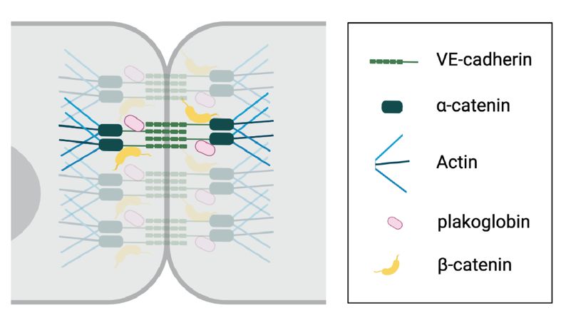
adhesion between cells. Vascular endothelial (VE)-cadherin is one of the most important
adherence junction proteins. VE-cadherin molecules are organized in dimers and attach cells
by binding to adjacent VE-cadherin molecules in a zipper-like manner. The intracellular tails
of VE-cadherin molecules interact with β-catenin and plakoglobin that in turn bind to α-
catenins, which interact with actin filaments (160) (Figure 3). Stimulation of endothelial cells
with inflammatory mediators such as thrombin, histamine, or vascular endothelial growth
factor (VEGF) causes phosphorylation of the intracellular tail of VE-cadherin and leads to its
internalization, with increased permeability as a consequence (160–162).
Figure 3. VE-cadherin organization. Endothelial cells are connected by VE-cadherin (green)
adherence molecules. The intracellular tail of VE-cadherin binds to β-catenin and plakoglobin,
which interact with actin-binding α-catenins.
9Endothelial cell activation, induced by inflammatory mediators, leads to upregulation of adhesion molecules such as intercellular adhesion molecule 1 (ICAM-1), vascular cell adhesion protein 1 (VCAM-1), and E-selectin on the endothelial cell surface (163,164). This allows for binding of immune cells and margination along the endothelium, which facilitates their extravasation into infected sites. Endothelial activation also causes aggregation of platelets and constriction of the blood vessels (164). Endothelial cell dysfunction refers to the inability of endothelial cells to maintain the above-mentioned functions and is often a result of continuous endothelial cell activation (158). 1.2.4 Inflammation Inflammation is a fundamental response to any insult threatening to disturb the homeostasis. Inducers of inflammation can be of exogenous origin, as during an infection, or endogenous, as result of for example tissue injury (165). More specifically, inflammation describes a state of vasodilation and an increase in permeability between the endothelial cells lining the blood vessels. This process, commonly referred to as increased vascular permeability, causes leakage of blood plasma out from the circulation, into the surrounding tissue (165,166). A healthy inflammatory response is well-regulated and actively resolves after some time, when the infection has been eradicated. In some cases, however, the inflammation develops into an uncontrolled process, with immunopathology as a consequence (166,167). This has for example been described in cytokine release syndrome (CRS), which is an acute inflammatory state that can develop as a side-effect of chimeric antigen receptor modified (CAR)-T cell treatment (168). An inflammatory response is mediated and regulated by a wide range of soluble mediators such as cytokines. 1.2.4.1 Cytokines and other markers of inflammation Cytokines are signaling proteins that allow communication between cells. All nucleated cells can produce as well as respond to cytokines (169). A cytokine binds to one or several specific receptors on the target cell and initiates an intracellular signaling cascade. Cytokine signaling can be autocrine, paracrine, or endocrine. Autocrine signaling refers to when a cell secretes a cytokine that then binds to receptors on the same cell. Paracrine signaling, on the other hand, refers to cytokine signaling between neighboring cells. Endocrine signaling describes a more global form of paracrine signaling, in which a cytokine that has reached the blood stream binds to cells in a different tissue (170). The effects of a cytokine are influenced by different factors, including the kinetics, half-life and location of the cytokine as well as the expression of its receptors (170). Cytokines can be separated into pro-inflammatory and anti-inflammatory based on their functions. Typical pro-inflammatory cytokines include interleukin (IL)-1β, IL-6, and tumor necrosis factor (TNF) (166). These cytokines can among other things elicit fever and stimulate the secretion of acute phase proteins such as C-reactive protein (CRP) and ferritin from the liver (171,172). These cytokines also have a role in activating the endothelium (164). Other pro-inflammatory cytokines exert important functions that in different ways support the 10
maturation, expansion, or function of T cells and NK cells. These include for example IL-2,
IL-12, IL-15, and IL-18. IL-2 and IL-15 are mainly known for stimulating activation and
proliferation of T cells and NK cells and IL-12 and IL-18 are strong stimulators of IFN-g
production (173–181). B cell-activating factor (BAFF) is, as the name implies, an important
factor for the survival, maturation and activation of B cells (182). Moreover, IL-10 is an anti-
inflammatory cytokine that suppresses the production of pro-inflammatory cytokines and
chemokines in monocytes and macrophages (183).
IFNs are key cytokines in antiviral immunity and are divided into type I, type II and type III
IFNs. Type I IFNs exist in many different forms, out of which IFN-a (existing in 13 different
subtypes) and IFN-b are the most important (184). IFN-g is the only type II IFN and has
important roles in stimulating the effector functions of macrophages and T cells (184). Type
III IFNs include three IFN-l subtypes. IFN-l shares many functions with the type I IFNs but
particularly controls antiviral responses at mucosal surfaces (185).
With their diverse functions and regulated expression, cytokines are often useful biomarkers in
different disease syndromes. However, also other inflammation markers, such as CRP, ferritin,
and complement factors including C5a can be used as biomarkers in inflammatory diseases.
The soluble IL-2 receptor a, also known as soluble CD25 (sCD25), is another marker that is
often increased in blood during inflammation. CD25 is upregulated on activated lymphocytes,
in particular T cells, and is shed into sCD25 (186,187).
1.2.4.2 IL-6: signaling and function
IL-6 was first identified as "B cell stimulating factor 2", after the discovery of a soluble factor
produced by T cells that stimulated antibody production in B cells (188). IL-6 is a pro-
inflammatory cytokine mainly produced by T cells, endothelial cells and antigen-presenting
cells such as monocytes, dendritic cells and macrophages (189,190). IL-6 is pleiotropic,
meaning it has a wide range of functions on different cells. For example, it induces the acute
phase response, elicits fever, and promotes B cell differentiation and polarization of T cells
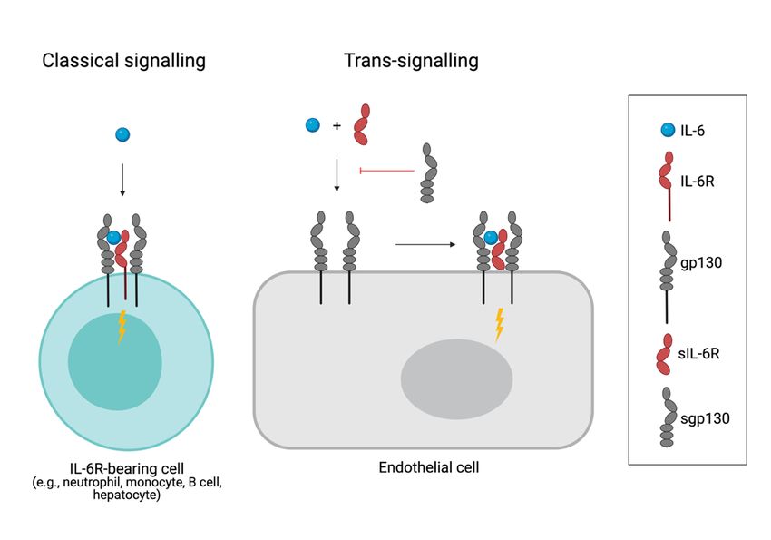
(189,191). IL-6 signals via the IL-6 receptor (IL-6R) in complex with glycoprotein 130 (gp130)
(189,192) (Figure 4). Upon ligation, dimerization of gp130 induces downstream signaling via
the JAK family kinases, causing phosphorylation of STAT1 or STAT3 (Figure 4).
While all cells of the body express gp130, IL-6R expression is restricted only to hepatocytes
and certain immune cells, including neutrophils and B cells (189,192). However, the actions of
IL-6 are not limited to IL-6R-bearing cells as IL-6 also can bind to the soluble form of IL-6R
(sIL-6R), shed from immune cells, and in complex with sIL-6R bind to any gp130 expressing
cell. This form of IL-6 signaling, referred to as trans-signaling, allows for IL-6 to exert its
biological effects also on cells lacking IL-6R (189) (Figure 4). Soluble gp130 (sgp130), created
by alternative splicing or shedding, can bind to the IL-6:sIL-6R complex and inhibit its binding
to membrane gp130. Thus, sgp130 is an inhibitor of trans-signaling (Figure 4). Studies showing
lack of responses to IL-6 in endothelial cells have led to the view that endothelial cells do not
express IL-6R (190,193). However, a few studies have demonstrated a very low IL-6R
11You can also read


