Modulation of Host Death by Chlamydia trachomatis - the Role of the Chlamydia-specific Protease CPAF
←
→
Page content transcription
If your browser does not render page correctly, please read the page content below
Modulation of Host Death by Chlamydia
trachomatis — the Role of the Chlamydia-
            specific Protease CPAF

                       Dissertation
          Zur Erlangung des akademischen Grades
           Doctor rerum naturalium (Dr. rer. nat.)

 vorgelegt dem Rat der Biologisch-Pharmazeutischen Fakultät
           der Friedrich-Schiller-Universität Jena

                            Von
                       Hangxing Yu
                geboren am 13 Oktober 1982
              in der Provinz Shandong, China
Modulation of Host Death by Chlamydia
trachomatis — the Role of the Chlamydia-
         specific Protease CPAF

                    Dissertation
             For obtaining the degree of
        Doctor rerum naturalium (Dr. rer. nat.)

       at the Faculty of Biology and Pharmacy,
          Friedrich-Schiller-University Jena

                    Submitted by
                    Hangxing Yu
      Master of Science in Microbiology (M. Sc.)
             Born on October 13th, 1982
            in Shandong Province, China
Reviewers:

  1. Prof. Dr. med. Eberhard Straube

  Institute of Medical Microbiology
  Friedrich-Schiller University Jena

  2. Prof. Dr. Hans Peter Saluz
  Cell and Molecular Biology Department
  Hans-Knöll Institute, Jena

  3. Prof. Dr. med. Andreas Essig
  Medical Microbiology and Hygiene Institute

  University Hospital, Ulm
Content                                                                                                                                        I

Content

Content ...................................................................................................................................I

Abstract.................................................................................................................................. 1

Zusammenfassung ................................................................................................................. 3

Abbreviations ........................................................................................................................ 5

1                     Introduction ................................................................................................... 7

    1.1               Chlamydiae.................................................................................................... 7

       1.1.1          Taxonomy and pathogenesis ..........................................................................7

       1.1.2          Developmental cycle ......................................................................................8

       1.1.3          Chlamydia persistence....................................................................................9

       1.1.4          Chlamydial effector proteins ........................................................................10

    1.2               Cell death..................................................................................................... 11

       1.2.1          Apoptosis......................................................................................................11

       1.2.2          Necrosis ........................................................................................................14

    1.3               Chlamydia regulates host cells death pathway ............................................ 15

       1.3.1          Chlamydia inhibits apoptosis .......................................................................15

       1.3.2          Chlamydia induces cell death .......................................................................17

       1.3.3          Chlamydial persistence regulate cell death ..................................................17

    1.4               Aim of this work.......................................................................................... 19

2                     Material and methods .................................................................................. 20

    2.1               Material........................................................................................................ 20

    2.2               Methods ....................................................................................................... 25

       2.2.1          Cell culture and Chlamydia organisms.........................................................25

       2.2.2          Infection and induction of persistence..........................................................25

       2.2.3          Immunofluorescence staining of Chlamydia trachomatis............................26
Content                                                                                                                               II

      2.2.4    Detection of cell death by PI/Hoechst staining and flow cytometry ............26

      2.2.5    Apoptosis induction and assay for nuclear morphology ..............................26

      2.2.6    Localization of active caspase-3, cytochrome c, and AIF............................27

      2.2.7    Detection of the caspase-3 /7 activity...........................................................27

      2.2.8    Immunoblotting ............................................................................................27

      2.2.9    Cell free degradation assay...........................................................................28

      2.2.10   Column Chromatography .............................................................................28

      2.2.11   2-D Gel Electrophoreses...............................................................................29

      2.2.12   Mass Spectrometry .......................................................................................29

      2.2.13   Enzyme-linked immunosorbent assay (ELISA) for HMGB-1 and PARP-1 30

      2.2.14   PARP-1 RNA interference and transfection.................................................30

      2.2.15   Lactacystin treatment and Chlamydia replication assay...............................31

      2.2.16   RNA extraction and reverse transcription (RT)-PCR analysis.....................31

3              Results ......................................................................................................... 33

    3.1        Regulation of host cell death by C. trachomatis active infection................ 33

      3.1.1    C. trachomatis active infected cells induce necrotic-like cell death ............33

      3.1.2    Resistance against apoptotic stimuli by C. trachomatis infected cells.........34

      3.1.3    Caspase-3 is not activated during C. trachomatis infection .........................37

      3.1.4    Mitochondrial factors are not activated during C. trachomatis infection.....39

      3.1.5    PARP-1 is cleaved to a multi-form during C. trachomatis infection ...........40

      3.1.6    PARP-1 cleavage depends of chlamydial but not host protein synthesis.....41

      3.1.7    Induction of PARP-1 cleavage by chlamydial proteasome-like activity
               factor (CPAF) ...............................................................................................42

      3.1.8    Cleaved PARP-1 in infected cells loses its polymerase activity ..................45

      3.1.9    Silencing of PARP-1 induces host cell death without inhibition of
               chlamydial developmental cycle ..................................................................46

      3.1.10   CPAF also contributes to HMGB-1 degradation in infected cells ...............48
Content                                                                                                                                      III

       3.1.11         The effect of lactacystin, a CPAF inhibitor, on PARP-1/HMGB-1 cleavage
                      and chlamydial replication............................................................................50

    3.2               Regulation of host cell death by C. trachomatis persistent infection.......... 52

       3.2.1          IFN-γ inhibition of C. trachomatis D infectivity and induction of
                      persistence ....................................................................................................52

       3.2.2          Differential protein expression during persistent infection ..........................54

       3.2.3          Chlamydia persistence resists on apoptotic stimuli......................................55

       3.2.4          Nuclear proteins keep intact during C. trachomatis infection......................57

       3.2.5          Expression level of CPAF correlates with suppression of host proteins
                      degradation during persistent infection ........................................................58

4                     Discussion.................................................................................................... 62

    4.1               Chlamydia regulated host cell death pathways............................................ 62

    4.2               PARP-1 cleavage during C. trachomatis infection ..................................... 63

    4.3               CPAF causes the PARP-1 cleavage............................................................. 65

    4.4               HMGB-1 degradation in the late stage of infection .................................... 67

    4.5               Chlamydial persistent infection ................................................................... 68

    4.6               Persistent infection resists on apoptotic stimuli .......................................... 69

    4.7               Persistence regulates death pathways by different CPAF expression profile
                      70

5                     Conclusion ................................................................................................... 72

6                     References ................................................................................................... 74

7                     Acknowledgement ....................................................................................... 85

Statement ............................................................................................................................. 88
Abstract                                                                                       1

Abstract
Chlamydia is confirmed to modulate host cell death pathways to complete its own
developmental cycle. A balance between pro- and anti- apoptotic influences by Chlamydia
has been postulated.

In this study, the effect of Chlamydia trachomatis on activation of host cell death pathways
was investigated. C. trachomatis infection induced caspase-3-independent cell death,
without the stimulation of apoptotic factors like cytochrome c and apoptotic induction
factor (AIF). On the other hand, after treatment with staurosporine, the activated caspase-3
and the subsequent apoptotic nuclear fragments displayed in uninfected cells were
inhibited by chlamydial infection.

Poly (ADP-ribose) polymerase-1 (PARP-1) is known to play an important role on
regulating apoptosis and necrosis. During necrosis, it will be over-activated and degraded
differently from that observed during apoptosis, which exhibited the signature of 89 kDa
and 24 kDa fragments. After Chlamydia infection, however, PARP-1 was cleaved to
necrotic-like multi-fragment independent of caspase-3 activation. Strikingly, this cleavage
was accompanied by a highly decreased enzymatic activity. PARP-1 silencing by siRNA
in the host cells resulted in cell death similar to that induced by Chlamydia infection, but
has no effect on chlamydial replicaction.

Chlamydial but not host cell protein synthesis contributed to this PARP-1 cleavage. Cell
free degradation assay confirmed that this proteolytic activity only existed in cytosolic
extractions of infected cells. The purified proteolytic protein fraction after column
chromatography exhibited a 29 kDa fragment by Coomassie staining. 2D gel
electrophoresis combined with mass spectrometry proved that this 29 kDa fragment
corresponds to the NH2-terminal portion of chlamydial proteolytic activity factor (CPAF).

The high mobility group box-1 (HMGB-1) protein, which is known to be released to the
extracellular matrix to induce inflammation during necrotic cell death, was also degraded
by CPAF in the late stage of the chlamydial infectious cycle. This gives the suggestion that
Chlamydia may evade the host immune system by degrading this inflammatory factor.

Under the stressful conditions like exposure to IFN-γ, C. trachomatis underwent a special
form so called “persistence”, which is characterized by enlarged pleomorphic RBs and
reduced inclusion size, with minimal cultivability and low infectivity. The persistent
Abstract                                                                                         2

Chlamydia also exhibited the ability of apoptosis inhibition which ensures the long-term
persistence in host cells.

During persistent chlamydial infection, the nuclear proteins PARP-1 and HMGB-1 were
not degraded, whereas cell free degradation assays with the cytosolic extraction from
persistently infected cells showed the activity to cleave both proteins, suggesting the CPAF
translocation to the host nucleus was inhibited during persistence. The pro-apoptotic BH-3
proteins, degraded by CPAF during active infection, were only slightly degraded during
persistence. RT-PCR gave the evidence that the expression of CPAF was highly decreased
during persistent infection.

Our results gave the convincible evidence that the Chlamydia-secreted protease CPAF, as a
ubiquitous protease in Chlamydia-infected cells, plays an important role in the regulation
of host cell death pathways and fulfilling the requirements for chlamydial developmental
cycle. During active infection this protease degrades many host factors, on the one hand,
cleavage of nuclear proteins could induce the host cell instability and subsequent cell death,
on the other hand, the degradation of pro-apoptotic BH-3 protein protect the infected cells
against apoptotic stimuli. For the long-term persistence in host cells, Chlamydia highly
decreases the expression of CPAF, which only existed in the cytosol but is not translocated
to the nucleus. The nuclear proteins kept intact which may make sure the stability of the
host cells, whereas slight degradation of pro-apoptotic BH-3 protein still support the
persistent infection resistance to apoptosis.
Zusammenfassung                                                                             3

Zusammenfassung
Chlamydien sind obligat intrazelluläre Bakterien, die die Zelltod-Signalwege ihrer
Wirtszellen modulieren, um ihren eigenen Eintwicklungszyklus vollenden zu können. Es
wurde ein Gleichgewicht zwischen zelltodinduzierenden und –inhibierenden Einflüssen
von Chlamydien postuliert.

In dieser Studie wurde der Effekt von Chlamydia trachomatis auf die Aktivierung der
Zelltod-Signalwege der Wirtszelle untersucht. Die Infektion von HeLa-Zellen mit
C. trachomatis induzierte einen Caspase-3-unabhängigen Zelltod ohne Stimulierung
verschiedener apoptotischer Faktoren, wie Cytochrom c und Apoptose-Induktions-Faktor
(AIF). Auf der anderen Seite inhibierte die chlamydiale Infektion die durch Staurosporin
verursachte Caspase-3-Aktivierung und die anschließende Bildung apoptotischer nukleärer
Fragmente.

Poly (ADP-ribose) Polymerase-1 (PARP-1) spielt eine wichtige Rolle bei der Regulation
von Apoptose und Nekrose. Während eines nekrotischen Zelltodes ist sie überaktiviert und
zeigt nachfolgend ein anderes Degradierungsmuster im Vergleich zur Apoptose, welche
durch die Spaltung von PARP-1 in 89 und 24 kDa-Fragmente gekennzeichnet ist. Nach
Infektion mit Chlamydien hingegen wurde PARP-1 in mehrere Fragmente gespalten und
zeigte ein Caspase-3-unabhängiges Muster. Auffallend war, dass diese Spaltung mit einer
starken Reduktion der enzymatischen Aktivität einherging. Die Inhibition der PARP-1-
Synthese in HeLa-Zellen mit Hilfe von siRNA induzierte einen Zelltod. Diese Ergebnisse
weisen auf eine Rolle der PARP-1-Degradation bei der Induktion eines Zelltodes durch
Chlamydien hin.

Die Spaltung von PARP-1 ist abhängig von der chlamydialen Proteinsynthese, jedoch nicht
von der Wirtszellproteinsynthese. Proteolytische Assays im zellfreien System bestätigten,
dass diese Aktivität nur in cytosolischen Extrakten infizierter Zellen existierte. Durch
Säulenchromatographie aufgereinigte proteolytische Fraktionen von cytosolischen
Extrakten zeigten nach Coomassie-Färbung ein 29 kDa-Fragment. Die Analyse mittels 2D-
Gelelektrophorese und Massenspektrometrie bewiesen, dass dieses 29 kDa-Fragment dem
NH2-terminalen Teil des chlamydial proteolytic activity factor (CPAF) entspricht.

Das high mobility group box-1 (HMGB-1) Protein, welches während eines nekrotischen
Zelltods in die extrazelluläre Matrix abgegeben wird und als proinflammatorischer Faktor
wirkt, wurde während der späten Phase des chlamydialen Infektionszyklus ebenfalls durch
Zusammenfassung                                                                                 4

CPAF     degradiert.   Durch    die   HMGB-1-Degradation       könnten    Chlamydien     der
Entzündungsreaktion und Abwehrmechanismen des Immunsystems entgehen.

Unter Stressbedingungen, z. B. der Stimulation von infizierten Zellen durch IFN- γ,
können Chlamydien intrazellulär persistierende Formen entwickeln. Diese sind durch
vergrößerte, pleomorphe Retikularkörperchen charakterisiert, welche sich kaum noch
replizieren und sich nicht in infektiöse Formen umwandeln. Diese Chlamydienformen
zeigten ebenfalls die Fähigkeit zur Inhibition der Apoptose, welche offensichtlich die
Langzeit-Persistenz in den Wirtszellen gewährleistet.

Während der persistenten chlamydialen Infektion wurden die nukleären Proteine PARP-1
und HMGB-1 nicht degradiert. Zellfreie proteolytische Assays zeigten hingegen, dass der
cytosolische Extrakt persistent infizierter Zellen beide Proteine spalten konnte. Dies deutet
darauf hin, dass die Translokation von CPAF in den Zellkern der Wirtszelle während der
Persistenz unterdrückt wird. Die proapoptotischen BH3-Proteine, welche während der
replikativen Infektion durch CPAF abgebaut wurden, waren während der Persistenz nur
geringfügig degradiert. Mittels RT-PCR konnte bewiesen werden, dass die Expression von
CPAF während der Persistenz stark reduziert war.

Die hier dargestellten Ergebnisse zeigen, dass die durch Chlamydien sezernierte Protease
CPAF eine wichtige Rolle bei der Regulation von Zelltod-Signalwegen der Wirtszelle und
bei der Komplettierung des chlamydialen Entwicklungszyklus spielt. Während der aktiven
Infektion spaltet diese Protease eine Reihe von Wirtszellproteinen. Einerseits kann die
Spaltung von Kernproteinen zur Instabilität der Wirtszelle und zum anschließenden Zelltod
führen. Andererseits schützt die Degradation von proapoptotischen BH3-Proteinen die
infizierten Zellen gegenüber Apoptose induzierenden Stimuli. Für die Langzeit-Persistenz
in ihren Wirtszellen reduzieren Chlamydien die Expression von CPAF, welches unter
diesen Bedingungen nur in geringen Mengen im Cytosol vorkommt und nicht in den
Zellkern transloziert wird. Die geringgradige Degradation proapoptotischer BH3-Proteine
im Cytosol kann die Apoptose-Resistenz der infizierten Zelle aber auch bei einer
persistenten Infektion aufrechterhalten.
Abbreviations                                                                         5

Abbreviations

AIF             Apoptosis-inducing factors

BCIP/NBT        5-bromo-4-chloro-3-indolylphosphate    toluidine   salt-p-nitroblue
                tetrazolium chloride

BGM             Buffalo Green Monkey

BSA             Bovine serum albumin

CADD            Chlamydia protein associated with death domain

cDNA            Complementary deoxyribonucleic acid

CE              Cytosolic extracts

cHSP60          Chlamydial 60 kDa heat shock protein

cIAP            Inhibitor of apoptosis protein

CPAF            Chlamydial protease-like factor

DAPI            4'-6-Diamidino-2-phenylindole

DBD             DNA-binding domain

DISC            death inducing signalling complex

DNA             Deoxyribonucleic acid

EB              Elementary body

ELISA           Enzyme-liked immunosorbent assay

GAPDH           Glyceraldehyde-3-phosphate dehydrogenase

GPIC            Guinea pig inclusion conjunctivitis

HMGB-1          High mobility group box 1-protein

IAP             Inhibitor of apoptosis

IDO             Indoleamine 2, 3-dioxygenase

IFN-γ           Interferon gamma

IFU             Inclusion-forming units

IgG             Immunoglubulin G

LGV             Lymphogranuloma venereum
Abbreviations 6 m.o.i. Multiplicity of infection MOMP Major outer membrane protein mRNA Message ribonucleic acid NE Nuclear extracts NFκB Nuclear factor kappa B PARP-1 Poly (ADP-ribose) polymerase-1 PBS Phosphate-buffered saline PCR Polymerase chain reaction PDH Pyruvate dehydrogenase p.i. Post infection PI Propidium iodide PI3K Phosphoinositide-3 kinase PKCδ Protein kinase C δ PMF Peptide mass fingerprint RB Reticulate body RLU Relative light units RT-PCR Reverse transcriptase polymerase chain reaction siRNA Small interfering RNA TBS Tris-buffered saline TNF α Tumor necrotic factor α TNFR Tumor necrosis factor receptor TTSS Type III secretion system
Introduction                                                                                 7

1 Introduction

1.1 Chlamydiae

Chlamydiae are gram-negative obligate intracellular bacteria which target epithelial cells
of mucosae and replicate within specialized parasitophorous vacuoles. Different species of
Chlamydia infect a variety of hosts with a wide range of tissue tropisms and varied
diseases.

1.1.1 Taxonomy and pathogenesis

Chlamydiae have been placed in their own order, Chlamydiales, with the family of
Chlamydiaceae, which was divided in two genera, Chlamydia and Chlamydophila,
containing altogether nine species (Fig. 1. 1). The genus Chlamydia consists of three
species, namely C. trachomatis, C. suis and C. muridarum, whereas Cp. psittaci, Cp.
abortus, Cp. felis, Cp. caviae, Cp. pecorum and Cp. pneumoniae were placed under the
genus Chlamydophila. Two of these species, C. trachomatis and Cp. pneumoniae, are
common pathogens in humans, whereas the other species usually occur in animals.

                  Fig. 1. 1 The classification of Chlamydia (Everett, K. D., 2001)
Introduction 8 C. trachomatis causes severe infections of the epithelial tissue of eyes and urogenital tract. It is divided into three groups of serovars. Serovar A to C is the leading cause of preventable blindness in developing countries. Serovar D to K is responsible for the majority of sexually transmitted infections; ascending infection by these serovars of the female upper genital tract leads to ectopic pregnancy and tubal infertility (Carlin, J. M., 1995). These infections are also associated with reactive arthritis, which develops in 1 to 3% of patients after genital chlamydial infection (Wollenhaupt, J., 1990). Serovar L1 to L3 shares the unique ability to pass through epithelia, after which they disseminate, invade, and destroy lymphatic tissue and cause Lymphogranuloma Venereum (LGV) (Mabey, D., 2002). Cp. pneumoniae is a respiratory pathogen that causes acute and chronic respiratory diseases. It is estimated to cause an average of 10% of community-acquired pneumonia cases and 5% of bronchitis and sinusitis cases (Kuo, C. C., 1995). Unresolved respiratory Cp. pneumoniae infection may contribute to the pathogenesis of chronic inflammatory lung diseases (Hahn, D. L., 1991; Blasi, F., 1993). In addition, it also acts as a possible risk factor for the development of atherosclerosis (Belland, R. J., 2004) or promoting the destabilization of atherosclerosis plaque (Rödel, J., 2004). 1.1.2 Developmental cycle All chlamydial species share a common biphasic developmental cycle (Fig. 1. 2), which involves two distinct morphological and functional forms: the smaller (≈ 0.3 µm) extracellular, metabolically inactive infectious particles called elementary bodies (EBs) and the larger (0.8~1.0 µm) intracellular metabolically active particles termed reticulate bodies (RBs) (Abelrahman, Y. M., 2005). The EB has a pear shape and possesses a rigid outer membrane with the DNA packed by histone-like proteins. The rigidity of EBs is based on cross-linking of three cysteine-rich proteins. The most important of these is the major outer membrane protein (MOMP), which represents up to 60% of the weight of the outer membrane. The RB possesses a fragile membrane lacking the extensive disulfide bonds with tendency to form pleomorphic outer envelope blebs. Intracellular growth of chlamydia is initiated by the invasion of non-phagocytic epithelial cells by infectious, metabolically inactive EBs. The internalized EBs replicate within a membrane-bound vacuole termed “inclusion” and differentiate into RBs. This includes the
Introduction                                                                                     9

DNA replication and reduction of the disulfide bridges of the outer membrane. RBs
multiply by binary fission. After 8 to 12 rounds of multiplication, RBs begin to convert
into EBs, including packing of the DNA and synthesis of late outer membrane proteins that
are disulfide bridged. At 48 to 72 h post infection (p.i.), depending primarily on the
infecting species, EBs progeny are eventually released through host cell lysis or extrusion
(Moulder, J. W., 1991; Wolf, K., 2000) (Fig. 1. 2).

                  Fig. 1. 2 Developmental cycle of Chlamydia. (Beatty, W. L., 1994)

1.1.3 Chlamydia persistence

In response to stress conditions, chlamydiae can switch to a special persistent form, which
is metabolically and morphologically altered and difficult to be eliminated by host cells
(Hogan, R. J., 2004). Persistent chlamydia has been described as an atypical “aberrant”
morphology, i.e., enlarged pleomorphic RBs and reduced inclusion size with minimal
cultivability and low infectivity resulting in a long-term relationship with the infected host
cell (Beatty, W. L., 1994) (Fig. 1. 2). The persistent chlamydial infection is thought to
Introduction 10 correlate with many chronic infections, including pelvic inflammatory disease, scarring trachoma and reactive arthritis (Hogan, R. J., 2004). In vivo, the host response against Chlamydia mostly depends on the inflammatory cytokine gamma interferon (IFN-γ). IFN-γ suppresses Chlamydia mainly through the induction of tryptophan degrading enzyme indoleamine 2, 3-dioxygenase (IDO). IDO is responsible for tryptophan catabolism, thus decreasing the intracellular concentration of tryptophan available for chlamydia (Beatty, W. L., 1993). Chlamydia deprived of tryptophan fail to complete secondary differentiation into infectious EBs, resulting in the loss of infectivity and cell-to-cell transmission and undergoing to the persistent form. However, different chlamydial species exhibit differential sensitivities to the effect of IFN-γ. For example, the genital serovars (D to K as well as L1 to L3) but not ocular serovars (A to C and Ba) of the C. trachomatis displayed the ability to resist on IFN-γ exposure. The differences in susceptibility of various chlamydial strains to IFN-γ have been explained by the differences in their ability to acquire exogenous tryptophan or to synthesize their own tryptophan (Morrison, R. P., 2000; Caldwell, H. D., 2003). Despite the evident importance of tryptophan catabolism, other mechanisms such as the inducible nitric oxide synthase effector pathway, iron deprivation and antibiotic treatment could also induce abnormal Chlamydia RBs. (Igietseme, J. U., 1998). 1.1.4 Chlamydial effector proteins Once internalized, chlamydiae stay inside the inclusion throughout the intracellular stage, which lasts for 2 to 4 days depending on the chlamydial species. Like other gram-negative pathogens, chlamydiae translocate ‘effector’ proteins into their host cytosol to modulate cellular functions. Now it is confirmed that all Chlamydiae code for the core components of a type III secretion system (TTSS), a protein transport system used by gram-negative bacteria to translocate proteins into the cytoplasm of the host cell, suggesting Chlamydia may deliver effector proteins to the inclusion membrane and into the host cell cytoplasm by type III secretion (Peter, J., 2007; Clifton, D. R., 2004; Stephens, R. S., 1998; Hsia, R- c., 1997). So far, evidence for the secretion of chlamydial effector proteins by type III secretion has been limited to proteins associated with the inclusion membrane, such as CopN (Fields, K. A., 2000) and members of the family of inclusion membrane proteins (Inc) (Subtil, C. Parsot, 2001).
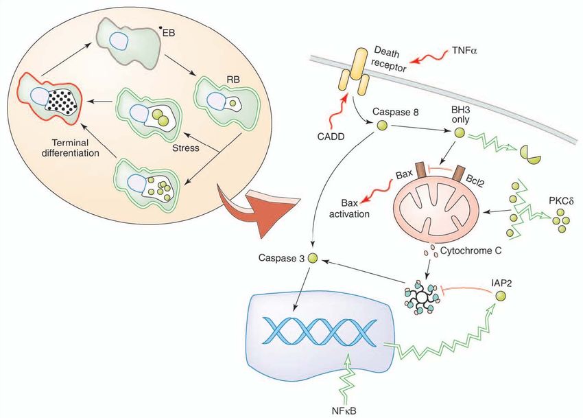
Introduction                                                                                     11

It should be noted that chlamydial effector proteins also access the cytoplasm of infected
cells via TTS-independent mechanisms. For example, chlamydial protease-like activity
factor (CPAF) contains a domain characteristic of bacterial Tail-specific proteases (Tsp)
(Shaw, A. C., 2002) and is secreted to the inclusion lumen before translocation into the
cytoplasm of the infected cells (Heuer, D., 2003).

Throughout the infectious cycle Chlamydia modulate many cellular functions, which
require the activity of chlamydial effector proteins, including subversion of the
cytoskeleton to facilitate intracellular redistribution of newly internalized EBs, inhibition
of apoptosis to ensure intracellular survival during the developmental cycle, induction of
cell death to release chlamydial progeny upon completion of the cycle, and evasion of host
immune system recognition (Carabeo, R. A., 2002). For example, the Incs are probably
central regulators of bacterial-host interactions. IncA mediates homotypic fusion of
inclusions potentially by forming a SNARE-like fusogenic intermediate between adjacent
inclusions (Hackstadt, T., 1999; Delevoye, C., 2004). IncG sequesters the host protein 14-
3-3β and its pro-apoptotic binding partner phosphor-BAD (Verbeke, P., 2006).

Another prominent function of these effector proteins is to interfere with the host cell death
programs which are central to innate immune responses. During middle stage of infection,
the CPAF degrades pro-apoptotic BH-3 proteins to shut down the infected cell’s ability to
undergo apoptosis in response to intrinsic and extrinsic stimuli. Finally, Chlamydia
infection significantly impacts the cell cycle of infected cells by cleavage of the mitotic
cyclin B1and delays in cytokinesis (Balsara, Z. R., 2006; Greene, W., 2003). These later
functions lead to genomic instability, which in conjunction with the strong anti-apoptotic
effect of chlamydial infection may explain how Chlamydia modulate host cell death
programs to fulfil its unique intracellular parasite lifestyle.

1.2 Cell death

In general, cellular homeostasis is maintained by a balance between proliferation and cell
death. Two major types of cell death are described as apoptosis and necrosis.

1.2.1 Apoptosis

Apoptosis is a well-conserved process that multicellular organisms use for control of
development and homeostasis as well as the removal of unwanted cells recognized by the
Introduction                                                                                     12

immune system. Common morphological and biochemical features of apoptosis include
nuclear chromatin condensation, cytoplasmic shrinking, dilated endoplasmic reticulum,
blebbing of plasma membrane, phosphatidylserine asymmetry, internucleosomal DNA
fragmentation, reduction of mitochondrial transmembrane potential, intracellular
acidification, etc (Vaux, D. L., 1994; Wyllie, A. H., 1997; Golstein, P., 1991).

Apoptosis can be induced through both extrinsic and intrinsic pathways (Hengartner, M.
O., 2000; Krammer, P. H., 2000; Green, D. R., 1998; Salvesen, G. S., 2002). The intrinsic
pathway involves the activation of several pro-caspases followed by the mitochondria
released apoptotic factors. The extrinsic pathway involves the activation of the death
receptors, FAS and tumor necrosis factor receptors (TNFR). In most cases, both pathways
require the release of mitochondrial cytochrome c, which in turn binds to Apaf-1 and
caspase 9 to form an apoptosome, the caspase-activating signaling complex (Ekert, P. G.,
2005). Subsequently, caspases get activated and cleave cystein-containing proteins like
lamins, poly (ADP-ribose) polymerase 1 (PARP-1), histones, nucleolin, and fodrin, thus
leading to cell death (Fig. 1. 3). Now, the cleavage of PARP-1 in fragments of 89 kDa and
24 kDa by activated caspase-3 and 7 has become a useful hallmark of apoptosis.

The release of cytochrome c is regulated by Bcl-2 family proteins. This family consists of
three groups of proteins: the pro-apoptotic subfamily (Bax, Bak and Bok), which directly
alters the integrity of the mitochondrial membrane and causes cytochrome c release; anti-
apoptotic proteins Bcl-2 and Bcl-xl, which inhibit cytochrome c release; and the BH-3 only
proteins that exhibit pro-apoptotic activity by inhibition of Bcl-2 or Bcl-xl. c-FLIP is a
critical negative regulator of the extrinsic cell death pathway. It functions as a competitive
inhibitor of pro-caspase 8. Upon death receptor stimulation, c-FLIP can be efficiently
recruited to the death inducing signalling complex (DISC) and inhibit pro-caspase 8
activation at the DISC (Zhang, N., 2005).

Two types of cells have been identified with regards to the requirement for a mitochondrial
amplification loop during apoptosis. In type II cells (HeLa, Jurkat, and human CEM T-cell
line), apoptosis inducers can activate caspase-3 only through a mitochondrion-dependent
step. In type I cells (human B lymphoblast cell line SKW6.4, H9, BJAB), caspase-8
recruitment and activation by cell surface receptors can lead directly to caspase-3
activation without help from mitochondria (Barnhart, B. C., 2003).
Introduction                                                   13

               Fig. 1. 3 Apoptotic pathway (Zhang, N., 2005)
Introduction 14 1.2.2 Necrosis In contrast to apoptosis, necrosis has generally been considered as an unregulated form of cell death which is associated with acute injury to cells. It results in plasma membrane disintegration and leakage of cellular contents to stimulate inflammation (Cohen, G. M., 1997; Fiers, W., 1999). Necrosis has long been described as a consequence of extreme physico-chemical stress, such as heat, osmotic shock, mechanical stress, freeze-thawing and high concentration of hydrogen peroxide. Under these conditions, cell death occurs quickly due to the direct effect of the stressor on the cell, and therefore this cell death process has been described as accidental and uncontrolled. However, recently many different cellular stimuli (TNF on certain cell lines, dsRNA, ATP depletion, ischemia) have been shown to induce a necrotic process that follows defined steps and signalling events reminiscent of a true cell death program (Vanden Berghe, T., 2007). This induced necrotic cell death results from extensive crosstalk between several biochemical and molecular events at different cellular levels, and it is similarly controlled like apoptosis. It is important to distinguish necrosis from other forms of cell death, particularly because it is often associated with unwarranted loss of cells in human pathologies. It can also lead to local inflammation due to release intracellular factors from dead cells; the so-called damage associated molecular patterns that alert the innate immune system. Now the different cleavages of PARP-1 have become the method to distinguish apoptosis and necrosis. During necrosis, the PARP-1 is cleaved to the multi-fragment form (72, 55 and 42 kDa) by the lysosomal proteases (Gobeil, S., 2001). This necrotic PARP-1 cleavage could also induce the release of the high mobility group box 1 protein (HMGB-1), which further stimulates monocytes to secrete a subset of pro-inflammatory cytokines and act as an important inflammatory factor (Ditsworth, D., 2007; Müller, S., 2001). However, there is no other clear biochemical definition of necrotic cell death and consequently no specific biochemical markers that unambiguously discriminate necrosis from apoptosis. Another problem is that even the interpretation of dying cell morphology may be complex, because in the absence of phagocytosis apoptotic cells proceed to a stage called secondary necrosis, which shares many features with primary necrosis.
Introduction                                                                                    15

1.3 Chlamydia regulates host cells death pathway

As an intracellular pathogen, Chlamydia replicates strictly in a vacuole in the host cytosol.
Thus, it is essential to maintain the integrity of host cells during its developmental cycle
not only for supplying the nutrients but also for protecting the intracellular organisms from
host phagocytosis and antigen-specific immune effectors. However, at the later stage, the
host cells rupture benefits on the release of elementary bodies to start a new infectious
cycle. Therefore, modulation of the host cell death could be a relevant component of the
chlamydial developmental cycle.

1.3.1 Chlamydia inhibits apoptosis

The anti-apoptotic activity of Chlamydia has consistently been demonstrated by its ability
to inhibit chemically and spontaneously induced apoptosis during mid to late stages in the
developmental cycle. The paper in 1998 first described that, C. trachomatis-infected cells
are resistant to numerous experimental apoptotic stimuli, exhibiting inhibition of caspase
activation and blockage of cytochrome c release from mitochondria (Fan, T., 1998). Later,
several studies have shown that epithelial cells or monocytes that are infected with C.
trachomatis or Cp. pneumoniae are protected against cell death stimuli (D. Dean, 2001;
Fischer, S. F., 2001, 2004; Rajalingam, K., 2001; Greene, W., 2004).

An explanation for these observations could be the proteolytic cleavage of a wide range of
pro-apoptotic BH-3 only proteins (Fischer, S. F., 2004). It is now known that Chlamydia
are capable to degrade the pro-apoptotic proteins Puma, Bad, Noxa and tBid (Ying, S.,
2005), a family of proteins that share this Bcl-2 homology domain. The secreted
chlamydial protease CPAF was confirmed to be responsible for this degradation, thus
ensuring a complete shut down of the infected cell’s ability to undergo apoptosis in
response to intrinsic and extrinsic stimuli.

Besides the BH-3 only proteins degradation, chlamydial infection also results in activation
of the phosphoinositide-3 kinase (PI3K) pathway. During infection with C. trachomatis,
activation of PI3K leads to Akt activation and in turn to phosphorylation of a pro-apoptotic
protein, Bad. C. trachomatis prevents phosphorylated Bad from binding to mitochondria
by sequestering their binding partners, 14-3-3β proteins, via a chlamydial inclusion
membrane protein IncG (Verbeke, P., 2006).
Introduction                                                                                  16

Additionally, Chlamydia appears to be capable of accumulating diacylglycerol in the
inclusion, which recruits and sequesters pro-apoptotic effector protein kinase C δ (PKCδ).
When PKCδ is activated, it is cleaved to release a catalytically active fragment that
translocates to the mitochondria where it promotes the release of cytochrome c and
subsequently activates of the apoptotic pathway (Tse, S. M., 2005). Thus, chlamydial
recruiting PKCδ could protect the mitochondria against the death signal.

Anti-apoptotic gene regulation through nuclear factor NF-κB-mediated gene activation is
an anti-apoptotic mechanism used by other pathogens, but conflicting results exist for
Chlamydia. Cp. pneumoniae infection of a human monocytic cell line induced expression
of nuclear factor kappa B (NF-κB) and the inhibitor of apoptosis 2 (IAP2). Inhibition of
NF-κB by proteosome inhibitor MG132 resulted in inhibition of IAP2 and induction of
apoptosis (Wahl, C., 2003). However, other studies is failed to demonstrate the function of
NF-κB activation in C. trachomatis inhibition of apoptosis. Neither chemical inhibition nor
gene deletion of NF-κB had any effect on anti-apoptotic activity of C. trachomatis (Xiao,
Y., 2005; Fischer. S. F., 2003).

                 Fig. 1. 4 The inhibition of apoptosis by Chlamydia (Miyairi, I., 2006)
Introduction 17 1.3.2 Chlamydia induces cell death Cytotoxicity associated with chlamydial infection has been reported for many years. Chlamydia infection was thought to be lytic for the host cells. Productive cell culture infections ultimately result in host cell lysis (Abdelrahman, Y. M., 2005). Cell death seems to occur in uninfected neighboring cells or in infected cells at later stages of infection. Though the cells displayed some morphological characteristics of apoptotic death, there is no classical caspase activation (Ojcius, D. M., 1998; Schoier, J., 2001; Dumrese, C., 2005). Another publication suggested an additional possibility, namely, that Cp. pneumoniae induces a form of cell death which displays some apoptotic features, but is more consistent with necrosis. The term “aponecrosis” has been proposed to characterize this special death form induced by Chlamydia infection (Dumrese, C., 2005). These findings suggest that an ongoing death stimulus by chlamydial infection exists to induce the host cell death in the late stage of infection. It is indicated that the pro-apoptotic protein Bax is activated during Chlamydia infection with the guinea pig inclusion conjunctivitis (GPIC) serovar of Cp. psittaci (J. L. Perfettini, 2002), giving an explanation for the Chlamydia induced caspase-independent cell death. One Chlamydia protein associated with death domain (CADD)— an iron-containing redox enzyme unique to chlamydial species, can induce cell death when ectopically expressed in mammalian cells by interacting with death domains of tumor necrosis factor (TNF) family of receptors (Stenner-Liewen, F., 2002). Host cytokines such as tumor necrotic factor α (TNF-α) (Jendro, M. C., 2004) have been implicated as sources of pro-apoptotic stimuli. Cp. pneumoniae infection is also known to modulate oxLDL-induced endothelial cell death, decreasing apoptosis and promoting necrosis (Nazzal, D., 2006). In addition, Chlamydia- induced necrosis might be associated with a heterogeneous family of clostridia cytotoxin homologues that are coded by the genomes of C. trachomatis and Cp. psittaci (Belland, R. J., 2001). 1.3.3 Chlamydial persistence regulate cell death During persistent infection, the antigenic profile of Chlamydia differs from that of acute infection. Beatty et al. (1993, 1994) proposed that during chlamydial persistence, the reduced levels of MOMP, an immunoprotective antigen, could enable chlamydiae to avoid the development of protective immunity. On the other hand, steady of chlamydial 60 kDa heat shock protein (cHSP60) levels would promote immunopathology of persistent
Introduction                                                                                    18

chlamydia. Hypersensitivity reactions are known to be elicited by cHSP60 (Morrison, R.
P., 1989, 1991; Patton, D. L., 1994), which is confirmed to induce apoptosis (Equils, O.,
2006); suggesting that an apoptotic block may favour a chlamydial form that expresses
inflammation-inducing proteins.

It has been reported that cells infected with C. trachomatis in the presence of IFN-γ resist
apoptosis due to external ligands, via inhibition of caspase activation (Dean, D., 2001).
During persistent chlamydial infection, cytochorome c remained sequestered in the
mitochondrial fraction after apoptotic induction (Deborah, D., 2001). The mechanism of
protection in persistently infected cells seems to be the same as that observed in the
absence of IFN-γ.

Furthermore, anti-apoptotic activity is prolonged during chlamydial persistence, which
strengthens the hypothesis that active chlamydial metabolism maintains host cell integrity
and contributes to intracellular survival (Dean, D., 2001; Perfettini, J. L., 2002). However,
the precise mechanisms and factors that Chlamydia uses to modulate host cell death
pathways during different life styles still need to be further investigated.
Introduction                                                                                     19

1.4 Aim of this work

In summary, to fulfill the long-term intracellular survive, Chlamydia is confirmed to inhibit
the death stimuli for the host cells during the developmental cycle and spontaneously
induce some kind of cell death to release the reproduced EBs. When this pathogen is under
the conditional stress like exposure to IFN-γ, it goes to a persistent form, which also
exhibits abilities to protect the host cells against death stimuli and prevent the elimination
by the host immune system. However, until now, the precise factors and mechanisms of
how Chlamydia regulates host cell death pathways to fulfill its specific developmental
cycle and the differences on this regulation between active and persistent infection are still
unclear. It is unclear how the balance between cell death induction versus apoptosis
inhibition is regulated in Chlamydia infected cells and which factors are involved.

The objective of my work can be described as follows:

   1. To characterize the host cell death induced by C. trachomatis and to illuminate the
       mechanisms which Chlamydia used to regulate host death pathways.

   2. To detect the factors like nuclear proteins PARP-1 and HMGB-1, which are
       involved in keeping host cell integrity and homeostasis, and to investigate the
       function of these factors in Chlamydia induced host cell death.

   3. To compare the differences of cell death regulation between active and persistent
       infection, and explain how Chlamydia can modify host cell death pathways under
       different conditions.
Material and methods 20
2 Material and methods
2.1 Material
Strain Company
C. trachomatis serovar D strain IC Cal 8 Institute of Ophthalmology, London, United
Kingdom
Cell lines and culture medium Company
BGM cell American Type Culture Collection, ATCC;
Manassas, VA
Fetal calf serum PromoCell, Heidelberg, Germany
HeLa cell ATCC; Manassas, VA
Minimal Essential Medium (OptiMEM, Invitrogen, Karlsruhe, Germany
Gibco)
Panserin medium Panbiotech
SF-3 medium Cytogen, Lohmar, Germany
Material Company
25-cm2 flasks CellStar; Greiner Bio-One GmbH,
Frickenhausen, Germany
6-well plates Nunclon TM, Roskilde, Denmark
8-well LabTec chamber Nunclon TM, Roskilde, Denmark
96-well plates Nunclon TM, Roskilde, Denmark
AKTA purifier 10 instruments Pharmacia Biotech Inc
IPG dry strips GE Healthcare Life Sciences
Mono Q ion exchange column Amersham Pharmacia Biotech Inc.
Semidry transblot system Bio-Rad, Munich, GermanyMaterial and methods 21
Buffer and solution Compound
Bjerrum and Schafer-Nielsen Transfer 48mM Tris, 39 mM glycine, 1.3 mM SDS,
buffer 20% methanol (pH ~9)
Blocking buffer 4% bovine serum albumin, 0.05% Tween,
TBS
Buffer B for Cell free degradation 0.5 mM NaCl, 1% Triton X-100 in 20 mM
Tris (pH 8.0)
Buffer A for chromatography 0.01 M Tris, pH 7.2
Buffer B for chromatography 0.01 M Tris, 1 M NaCl, pH 7.2)
Equilibration buffer 6 M urea, 30% (w/v) glycerol and 2% (w/v)
SDS in 0.05 M Tris-HCl buffer, pH 8.8.
Gel fixing solution 40% methanol, 7% glacial acetic acid
Gel staining solution 10% methanol, 0.1% CBB G-250, 1.6% o-
phosphoric acid, and 10% (w/v) ammonium
sulfate
HiPerfect transfection reagent Qiagen, Hilden, Germany
IPG blocking buffer 7 M urea, 2 M thiourea, 2% CHAPS,
0.002% bromophenol blue, 0.5% IPG buffer,
1.2% De-Streak Reagent
Laemmli sample buffer Bio-Rad, Munich, Germany
Neutralised solution 0.1 M Tris-base titrated to pH 6.5 with o-
phosphoric acid
Saccharose phosphate buffer PBS with 0.2 M saccharose and 2% FCS
Chemical Company
Aprotinin Sigma, US
Bovine serum albumin (BSA) Sigma, US
Chloroamphenicol Biocat, Heidelberg, Germany
Cycloheximide Biocat, Heidelberg, Germany
DAPI Invitrogen
DMSO Sigma, USMaterial and methods 22
Gel strengthener Molecular Probes, Leiden, The Netherlands
Hoechst 34580 Invitrogen, Karlsruhe, Germany
Lactacystin Enzo Life Sciences, Lörrach, Germany
Leupeptin Serva, Heidelberg, Germany
Methanol Mallinckrodt Baker, Deventer, Holland
Paraformaldehyde Sigma, US
Phenylmethylsulfony fluoride (PMSF) Serva, Heidelberg, Germany
Propidium iodide (PI) Biotium, purchased from Biotrend, Cologne,
Germany
Protein standard Mark 12 Invitrogen, Karlsruhe, Germany
SDS-PAGE Standards Broad Range Bio-Rad, Munich, Germany
Sodium dodecyl sulfate (SDS) Sigma, US
Staurosporine Roche Applied Science, Mannheim,
Germany
Sulforhodamine 101-annexin V Biotium, Hayward, CA
SYBR Green I (Molecular Probes; MoBiTec; Göttingen,
Germany
TBS-tween Sigma, US
Trypsin Biochrom AG
Kits Company
Annexin V-FLUOS staining kit Roche Applied Science, Mannheim,
Germany
Caspase-Glo® 3/7 Assay Promega, Mannheim, Germany
DEVD-NucViewTM caspase-3 Biotium, Hayward, CA
HMGB-1 enzyme-linked immunosorbent Shino-test, Kanagawa, Japan
assay
HP validated small interfering RNA Invitrogen, Carlsbad, CA
(siRNA)Material and methods 23
Promega reverse transcription system Promega, Mannheim, Germany
Rnase-Free Dnase Set Qiagen, Hilden, Germany
RNeasy Mini Kit Qiagen, Hilden, Germany
Universal colorimetric PARP-1 assay kit R&D Systems, Wiesbaden-Nordenstadt,
Germany
Antibody Company
5-bromo-4-chloro-3-indolylphosphate Sigma Fast, Sigma-Aldrich
toluidine salt-p-nitroblue tetrazolium
chloride (BCIP/NBT substrate)
Alkaline phosphatase-conjugated goat anti- Dianova, Hamburg, Germany
rabbit/mouse IgG
Mouse monoclonal antibody to chlamydial Kindly provided by Dr. V. Forsbach-Birk,
CPAF Institute of Medical Microbiology,
University of Ulm, Germany
Mouse monoclonal antibody to Chlamydia Acris, Hiddenhausen, Germany
trachomatis heat shock protein 60
(cHSP60)
Mouse monoclonal antibody to Chlamydia Acris
trachomatis (MOMP)
Rabbit polyclonal antibody to human Bad Cell-Signalling Tech
Rabbit polyclonal antibody to human Bim Sigma, US
(BOD)
Rabbit polyclonal antibodies to GAPDH Santa Cruz Biotechnology
Rabbit polyclonal antibody to human Abcam, Cambridge, UK
HMGB-1
Rabbit polyclonal antibody to IDO Santa Cruz Biotechnology
Rabbit polyclonal antibody to human Sigma, US
Puma/bbc3, N-terminal
Taxas-conjugated goat anti-rabbit/ mouse Santa Cruz Biotechnology
IgG (H+L)Material and methods 24
Primer Sequence
16S rRNA of Mycoplasma 5’-ccagactcctacgggaggca-3’
5’-tgcgagcatactactcaggc-3
cIAP-1 5’-cacatgcagctcgaatgagaa-3’
5’-tccaaggcagatttaaccacagg-3’
cIAP-2 5’-catgcagcccgctttaaaacattc-3’
5’-catttccacggcagcattaatcac-3’
HP validated small interfering RNA Qiagen, Hilden, Germany
(siRNA) targeting PARP-1 (SI02662989)
nonsilencing All Stars Negative Control Qiagen, Hilden, Germany
siRNA
PDH 5’-ggtatggatgaggacctgga-3’
5’-cttccacagccctcgactaa-3’
Machine Company
Branson Sonifier W-250 Heinemann, Germany
Confocal laser scanning microscope (TCS Leica, Heidelberg, Germany
SP5)
Confocal laser scanning microscope (Zeiss) Zeiss, Jena, Germany
Ettan DALT System GE Healthcare Life Sciences
FACSCalibur flow cytometer BD Biosciences, Heidelberg, Germany
Fluorescence microscope Axioskop, Zeiss, Jena, Germany
IPGphorTM GE Healthcare Life Sciences
Semidry transblot system Bio-Rad, Munich, Germany
Protean II xi multicell system Bio-Rad, Munich, Germany
TRIO-Thermoblock Biometra, Göttingen, Germany
Ultraflex I device Bruker Daltonics, Bremen, GermanyMaterial and methods 25 Software Company Cell QuestPro BD Biosciences, Heidelberg, Germany Labscan software 5 GE Healthcare Life Sciences MASCOT interface (MASCOT 2.1.0) Matrix Science, London, UK ProFound Genomic Solutions, USA 2.2 Methods 2.2.1 Cell culture and Chlamydia organisms HeLa cells (ATCC CCL-2) were grown in minimal essential medium with 10% fetal calf serum (FCS). C. trachomatis serovar D strain IC Cal 8 (obtained from the Institute of Ophthalmology, London, United Kingdom) was propagated in buffalo green monkey (BGM) cells. C. trachomatis D were inoculated onto cell monolayers in 25-cm2 flasks and centrifuged at 2,000 × g for 45 min at 37oC. The inoculum was removed and replaced with serum-free SF-3 medium. Infected cells were collected in phosphate-buffered saline (PBS) with 0.2 M sucrose and 2% FCS 48 h after infection and lysed by sonication. The chlamydial suspension was centrifuged at 800 × g to remove cellular debris. Supernatants were stored at -70oC. Infectivity titers of chlamydial stocks were quantified by titrating the number of inclusion-forming units (IFU) per milliliter in BGM cells. Mycoplasma contaminations in cell cultures were excluded by PCR targeting the 16S rRNA gene of Mycoplasma, Acholeplasma, and Ureaplasma species (Hotzel, H., 1998). 2.2.2 Infection and induction of persistence For infection experiments, HeLa cells were grown in 35-mm-diameter culture wells (six- well plates) or 11-mm-diameter culture tube containing a glass coverslip (shell-vials) to c. 70% confluence. The cells were inoculated with C. trachomatis at a multiplicity of infection (m.o.i.) of 5. For mock-infected cultures diluted harvests of uninfected BGM cells were added. After centrifugation at 4,000 × g at 37oC for 45 min, the inoculum was decanted, and the cells were further incubated with OptiMEM containing 10% FCS.
Material and methods                                                                           26

To induce the persistence, HeLa cells were preincubated 48 h before infection with
different concentration of IFN-γ. After infection, the medium were changed and new IFN-γ
was added.

For inhibition experiments, the prokaryotic protein synthesis inhibitor chloroamphenicol
(60 µg/ mL) or the eukaryotic protein synthesis inhibitor cycloheximide (10 µg/ mL) were
added to the cultures directly after infection.

2.2.3 Immunofluorescence staining of Chlamydia trachomatis

Infected HeLa cell monolayers grown on coverslips were fixed with methanol at 48 h post
infection. Samples were incubated with a FITC-conjugated antibody to the C. trachomatis
major outer membrane protein (MOMP).

The number of inclusions per shell vial was calculated from determination of inclusions in
20 randomly selected × 400 microscopic fields under a fluorescence microscope.

2.2.4 Detection of cell death by PI/Hoechst staining and flow cytometry

HeLa cells grown and infected in 8-well LabTec chambers were washed with PBS at 48 h
after infection and fixed with 4% paraformaldehyde for 30 min. Then the cells were
incubated with 10 µM of Hoechst 34580 to stain DNA and propidium iodide (PI) to stain
dead cells. Images were taken with a confocal laser scanning microscope.

For the identification of cell death by flow cytometry, the Annexin V-FLUOS Staining Kit
was used to detect Phosphatidylserine exposure and PI incorporation. Culture supernatants
were collected and mixed with trypsinized cells. After centrifugation at 3000 × g for 4 min,
samples were washed in PBS and resuspended in the supplied buffer containing annexin
V-FLUOS labeling reagent and PI. After 20 min incubation at room temperature, labeled
cells were analyzed with a FACSCalibur flow cytometer and Cell QuestPro Software.
Then thousand cells were scored for each sample.

2.2.5 Apoptosis induction and assay for nuclear morphology

To induce apoptosis, cells with or without chlamydial infection were treated with 1 μM
staurosporine for 4 h. For nuclear morphology assays cells grown on coverslips were used
for infection. After apoptosis induction cells were washed twice with PBS, fixed with
methanol for 5 min at room temperature, and stained with 1 µM DAPI at 37 oC for 30 min.
Material and methods                                                                             27

After being washed three times with PBS, nuclear fragmentation was examined under a
fluorescence microscope. Nuclei in 10 randomly selected visual fields were counted and
the percentage of apoptotic cells was calculated.

2.2.6 Localization of active caspase-3, cytochrome c, and AIF

For the detection of active caspase-3 HeLa cells were cultured in 8-well LabTek chambers.
Mock-infected and infected cells with or without staurosporine treatment were incubated
with a caspase-3-specific chromogenic substrate (NucView 488 Caspase-3 Assay Kit),
FITC conjugated-annexin V, and Hoechst 34580. The procedures were performed as
described by the manufacture. Fluorescence-labeled samples were examined under a
confocal laser scanning microscope.

For cytochrome c and AIF staining HeLa cells grown and infected on coverslips (shell vial
cultures) were washed in PBS and fixed with 4% paraformaldehyde for 15 min at room
temperature. The slides were incubated with 1:100 dilutions of rabbit polyclonal antibodies
to cytochrome c or AIF for 1 h at 37 oC. As secondary antibody Texas Red-conjugated
goat anti-rabbit IgG (H+L) was used at a dilution of 1:50. Washings between antibody
addition were performed with PBS three times. Finally, the cells were stained with Hoechst
34580. Images were taken with a confocal laser scanning microscope.

2.2.7 Detection of the caspase-3 /7 activity

Cells were grown and infected on 96-well microtiter plates. The caspase-3/7 activity was
                                ®
measured using a Caspase-Glo        3/7 Assay (Promega). The assay provides a luminogenic
caspase-3/7 substrate, which contains the tetrapeptide sequence DEVD, in a reagent
optimized for caspase activity, luciferase activity and cell lysis. Following caspase-3/7
cleavage, a substrate for luciferase is released, resulting in the luciferase reaction and the
production of light. Luminescence was measured by a plate-reading luminometer, and the
activity of caspase-3/7 is presented as the number of relative light units (RLU).

2.2.8 Immunoblotting

Cells were lysed in radioimmunoassay buffer for 30 min on ice. Cell lysates were
centrifuged in a microcentrifuge at 5,000 g at 4 oC for 15 min, and the supernatants were
mixed with an equal volume of Laemmli sample buffer. The samples were electrophoresed
You can also read


