THE DEVELOPMENT AND POTENTIAL OF SPINAL CORD STEM AND PROGENITOR CELLS - From the Department of Neuroscience Karolinska Institutet, Stockholm ...
←
→
Page content transcription
If your browser does not render page correctly, please read the page content below
From the Department of Neuroscience
      Karolinska Institutet, Stockholm, Sweden

THE DEVELOPMENT AND POTENTIAL OF
SPINAL CORD STEM AND PROGENITOR
             CELLS

                     Xiaofei Li

                   Stockholm 2018
All previously published papers were reproduced with permission from the publisher.
Published by Karolinska Institutet.
Printed by E-Print AB, 2018
© Xiaofei Li, 2018
ISBN 978-91-7831-167-5
THE DEVELOPMENT AND POTENTIAL OF SPINAL
CORD STEM AND PROGENITOR CELLS

THESIS FOR DOCTORAL DEGREE (Ph.D.)

ACADEMIC DISSERTATION
For the degree of PhD at Karolinska Institutet

This thesis will be defended in public at the Samuelssonsalen Lecture Hall, Tomtebodavägen
6, Karolinska Institutet, Stockholm, Sweden

Friday the 21st of September, 2018, 9:30.

By

Xiaofei Li

Principal Supervisor:                            Opponent:
Dr. Nicolas Guérout                              Prof. Cindi Morshead
Normandie University                             University of Toronto
Department of Medicine                           Department of Surgery
Division of EA3830                               Division of Anatomy

Karolinska Institutet                            Examination Board:
Department of Neuroscience                       Prof. Erik Sundström
                                                 Karolinska Institutet
Co-supervisor(s):                                Department of Neurobiology, Care Sciences and
Dr. Fanie Barnabé-Heider                         Society
Karolinska Institutet
Department of Neuroscience                       Prof. Thomas Perlmann
                                                 Karolinska Institutet
Prof. Jonas Frisén                               Department of Cell and Molecular Biology
Karolinska Institutet
Department of Cell and Molecular Biology         Dr. Jean-Philippe Hugnot
                                                 Université Montpellier 2
Dr. Ola Hermanson                                Department of Neuroscience
Karolinska Institutet
Department of Neuroscience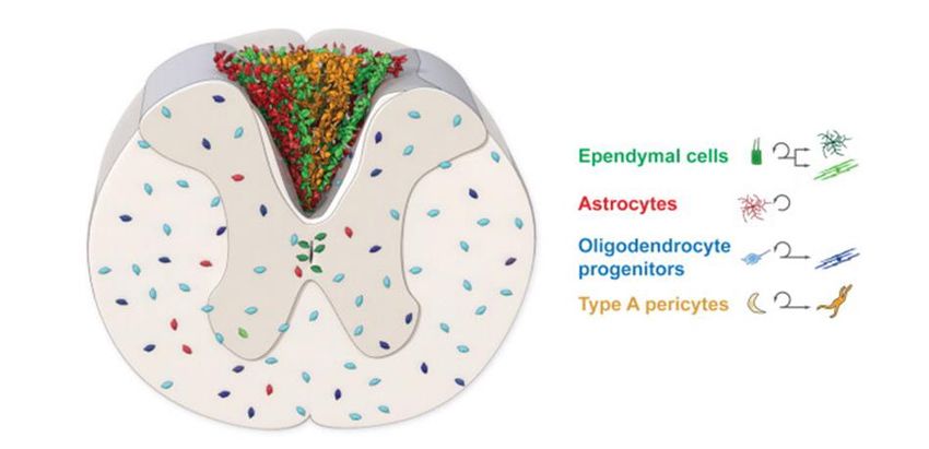
Dedicated to my beloved mother, father and my grandmother
                                             致我的家人
POPULAR SCIENCE SUMMARY
Spinal cord controls the communication between the brain and the body and coordinates the
motor movements and sensory functions. When an injury occurs to the spinal cord, the signal
bridge between the brain and the body is impaired, resulting in partly or fully functional loss
of the body below the lesion level. The main reason is that after spinal cord injury (SCI), the
neurons and other helper cells in the spinal cord die, and this cell loss is a continuous process
even long after the injury has occurred. There is no cure so far for SCI, but several new
treatments based on stem cell therapy have been studied in this thesis.

Stem cells are cells that can generate more identical cells to themselves, and can also become
other cell types as “self-reproductive transformers”. After SCI, spinal cord stem cells can
activate the self-production system to produce more stem cells, move to the lesion area, and
also become other helper cells to compensate for the cell loss. However, we found that the
capacity of spinal cord stem cells decreases dramatically during aging, and the ways they
contribute to self-recovery are different at young and mature age, suggesting that the current
standard treatments for SCI need to take age into more consideration. Moreover, the
regeneration capacity of stem cells and other helper cells at adult age has been largely
determined during embryonic development, but many current studies only focus on the adult
stage and overlook the link between development and regeneration. Here we found a gene,
called FoxJ1, which regulates the embryonic development of the spinal cord, and is essential
to maintain the stem cell potential in adulthood and after SCI. Targeting this gene could be a
novel approach to recruit more spinal cord stem cells and provide better SCI treatments.

Furthermore, a severe SCI patient received the transplantation of cells extracted from part of
his own olfactory system in the brain, which made him the first severe SCI patient walk again
with limited side effects. The surgery was successful, but the reasons were unknown, leading
to a difficulty in achieving the same effect on other SCI patients. We studied the mechanisms
behind and found that this type of cells originally from the olfactory system cannot generate
other cells after being transplanted into the injured spinal cord, but they can stimulate the spinal
cord stem cells to generate more stem cells and give rise to a more substantial number of
beneficial helper cells. Moreover, it is widely believed that there are no newborn neurons in
the adult spinal cord, resulting in the permanent loss of neurons in case of SCI. However, our
study found that after this cell therapy, there are newborn neurons in the spinal cord after SCI.
We suggest that a further study on the molecules produced by this cell therapy will be of high
importance for developing new cell therapies for SCI.

Altogether, this thesis sheds some light on the understandings about how the spinal cord is
developed, and how this developmental process influences the stem cell potential at adulthood
and after SCI. This thesis provides some inspirations to target specific cell types and genes for
new therapeutic possibilities for SCI.
作为中枢神经系统的一部分,脊髓控制着运动和感觉,是大脑和躯体的沟通的桥梁。
如果脊髓发生损伤,大脑和身体之间的信号传输就会受到阻碍,导致在损伤位置以下
甚至全部躯体功能的丧失。这主要是由于损伤发生后,脊髓中的神经元和其他的辅助
细胞大量死亡,而且细胞死亡过程会一直持续到损伤发生后的很长一段时间。目前脊
髓损伤尚无法治愈,但本文研究了几种基于干细胞的新疗法。

干细胞是一种能自我更新,并可以分化成其他细胞类型的“变形金刚”。脊髓损伤后,
脊髓干细胞能够激活自我更新的能力,产生更多的干细胞,并且能迁移到病变区域,
分化成其他类型的细胞以补偿损伤造成的细胞死亡。然而,我们发现在机体衰老的过
程中,脊髓干细胞的这种自我更新和变化能力急剧下降。因此在年轻和年老时期,脊
髓的自我恢复的方式大不相同,提示当前脊髓损伤的治疗方案需要更进一步考虑年龄
的影响。此外,成年期干细胞和其他辅助细胞的再生能力在胚胎发育过程中已基本被
确定,但目前许多研究却仅关注成体期,忽略了胚胎发育和组织再生的关联。本论文
中,我们发现了一种叫 FoxJ1 的基因,它能调节脊髓的胚胎发育,并且在成年期,当
脊髓发生损伤后,对维持干细胞的潜能至关重要。靶向这个基因可能会带来新的基因
疗法,刺激并获得更多的脊髓干细胞,从而提供更好的脊髓损伤治疗的可能性。

此外,一名严重脊髓损伤患者接受了细胞移植手术,细胞来源于他自己的嗅觉系统。
这个手术使他成为第一个能重新获得行走能力的严重脊髓损伤患者,而且术后副作用
很小。但由于机理不明,这个手术的成功并不能使其他脊髓损伤的患者获得同样的疗
效。我们对这种疗法进行了研究,发现虽然这种类型的细胞在移植到受损的脊髓后并
不能产生其他细胞,但是它们可以刺激脊髓干细胞产生更多的干细胞并分化出大量有
益的辅助细胞。更有趣的是,人们普遍认为,成年脊髓中不能再出生出新的神经元,
因此脊髓损伤导致的神经元丧失是永久的。然而,我们的研究发现,在经过这种细胞
疗法之后,损伤的脊髓出现了新生的神经元。因此,我们建议进一步研究究竟是这种
细胞产生的何种分子起的作用,这对于促进脊髓损伤的新细胞疗法的发现非常重要。

总之,本文对研究脊髓发育以及脊髓发育过程如何影响成年和脊髓损伤后的干细胞潜
能提供了新的思路,并为脊髓损伤的细胞和基因疗法提供了一些新启示,从而为脊髓
损伤的治疗提供了新的可能性。
ABSTRACT
Spinal cord injury (SCI) is an incurable condition, which is mainly due to the highly limited
regenerative potential of the adult spinal cord. The discovery of ependymal cells as the source
of spinal cord stem cells raises hopes for the development of new therapies, while cell
transplantations for SCI also provide promising means for potential treatments. However,
curing SCI has been proven difficult as the potential of these progenitors and ependymal stem
cells is still understudied. Moreover, the development of the spinal cord is a key factor
influencing the regenerative potential of neural stem/progenitor stem cells, but the link between
the development of spinal cord progenitors and adult spinal cord regeneration has been largely
overlooked. By using different transgenic mouse lines and biomedical techniques, we studied
the neural progenitors and stem cells during spinal cord development, after SCI and after cell
transplantation in this thesis.

FoxJ1 is traditionally regarded as a transcription factor involved in ciliogenesis and a specific
marker for ependymal cells. In Paper I, however, we discovered that FoxJ1 is transiently
expressed in neuronal and glial progenitors, which will further give rise to subsets of
interneurons, two subsets of astrocytes and all ependymal cells. FoxJ1 is required for the
maintenance of stemness of the progenitors during development and the stem cell potential
during adulthood. After SCI, FoxJ1 is required for the normal stem cell potential, proliferation
and migration of ependymal cells to promote regeneration.

After the early developmental stage, in Paper II, we observed that the stem cell potential is
fully confined to ependymal cells from P10 in mice, and the potential of self-renewal and
oligodendrocytic differentiation decreases over time. Juvenile ependymal cells have higher
stem cell potential after SCI than adult ones, but their contribution to the glial scar formation
in vivo is lesion size- and age-dependent. We found that the resident astrocytes and stromal
derived pericytes show higher regenerative potential at the juvenile stage, and ependymal cells
serve as a backup regeneration candidate after SCI.

Clinically in the adult spinal cord, the transplantation of bulbar olfactory ensheathing cell
(bOEC) has shown significant functional recovery in SCI patients, but the mechanisms are not
elucidated. In Paper III, we found that after SCI, bOEC transplantation increases the
proliferation and self-renewal potential of ependymal cells. The transplantation of bOECs
promotes higher astrocytic differentiation of ependymal cells but reduces the axonal growth
inhibitors after SCI. The microenvironment of the injured spinal cord is enriched after bOEC
transplantation in terms of less axonal growth inhibitor, a higher level of neurotrophic factors
and better neuronal survival. Unexpectedly, we found newborn neurons after SCI with bOEC
transplantation, challenging the current central stream theory that there is no neurogenesis after
SCI.

Altogether, this thesis provides new insights into the potential of ependymal cells and
progenitors during development, regeneration after SCI and after transplantation for SCI
treatment, and hopefully can contribute to new therapeutic approaches.
LIST OF SCIENTIFIC PAPERS
 I. Li, X.*, Floriddia, E. M.*, Toskas, K., Chalfouh, C., Honore, A., Aumont, A.,
    Vallières, N., Lacroix, S., Fernandes, K.J.L., Guérout, N., & Barnabé-Heider,
    F. (2018). FoxJ1 regulates spinal cord development and is required for the
    maintenance of spinal cord stem cell potential. Experimental cell research,
    368(1), 84-100.

 II. Li, X.*, Floriddia, E. M.*, Toskas, K., Fernandes, K. J., Guérout, N., &
     Barnabé-Heider, F. (2016). Regenerative potential of ependymal cells for
     spinal cord injuries over time. EBioMedicine, 13, 55-65.

III. Li, X.*†, Honore, A.*, Habib N.*, Chalfouh, C., Delarue, Q., Giovanni M.D.,
     Vaudry, D., Marie, J.P., Barnabé-Heider F., Regev, A.†, Guérout, N.† (2018).
     The transplantation of olfactory ensheathing cells promotes endogenous stem
     and progenitor cell reactivity and induces neurogenesis after spinal cord
     injury. Manuscript

     (* Co-first authors; † Co-correspondence)

    Papers not included in this thesis:
IV. Guérout, N., Li, X., & Barnabé-Heider, F. (2014). Cell fate control in the
    developing central nervous system. Experimental cell research, 321(1), 77-
    83.

V. Wicher, G., Wallenquist, U., Lei, Y., Enoksson, M., Li, X., Fuchs, B.,
   Hamdeh, S.A., Marklund, N., Hillered, L., Nilsson, G., & Forsberg-Nilsson,
   K. (2017). Interleukin-33 promotes recruitment of microglia/macrophages in
   response to traumatic brain injury. Journal of neurotrauma, 34(22), 3173-
   3182.
CONTENTS
1 Introduction ..................................................................................................................... 1
1.1 The development of spinal cord ............................................................................ 2
1.1.1 Neurogenesis during spinal cord development ........................................ 2
1.1.2 Gliogenesis during spinal cord development ........................................... 4
1.1.3 The development of spinal cord stem cells .............................................. 6
1.2 Spinal cord stem/progenitor cells at juvenile stage and adulthood ...................... 7
1.3 Spinal cord injury .................................................................................................. 9
1.3.1 Scar formation and the response of neural stem/progenitor cells ............ 9
1.3.2 The regenerative potential of spinal cord cells after SCI over time ......11
1.4 Regenerative approaches .....................................................................................13
1.4.1 Modulating endogenous stem/progenitor cells ......................................13
1.4.2 Cell transplantation therapies ..................................................................15
2 Aims of the thesis ..........................................................................................................21
3 Materials and Methods ..................................................................................................22
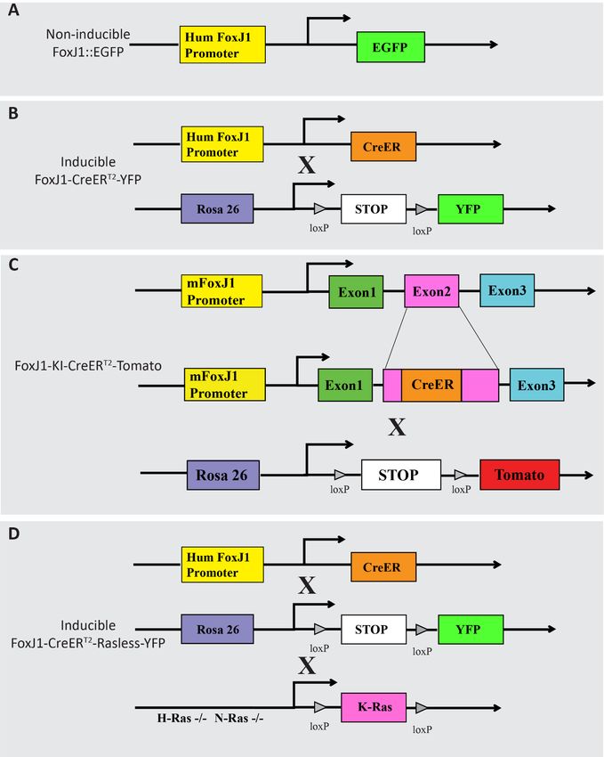
3.1 Animals ................................................................................................................22
3.2 Surgical procedure and postoperative care .........................................................25
3.2.1 Spinal cord injury ....................................................................................25
3.2.2 Transplantation of olfactory ensheathing cells .......................................25
3.3 Tissue preparation and sectioning .......................................................................26
3.4 In situ hybridization (ISH) ..................................................................................26
3.5 Cell culture...........................................................................................................27
3.5.1 Neural stem cell culture and neurosphere assay.....................................27
3.5.2 Differentiation assay ...............................................................................27
3.5.3 Bulbar olfactory ensheathing cell culture ...............................................27
3.6 Immunohistochemistry and Immunocytochemistry ...........................................28
3.7 Image acquisition and tissue analysis .................................................................28
3.8 qPCR ....................................................................................................................29
3.9 DroNc-Seq single nucleus sequencing ...............................................................29
3.10 Statistical analysis................................................................................................29
4 Results & Discussion ....................................................................................................32
4.1 FoxJ1 is required for spinal cord development and the maintenance of
spinal cord stem cell potential .............................................................................32
4.2 The regenerative potential of ependymal cells for SCI over time .....................34
4.3 The transplantation of bOECs promotes the regenerative potential of
endogenous progenitors after SCI .......................................................................36
5 Conclusions ...................................................................................................................40
6 Future perspectives ........................................................................................................41
6.1 FoxJ1 regulates spinal cord development and stem cell potential after SCI .....41
6.2 Juvenile and adult ependymal cells have different regenerative
mechanisms .........................................................................................................426.3 bOEC transplantation effects on the regenerative potential of endogenous
stem/progenitor cells ........................................................................................... 43
7 Acknowledgements ....................................................................................................... 44
8 References ..................................................................................................................... 49LIST OF ABBREVIATIONS BDNF Brain-Derived Neurotrophic Factor bHLH basic Helix–Loop–Helix BMP Bone Morphogenetic Proteins bOEC bulbar Olfactory Ensheathing Cell CNS Central Nervous System CNTF Ciliary Neurotrophic Factor CSF Cerebral-Spinal Fluid DFT Dorsal Funiculus Transection DH Dorsal Hemisection DMEM/F12 Dulbecco's Modified Eagle's/Ham's F12 medium E Embryonic day EGFP Enhanced Green Fluoresce Protein ESCs Embryonic Stem Cells FBS Fetal Bovine Serum FGF Fibroblast Growth Factors FoxJ1 Forkhead Box Protein J1 GDNF Glial cell-Derived Neurotrophic Factor GGF Glial Growth Factor IGF1 Insulin-like Growth factor 1 iPSCs induced-Pluripotent Stem Cells ISH In Situ Hybridization JAG2 Ligand Jagged2 MSCs Mesenchymal Stem Cells NECs Neural Epithelial Cells NGF Nerve Growth Factor NSCs Neural Stem Cells
OPCs   Oligodendrocyte Precursor Cells

P      Postnatal day

PCR    Polymerase Chain Reaction

RA     Retinoic Acid

RGCs   Radial Glial Cells

SCI    Spinal Cord Injury

Shh    Sonic HedgeHog

SVZ    SubVentricular Zone

VEGF   Vascular Endothelial Growth Factor

WT     WildType
1 INTRODUCTION
 The central nervous system (CNS) consists of the brain and the spinal cord. The spinal cord
 serves as a key bridge for the communication between the brain and the body. Notably, it
 serves both as the final output command for the elaboration of movements, such as walking
 or breathing, as well as the first input connection for most sensory modalities. When injury
 occurs to the spinal cord, the lesion partly or completely disrupts the signal transduction
 along the CNS, leading to permanent functional impairment in motor and sensory system
 below the lesion, and typically resulting in neuropathic pain, paralysis, and the dysfunction
 or disorder of the digestion system and reproductive system (Westgren and Levi, 1998).
 Spinal cord injury (SCI) is currently a chronic incurable disease without clear epidemiology
 statistics. However, it is estimated that 9.2 to 246 cases per million of the population a year
 varying from different regions (Li et al., 2016; Siddiqui et al., 2015). The majority of SCI
 patients are at the age between 10-40 years old at the time of injury (Siddiqui et al., 2015)
 and the life quality and expectancy of these young people are extensively and deeply
 influenced (Center, 2015; Li et al., 2016). Recently, new studies based on stem cell
 therapies have opened up new promising avenues. Indeed, by manipulating endogenous
 stem and progenitor cells or by cell transplantation after SCI, these cells can contribute to
 spinal cord regeneration, including tissue repair and functional recovery (Assinck et al.,
 2017; Gregoire et al., 2015).

 Stem cells are cell types with unlimited self-renewal potential and multipotent capacity.
 Self-renewal is the process that stem cells divide to generate more identical themselves,
 maintaining the stem cell pool throughout life, while differentiation is the process by which
 more specialized cells are formed from stem cells. Self-renewal and differentiation occur
 spontaneously during organ development from embryonic stages to adulthood and can be
 specifically mobilized under different pathological conditions. In general, the most potent
 stem cells are found in early embryonic stages, and the stem cell properties decrease during
 development and aging (Silva-Vargas et al., 2013). Other progenitor cells, on the other
 hand, do not share all the characteristics of stem cells, but can also contribute to
 regeneration after SCI (Sabelstrom et al., 2014; Stenudd et al., 2015).

 After embryonic development, except for tissues with fast turnover such as skin or intestine
 (Li and Clevers, 2010), adult stem cells maintain their population by self-renewal with
 relatively low division rate and rarely generate fast dividing progenitor cells. In the CNS,
 adult neural stem cells are mainly restricted in the ventricular-subventricular zone of the
 lateral ventricles, the subgranular zone of the hippocampus in the brain, and in the central
 canal of the spinal cord, with limited self-renewal and differentiation potential (Seaberg
 and van der Kooy, 2003). Although stem and progenitor cells give hope for the recovery
 after SCI or other neurodegenerative diseases, the regeneration capacity of the adult spinal
 cord is insufficient (Gregoire et al., 2015). Moreover, the capacity of adult stem and
 progenitor cells after injury is mostly influenced or determined during the developmental
 process, but the link and the mechanisms are still largely unknown. Therefore, to further

                                                                                              1
develop new SCI therapies, it is crucial to understand the cellular and molecular properties
of stem and progenitor cells during both embryonic development and adult regeneration
(Becker and Diez Del Corral, 2015; Gage and Temple, 2013).
1.1 THE DEVELOPMENT OF SPINAL CORD
1.1.1 Neurogenesis during spinal cord development
The spinal cord comprises the caudal region of the CNS and serves as a bridge, through
which the motor and sensory information travel between the brain and the periphery. At
the early stage of spinal cord development before embryonic day (E) 9 in mice, neural
epithelial cells (NECs) are under fast proliferation and by migrating away, they expand the
dorso-ventral and rostro-caudal dimensions of the spinal cord. Later on, NECs start to lose
their epithelial properties and acquire features associated with glial cells and termed radial
glial cells (RGCs) (Kriegstein and Alvarez-Buylla, 2009). RGCs are found to be neural
precursors throughout the whole CNS, and give rise to neurons and glial cells during neural
development, following a spatial-temporal manner (generating neurons first, then glial cells
later). This cell fate specification process is primarily determined at an early developmental
stage, neural patterning, the biological process by which cells in the developing CNS
acquire distinct identities according to their specific spatial positions. At the molecular
level, the key player of neural patterning is morphogens, soluble secreted signaling
molecules from roof-plate or floor-plate that follow gradient distribution. These
morphogens include fibroblast growth factors (FGF), retinoic acid (RA), Sonic hedgehog
(Shh), bone morphogenetic proteins (BMP) and Wnts, and govern the arrangements of
subtypes of neural precursors and direct their cell fate specification (Gurdon and Bourillot,
2001; Kiecker and Lumsden, 2012).
In the dorsal spinal cord, the regulation of neurogenesis is still not well understood, but
many experiments have suggested that two of the key players, BMP and Wnts, determine
the dorsal patterning. BMP and Wnt proteins are secreted by the roof plate and therefore
are expressed in a graded manner from the dorsal (high) to the ventral (low) developing
spinal cord. Progenitors respond to a specific concentration of these proteins by the
expression or repression of a specific combination of transcription factors (TFs). These
TFs, in turn, direct the determination of progenitor cells. Therefore, the dorso-ventral
gradient of morphogens directs progenitors to differentiate into six distinct position-
dependent domains of postmitotic dorsal neurons (dI1-6) between E10 and 12.5 in mice
(Figure 1) and (Alaynick et al., 2011; Liem et al., 1997). Genetic studies showed that
disturbed expression of BMP- or Wnt- associated factors, such as Lmx1a, Gdf7,
Wnt1/Wnt3a perturb neuronal cell fate determination in different domains in the dorsal
spinal cord during development (Chizhikov and Millen, 2004; Lee et al., 2000; Liem et al.,
1997; Millonig et al., 2000; Muroyama et al., 2002). However, the study on dorsal spinal
cord neurogenesis still faces many challenges. Genetic inactivation of individual TFs often
results in normal spinal cord development (Kiecker and Lumsden, 2012) , which is mainly
2because the roof plate secretes several types of Wnts and a large number of BMPs, therefore
single inactivation can be compensated by the other TFs.
Neurogenesis of ventral spinal cord is relatively better studied than the dorsal half as more
TFs have been investigated by the use of transgenic animals. Probably it is because that
this system serves primarily as motor control and motor functions and is easier to study for
phenotypes. The patterning is essentially regulated by Shh, a morphogen released from the
notochord and by the floor plate during ventral spinal cord development. During the
patterning of ventral spinal cord, Shh is expressed in a gradient manner, from ventral (high)
to dorsal (low) found to be declining when distributed from ventral to dorsal (Chamberlain
et al., 2008). As a consequence, segmental expression of the homeodomain and basic helix–
loop–helix (bHLH) TFs subsequently engage in cross-repressive interactions to refine
domain boundaries. For example, the expression of the homeobox protein IRX3 is
repressed by Olig2, and NKX2.2–PAX6 leads to reciprocal repression (Rowitch and
Kriegstein, 2010). Moreover, the concentration and exposure time of Shh regulates the
expression of distinct TFs during spinal cord development, such as Olig 2 and Nkx2.2,
which further direct the specification of subtypes of neurons in the developing ventral
spinal cord following a temporal-spatial manner (Dessaud et al., 2008; Dessaud et al., 2007;
Lupo et al., 2006). The Shh gradient activates or represses a number of TFs, giving rise to
spatially segregated progenitor domains: FP (Foxa2), p3 (Nkx 2.2), pMN (Olig2), p2
(Nkx6.1 and Irx3), p1 (Nkx6.2) and p0 (Dbx1), where the listed genes encode TFs that
specify each domain (Alaynick et al., 2011; Guerout et al., 2014). Progenitors in each
domain further differentiate into distinct neuronal subtypes, which are traditionally
categorized as motor neurons derived from the progenitors in the pMN domain, and V0,
V1, V2 and V3 interneurons derived from progenitors in p0–p3 domains respectively
(Figure 1). Importantly, each of these genetically or developmentally defined progenitor
types give rise to functionally-distinct neuronal types. Indeed, neurons originating from a
similar progenitor domain often share similar position, morphology, projection profile,
electrophysiological properties. In other words, the progenitor cell origin of a neuron has
become a good predictor of its functional properties and functional role in the mature circuit
(Alaynick et al., 2011; Kiehn, 2016). More recently, taking advantages of transgenic animal
models, studies in developmental biology have shown that each population of interneurons
(V0, V1 V2, and V3) can be even further divided into subgroups based on distinct gene
expression profiles (Bikoff et al., 2016; Lu et al., 2015). Even though there are overlaps of
makers for different subtypes of interneurons, subpopulations of interneurons can now be
defined by combinatorial expression of TFs. For instance, V0 interneurons express Pax2,
Pax6, and Evx1; V1 interneurons express Pax2, Evx1, Nurr1; and V2 interneurons express
Pax2 and Chx10 (Francius et al., 2013).
Nevertheless, the TFs involved in neurogenesis and neurodevelopment is under active
studies and characterizations. Forkhead Box protein J1 (FoxJ1), for instance, is a
transcription factor mostly involved in ciliogenesis and previously considered as a specific
marker for adult ependymal cells in the spinal cord. However, we unexpectedly found that
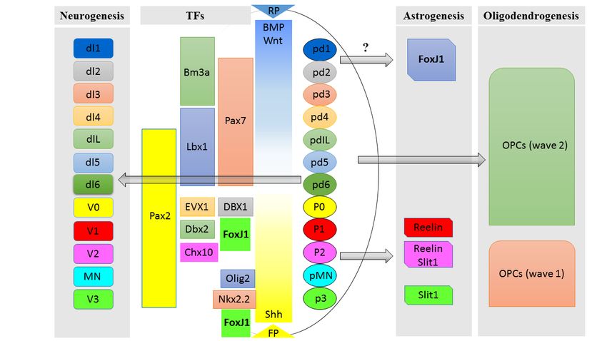
3FoxJ1 is also involved in spinal cord neurogenesis and development. We used FoxJ1 lineage tracing animal models and some of the combinations of TFs mentioned above to identify further the subtypes of neurons derived from FoxJ1 progenitor cells, and we found that FoxJ1 is transiently expressed by the progenitors of V1 and V2 interneurons during embryonic development, which will be further discussed in detail later (Figure1; Paper I/Li et al., 2018). 1.1.2 Gliogenesis during spinal cord development Gliogenesis in the developing CNS takes place after neurogenesis at the later stage of embryonic development after neurogenesis. Recent genetic studies have shown that similar to neurogenesis, most glial cells, including astrocytes, oligodendrocytes, and ependymal cells also differentiate in a spatial-temporal manner (Hochstim et al., 2008; Tsai et al., 2012; Xie et al., 2012). Astrocytes are the largest population of glial cells in the CNS, which guide the migration of developing axons and play an essential role in releasing signals for synapse formation and function (Barres, 2008; Powell and Geller, 1999; Sofroniew and Vinters, 2010). In the developing spinal cord, the dorso-ventral axis is segmented into distinct domains under the influence of morphogens during neural patterning as described above. After early embryonic neurogenesis, neural tube progenitor domains switch to a glial fate at around E12.5 in mice. In the ventral spinal cord, progenitors from p1, p2, and p3 domains, which give rise to neurons during neurogenesis at E10-E14 in mice, have been shown to be the precursors of three molecularly heterogeneous subpopulations of astrocytes, VA1-3 astrocytes during gliogenesis. These subtypes of astrocytes are specified by the gradients of BMP and Shh, as well as the combinatorial expression of transcription factors Pax6 and Nkx6.1 (Hochstim et al., 2008). After differentiation, VA1-3 astrocytes can be further characterized by their expression of the axon and the neuronal migration factors Slit1 and Reelin, and their migration to the lateral white matter of the spinal cord, mirroring the localization of the progenitors in the p1–3 domains (Hochstim et al., 2008). Furthermore, a segmental model of astrocyte specification and migration was proposed by a recent study. By using a number of transgenic mouse lines, progenitors from different domains expressing distinct transcription factors such as Pax3, Msx3, Dbx1, Olig2, Ngn3, and Nkx 2.2 along the dorso-ventral axis were fate mapped (Tsai et al., 2012). Astrocytes derived from each domain migrate radially, according to the dorso-ventral position of their neuroepithelial precursor. Postnatally, different subtypes of astrocytes have insufficient migratory potential in both intact and injured spinal cord. Therefore, astrocytes are regionally allocated into the spinal cord according to a segmental template with heterogeneous subtypes (Tsai et al., 2012). Besides, our study recently showed that FoxJ1 is transiently expressed by progenitors of specific subsets of astrocytes during embryonic spinal cord development. One subpopulation of FoxJ1 progenitor-derived astrocytes migrates to the lateral white matter, similarly to Reelin-Slit V1-V3 astrocytes. The other 4
FoxJ1 progenitor-derived astrocytes migrate to the dorsal funiculus, explicitly labeling a
subset of astrocytes with unreported origin from Pax3+ astrocytes (Paper I/Li et al., 2018).

Oligodendrocytes are myelin-forming cells in the CNS, differentiated from
oligodendrocyte precursor cells (OPCs). Studying the development of oligodendrocytes
and OPCs helps to understand the regulations of myelin production and the regeneration
capacity in CNS diseases and injuries. Indeed, during the progress of most of SCI, the
necrosis and apoptosis of oligodendrocytes, the failure of sufficient production of
oligodendrocytes from OPCs, and the continuous myelin degeneration are crucial factors
that result in pathology and functional loss. During development, RGCs in the ventricular
zone undergo asymmetric division, giving rise to one daughter cell retaining contact with
the ventricular and pial surface, and another daughter cell (OPC) migrating to the gray or
white matter (Pringle and Richardson, 1993). Even though the origin of OPCs in the spinal
cord is not very well known due to the limitation of genetic animal models, OPCs are found
to originate from several domains after neural patterning. In the ventral spinal cord, under
the effects of BMP and Shh, the transcription factor Olig2 is specifically expressed in the
pMN domain and is phosphorylated before and during neurogenesis, which results in the
differentiation of progenitors to motor neurons. However, after E12 in mice, Olig2
undergoes dephosphorylation and heterodimerize with neurogenin 2, contributing to the
cell fate change from neural to oligodendrocyte fate (Li et al., 2011). Furthermore, the
Notch ligand Jagged2 (JAG2), a Shh-regulated factor transiently expressed in motor neuron
progenitors (pMNs) leads to motor neuron to OPC cell fate switch (Rabadan et al., 2012).
Interestingly, the disruption of Shh signaling does not deplete all OPC production in the
dorsal spinal cord, suggesting that FGF or other factors may compensate for the loss of Shh
at least partly (Cai et al., 2005; Vallstedt et al., 2005). After the production of OPCs in the
ventral domain pMN, the second wave of OPC production starts in the dorsal spinal cord
at around E15. This process is determined by transcription factors including Pax7 and
Dbx1, potentially from dI3-dI5 domains (Fogarty et al., 2005; Vallstedt et al., 2005). OPCs
derived from dorsal and ventral spinal cord are respectively around 20% and 80% during
development (Tripathi et al., 2011). From E18 in mice, OPCs start generating mature
myelinating oligodendrocytes, reaching a peak at 2-4 weeks after birth and continue until
eight months postnatally (Figure 1) and (Rivers et al., 2008).

Ependymal cells are multi-ciliated glial cells lining the ventricular system, forming a
continuous cellular barrier between the cerebral-spinal fluid (CSF) and the adjacent
parenchyma. The cilia beating of ependymal cells is responsible for CSF flow, brain
homeostasis, and normal functions of adult neural stem cells in the spinal cord (Guerout et
al., 2014). Ependymal cells have been categorized into three subtypes by their
morphological and molecular features (tanycytes, cuboidal and radial ependymal cells) but
the developmental process and the functional differences of ependymal cells are largely
unknown (Meletis et al., 2008; Robins et al., 2013; Spassky et al., 2005). Factors such as
homeobox transcription factor Six3, Numb/Numlike proteins, and SNX27 were suggested
to be crucial for ependymal layer formation in the brain (Kuo et al., 2006; Lavado and

                                                                                             5
Oliver, 2011; Wang et al., 2016b). During spinal cord development, Shh is essential for ependymal cell differentiation, and deletion of Shh leads to the absence of ependymal layer (Yu et al., 2013). Moreover, FoxJ1 is found to be expressed explicitly in adult spinal cord ependymal cells, and have been used as a spinal cord neural stem cell marker (Barnabe- Heider et al., 2010; Meletis et al., 2008). In the developing spinal cord, the first FoxJ1+ ependymal cells appear earliest at E15.5 in mice by expressing FoxJ1 around the ventricular/ central canal, and quickly expand their population and almost entirely occupy the central canal by birth. Around postnatal day 10 (P10), FoxJ1 is restricted to the ependymal cells which have fully formed the ependymal layer around the central canal, displaying the unique stem cell source of the spinal cord (Li et al., 2016) /(Paper II). Figure 1. The developing spinal cord is regulated by morphogens and various transcription factors. During the development of the spinal cord, morphogens are secreted from the floor plate and roof plate, and distributed in a gradient manner, regulating the expression of a number of transcription factors (middle panel). During neurogenesis, specific transcription factors define domains of progenitors along the dorsal-ventral axis and further give rise to distinct neuronal subtypes (left panel), while other transcription factors in specific domains regulate the development of astrocytes and oligodendrocyte precursor cells during gliogenesis (right panel). The transcription factor FoxJ1 (in bold font with green background), is mainly expressed in the p1- p2 domains and the floor plate during early development and found to be continuously occupying the central canal from E15 onwards, which further regulates the progenitors to give rise to V1 and V2 interneurons, a subset of astrocytes and ependymal cells. 1.1.3 The development of spinal cord stem cells Embryonic stem cells from neuroepithelium of ectodermal origin are the most potent stem cells during early neurodevelopment. However, the stem cell potential starts to decline when neuroepithelial cells begin to specialize into different RGCs around E9-10 in mice. In the brain, adult neural stem/progenitor cells share a common progenitor pool with different types of embryonic progenitors that give rise to cells of different regions of the 6
brain, including the cortex, striatum, and septum during development. However, this
      lineage relationship among all the progenitors is lost before E15.5 in mice. Indeed, adult
      neural stem cells were allocated and specified early in embryonic development, and the
      lineages between adult and embryonic stem cells diverge during mid-embryonic
      development (Fuentealba et al., 2015). Moreover, the majority of adult cells with
      stem/progenitor cell potential in mice are derived between E13.5 and E15.5 in the brain
      and between E15.5 and P0 in the spinal cord, and remain mostly quiescent in the adulthood
      (Bond et al., 2015; Fuentealba et al., 2015; Li et al., 2016). Even though these stem cells
      remain mostly quiescent during postnatal development and adulthood, they can be
      reactivated under certain conditions, such as injury or stroke (Bond et al., 2015; Fuentealba
      et al., 2015). In the spinal cord, it is still not clear how the adult neural stem cells obtain
      their potential during development, but during neurogenesis and gliogenesis, RGCs and
      other intermediate precursor cells maintain their stem cell potential to both generate their
      population and give rise to specific differentiated progeny (Temple, 2001). However, the
      frequency of stem cells is diluted by the production of restricted progenitors and
      differentiated cells, dropping to 10% at E12 and only 1% at P1 in the spinal cord (Kalyani
      et al., 1997; Kalyani et al., 1998). The spinal cord stem cell potential is gradually restricted
      from progenitors to ependymal cells since they begin to appear as differentiated cells at
      E15. After the first wave of ependymal cells, they quickly take over the full stem cell
      population within three weeks in mice till around P10 (Li et al., 2016/ Paper II). Using
      genetic labeling of ependymal cells by FoxJ1-CreER transgenic mice and cell culture
      studies have shown that the first ependymal cells have stem cell potential as early as E15
      while almost 100% stem cells are ependymal cell-derived at P10. This data suggest that
      embryonic spinal cord stem and progenitor cells from other sources rapidly lose their stem
      cell potential during development, and the only source of stem cells after birth is confined
      to ependymal cells around the central canal (Li et al., 2016/ Paper II).

1.2     SPINAL CORD STEM/PROGENITOR CELLS AT JUVENILE STAGE AND
        ADULTHOOD
      After embryonic and early postnatal development of the spinal cord, neurogenesis and
      gliogenesis have been mostly completed. Neurons, astrocytes, oligodendrocytes have been
      distributed in gray matter or white matter while ependymal cells are restricted around the
      central canal. Spinal cord astrocytes share similarities with brain astrocytes, including
      neurotrophic factor secretion, contribution to metabolism and maintenance of homeostasis
      (Sofroniew and Vinters, 2010). Besides, spinal cord astrocytes are involved in blood spinal
      cord barrier formation and prevent the influx of antigens or molecule at a certain size to the
      parenchyma of the spinal cord (Bartanusz et al., 2011). However, unlike subtypes of
      astrocytes (or GFAP-expressing glial cells) that can give rise to neurons during embryonic
      development and are involved in adult neurogenesis in the brain, spinal cord astrocytes do
      not have stem cell potential in both healthy and injured spinal cord (Barnabe-Heider et al.,
      2010). In the intact adult spinal cord, in vivo astrocytes are under low proliferation rate and
      do not give rise to non-astrocytic progeny. Moreover, fate-mapping experiments showed

                                                                                                    7
that astrocytes form a small number of primary neurospheres but are not capable of being passaged, suggesting that spinal cord astrocytes do not have real stem cell potential (Barnabe-Heider et al., 2010; Buffo et al., 2008). It is still largely unknown how aging affects astrocytes in the spinal cord over time. Previous data about the effects of aging in the brain showed that astrocytes in the neurogenic zone, the subventricular zone (SVZ), are less dividing over time, as well as undergo morphology changes and express a lower level of GFAP, DCX and S100 (Capilla-Gonzalez et al., 2014). Oligodendrocytes produce myelin, an insulating sheath required for the saltatory conduction of electrical impulses along axons in the CNS. They are derived from oligodendrocyte progenitor cells (OPCs) under physiological condition (Ffrench-Constant and Raff, 1986; Raff et al., 1983a; Raff et al., 1983b). OPCs are distributed in both gray and white matter throughout the CNS. During the differentiation of OPCs into oligodendrocytes, multiple processes are extended, and axons are ensheathed, and then the oligodendrocytes proceed to generate the concentric layers of the modified cell membrane that compose myelin (Sherman and Brophy, 2005). Even though OPCs are proliferating lifelong to generate new oligodendrocytes in mice, it was shown that NG2+ and Olig2+ OPCs are not multipotent and restricted to the oligodendrocyte lineage (Barnabe-Heider et al., 2010; Kang et al., 2010). During aging, the myelination of CNS becomes less efficient, due to both the impairment of OPC recruitment and the differentiation of OPCs to oligodendrocytes, which was found to be associated with the changes of environmental signals and epigenetic changes within OPCs (Hinks and Franklin, 2000; Shen et al., 2008; Sim et al., 2002; Tang et al., 2000). Ependymal cells are lining the central canal in adulthood with a low proliferation rate in vivo. During postnatal development, ependymal cells are contributing to the elongation of the central canal and the flow of cerebrospinal fluid (Alfaro-Cervello et al., 2012; Sabourin et al., 2009). Similarly, ependymal cells at adulthood undergo symmetric division, but all their progeny remain in the ependyma, suggesting that their primary role in adults is ependymal cell maintenance (Barnabe-Heider et al., 2010). Despite the low proliferation rate in the intact spinal cord, it was shown that ependymal cells are capable of generating neurospheres, with the self-renewal potential over many passages (Barnabe-Heider et al., 2010). Moreover, even though ependymal cells do not give rise to other cell types in the intact spinal cord, the neurospheres derived from ependymal cells in vitro display multipotent phenotype by differentiating into astrocytes, oligodendrocytes, and neurons (Barnabe-Heider et al., 2010; Li et al., 2018; Li et al., 2016; Meletis et al., 2008). In the aging adult brain, ependymal cells were found to undergo morphology changes, including a higher number of intermediate filaments in the cytoplasm, presenting larger lipid droplets, and concentrated cilia in limited areas (Capilla-Gonzalez et al., 2014). In the spinal cord, the stem cell potential of ependymal cells declines quickly over time from juvenile to adulthood in vitro, by displaying less self-renewal capacity and a dramatic decrease of oligodendrocytic differentiation potential (Barnabe-Heider et al., 2010; Li et al., 2018; Li et al., 2016). 8
The spinal cord is generally considered as a non-neurogenic region in the CNS under
      normal condition or after injury (Barnabe-Heider et al., 2010; Horner et al., 2000; Shechter
      et al., 2007). However, a recent study showed that taking advantage of the new technique
      Div-sequencing which better preserves the integrity of neurons and their RNA content,
      19% of the analyzed proliferative cells are immature neurons which can be still detected up
      to 25 days or longer after survival in the spinal cord. Further analysis showed that these
      rare newborn neurons in the intact spinal cord are due to GABAergic neurogenesis (Habib
      et al., 2016). Although the function and the origin of these newborn neurons and the spinal
      cord neurogenesis capacity between young and aging animals are still not clear, this study
      opens a new way to study adult neurogenesis and provides more insights on the overlooked
      neural stem cell potential of the spinal cord.

1.3     SPINAL CORD INJURY

1.3.1 Scar formation and the response of neural stem/progenitor cells
      Spinal cord injury (SCI) leads to massive cell death and a loss of motor function and
      sensory inputs below the injury level. CNS intrinsic neural cells (neurons, astrocytes,
      OPCs, ependymal cells), CNS intrinsic non-neural cells (microglia, pericytes, etc.) and
      immune cells from the blood are affected by and respond to an injury differently (Burda
      and Sofroniew, 2014). Upon SCI, in the acute phase, the immediate damage of spinal cord
      leads to the loss of neurons and glial cells, including oligodendrocytes that should be
      remyelinating the surviving neurons. As a consequence, the loss of neurons after SCI leads
      to the dysfunction of the motor and sensory system (Ahuja et al., 2017). During the sub-
      acute phase, inflammation and ongoing necrotic neurons and glial cells lead to the
      production and secretion of free radicals, chemokines, and cytokines that activate
      microglial cells (Ahuja et al., 2017). Together with other inflammatory cells such as
      activated macrophages, polymorphonuclear cells, and lymphocytes, microglia infiltrate the
      lesion site for further inflammatory response and contribute to ongoing apoptosis of
      neurons and oligodendrocytes (Ahuja et al., 2017; Hausmann, 2003). After the sub-acute
      phase of SCI, Wallerian degeneration, an ordered process of axonal death is undertaken
      (Alizadeh et al., 2015; Waller, 1850). After the injury on the nerve fiber, the axonal skeleton
      disintegrates, and the axonal membrane breaks apart, which leads to axonal degeneration
      and the release of myelin debris. Myelin debris has found to be the source of axonal
      regeneration inhibitors, such as neurite outgrowth inhibitor A (Nogo‑A), oligodendrocyte-
      myelin, glycoprotein (OMgp) and myelin-associated glycoprotein (MAG) (Filbin, 2003).
      However, due to the slow infiltration of immune cells and the low capacity of
      oligodendrocytes to clear myelin, there is accumulation of myelin in the CNS tissue after
      injury. This accumulated myelin in turn leads to the apoptosis of oligodendrocytes and
      further contributes to the failure of remyelination and regeneration (Ahuja et al., 2017;
      Barres et al., 1993; Franklin and Ffrench-Constant, 2008; Vargas and Barres, 2007).

      Following SCI, a scar is formed, which is composed of a fibrotic component core and a
      glial scar surrounding it. The fibrotic lesion core is formed by perivascular cells, including

                                                                                                   9
type-A pericytes (Goritz et al., 2011; Klapka and Muller, 2006; Soderblom et al., 2013), while the glial scar is generated by astrocytes derived from resident astrocytes and ependymal cells-derived astrocytes (Barnabe-Heider et al., 2010; Meletis et al., 2008). The fibrotic scar is traditionally believed to be nonfunctional or have adverse long-term effects and impacts on axon regeneration (Burda and Sofroniew, 2014; Zukor et al., 2013). However, new studies have shown that the absence of type-A pericytes or ependymal cells- derived astrocytes prevents the sealing of wound and therefore worsen SCI outcomes (Goritz et al., 2011; Sabelstrom et al., 2013). Moreover, even though it was widely believed that reactive astrocytes migrate to the injury site and contribute to glial scar formation, recent fate-mapping and live imaging studies showed that astrocytes do not migrate to the lesion site after SCI and most of the astrocytes are not migratory after brain injury (Bardehle et al., 2013; Tsai et al., 2012). Instead, astrocytes are under massive proliferation and upregulate the GFAP expression to further participate in the glial scar formation within their previous locations (Barnabe-Heider et al., 2010; Tsai et al., 2012). The function of the glial scar is under debate regarding its role in attenuating axonal regrowth, but it has been shown that the glial scar also serves as a barrier to block inflammation and immune cell infiltration to the lesion and prevent further tissue damage (Faulkner et al., 2004; Herrmann et al., 2008; Okada et al., 2006; Sabelstrom et al., 2013; Wanner et al., 2013). By impairing the formation of glial scar by transgenic mouse models to block cell cycle of astrocytes- producing ependymal cells or to kill proliferative astrocytes, previous studies have shown that the significant loss of glial scar leads to worsened secondary injury to the tissue and the loss of axonal regeneration (Anderson et al., 2016; Sabelstrom et al., 2013). Neural stem/progenitor cells, including ependymal cells, astrocytes, and OPCs are highly proliferative after SCI and display different cellular responses (Figure 2). Ependymal cells were found to be the only cell type displaying multi-potency after SCI (Barnabe-Heider et al., 2010; Meletis et al., 2008). Ependymal cells rarely divide around the central canal and can only generate a small number of neurospheres in cell culture under physiological condition. After SCI, ependymal cells divide rapidly and differentiate into other glial cells in vivo, and these differentiated progenies leave the central canal and migrate to the lesion side. Half of the ependymal cells differentiate into astrocytes in the glial scar and also produce a few oligodendrocytes that myelinate axons (Barnabe-Heider et al., 2010). Moreover, ependymal cells from an injured spinal cord generate a significantly higher number of neurospheres in vitro and can be passaged on with higher self-renewal capacity than those from the non-injured condition. Differentiation assays showed that ependymal cell-derived neurospheres have higher potential to generate oligodendrocytes and neurons in vitro after SCI. This observation suggests that the stem cell potential of these neural stem cells is activated by SCI, regarding self-renewal and differentiation (Li et al., 2016/ Paper II). Resident astrocytes dramatically increase their proliferation after SCI and upregulate the expression of GFAP near the lesion site and surrounding area, forming part of the glial scar that prevents inflammation and the infiltration of immune cells. However, astrocytes can only give rise to more astrocytes and do not acquire stem cell potential after SCI, neither 10
in vivo nor in vitro (Barnabe-Heider et al., 2010). Unlike the astrocyte-derived astrocytes
in the glial scar that forms a barrier to inhibit secondary tissue damage, ependymal cell-
derived astrocytes contribute to glial scar formation and reside in the center of the scar,
surrounding fibroblast-like stromal cells that make up the core of the forming scar
(Barnabe-Heider et al., 2010; Camand et al., 2004; Goritz et al., 2011; Krikorian et al.,
1981; Sabelstrom et al., 2013; Shearer and Fawcett, 2001; Windle and Chambers, 1950). It
was shown that blocking the cell cycle of ependymal cells leads to enlarged cyst at the
lesions, resulting in injuries growing deeper and an increased loss of neurons, partly due to
the decreased production of neurotrophic factors by ependymal cells. These findings
suggest that ependymal cells act as a scaffold to reinforce the injured spinal cord by
restricting secondary enlargement of lesions (Sabelstrom et al., 2013). OPCs are the most
proliferative glial cells in the intact spinal cord, and their proliferation is even more
increased after SCI. OPCs and ependymal cells generate remyelinating oligodendrocytes
after SCI, but OPCs cannot self-renew in vitro and do not give rise to other cell types in
vivo. However, recent studies using transgenic lineage tracing mouse models showed that
OPCs are the major cell type that contributes to new myelin formation following SCI (Hesp
et al., 2015). OPCs also produce the majority of myelinating Schwann cells in the injured
spinal cord, while the contribution to myelination by invading peripheral myelinating
Schwann cells after SCI is very limited (Assinck et al., 2017).
Figure 2. The response of endogenous cells after SCI (dorsal funiculus lesion). After a dorsal funiculus
lesion, the glial scar is formed with cells produced by ependymal cells (green), astrocytes (red) and pericytes
(yellow). After SCI, the ependymal cells self-renew, differentiate, and the progeny migrate to the lesion site.
Astrocytes (red), OPCs (blue) and pericytes (yellow) also self-renew and give rise to astrocytes,
oligodendrocytes and stromal cells, respectively. Adapted from Sabelstrom et al., 2014.
1.3.2 The regenerative potential of spinal cord cells after SCI over time
Even though neural cells intensively respond to SCI with their specific potential, the self-
recovery potential of the spinal cord after injury seems to decline during aging (DeVivo et
al., 1990; Furlan et al., 2010; Wyndaele and Wyndaele, 2006). It was reported that the
potential of neural stem cells decreases during aging and in aged animals after traumatic
brain injury, which is due to the quiescence but not loss of neural stem cells, and also due
to the change of their differentiation potential (Bouab et al., 2011; Conover and Shook,
2011; Sun et al., 2005). Indeed, astrocytes and ependymal cells significantly change their
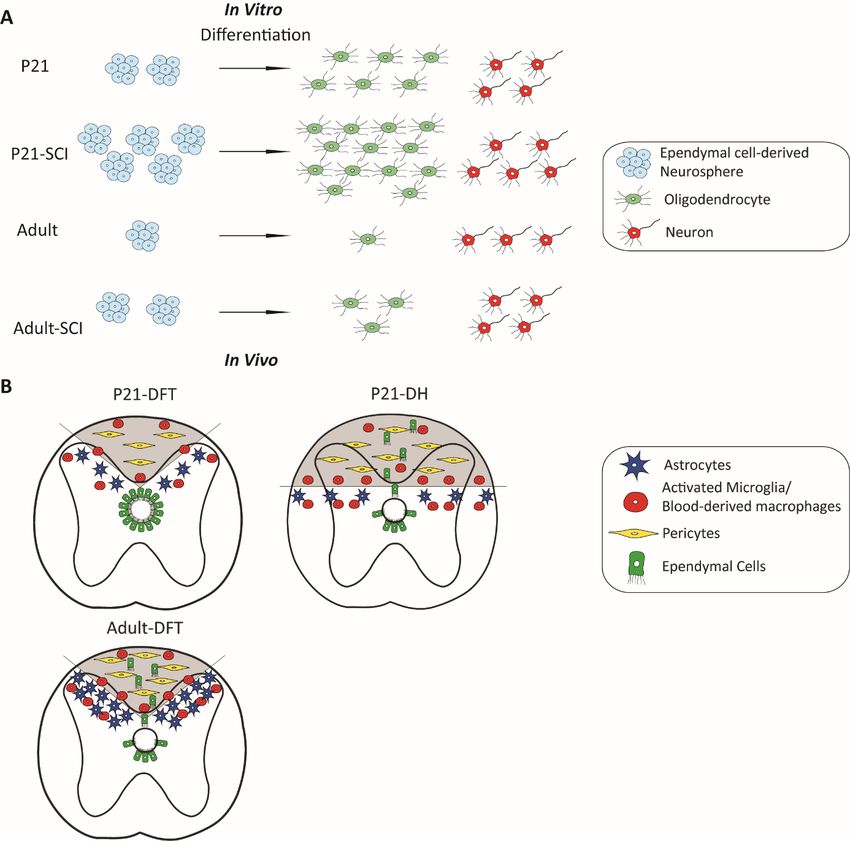
11morphology and molecular signatures during brain aging, and their proliferation capacity decreases over time (Capilla-Gonzalez et al., 2014). After SCI, there is a higher expression of astroglial and inflammatory markers near and at the lesion site in the aged animals, and the mammalian CNS undergoes an age-dependent decline in axonal regeneration and becomes less regenerative (Geoffroy et al., 2016). Besides the changes in the microenvironment, the intrinsic regenerative potential of stem/progenitor cells is also age-dependent. At a very young age (P10), all the spinal cord stem cell potential is entirely confined to ependymal cells, but the self-renewal capacity significantly decreases at the juvenile stage and even more in adulthood in mice. The differentiation capacity of ependymal-derived neurospheres to oligodendrocytes also declines over time (Li et al., 2016). Similarly, the recruitment and differentiation capability of OPCs in aged animals largely decrease (Hinks and Franklin, 2000; Kuhlmann et al., 2008; Sim et al., 2002). These findings suggest that the self-recovery capacity decreases with increased aging is partly due to the decreased endogenous remyelination potential by ependymal cells and APCs (Figure 3; Paper II). After the insult to a spinal cord, astrocytes, ependymal cells and type A pericytes rapidly proliferate and contribute to the scar formation in adult (Barnabe-Heider et al., 2010; Goritz et al., 2011; Sabelstrom et al., 2013). At a younger age, these cell types are more proliferative and pro-regenerative compared to adult cells. After SCI, the stem cell potential of juvenile ependymal cells is more activated than those in adults in vitro, by showing greater self-renewal capacity and more oligodendrocyte differentiation. We found that similar lesions are sealed more efficiently in young animals compared to adults. Juvenile lesions have a smaller fibrotic core and smaller glial scar, as well as less infiltration of microglia and blood-derived macrophages (Figure 3). Interestingly, even though ependymal cells have a higher stem cell potential in juvenile mice, they are acting as a backup reserve and contribute to scar formation only when the lesion is larger and need more cells to be sealed (Li et al., 2016/ Paper II). Ependymal cells are required for restricting enlargement of the lesion in adult (Sabelstrom et al., 2013), but the same transgenic mouse model in which the cell cycle of ependymal cells is blocked showed a different phenotype in juvenile animals. The juvenile spinal cord is sealed so efficiently by other cells that blocking ependymal cell proliferation does not lead to deeper lesions nor the formation of a cyst at the lesion site. The area of glial scar and the lesion core are also smaller even when ependymal cells are not able to proliferate. This analysis is in line with the clinical studies that have shown that juveniles have better functional recovery than adults in human (DeVivo et al., 1990; Furlan et al., 2010; Wyndaele and Wyndaele, 2006). 12
Figure 3. Ependymal cells and resident cells respond to SCI. A) Under both physiological and injured
      conditions, ependymal cells show different self-renewal and differentiation potential in juvenile mice
      compared to adult mice in vitro. B) The response of ependymal cells (green) is age- and lesion size-dependent.
      Response to SCI of other endogenous cell types is illustrated as follows: resident astrocytes (purple), immune
      cells (red) and pericytes (yellow). Adapted from Li et al., 2016.

1.4     REGENERATIVE APPROACHES

1.4.1 Modulating endogenous stem/progenitor cells
      Endogenous glial cells play important roles in maintaining homeostasis of the spinal cord.
      Even though OPCs are the most proliferative cell type throughout the entire parenchyma
      of the spinal cord, astrocytes and ependymal cells are also under massive proliferation after
      SCI and contribute to the self-repair of the spinal cord.

                                                                                                                 13
You can also read