Genetic analysis of the catalytic activity of Integrin-linked kinase (ILK) in vivo - Dissertation zur Erlangung des Doktorgrades der Fakultät für ...
←
→
Page content transcription
If your browser does not render page correctly, please read the page content below
Dissertation zur Erlangung des Doktorgrades
        der Fakultät für Chemie und Pharmazie
      der Ludwig-Maximilians-Universität München

Genetic analysis of the catalytic activity of
   Integrin-linked kinase (ILK) in vivo

                   Anika Lange
                        aus
                 Osterburg/Altmark
                       2010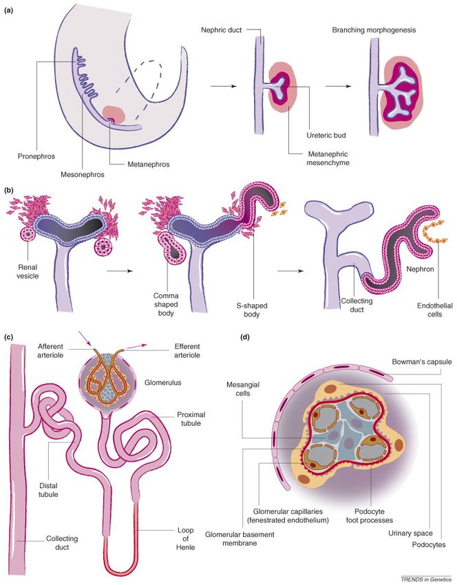
Erklärung

Diese Dissertation wurde im Sinne von § 13 Abs. 3 der Promotionsordnung vom
29. Januar 1998 von Herrn Prof. Dr. Reinhard Fässler betreut.

Ehrenwörtliche Versicherung

Diese Dissertation wurde selbstständig, ohne unerlaubte Hilfe erarbeitet.

München, am

                                                    ----------------------------
                                                          Anika Lange

Dissertation eingereicht am: 22.01.2010

1. Gutachter                       Prof. Dr. Reinhard Fässler

2. Gutachter                       Prof. Dr. Christian Wahl-Schott

Mündliche Prüfung am: 04.03.2010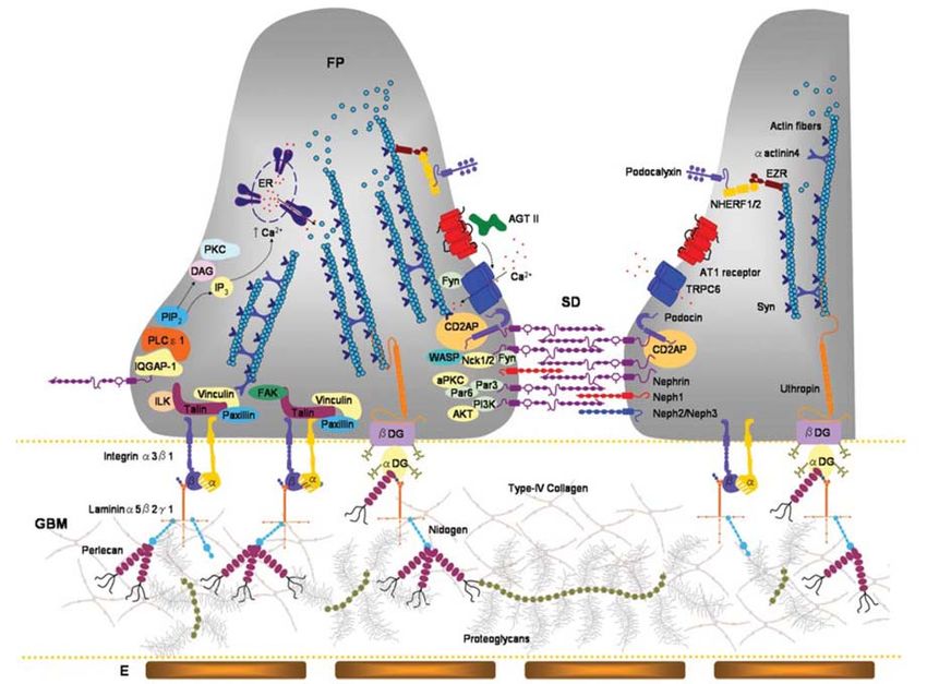
Für meine lieben Eltern
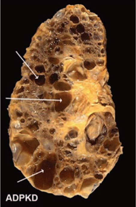
Table of Contents
Table of Contents
Table of Contents ............................................................................................................... I
List of Publications ......................................................................................................... III
Abbreviations .................................................................................................................. IV
Summary.......................................................................................................................... VI
Introduction....................................................................................................................... 1
1. The integrin receptor family .................................................................................... 1
1.1 Structure of integrins.......................................................................................... 1
1.2 Integrins and their ligands ................................................................................. 4
1.3 Bidirectional regulation of integrin signaling................................................... 8
1.3.1 Inside-out signaling...................................................................................... 8
1.3.2 Outside-in signalling .................................................................................. 12
1.4 Assembly of integrin-dependent adhesion structures.................................... 16
1.4.1 The integrin-actin connection ................................................................... 18
1.5 The ILK/PINCH/parvin (IPP) complex.......................................................... 20
1.5.1 The molecular composition of the IPP complex...................................... 21
1.5.2 The biological functions of the IPP complex ........................................... 22
1.5.3 The putative kinase activity of ILK.......................................................... 24
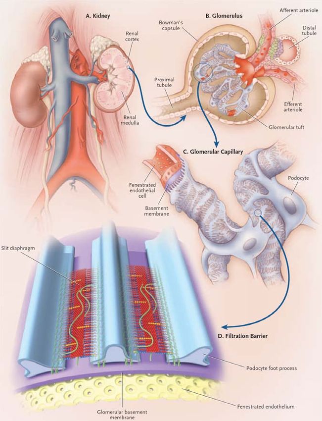
2. Kidney physiology................................................................................................... 26
2.1 Kidney development in mice ............................................................................ 30
2.1.1 Kidney morphogenesis – pronephros, mesonephros and metanephros 30
2.1.2 Mesenchymal signals initiate kidney development ................................. 33
2.1.3 Ureteric bud outgrowth and branching ................................................... 36
2.1.4 Tubulogenesis – MET ................................................................................ 42
2.1.5 Role of stroma in kidney development..................................................... 44
2.2 Renal abnormalities in humans ....................................................................... 46
2.2.1 The glomerulus and proteinuria............................................................... 49
2.2.2 Cystic kidney diseases................................................................................ 54
2.3 Integrins and kidney ......................................................................................... 56
2.3.1 ECM and its receptors in mammalian nephrogenesis............................ 56
I
Table of Contents

           2.3.2 Role of integrins and their binding partners in the development of the
           collecting system.................................................................................................. 59
           2.3.3 Role of integrins and their binding partners in the development and
           function of the glomerulus.................................................................................. 60
Aim of the Thesis............................................................................................................. 63
Short Summaries of Publications .................................................................................. 64
   Publication I: Local call: from integrins to actin assembly .................................... 64
   Publication II: How ILK and kindlins cooperate to orchestrate integrin signaling
   ....................................................................................................................................... 65
   Publication III: Integrin-linked kinase is an adaptor with essential functions
   during mouse development ........................................................................................ 66
   Publication IV: The ILK/PINCH/parvin complex: the kinase is dead, long live the
   pseudokinase! .............................................................................................................. 67
   Publication V: Integrin-mediated signals control microtubule dynamics required
   for plasma membrane targeting of caveolae ............................................................ 68
   Publication VI: Bacteria hijack integrin-linked kinase to stabilize focal adhesions
   and block cell detachment .......................................................................................... 68
References........................................................................................................................ 69
Acknowledgements ......................................................................................................... 83
Curriculum Vitae ............................................................................................................ 84
Supplements..................................................................................................................... 85

                                                                                                                                          II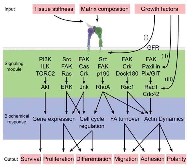
List of Publications

List of Publications

This thesis is based on the following publications, which are referred to in the text by
their Roman numerals (I-VI):

I.     Wiesner S, Lange A and Fässler R. Local call: from integrins to actin
       assembly. Trends in Cell Biology 2006, 16

II.    Böttcher RT, Lange A and Fässler R. How ILK and kindlins cooperate to
       orchestrate integrin signaling. Current Opinion in Cell Biology 2009, 21(5):
       670-5

III.   Lange A, Wickström SA, Jakobson M, Zent R, Sainio K and Fässler R. Integrin-
       linked kinase is an adaptor with essential functions during mouse
       development. Nature 2009, 461: 1002-1006 .

IV.    Wickström SA, Lange A, Montanez E and Reinhard Fässler. The
       ILK/PINCH/parvin complex: the kinase is dead, long live the pseudokinase!
       EMBO J 2010, 29(2): 281-91.

The following publications were not the focus of my project but I contributed to them:

V.     Wickström SA, Lange A, Hess MW, Krüger M, Pfaller K, Mann M, Bloch W,
       Huber LA and Fässler R. Integrin-mediated signals control microtubule
       dynamics required for plasma membrane targeting of caveolae. Submitted
       manuscript

VI.    Kim M, Ogawa M, Fujita Y, Yoshkawa Y, Nagai T, Koyama T, Nagai S, Lange
       A, Fässler R and Sasakawa C. Bacteria hijack integrin-linked kinase to
       stabilize focal adhesions and block cell detachment. Nature 2009, 459: 578-
       583.

                                                                                         III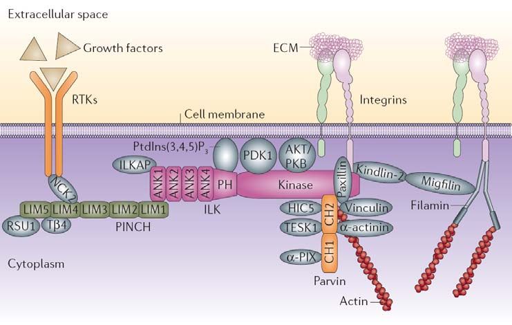
Abbreviations

Abbreviations
ADAM       a disintegrin and metalloproteinase
ADP        adenosine diphosphate
Akt        RAC-alpha serine/threonine protein kinase
APC        adenomatous polyposis coli
Arp2/3     actin-related protein 2/3 complex
ATP        adenosine triphosphate
BMP        bone morphogenetic protein
Cdc42      cell division cycle 42
CH         calponin homology
Cre        cyclization recombinase
DAG        diacylglycerol
E          embryonic day
ECM        extracellular matrix
EGF        epidermal growth factor
ERK        extracellular signal-regulated kinase
FA         focal adhesion
F-actin    filamentous actin
FAK        focal adhesion kinase
FERM       4.1, ezrin, radixin, moesin
FGF        fibroblast growth factor
FGFR       fibroblast growth factor receptor
FP         foot process
GAP        GTPase activating protein
GBM        glomerular basement membrane
GDI        GDP dissociation inhibitor
GDNF       glial-cell-derived neurotrophic factor
GDP        guanosine diphosphate
GEF        guanine nucleotide exchange factor
GFR        growth factor receptor
GPCR       G protein coupled receptor
Gsk-3β     glycogen synthase kinase-3β
HGF        hepatocyte growth factor
ICAM       intercellular adhesion molecule
ILK        integrin-linked kinase
IP3        inositol triphosphate
IPP        ILK/PINCH/parvin
JNK        c-jun N-terminal kinase
MAPK       mitogen-activated protein kinase
MET        mesenchymal-to-epithelial transition
MIDAS      metal-ion-dependent adhesion site
MLC        myosin light chain
MM         metanephric mesenchyme
MMP        matrix metalloproteinase
Nck2       noncatalytic region of tyrosine kinase, adaptor protein 2
NS         nephrotic syndrome

                                                                                 IV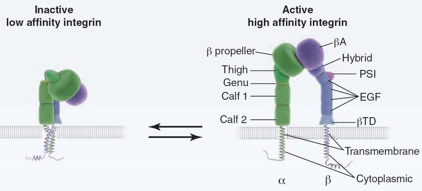
Abbreviations

PCP     planar cell polarity
PH      pleckstrin homology
PI3K    phosphatidylinositol 3-kinase
PINCH   particularly interesting new Cys-His rich protein
PKC     protein kinase C
PKD     polycystic kidney disease
PLC     phospholipase C
PTB     phosphotyrosine binding
Ret     rearranged during transformation
RGD     arginine, glycine, aspartate
RhoA    Ras homology gene family, member A
ROCK    Rho-associated protein kinase
SD      slit diaphragm
SH2     src homology 2
Src     Rous sarcoma oncogene
TGFβ    transforming growth factor β
UB      ureteric bud
VCAM    vascular cell-adhesion molecule
WD      wolffian duct
Wnt     wingless-type MMTV integration site family member
WT1     wilms tumour 1

                                                                       V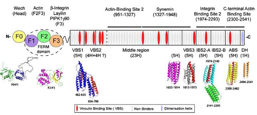
Summary

Summary

Integrins are α/β heterodimeric transmembrane receptors that can bind to the extracellular
matrix (ECM) and thereby mediate cell adhesion. Integrins are also signal transducing
receptors. Since integrins are devoid of enzymatic activity and actin binding capability
they depend on the recruitment of adaptor, scaffolding and signalling proteins in order to
couple to the cytoskeleton and to propagate signals that regulate a variety of cellular
processes including proliferation, survival, differentiation, and migration. One central
constituent of this multiprotein complex is integrin-linked kinase (ILK), which is
recruited to β1 and β3 integrin-mediated adhesion complexes. ILK consists of an ankyrin,
pleckstrin homology (PH) and kinase domain and has been shown to directly bind the
cytoplasmic parts of these integrin subunits. At the beginning of my thesis it has also
been thought that ILK possesses kinase activity towards substrates such as Akt and Gsk-
3β. However, the kinase activity of ILK and its physiological relevance was controversial
due to several reasons: (1) ILK lacks well conserved residues that are important for
kinase activity, (2) deletion of ILK in several cell types such as keratinocytes, fibroblasts
or chondrocytes failed to diminsh or ablate phosphorylation of key substrates such as Akt
or Gsk-3β, (3) genetic studies in C.elegans and D.melanogaster failed to confirm a kinase
function of ILK in vivo.
Due to this controversy, it was important to determine whether the catalytic activity of
ILK exists in a mammalian model system. To this end I established knock-in mouse
strains with point mutations in ILK that were reported to convert ILK kinase activity in
vitro either into a constitutive-dead or constitutive-active kinase. Surprisingly, knock-in
mice carrying mutations in the putative PH domain (R211A, required for kinase activity)
or in the autophosphorylation site (S343A; required for kinase activity, or S343D; renders
ILK constitutive-active) do not show any obvious phenotype or changes in Akt or Gsk-3β
phosphorylation or actin organization. In contrast, mice carrying point mutations in the
potential ATP-binding site (K220A/M; required for catalytic activity) die shortly after
birth due to kidney agenesis. This phenotype does not result from impaired kinase
activity, as the mutations did not alter the phosphorylation levels of reported ILK
substrates in vivo. In addition, no evidence of kinase activity was detected in vitro.

                                                                                          VI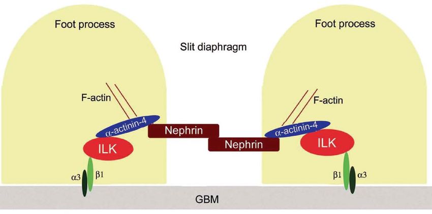
Summary

However, these mutations selectively impair the interaction of ILK and its key binding
partner α-parvin. In line with this, similar kidney defects occur also in α-parvin null mice.
On the basis of this study, it is now clear that the proposed kinase activity does not exist
and thus is neither playing a role in mammalian development or adult life nor the
integrin-actin linkage. However, the adaptor function of ILK is crucial for mammalian
kidney development. Thus, my studies allow the conclusion that the kinase domain of
ILK has been mutated in evolution to provide a novel and essential, non catalytic
function to integrins.

                                                                                         VII
Introduction
Introduction
1. The integrin receptor family
Integrins are α/β heterodimeric type I transmembrane glycoproteins that mediate the
attachment of cells to the extracellular matrix (ECM) and to other cells. The receptor
family is evolutionarily highly conserved. Integrins have been identified in mammals,
chicken and zebrafish, as well as in lower eukaryotes, including sponges, the nematode
C.elegans (two α and one β subunit, generating two integrins) and the fruitfly D.
melanogaster (five α and one β subunit, generating five integrins) (Johnson et al., 2009).
The name “integrin” was coined in the 1980ties to denote the importance of these
receptors for maintaining the integrity of the cytoskeletal-ECM linkage (Hynes, 2004;
Tamkun et al., 1986). The majority of integrins links the ECM to the actin cytoskeleton,
while integrin α6β4 connects to the intermediate filament system.
Integrins present a bi-directional conduit for mechanochemical information across the
cell membrane, as they provide a major mechanism to connect the inside of the cell with
the extracellular environment. Their activation triggers a large variety of signal
transduction events that affect cell behaviors such as adhesion, proliferation, survival or
apoptosis, shape, polarity, motility, haptotaxis, gene expression, and differentiation,
mostly through modulating the cytoskeleton (Takada et al., 2007). Notably, integrins do
not possess enzymatic or actin-binding activity of their own.
1.1 Structure of integrins
Integrin α- and β-subunits have large extracellular domains (approximately 800 amino
acids) that contribute to ligand binding, single transmembrane domains (approximately
20 amino acids) and short cytoplasmic tails (13 to 70 amino acids, with the exception of
β4, which has a length of approximately 1.000 amino acids). While there is a striking
sequence homology among the different β-subunit cytoplasmic tails, the α-subunit tails
are highly divergent apart from a conserved GFFKR motif next to the transmembrane
region, which is important for association with the β-tail (Takada et al., 2007). The
extracellular domain of the heterodimer consists of a ligand-binding head domain
1Introduction
standing on two long legs (Figure 1). The extracellular domains can also associate
laterally with other proteins such as tetraspanins, growth factor receptors, matricellular
proteins, and matrix proteases or their receptors at the cell surface (Miranti and Brugge,
2002).
Figure 1: Integrin architecture and schematic representation of integrin activation
(Moser et al., 2009b).
Specific contacts between the ectodomain, the transmembrane domain and cytoplasmic
domains keep the integrin in its bent, inactive conformation. During integrin activation
the integrin legs, transmembrane domain and cytoplasmic domains separate, resulting in
an extended integrin conformation. βTD, β tail domain; EGF, epidermal growth factor
domain; PSI, plexin/semaphorin/integrin domain
The ectodomain of an integrin α-subunit is composed of a seven-bladed β-propeller,
which is connected to a thigh, a calf-1 and a calf-2 domain, together forming the leg
structure that supports the integrin head. The last blades of the β-propeller contain EF-
hand domains that bind Ca2+ -ions and thereby affect ligand binding (Humphries et al.,
2003). Nine of the integrin α chains contain an additional I domain (Table 1), referred to
as a von Willebrand factor A domain that almost always constitutes the ligand binding
site. Ligand binding occurs via a coordinating Mg2+ -ion in the so-called metal-ion-
dependent-adhesion site (MIDAS) motif (Barczyk et al., 2009; Moser et al., 2009b).
The β-subunit is composed of a βA (I) domain, which is analogous to the I domain of the
α-subunit, a hybrid domain, a PSI (plexin/semaphorin/integrin) domain, four epidermal
2Introduction
growth factor (EGF) domains and a membrane proximal β tail domain (βTD). In integrins
lacking an I domain, ligands bind to a crevice between the αβ-subunit interface, where
they interact with a metal ion-occupied MIDAS within the β-subunit and the propeller
domain of the α-subunit (Figure 1) (Moser et al., 2009b).
Most integrin β cytoplasmic domains (also called integrin tails) contain one or two motifs
that are part of a canonical recognition sequence for phosphotyrosine-binding (PTB)
domains: a membrane proximal NPxY motif and a membrane distal NxxY motif. These
protein sequences are present in a wide variety of signaling and cytoskeletal proteins, and
play a crucial role in integrin activation.
Integrins can exist in low-, intermediate-, and high-affinity states. Based on structural and
electron microscopy studies, it is believed that integrins are in a low-affinity state when
their extracellular domains are bent and in a high-affinity state when the extracellular
domains are extended (Figure 1). Two models have been proposed for the affinity
change. In both, the inactive integrin is in a bent conformation, with the headpiece facing
the membrane. In the “deadbolt model” the bent conformation is maintained in the
activated integrin, but piston-like movements of the transmembrane regions cause sliding
of the extracellular stalks of the α- and β-subunits. As a consequence, this sliding disrupts
the interaction between the headpiece and the β stalk just beyond the membrane (Arnaout
et al., 2005). In the “switchblade model”, dissociation of the α and β cytoplasmic and
transmembrane regions leads to dislocation of an EGF-like repeat in the β stalk, which
causes the head region to extend outwards in a switchblade-like movement (Arnaout et
al., 2005). Support for “the switchblade model” came from the crystal structure of
integrin αVβ3, which revealed a bent conformation of the head region associated with
low-affinity for the ligand. It was therefore proposed that the bent form does not bind to a
ligand and that activated integrins have an extended form (switchblade model) (Takada et
al., 2007). However, the bent conformation does not always seem to be inactive,
especially in the context of binding to small ligands (Askari et al., 2009).
The transmembrane domains have a key role in integrin activation. The transmembrane
domains of inactive integrins are engaged in a coiled-coil interaction between canonical
GxxxG dimerization motifs in each subunit. Separation of integrin transmembrane
domains is a requirement for integrins to adopt the high-affinity state (Moser et al.,
3Introduction
2009b). The role of integrin cytoplasmic tails in regulating integrin affinity has been
extensively studied in the rapidly activated leukocyte-specific β2 and platelet-specific
aIIbβ3 integrins. High integrin affinity has been shown to be associated with separation
of the α and β cytoplasmic tails. The separation is most likely achieved by binding of
cytoplasmic proteins to the β-tail and will be discussed later.
1.2 Integrins and their ligands
Integrin heterodimers are composed of non-covalently associated α- and β-subunits
(Hynes, 2002). In vertebrates, the family is comprised of 18 α-subunits and 8 β-subunits
that can assemble into 24 different heterodimers. Some subunits appear only in a single
heterodimer, whereas 12 integrins contain the β1 subunit and five contain αV (Figure 2
and Table 1). Integrins assemble in the endoplasmatic reticulum and are transported to
the plasma membrane as heterodimers.
Figure 2: Representation of the integrin receptor family grouped by their main
ligand specificity (Barczyk et al., 2009).
Vertebrates possess 18 α- and 8 β-subunits, which give rise to 24 heterodimers that can
be assembled into four distinct ligand binding classes.
4Introduction
Table 1: Characteristics of human integrin α-subunits (Barczyk et al., 2009).
CI, cleavage; αI, αI domain
Integrin α chain Cl αI Prototypic ligands Additional
characteristics ligands
α1β1 (CD49a, 1151 aa - X collagens (collagen IV > collagen I; semaphorin 7A,
VLA1) collagen IX) laminin
α2β1 (CD49b, 1181 aa - X collagens (collagen I > collagen IV; E-cadherin,
VLA2) collagen IX) endorepellin,
laminin
α3β1 (CD49c, 1051 aa, splice X - laminins (LN-511 > LN-332 > LN- -
VLA3) variants α3A and 211)
α3B
α4β1 (CD49d, 1038 aa - - fibronectin, VCAM-1 -
VLA4)
α5β1 (CD49c, 1049 aa X - fibronectin (RGD) endostatin
VLA5)
α6β1 (CD49f, 1073 aa, splice X - laminins (LN-511 > LN-332 > LN- -
VLA6) variants α6A and 211 > LN-411)
α6B
α7β1 1137 aa, splice X - α7X1β1: laminins (LN-511 > LN- -
variants X1, X2, 211 > LN-411 > LN-111)
α7A and α7B α7X2β1: laminins (LN-111 > LN-
211 > LN-511)
α8β1 1025 aa X - fibronectin, vitronectin, -
nephronectin (RGD)
α9β1 1035 aa - - tenascin-C, VEGF-C, VEGF-D osteopontin
α10β1 1167 aa - X collagens (collagen IV > collagen -
VI > collagen II; collagen IX)
α11β1 1188 aa, serted - X collagens (collagen I > collagen IV; -
domain 21 aa collagen IX)
αLβ2 (CD11a) 1170 aa - X ICAM-1, -2, -3, -5 -
αMβ2 (CD11b) 1153 aa - X iC3b, fibrinogen + more -
αXβ2 (CD11c) 1163 aa - X iC3b, fibrinogen + more -
αDβ2 (CD11d) 1162 aa - X ICAM-3, VCAM-1 -
αIIbβ3 (CD41, 1039 aa X - fibrinogen, fibronectin (RGD) -
GpIIb)
α6β4 X - laminins (LN-332, LN-511) -
αVβ1 (CD51) 1048 aa X - fibronectin, vitronectin (RGD) -
αVβ3 X - vitronectin, fibrinogen, fibronectin tumstatin
(RGD)
αVβ5 X - vitronectin (RGD) -
αVβ6 X - fibronectin, TGF-β-LAP (RGD) -
αVβ8 X - vitronectin, TGF-β-LAP (RGD) -
αEβ7 (CD103, 1178 aa X X E-cadherin -
HML-1)
α4β7 - - MadCAM-1, fibronectin, VCAM-1 -
5Introduction
Table 2: Characteristics of human integrin β-subunits (Barczyk et al., 2009).
Integrin β chain Charateristics Notes
β1 (CD29, GpIIa) 798 aa, splice variants β1A-D, Splice variants β1B and β1C not
present in mice, minor variants
with unclear function
β2 (CD18) 769 aa -
β3 (CD61, GpIIIa) 788 aa, splice variants β3A, β3B β3A major form
and β3C
β4 (CD104, TSP-180) 1875 aa, splice variants β4A-E β4A and β4B major forms,
similar function
β5 799 aa, splice variants β5A, β5B Both splice variants have similar
functions
β6 788 aa -
β7 (LPAM-1, βP) 798 aa -
β8 769 aa -
Integrins are grouped into subgroups based on ligand-binding properties (Figure 2) or
based on their subunit composition (Table 1 and 2). It is possible to cluster integrin-
ligand combinations into four main classes, reflecting the structural basis of the
molecular interaction: collagen-binding integrins, laminin-binding integrins and RGD
(arginine, glycine, aspartate)-binding integrins (Humphries et al., 2006). Leukocyte-
specific integrins establish cell-cell contacts with endothelial cells by interacting with
cellular counter-receptors such as intercellular adhesion molecules (ICAMs) and vascular
cell adhesion molecules (VCAMs) (Ley et al., 2007).
All five αV containing integrins and two β1 integrins (α5β1, α8β1) share the ability to
recognize ligands containing an RGD tripeptide active site. RGD constitutes the minimal
integrin recognition sequence in ligands such as fibronectin, vitronectin, tenascin,
osteopontin and fibrinogen. α4β1, α4β7, α9β1, the four members of the β2 subfamiliy and
αEβ7 recognize related sequences in their ligands. α4β1, α4β7 and α9β1 bind to an acidic
motif, termed “LDV” (lysine, aspartate, valin) that is functionally related to RGD.
Fibronectin contains the prototype LDV ligand in its type III connecting segment region;
other ligands (such as VCAM-1 and MadCAM-1) employ related sequences. Four α-
subunits containing an αA-domain (α1, α2, α10, α11) combine with β1 and form a
distinct laminin/collagen-binding subfamily. The fourth group includes three β1 integrins
6Introduction
(α3β1, α6β1 and α7β1) and α6β4 that are highly selective laminin receptors which do not
have an αA-domain (Humphries et al., 2006).
Other integrin ligands include milk fat globule-EGF factor 8 (MFGE8) and complement
factor iC3b, which facilitate phagocytosis of apoptotic cells and pathogens, respectively;
the latency-associated peptide of transforming growth factor β (TGFβ), which regulates
the activation of TGFβ and some of the a disintegrin and metalloproteinase (ADAM)
family members and matrix metalloproteinase-2 (MMP-2) which participate in ECM
remodeling during cell adhesion and migration (Table 1) (Legate and Fassler, 2009).
Integrin ligands can also be generated by proteolysis. Endostatin (derived from collagen
XVIII), endorepellin (derived from perlecan) and tumstatin (derived from collagen α3)
are the best-known examples (Bix and Iozzo, 2005; Wickstrom et al., 2005). In addition,
integrins can bind snake toxins, and certain viruses and bacteria. Some of these
interactions occur outside the regular ligand-binding sites in the integrins and display
distinct binding characteristics compared with the binding of physiological ligands
(Barczyk et al., 2009).
Alternative splicing of mRNA leads to additional complexity of the integrin family.
Variants of both the extracellular and cytoplasmic domains have been reported.
Alternative extracellular domains may account for different ligand-binding affinities or
variations in the state of activation, while variants of the cytoplasmic domain may
modulate integrin activity, cytoskeletal associations and/or signaling events (van der Flier
and Sonnenberg, 2001). The best studied are the four cytoplasmic variants of the β1
subunit: β1A, β1B, β1C and β1D. Integrin subunit β1A is present in all tissues except
mature cardiac and skeletal muscle, which instead express the highly homologous β1D
variant. However, β1A and β1D are not functionally equivalent in embryonic
development. The replacement of β1A by β1D results in embryonic lethality in mice,
whereas replacement of β1D with β1A does not lead to severe abnormalities in striated
muscles in vivo (Baudoin et al., 1998).
Each of the 24 vertebrate integrins appears to have a specific, non-redundant function.
This is in part apparent from their ligand specificities but is best proven by the distinct
phenotypes of the knockout mice of single integrin subunits. The phenotypes reflect the
different functions of individual integrins and range from a complete block in
7Introduction
preimplantation development (β1) through major developmental defects (α4, α5, αV, β8)
to perinatal lethality (α3, α6, α8, αV, β4, β8) and defects in leukocyte function (αL, αM,
αE, β2, β7), inflammation (β6), homeostasis (αIIb, β3, α2), bone remodeling (β3), and
angiogenesis (α1, β3) as well as others (Bouvard et al., 2001; Hynes, 2002). Generation
of tissue-specific integrin knockout mice that gave rise to severe phenotypes during
embryonic development provided further insight to the specific function of a given
integrin.
1.3 Bidirectional regulation of integrin signaling
Integrin receptors possess the rare ability to signal bidirectionally across the plasma
membrane. Ligand binding triggers signal transduction into the cell through the
recruitment of adaptor and signaling proteins that establish a connection to actin and
various signal transduction pathways (“outside-in” signaling) which is important for
example in cell spreading and cell migration. Conversely, intracellular non-integrin
mediated signals can induce changes in integrin conformation and activation that alters its
ligand-binding affinity in a process termed “inside-out” signaling or integrin activation.
Integrin clustering follows the engagement of integrins triggered by the naturally
multivalent nature of ECM, and it promotes the localized concentration of intracellular
signaling molecules.
1.3.1 Inside-out signaling
Inside-out activation of integrins relys on the binding of cytoplasmic ligands to specific
sites within the integrin tails. This induces conformational changes that are transmitted to
the extracellular ligand-binding domains via the transmembrane domains and stalk
regions. The β-integrin interacting proteins talin and kindlin have emerged as important
regulators of integrin activation.
Talin orthologs have been identified in all multicellular eukaryotes studied; vertebrates
encode two talin isoforms, talin1 and talin2, whereas lower eukaryotes encode only a
single talin isoform corresponding to talin1 (Moser et al., 2009b). Talins are ~ 270 kD
8Introduction
proteins, composed of an N-terminal 47 kD head domain and a C-terminal flexible rod
domain and form dimers. The talin head consists of a FERM (4.1, ezrin, radixin, moesin)
domain composed of three subdomains (F1-F3) and an F0 subdomain with no homology
to known domains (Figure 3). The F3 subdomain resembles a phosphotyrosine-binding
(PTB) domain and binds to the conserved membrane proximal NPxY motifs in β integrin
tails (Calderwood et al., 2002), but also to phosphatidylinositol 4-phosphate 5-kinase γ
(PIPK1γ), and the hyaluronan receptor layilin. The talin rod domain is made of a series of
domains composed of helical bundles that contain multiple binding sites for the F-actin-
binding protein vinculin, and a second integrin binding site (Figure 3) (Critchley and
Gingras, 2008). As talin binds to integrin cytoplasmic domains, vinculin and actin
filaments it is suggested that it forms an important link between the cytoskeleton and the
ECM (Critchley and Gingras, 2008).
Figure 3: Schematic representation of the domain structure of talin (Roberts and
Critchley, 2009).
Talin binds to β integrin cytoplasmic tails and regulates integrin activation in cooperation
with kindlin proteins. In addition, talin can bind to actin and vinculin and thereby links
the ECM to the actin cytoskeleton.
Talin's role in integrin activation was originally demonstrated by the ability of its F3
domain to activate αIIbβ3 integrin when expressed in CHO cells (Calderwood et al.,
2002; Calderwood et al., 1999). Knock-out and knock-down experiments subsequently
9Introduction
reinforced the notion that talin is a key component of integrin affinity regulation
(Nieswandt et al., 2007; Petrich et al., 2007; Tadokoro et al., 2003).
Inside-out signaling has been extensively studied in circulating blood cells such as
platelets, leukocytes and lymphocytes which present integrin αIIbβ3 or β2 integrins on
their surfaces. In these cells, integrin activation has to be rapid and tightly controlled as
constitutively active αIIbβ3 for example, would trigger pathologic thrombus formation
causing strokes, myocardial infarction and other embolic events. This tight regulation is
achieved by controlling the binding of integrin-activating proteins such as talin. NMR
studies revealed an intramolecular autoinhibitory interaction between the talin C-terminus
and its PTB domain that masks the integrin binding pocket. The precise mechanism that
disrupts this autoinhibition requires further investigation, although the small GTPase
Rap1 and its binding partner RIAM have been shown to play a key role in talin activation
(Figure 4). In addition, PIPK1γ90 and PIP2 have also been implicated in this process
(Roberts and Critchley, 2009). The talin-integrin interaction might additionally be
controlled on the receptor level through phosphorylation of the β integrin tail. The
tyrosine residue within the β1 and β3 integrin NPxY motif can be phosphorylated by Src
family kinases. When this tyrosine is mutated to phenylalanine it reverses the integrin-
dependent spreading and migration defects in v-Src-transformed cells, suggesting that
phosphorylation might inhibit talin binding (Moser et al., 2009b; Sakai et al., 2001).
10Introduction
Figure 4: Agonist stimulation triggers integrin activation (Han et al., 2006).
Agonist receptors like G-protein coupled receptors or tyrosine-kinase coupled receptors
induce the formation of diacylglycerol (DAG) and increased Ca2+ leading to the
activation and/or translocation of active GTP bound Rap1 to the plasma membrane via
activation of protein kinase C (PKC) or a Rap guanine nucleotide exchange factor (Rap-
GEF). At the plasma membrane, activated Rap interacts with RIAM, leading to the
recruitment of talin to form the integrin activation complex.
Mutations and truncations of the β3 integrin tail C-terminal to the the talin binding site
decrease integrin affinity for ligands (Ma et al., 2006), which raised the possibility that
additional factors besides talin also affect the affinity states of integrins. Indeed, recent
work showed that talin alone is not sufficient for integrin activation and that kindlin
proteins are as important in mediating this function (Montanez et al., 2008; Moser et al.,
2009a; Moser et al., 2008; Ussar et al., 2008). Kindlins are essential components of the
integrin adhesion complex, which bind to the membrane distal NxxY motif of β1, β2 and
β3 integrins (Böttcher et al., 2009; Meves et al., 2009). As kindlins and talin bind distinct
regions of the β integrin tail, they most likely cooperate to regulate integrin affinity.
Although kindlins are not sufficient to shift integrins to a high-affinity state, they
facilitate talin function. Conversely, talin depends on kindlins to promote integrin affinity
because talin-head overexpression failed to induce activation of αIIbβ3 in kindlin-
depleted CHO cells. Thus, kindlins require talin, and talin alone is not sufficient to
increase integrin affinity (Moser et al., 2009b).
For more information about the kindlin protein family, the reader is referred to the second
publication (review) presented in this PhD thesis.
11Introduction
1.3.2 Outside-in signalling
Extracellular ligand binding to integrins is believed to induce conformational changes
within the integrin heterodimer, including the outward swing of the hybrid domain,
separation of the α and β “leg” domains and separation of the transmembrane and
cytoplasmic domains. This leads to the interaction of the cytoplasmic tails with
intracellular signaling molecules (“switchblade” model) (Arnaout et al., 2005). Once
integrins are activated and clustered they are able to transmit the vast array of
intracellular changes collectively referred to as “outside-in” signaling. Interestingly, up to
now, around 156 components have been described that build up the so called “integrin
adhesome”. Theoretically, interactions among those components could give rise to 690
interactions (Zaidel-Bar et al., 2007).
Integrin activation leads to downstream signaling events that can be divided into three
temporal stages (Figure 5) (Legate et al., 2009). The immediate effects of integrin
activation are the up-regulation of lipid kinase activity that increases the local
concentration of the phosphoinositide second messengers PtdIns-4, 5-P2 and PtdIns-3, 4,
5-P3 as well as rapid phosphorylation of specific protein substrates. Within several
minutes these changes then lead to the activation of diverse signaling pathways.
12Introduction
Figure 5: Integrin activation leads to downstream signaling events that can be
divided into three temporal stages (Legate et al., 2009).
The immediate effects are increased lipid kinase activity and increased tyrosine
phosphorylation. Short-term changes consist of cytoskeletal rearrangements and long-
term effects are regulation of various signaling pathways and gene expression.
One important event in integrin-mediated signaling is cell adhesion-dependent
phosphorylation of key focal adhesion proteins such as FAK and Src. Phosphorylation of
FAK at tyrosine-397 creates a docking site for the SH2 domain of Src family kinases.
Binding of Src to the FAK phosphotyrosine-397 site releases an autoinhibitory
interaction and consequently activates Src. The activated FAK/Src complex in turn
phosphorylates components of focal adhesions including FAK, paxillin and p130Cas,
resulting in the recruitment of additional signaling intermediates including Grb2 and
activation of downstream signaling pathways such as the Ras/MAPK signaling pathway.
Another central event is the activation of Rho family GTPases and other actin regulatory
proteins, which drive the reorganization of the actin cytoskeleton allowing cells to adopt
their characteristic shape and initiate migration. Long-term consequences of integrin
outside-in signaling are the activation of proliferation and survival pathways, leading to
the induction of genetic programs to control cell fate (Legate et al., 2009).
13Introduction
Importantly, other signaling pathways such as growth factor signaling interact with
integrin-mediated signaling on multiple levels. They can regulate integrin affinity for
their ligands, regulate the activity of the integrin-associated signaling proteins, and
control the activity of the downstream effectors such as ERK, Akt and JNK and the Rho
GTPases.
In addition, key protein complexes such as the integrin-linked kinase
(ILK)/PINCH/parvin (IPP) complex are recruited to cell-ECM contacts. The IPP complex
connects integrin signaling with growth factor signaling through interaction of PINCH
with Nck2 and can connect integrins to the actin cytoskeleton through direct binding of
parvin proteins to F-actin or through interaction of ILK with paxillin (Legate et al.,
2006). In addition, ILK has initially been identified as a true kinase and later shown to
directly phosphorylate substrates such as Akt and Gsk-3β and thereby regulate cell
survival and cell proliferation (McDonald et al., 2008). However, ILK lacks well
conserved amino acid residues that are required for eukaryotic kinase activity. Therefore
its kinase activity and the physiological relevance was hotly debated (Legate et al., 2006).
Interestingly, integrin-dependent processes are strongly influenced by mechanical
properties of the matrix such as rigidity and tensile strength. Conversely, endogenous
tension (cell contractility) is transmitted through integrins to the ECM to influence matrix
rigidity. This occurs through the recruitment of cytoplasmic proteins that induce
downstream effectors involved in regulating matrix deposition or remodeling (Berrier and
Yamada, 2007).
In summary, the composition of the ECM, its mechanical properties and the growth
factor environment regulate the outside-in signaling by integrins in cooperation with
growth factor receptors (Figure 6).
14Introduction
Figure 6: Examples of signaling pathways located downstream from integrin
activation and their possible crosstalk (Legate et al., 2009).
Growth factor signaling interacts with integrin-mediated signaling on multiple levels: by
regulating integrin affinity for ligands (I), by regulating the activity of the integrin-
associated signaling proteins such as FAK, Src and PI3K (II), and by regulating the
activity of the downstream effectors such as ERK, Akt and JNK and the Rho GTPases
(III). The central signaling module downstream of integrins is the Src/FAK complex,
which activates ERK and JNK to regulate cell survival, proliferation and differentiation.
In addition, through activation of Crk/Dock180 or alternatively PIX/GIT pathways, the
Src/FAK complex regulates Rho GTPase activity, resulting in cytoskeletal reorganization
and regulation of cell migration, adhesion and polarity. Integrins also activate PI3K,
which in collaboration with ILK and mTOR is thought to regulate cell survival through
Akt.
Growth factor receptor (GFR), PI-3-kinase (PI3K), Integrin-linked kinase (ILK),
mammalian target of rapamycin complex (TORC), Focal Adhesion kinase (FAK),
extracellular signal-regulated kinase (ERK), Crk-associated substrate (Cas), Janus kinase
(JNK), dedicator of cytokinesis 1 (DOCK180), PAK interactive exchange factor (PIX),
G-protein-coupled receptor kinase-interacting protein (GIT).
15Introduction
1.4 Assembly of integrin-dependent adhesion structures
Matrix adhesions are highly dynamic structures that organize around activated integrin
clusters. All subtypes of matrix adhesions are areas of very close contact between the
plasma membrane and the substrate. The number of adhesions, their morphology and
molecular composition vary widely between cell types.
The current nomenclature differentiates between five different types of integrin-
containing cell-substrate adhesion structures: focal complexes, focal adhesions (FAs),
fibrillar adhesions, podosomes and 3D-matrix adhesions. Focal complexes are small
(~100 nm in diameter), dot-like transient matrix contact structures that provide early cell
attachment at the leading edge. If stabilized, they will subsequently mature and form FAs
(size around 1-5 µm). The molecular nature of this transition is still enigmatic, even
though differences in protein composition, phosphorylation status and dynamics were
detected. The LIM-domain protein zyxin, for example, constitutes a distinctive protein
marker that localizes to FAs but not to the nascent focal complexes (Zaidel-Bar et al.,
2003).
Focal adhesions are structures that are predominantly found in resting cells or in areas of
cells with low motiliy and display much slower turnover than focal complexes.
Structurally, mature FAs are elongated and localized at the termini of stress fibers. Stress
fibers consist of actin filament bundles that contain a multitude of accessory proteins,
including actin filament crosslinkers (such as α-actinin and filamin) and myosin II.
Myosin II possesses both actin-bundling activity (motor-independent) and contractile
activity. The presence of myosin II is responsible for the contractile nature of the stress
fibers such that FAs experience continuous pulling forces, which they in turn transmit to
the ECM through the associated integrins (Geiger et al., 2009). Interestingly, it has
recently been shown that the formation of focal complexes does not require myosin II
activity whereas both functions seem to be essential for adhesion maturation (Choi et al.,
2008).
FAs can subsequently transform into streak-like fibrillar adhesions which differ from FAs
in their characteristic morphology, consisting of elongated fibrils or array of dots, and
their distribution in more central areas under the cells. Certain integrin receptors are
16Introduction
preferentially concentrated at different cell-matrix adhesion structures. For example,
fibroblasts adhering to a 2D fibronectin matrix will form focal complexes and focal
adhesions that are rich in integrin αVβ3 (Figure 7). In the same cells integrin α5β1 is
often excluded from the focal adhesion core, but localizes to fibrillar adhesions (Berrier
and Yamada, 2007).
Podosomes are found naturally in osteoclasts or cells of hematopoietic origin. They
compare with focal complexes in both size and half-life, but are composed of a ring-like
assembly of matrix adhesion components surrounding an F-actin core (Gimona et al.,
2008).
The biological relevance of FAs was initially questioned, since equivalent structures to
these prominent 2D adhesion structures were not observed in tissues. However, FAs have
been found in cells at points of high fluid shear stress in blood vessels (Romer et al.,
2006).
Figure 7: Comparison of focal adhesions with fibrillar adhesions (Berrier and
Yamada, 2007).
Subsets of proteins are recruited to different adhesion structures suggesting that
adhesions may have signaling specificity. For instance, focal adhesions contain vinculin
and numerous tyrosine-phosphorylated proteins including FAK and paxillin. In contrast,
fibrillar adhesions contain high levels of tensin, low levels of protein tyrosine
phosphorylation, and integrin α5β1 instead of integrin αVβ3.
17Introduction
1.4.1 The integrin-actin connection
A central consequence of integrin outside-in signaling is the establishment of the
integrin-actin connection allowing cells to change their cell shape and to initiate
migration. This linkage is a network of transient, highly dynamic interactions between
FA proteins and F-actin. FA proteins that are involved in establishing and maintaining the
integrin-cytoskeleton linkage can roughly be divided into four classes: (1) integrin-bound
proteins that directly bind actin, such as talin, α-actinin and filamin; (2) integrin-bound
proteins that indirectly associate with/regulate the cytoskeleton, such as kindlin, ILK,
paxillin and FAK; (3) non-integrin-bound actin-binding proteins, such as vinculin; and
(4) adaptor and signaling molecules that regulate the interactions of the proteins from the
above mentioned groups (Legate et al., 2009).
Experiments utilizing novel imaging technologies such as total internal reflection
fluorescence microscopy and fluorescent speckle microscopy, in combination with
structural, biochemical, and in vivo data, point to talin, vinculin, α-actinin, and ILK as the
crucial structural elements of the integrin-actin linkage, as well as the main components
regulating FA growth.
The initial integrin-cytoskeleton linkage following fibronectin binding involves the
recruitment of talin to β integrins and the establishment of a 2-pN slip bond, which
provides the initial force applied by the cytoskeleton to the extracellular ligand (Jiang et
al., 2003). The importance of talin in connecting integrins to the actin cytoskeleton is
underlined by in vivo studies in mice. Mice lacking talin1 die during gastrulation due to a
defect in cytoskeletal organization and cell migration (Monkley et al., 2000). Ablation of
both talin1 and talin2 in skeletal muscle causes defects in myoblast fusion, sarcomere
assembly and maintenance of myotendinous junctions. Interestingly, talin1/2-deficient
myoblasts express functionally active β1 integrins suggesting that the defects are caused
by disruptions of the interaction of integrins with the actin cytoskeleton (Conti et al.,
2009).
Talin binding is rapidly followed by the recruitment of proteins such as vinculin to the
nascent adhesion. Vinculin binds to several sites in the talin rod that are normally buried
in helical bundles but may become exposed upon mechanical stretch. Expressing the talin
18Introduction
head domain, which does not bind vinculin, in talin-null cells, activates integrins but fails
to form detectable focal contacts (Zhang et al., 2008). This study suggests that talin
makes the initial contacts between integrins and actin, but it is not sufficient to maintain
this connection on its own. Vinculin is required to strengthen the linkage by acting as a
crosslinker and by stabilizing the talin-actin interaction by binding directly to both
proteins (Legate et al., 2009).
Recent studies have highlighted the importance of α-actinin, a binding partner of both
talin and vinculin, in linking integrins to the actin cytoskeleton. It has been shown that
the force-dependent strengthening of integrin-cytoskeleton linkages correlates with the
incorporation of α-actinin into integrin adhesion sites (Laukaitis et al., 2001).
ILK as a core scaffold protein connecting integrins with the actin cytoskeleton and with
growth factor signaling will be discussed in more detail below.
Most proteins that mediate the integrin-cytoskeleton linkage act by some means or other
on Rho GTPases. Mammalian Rho GTPases are a family of 20 signaling proteins, which
cycle between an active GTP-bound state and an inactive GDP-bound state. Three types
of proteins can regulate the cycling and thereby the activation of Rho GTPases: guanine
nucleotide exchange factors (GEFs), GTPase-activating proteins (GAPs), and guanine
nucleotide dissociation inhibitors (GDIs). The most important regulators of actin
dynamics downstream of integrins are RhoA, Rac and Cdc42. The activation of RhoA,
Rac or Cdc42 leads to the assembly of contractile actin-myosin filaments, protrusive
lamellipodia and protrusive actin-rich filopodia, respectively (Etienne-Manneville and
Hall, 2002). Although Rac and Cdc42 lead to morphologically distinct protrusions at the
plasma membrane (lamellipodia and filopodia), they both initiate peripheral actin
polymerization through the Arp2/3 complex whereas RhoA stimulates actin
polymerization through formins (Jaffe and Hall, 2005).
Regulation of actomyosin-based contraction by RhoA, Rac and Cdc42 is antagonistic.
RhoA activates Rho-kinase (ROCK), which in turn phosphorylates and inactivates the
phosphatase that dephosphorylates myosin light chain (MLC), resulting in increased
contractility. Conversely, Rac activates PAK, which phosphorylates and inactivates MLC
kinase, leading to decreased contractility, which promotes cell spreading (Vicente-
Manzanares et al., 2005).
19Introduction
Interestingly, besides the remote actin nucleation upon integrin activation by Rho-family
GTPases, Butler et al. have shown that purified adhesion complexes possess the entire
machinery to actively assemble F-actin suggesting that Arp2/3 and formins might also be
recruited directly to matrix adhesion complexes through integrin-associated adaptor
proteins (Butler et al., 2006). For more details, the reader is referred to the first
publication (comment) presented in this PhD thesis.
In addition, mechanotransduction is an essential function of FAs that requires an intact
integrin-actin connection. Conversion of physical signals into chemical signals is critical
for many biological and pathological processes including morphogenesis, wound healing,
cancer, atherosclerosis and osteoporosis. In principle, mechanotransduction can be
achieved through force-induced protein conformational changes, modifications and/or
positional changes (Bershadsky et al., 2006; Orr et al., 2006). The ECM protein
fibronectin is so far the best-studied example. Friedland et al. provide evidence that the
major fibronectin-binding integrin, α5β1, undergoes a force-dependent conformational
transition. The emerging picture from these results is that initial low-tension binding of
integrin α5β1 to fibronectin involves association of the integrin with the RGD sequence,
which under force converts to a higher-strength, more readily cross-linked bond that
involves the synergy site. Only this second conformation can activate FAK and transmit
downstream signals (Friedland et al., 2009; Schwartz, 2009). Focal adhesion proteins that
serve as force sensors in cells include talin and p130Cas (Sawada et al., 2006).
Interestingly, also ILK has been recently shown to act as a cardiac stretch sensor,
fulfilling a structural role as a mechanical integration site that links membrane-bound β
integrins via β-parvin and α-actinin to the sarcomeric Z-disc to exert a functional role in
the regulation of cardiomyocyte contractility via Akt/VEGF signaling (Bendig et al.,
2006).
1.5 The ILK/PINCH/parvin (IPP) complex
The response of the cell to integrin ligation depends not only on the type of integrin
heterodimer but also on the molecular composition of the adhesion complex. The
20Introduction
ILK/PINCH/parvin (IPP) complex is a central constituent of at least β1 and β3 integrin
containing adhesion sites, from where it regulates multiple cellular processes.
1.5.1 The molecular composition of the IPP complex
The assembly of the IPP complex precedes cell adhesion, which indicates that these
complexes first form in the cytosol (Zhang et al., 2002). Interestingly, the stability of the
individual IPP components is dependent on complex formation (Fukuda et al., 2003).
ILK, which is ubiquitously expressed in mammalian tissues, is composed of three
structurally distinct domains. The N-terminus consists of four (five) ankyrin repeats
followed by a PH-like domain and a C-terminal kinase-like domain (Figure 8).
Interestingly, almost all adaptor proteins that bind either directly or indirectly to ILK
regulate the actin cytoskeleton and hence could be responsible for the shape change and
FA dysfunction associated with altered ILK expression.
The ankyrin repeats mediate the interaction between ILK and PINCH, a family of LIM
domain only containing proteins consisting of two members, PINCH-1 and PINCH-2.
Both PINCH proteins contain five LIM domains, the first of which is responsible for their
interaction with ILK (Chiswell et al., 2008; Tu et al., 2001; Tu et al., 1999). PINCH can
signal to receptor tyrosine kinases (RTKs) through the SH2-SH3 adaptor Nck2, thereby
PINCH couples growth factor signaling to integrin signaling (Vaynberg et al., 2005). The
PH domain of ILK has been shown to bind phosphatidylinositol-3,4,5-trisphosphate
(PtdIns(3,4,5)P3) (Delcommenne et al., 1998; Pasquali et al., 2007). The C-terminal
kinase-like domain binds several adaptor proteins including the parvins that consist of
three members; the ubiquitously expressed α-parvin (also known as actopaxin or CH-
ILKBP), -parvin (also known as affixin), which is primarily expressed in heart and
skeletal muscle, and γ-parvin, which is expressed exclusively in the haematopoietic
system (Chu et al., 2006; Nikolopoulos and Turner, 2000; Olski et al., 2001; Tu et al.,
2001; Yamaji et al., 2001). Parvins are characterized by an N-terminal polypeptide
stretch followed by two calponin homology (CH) domains arranged in tandem, of which
the second has been shown to mediate its interaction with ILK (Tu et al., 2001).
21Introduction
Figure 8: Anatomy of the IPP complex and its main binding partners (Legate et al.,
2006).
ILK consists of three structurally distinct domains, N-terminal ankyrin repeats (ANK), a
pleckstrin homology domain (PH) and a C-terminal kinase-like domain. Via ANK1, ILK
binds to the PINCH isoforms as well as to ILK associated phosphatase (ILKAP). The PH
domain is believed to bind phosphatidylinositol-3, 4, 5-trisphosphate (PtdIns(3, 4, 5)P3.
The kinase domain of ILK binds parvins, paxillin, kindlin-2, the cytoplasmic tails of β
integrins, and maybe the kinase substrate Akt/PKB and PDK1.
1.5.2 The biological functions of the IPP complex
The biological functions of the IPP complex proteins have been extensively studied in
several organisms and cell types. Genetic ablation of ILK or PINCH-1 in mice results in
embryonic lethality (Li et al., 2005; Sakai et al., 2003). Mice lacking ILK expression die
during peri-implantation due to a failure in epiblast polarisation, which is associated with
severe defects in F-actin organization at adhesion sites (Sakai et al., 2003). ILK-deficient
fibroblasts display defects in cell adhesion, spreading and migration due to a delay in the
formation of FAs, which also fail to mature and are poorly linked to a disorganized actin
cytoskeleton (Sakai et al., 2003; Stanchi et al., 2009). The defective maturation of ILK-
deficient FAs into fibrillar adhesions leads to defects in deposition of the fibronectin
matrix (Stanchi et al., 2009). Interestingly, this function requires the interaction of ILK
with α-parvin but not with PINCH-1 (Stanchi et al., 2009). The essential role of ILK in
22You can also read