A How-To Guide for Mode of Action Analysis of Antimicrobial Peptides
←
→
Page content transcription
If your browser does not render page correctly, please read the page content below
A How-To Guide for Mode of Action Analysis of Antimicrobial
Peptides

Downloaded from: https://research.chalmers.se, 2021-04-18 13:52 UTC

     Citation for the original published paper (version of record):
     Schäfer, A., Wenzel, M. (2020)
     A How-To Guide for Mode of Action Analysis of Antimicrobial Peptides
     Frontiers in cellular and infection microbiology, 10
     http://dx.doi.org/10.3389/fcimb.2020.540898

N.B. When citing this work, cite the original published paper.

research.chalmers.se offers the possibility of retrieving research publications produced at Chalmers University of Technology.
It covers all kind of research output: articles, dissertations, conference papers, reports etc. since 2004.
research.chalmers.se is administrated and maintained by Chalmers Library

                                           (article starts on next page)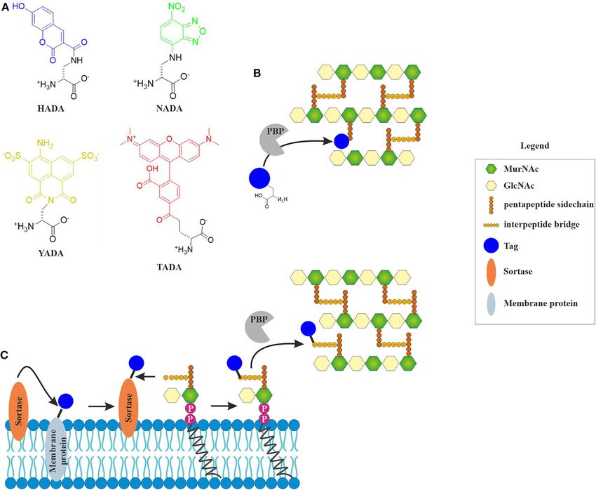
REVIEW
                                                                                                                                              published: 19 October 2020
                                                                                                                                        doi: 10.3389/fcimb.2020.540898

                                              A How-To Guide for Mode of Action
                                              Analysis of Antimicrobial Peptides
                                              Ann-Britt Schäfer and Michaela Wenzel*

                                              Division of Chemical Biology, Department of Biology and Biological Engineering, Chalmers University of Technology,
                                              Gothenburg, Sweden

                                              Antimicrobial peptides (AMPs) are a promising alternative to classical antibiotics in
                                              the fight against multi-resistant bacteria. They are produced by organisms from all
                                              domains of life and constitute a nearly universal defense mechanism against infectious
                                              agents. No drug can be approved without information about its mechanism of action.
                                              In order to use them in a clinical setting, it is pivotal to understand how AMPs work.
                                              While many pore-forming AMPs are well-characterized in model membrane systems,
                                              non-pore-forming peptides are often poorly understood. Moreover, there is evidence that
                                              pore formation may not happen or not play a role in vivo. It is therefore imperative to
                                              study how AMPs interact with their targets in vivo and consequently kill microorganisms.
                                              This has been difficult in the past, since established methods did not provide much
                            Edited by:        mechanistic detail. Especially, methods to study membrane-active compounds have
                        Florie Desriac,       been scarce. Recent advances, in particular in microscopy technology and cell biological
                    Université de Caen
                                              labeling techniques, now allow studying mechanisms of AMPs in unprecedented detail.
                    Normandie, France
                                              This review gives an overview of available in vivo methods to investigate the antibacterial
                        Reviewed by:
                  Octavio Luiz Franco,        mechanisms of AMPs. In addition to classical mode of action classification assays,
          Catholic University of Brasilia     we discuss global profiling techniques, such as genomic and proteomic approaches,
                          (UCB), Brazil
                        Yannick Fleury,       as well as bacterial cytological profiling and other cell biological assays. We cover
               Université de Bretagne         approaches to determine the effects of AMPs on cell morphology, outer membrane,
                   Occidentale, France
                                              cell wall, and inner membrane properties, cellular macromolecules, and protein targets.
                  *Correspondence:
                                              We particularly expand on methods to examine cytoplasmic membrane parameters,
                     Michaela Wenzel
                wenzelm@chalmers.se           such as composition, thickness, organization, fluidity, potential, and the functionality of
                                              membrane-associated processes. This review aims to provide a guide for researchers,
                    Specialty section:
                                              who seek a broad overview of the available methodology to study the mechanisms of
          This article was submitted to
                   Clinical Microbiology,     AMPs in living bacteria.
                 a section of the journal
                                              Keywords: antimicrobial peptides, antibiotics, mode of action, microscopy, membranes
      Frontiers in Cellular and Infection
                            Microbiology

          Received: 06 March 2020
      Accepted: 18 September 2020
                                              INTRODUCTION
        Published: 19 October 2020
                                              The discovery of antibiotics has been a major historical milestone. With formerly deadly diseases
                               Citation:      now being curable with a simple pill, life expectancy, and quality of life increased significantly.
  Schäfer A-B and Wenzel M (2020) A
                                              The golden age of antibiotics, characterized by the frequent discovery of new lead structures, lasted
    How-To Guide for Mode of Action
   Analysis of Antimicrobial Peptides.
                                              until the late 1980’s. Unfortunately, since the 1990’s antibiotic discovery has stagnated while the
          Front. Cell. Infect. Microbiol.     emergence of multi-resistant bacteria has resulted in untreatable superbugs (Goic-Barisic et al.,
                            10:540898.        2016; Mobarki et al., 2019). The urgent need for new antibiotics prompted a range of interesting
    doi: 10.3389/fcimb.2020.540898            alternative strategies and molecules (Spellberg et al., 2015; Silva et al., 2016; Singer et al., 2019).

Frontiers in Cellular and Infection Microbiology | www.frontiersin.org              1                                         October 2020 | Volume 10 | Article 540898
Schäfer and Wenzel                                                                                                       AMP Mode of Action Methods

   In order to tackle the antibiotic resistance crisis, novel                of its target structure. Different labeling approaches have been
compounds and novel mechanisms are essential. Bacteria possess               developed that allow either the detection of compounds in
a plethora of possible drug targets, yet only few are currently              subcellular fractions (e.g., cytosolic, membrane, and cell wall
clinically exploited. One promising class of new antibiotic                  fractions), or the microscopic visualization of antimicrobial
molecules are antimicrobial peptides (AMPs) (Silva et al., 2016;             molecules. Although being very useful, the chemical labeling of
Wang et al., 2016). These omnipresent compounds occur in all                 a molecule is bound to change its properties and can change
domains of life and constitute an effective host defense strategy            its antimicrobial activity or mechanism of action (Phetsang
(Baltzer and Brown, 2011). AMPs are usually defined as up                    et al., 2014, 2016; Omardien et al., 2018b; Stone et al.,
to 100 amino acids long, possess cationic, hydrophobic, and                  2018, 2019). Mass spectrometry-based label-free technologies
amphipathic properties, and typically target the bacterial cell              constitute an alternative, yet do not allow visualization
membrane. Despite these relatively common features, they are a               of compound localization. The individual advantages and
highly diverse class of molecules, both regarding their structures           disadvantages of common localization techniques are discussed
and mechanisms of action (Table 1). The best characterized                   in the following chapter.
AMPs are classical pore formers. Different models exist for
this mode of action, including the classical barrel stave, the               Radioactive Labeling
toroidal pore and carpet mechanisms as well as the newer                     Radioactive labeling is the oldest approach to labeling a molecule
molecular electroporation, sinking raft, and interfacial activity            for following its subcellular distribution and at the same
models (Miteva et al., 1999; Pokorny and Almeida, 2004; Chan                 time only minimally invasive to the compound’s structure:
et al., 2006; Wimley, 2010; Teixeira et al., 2012). Accordingly,             Radioactive isotopes are generally thought not to affect the
mechanisms of AMPs were predominantly investigated using                     chemical properties of a given compound. However, even the
model lipid systems (in vitro). However, more and more AMPs                  mass of an atom can affect its chemical bonds. Thus, mass
are being discovered that have more complex or more subtle                   isotopes can still change the behavior of a labeled molecule
interactions with bacterial membranes and do not form pores                  (Filiou et al., 2012; Fleming et al., 2014). Radioactive labeling
[e.g., MP196, cWFW, and daptomycin (Wenzel et al., 2014;                     is normally only suitable for antibiotics that can be produced
Scheinpflug et al., 2017; Gray and Wenzel, 2020a)], or do not                at least semi-synthetically, but it is also possible to obtain
target membranes at all (Brötz et al., 1998a; Graf et al., 2017;             radioactively labeled microbially produced antibiotics by growing
Mishra et al., 2018).                                                        the producer strain on a radioactive precursor (Atzrodt and
   The road to clinical approval can be long and rocky and                   Allen, 2011). Radioactive labeling allows very sensitive detection
elucidating the mechanism of action of a new antibiotic can be               of compounds in subcellular fractions (Perkins and Nieto, 1970;
challenging. Over the last years, a number of methods have been              Ishiguro et al., 1981), but it does not allow the visualization of
developed, adapted, and refined to investigate the mechanisms of             antibiotic localization. Due to these drawbacks and the overall
antibiotics in living bacterial cells. This is essential, since the in       trend to reduce the amount of radioactive material used in
vivo mechanism of a compound can be fundamentally different                  research, radioactive labels are typically no longer the method
from its action in artificial models or the molecule may have more           of choice for localizing antimicrobial molecules. However, it may
than one target, a relatively common feature for AMPs (Sass et al.,          still have its uses in some cases (e.g., for very small molecules that
2010; Müller et al., 2016b; Wenzel et al., 2019).                            are dwarfed by large fluorescence tags).
   In this review, we want to give an overview of the
tools available to investigate the in vivo mechanisms of both                Metal Labeling
membrane-active AMPs and AMPs with other targets. Thereby,                   A newer approach is metal labeling of antimicrobial compounds.
we do not aim to provide an exhaustive list of techniques                    This technique was largely inspired by a ferrocene-containing
or detailed summary of all recent technical developments. We                 derivative of the antimalarial drug chloroquine (Biot et al.,
rather want to provide a broad handbook for researchers, who                 2011). Since iron is an electron-dense metal, it should be
are more or less acquainted with mode of action studies, to                  possible to detect it by electron microscopy. However, iron
guide them through a range of possibilities for analyzing the                occurs in bacterial cells in relatively high concentrations,
mechanisms of their compounds. While this article is focused on              which could lead to a high background signal. This led to
techniques available for studying AMPs, most assays are perfectly            the development of a ruthenocene-containing derivative,
suitable to analyze other antibiotic molecules as well. We put               which was successfully employed to detect the compound
special emphasis on analyzing the bacterial cell envelope, but               in ultrathin sections of malaria parasites (Biot et al.,
also address other possible targets. Where possible, we selected             2012). A similar approach was then employed for a small
techniques that can be relatively easily adapted and tried to avoid          hexapeptide antibiotic by exchanging the N-terminal amino
very specialized niche techniques.                                           acid for ruthenocene. This allowed both the visualization
                                                                             of the peptide by electron microscopy and quantification
                                                                             in subcellular fractions by element analysis (Wenzel et al.,
COMPOUND LOCALIZATION                                                        2014).
                                                                                While in this case the activity and mechanism of action
Knowing where an antimicrobial compound accumulates in                       of the labeled compound were not notably compromised
the bacterial cell can give a first hint toward the localization             (Wenzel et al., 2014), it is well-possible that the addition

Frontiers in Cellular and Infection Microbiology | www.frontiersin.org   2                                    October 2020 | Volume 10 | Article 540898
Schäfer and Wenzel                                                                                                                              AMP Mode of Action Methods

TABLE 1 | Overview of different AMPs and antimicrobial proteins and their modes of action.

Peptide                    Peptide class                   Mode of action                                                                              References

Daptomycin                 Cyclic lipopeptide              Inserts into fluid membrane microdomains that harbor cell envelope synthesis                Müller et al., 2016b;
                                                           complexes; inhibits cell wall and membrane synthesis by displacing the MurG and             Grein et al., 2020
                                                           PlsX proteins; binds phosphatidylglycerol and undecaprenyl-bound cell wall
                                                           intermediates
Polymyxin B                Cyclic lipopeptide              Binds to lipopolysaccharides and permeabilizes the outer membrane; integrates into          Vaara, 1992; Fu et al.,
                                                           and permeabilizes the cytoplasmic membrane; inhibits respiration                            2019
Surfactin                  Cyclic lipopeptide              Membrane permeabilization; local destabilization of membrane packing at low                 Carrillo et al., 2003;
                                                           concentrations, detergent-like membrane solubilization at high concentrations               Henry et al., 2011
Bacitracin                 Cyclic peptide                  Binds to undecaprenylphosphate and leads to inhibition of wall teichoic acid and lipid      Ruhr et al., 1971
                                                           II synthesis
Gramicidin S               Cyclic beta-sheet peptide       Induces large-scale membrane phase separation and delocalizes peripheral                    Wenzel et al., 2018a
                                                           membrane proteins involved in cell division and cell envelope synthesis
Tyrocidine A               Cyclic beta-sheet peptide       Induces membrane phase separation and forms large transmembrane pores;                      Ristow et al., 1975;
                                                           interferes with DNA-binding proteins and probably induces DNA damage                        Wenzel et al., 2018a
Theta-defensin             Cyclic beta-sheet peptide       Membrane interaction of theta defensin leads to deregulation of autolytic enzymes           Wilmes et al., 2014
                                                           and indices autolysis
MP196                      RW-rich, cationic               Disturbs membrane organization and delocalizes cytochrome c, MurG, and MinD,                Wenzel et al., 2014
                           antimicrobial peptide           resulting in inhibition of respiration, cell wall synthesis, and cell division
                           (CAMP)
cWFW                       RW-rich, cyclic CAMP            Separates membrane lipids into fluid and rigid domains, resulting in separation of          Scheinpflug et al., 2017
                                                           integral and peripheral membrane proteins in the respective domains, in turn leading
                                                           to separation of multiprotein complexes
LL-37                      Alpha-helical CAMP              Membrane disruption by carpet mechanism                                                     Kościuczuk et al., 2012
Aurein 2.1                 Alpha-helical CAMP              Forms cation-selective transmembrane pores                                                  Cheng et al., 2009;
                                                                                                                                                       Wenzel et al., 2015b
Gramicidin A               Alpha-helical peptide           Na+ /K+ channel ionophore                                                                   Duax et al., 1996
Magainin                   Alpha-helical peptide           Forms a toroidal membrane pore                                                              Ludtke et al., 1996
Alamethicin                Alpha-helical peptaibol         Forms voltage-dependent ion channels                                                        Leitgeb et al., 2007
Vancomycin                 Glycopeptide                    Inhibits cell wall synthesis by binding to the D-Ala-D-Ala motif of lipid II                Schneider and Sahl,
                                                                                                                                                       2010
Nisin                      Type A lantibiotic              Binds to lipid II and uses it as a docking molecule to form a transmembrane pore            Breukink et al., 1999
Mersacidin                 Type B lantibiotic              Inhibits cell wall synthesis by binding lipid II                                            Brötz et al., 1998b
hBD3                       Beta defensin                   Interacts with membranes and displays low affinity for lipid II; probably localizes to      Sass et al., 2010
                                                           sites of active cell wall synthesis and sterically hinders the interaction of protein
                                                           complexes
Plectasin                  Fungal defensin                 Inhibits cell wall synthesis by binding to lipid II                                         Schneider et al., 2010
Microcin                   Lasso peptide                   Depolarizes bacterial membranes, stabilizes gel phase in bacterial membrane mimics,         Delgado et al., 2001;
                                                           RNA polymerase may be an additional target                                                  Rintoul et al., 2001,
                                                                                                                                                       2015
Valinomycin                Depsipeptide                    Potassium carrier ionophore                                                                 Duax et al., 1996
Teixobactin                Macrocyclic depsipeptide        Inhibits cell wall synthesis by binding bactoprenol-coupled cell wall precursors            Ling et al., 2015
ADEP                       Acyldepsipeptide                Deregulates the ClpP protease, leading to uncontrolled proteolysis of substrates like       Brötz-Oesterhelt et al.,
                                                           FtsZ, inhibiting cell division                                                              2005; Sass et al., 2011
Lysozyme                   Antibacterial protein           Lyses the peptidoglycan cell wall by hydrolyzing glycosidic bonds                           Aminlari et al., 2014
Actinonin                  Peptidomimetic                  Inhibits peptide deformylase leading to accumulation of formyl-methionine-capped            Chen et al., 2000
                                                           proteins

of a metallocene tag will influence the behavior of the                                     occur in bacterial cells in high concentrations) can easily be
compound in one way or another. However, metallocenes are                                   localized without additional labeling (Wenzel et al., 2013).
still considerably smaller than common fluorescence labels and                              Similarly, AMPs may be visualized with electron microscopy
thus less likely to severely change the antibiotic properties of                            without the need to chemically label them through specific
a molecule.                                                                                 detection with gold-labeled antibodies (Azad et al., 2011).
    Compounds that already contain a residue that can be                                    However, this approach requires that the peptide is immunogenic
visualized by electron microscopy (electron-dense metals) or                                enough to obtain specific antibodies, a property that is normally
detected by atomic spectroscopy (most elements that do not                                  not desired for antibiotic candidates.

Frontiers in Cellular and Infection Microbiology | www.frontiersin.org                  3                                            October 2020 | Volume 10 | Article 540898
Schäfer and Wenzel AMP Mode of Action Methods
Fluorescence Labeling more and more evidence is emerging that membrane-targeting
While metal labels only allow the visualization of antimicrobial AMPs do not uniformly attack the lipid bilayer but instead target
compounds in fixed cells, fluorescence labels allow live cell specific foci and that their interaction with bacterial membranes
imaging of antibiotic attacks on bacterial cells and even co- can be highly dynamic (Kandaswamy et al., 2013; Rangarajan
localization of the antimicrobial molecule with its target. It is et al., 2013; Müller et al., 2016b; Rashid et al., 2016). To date,
a relatively common approach and has aided several mode of fluorescence labeling remains the only technique that is suitable
action studies so far (Tiyanont et al., 2006; Pogliano et al., 2012; for capturing these dynamic interactions.
Scheinpflug et al., 2013; Chileveru et al., 2015; Müller et al.,
2016b; Omardien et al., 2018b). Most fluorophores have much FINDING THE PATHWAY
higher molecular weights than the average antibiotic. Direct
labeling with such large moieties may critically influence activity, While the localization of an antimicrobial compound within its
uptake, and mechanism of action (Katritzky and Narindoshvili, target cell helps narrowing down its potential molecular target,
2009; Müller et al., 2016b; Stone et al., 2019). Even very it does not give insight into the process or pathway that is
small fluorescence labels might already compromise antibacterial actually inhibited and basing hypotheses on localization alone
activity (Scheinpflug et al., 2013). Direct fluorescence labeling can be misleading. Thus, finding the primarily inhibited pathway
approaches can therefore be restricted to larger molecules, which is of crucial importance to proceed with detailed mode of action
are not severely affected by the addition of a fluorophore analysis and identifying the molecular target. Classically, this has
(Tiyanont et al., 2006; Chileveru et al., 2015). This generally been done by radioactive precursor incorporation studies, but
makes this approach better suited for AMPs than for small more recent alternatives include fluorescently labeled precursors
molecule antibiotics. and reporter gene fusion.
In any case, possible effects of the label on the compound’s
behavior need to be carefully assessed. This should not be Incorporation of Radioactive Precursors
limited to assaying antimicrobial activity alone but also extend Incorporation experiments with radioactively labeled precursors
to phenotypical characterization to ensure that the compound’s for the main cellular macromolecules (DNA, RNA, proteins,
mechanism of action has not notably changed. However, the lipids, peptidoglycan) are very sensitive. While radioactive
use of fluorescent labels always remains a trade-off between labeling is commonly sought to be avoided for safety and
their versatility in live cell microscopy and the possibility that environmental concerns, it is the only method that allows the
observations made with the labeled compound may not fully detection of macromolecules without altering their chemical
translate to its unlabelled original. structure and thus has the lowest risk of labeling-imposed
An alternative to direct the labeling of AMPs is artifacts. Custom synthesis of radioactively labeled molecules
immunolabelling with fluorescently labeled antibodies. While is possible, but commonly used isotopic precursors include
this approach does not affect the behavior of the compound and [14 C] glucosamine for peptidoglycan, [14 C]-thymidine for DNA,
still allows microscopic localization studies, it is not suitable for [3 H]-uridine for RNA, L-[14 C]-isoleucine and [3 H] glycine
live cell imaging, since it requires permeabilization and chemical for proteins, and [14 C]-acetate for lipids (Hofmann and
fixation of the cells (Choi et al., 2016). Eichenberger, 1998; Ling et al., 2015; Müller et al., 2016b).
Some of these labels can be combined in the same sample [e.g.,
Label-Free Detection [14 C] glucosamine and [3 H] glycine (Molenkamp and Veerkamp,
Label-free detection of antimicrobial compounds by mass 1976)], yet individual samples are more commonly used). It
spectrometry is an alternative approach that does not have the has to be noted that in order to assess incorporation into
drawback of compromised activity of labeled compounds. As macromolecules and not just uptake into cells, samples must be
long as the mass of the molecule of interest is known, it is precipitated [e.g., using trichloroacetic acid, prior to measuring
possible to detect the unlabeled compound in a complex mixture radioactivity (Wenzel et al., 2014)]. However, measuring whole
such as cell lysate (Ackermann et al., 1996; Deltombe et al., cells in parallel is a useful control for cellular uptake, since AMPs
2019). This can be used to directly detect and quantify antibiotic often depolarize the cell membrane, which may affect the activity
concentrations in subcellular fractions. Interestingly, a new of nutrient uptake systems.
approach called 3D imaging cluster Time-of-Flight secondary ion
mass spectrometry allowed the label-free detection and mapping Fluorescent Labeling of Cellular
of antibiotics in single cells of Escherichia coli (Tian et al., Macromolecules
2017). The relatively low spatial resolution of this technique An alternative to radioactive labeling of metabolites is constituted
does not allow the visualization of antibiotics to specific cell by fluorescent labeling. A range of fluorescent molecules have
structures and is therefore not well-suited for mode of action been developed that can be used to cover some of the major
studies yet. However, it gives hope that label-free tracking of metabolic pathways of bacterial cells. The simplest example for
antibiotics within bacterial cells might indeed become possible this is probably the expression of a fluorescent protein, such
at some point. as green-fluorescent protein (GFP), from a housekeeping or
However, one limitation that will always remain is that mass inducible promoter, which allows monitoring of active protein
spectrometry-based techniques do not allow visualization of synthesis in living bacterial cells (Gray et al., 2019). A more direct
antibiotics in living cells. This is an important limitation since approach is metabolic labeling of nascent peptide chains with the
Frontiers in Cellular and Infection Microbiology | www.frontiersin.org 4 October 2020 | Volume 10 | Article 540898Schäfer and Wenzel AMP Mode of Action Methods
amino acid analog L-homopropargylglycine (L-HPG), followed S. aureus, and also strains that were not developed as antibiotic
by fluorescent labeling of this molecule with Alexa-594 by click mode of action analysis tool can prove useful as such (Mesak
chemistry (Stempler et al., 2017; Gray et al., 2019). Several similar et al., 2008; Chanda et al., 2009; Mondal et al., 2010; Dengler and
probes have been described and specific reporters for certain McCallum, 2016; Bojer et al., 2017).
posttranslational modifications are available as well (Grammel New reporter gene tools are constantly developed and refined.
and Hang, 2013). For example, a modified luciferase reporter assay reporting
Incorporation of cell wall material can be monitored by on cell wall synthesis and DNA integrity enables antibiotic
fluorescent D-amino acids or sortase-mediated incorporation mode of action analyses and screening of new drugs against
of fluorescently labeled lipid II (Nelson et al., 2010; Kuru Mycobacterium tuberculosis (Naran et al., 2016). Efforts to enable
et al., 2012, 2015; Hsu et al., 2017). Similarly, fluorescently cost-efficient high through put screenings with reporter gene
labeled glycans can be incorporated into the Gram-negative fusions have recently resulted in the development of a phenomics
or mycobacterial outer membrane (Siegrist et al., 2015). These screening platform containing an E. coli reporter gene library
techniques are described in detail under 6.2 Cell wall and 6.1 enabling large-scale gene expression studies in a cost- and time-
Outer membrane, respectively. efficient manner (French et al., 2018).
Fluorescent nucleotide analogs that can be incorporated into
DNA or RNA have been developed for eukaryotic cells, but their PROFILING APPROACHES
suitability for bacterial cells has not yet been explored (Grammel
and Hang, 2013). While precursor incorporation and reporter gene experiments
Fluorescent labeling of metabolic precursors is superior to offer a great way to quickly identify the affected pathway, they
radioactive labeling in terms of safety and official regulations, can only scratch the surface. A much deeper understanding is made
be visualized in living bacterial cells, and in some cases allows possible by -omics approaches that allow global profiling on
further analysis of the labeled macromolecules, for example by genomic, transcriptomic, proteomic, and metabolomic level.
affinity purification of the tag followed by mass spectrometry These techniques are very useful for generating hypotheses about
(Grammel and Hang, 2013). However, it is an inherent limitation antibiotic mechanisms but can rarely stand all alone. While
of chemically modified precursors that they may not behave they offer a large amount of information, complex datasets also
exactly as the unlabeled molecule. This can be due to the size require a significant amount of time for analysis and may be
of the fluorescent tags, which are often larger than the precursor difficult to interpret for antimicrobial compounds with multiple
itself, or simply to changing the physicochemical properties of the or pleiotropic effects, which is often the case for AMPs.
target molecule (Siegrist et al., 2015). -omics approaches are certainly not a must in mode of action
analysis of “typical” AMPs that impair membrane integrity,
Reporter Gene Fusions but they allow an unmatched combination of breadth and
A simple alternative to precursor incorporation studies are depth of physiological insight and can be extremely valuable for
reporter gene fusions. Bacteria react to stress in a highly specific compounds with unknown/unusual mechanisms. The amount
manner. So much so that the stress response can be used as a of technical variations, especially in mass spectrometry-based
diagnostic tool to identify antibiotic mechanisms of action (see proteomics, is immense and we do not remotely attempt to
also four Profiling approaches) (Bandow et al., 2003). Based on cover them all. In the next chapter we want to present selected
this, specific reporters can be selected for mapping the inhibited techniques that have a well-established standing in antibiotic
pathway, analogous to precursor incorporation experiments mode of action studies.
(Urban et al., 2007). To this end, either the gene of interest or
only its promoter, is fused to a gene encoding a reporter protein, Genomic Profiling
whose expression can be visualized by calorimetric, fluorescent, Genomic-driven approaches have gained much attention in
or luminescent measurements. The most common reporter genes antibiotic drug discovery, mainly as sources for new antibiotic
encode firefly luciferase or beta-galactosidase, but fluorescent targets (Miesel et al., 2003; Freiberg et al., 2005). However,
proteins like GFP are also possible. genomic approaches can also aid in identifying antibiotic targets.
The main advantage of this method is that it does not need They can be roughly divided into two groups, screening existing
radioactive labeling, does not produce artifacts by chemical libraries and generating new mutant libraries.
modification of precursors, and does not require any major or For both E. coli (Baba et al., 2006) and B. subtilis (Koo
unusual equipment. However, the choice of reporter genes or et al., 2017), commercially available mutant collections exist
promoters requires solid knowledge of bacterial stress responses that comprise deletion strains of each non-essential gene.
and a new set of strains has to be constructed for each organism of These collections can be screened against hypersensitivity to or
interest. This also limits it to model organisms that are genetically resistance against an antibiotic of interest to discover potential
accessible. Thus, reporter gene approaches for antibiotic mode of resistance factors or target candidates, respectively. This has
action analysis are most common in the standard Gram-positive been systematically approached by Tamae et al. and Liu et al.,
and Gram-negative model organisms Bacillus subtilis and E. coli resulting in sensitivity patterns for close to 30 different antibiotic
(Table 2) (Bianchi, 1999; Hutter et al., 2004a; Urban et al., 2007; compounds (Tamae et al., 2008; Liu et al., 2010), which can be
Wenzel et al., 2014). However, reporter gene studies in general used as a reference for studying novel drug candidates (Tran
are common tools in many organisms, including pathogens like et al., 2011; Kang et al., 2012). One obvious limitation of this
Frontiers in Cellular and Infection Microbiology | www.frontiersin.org 5 October 2020 | Volume 10 | Article 540898Schäfer and Wenzel AMP Mode of Action Methods
TABLE 2 | Examples of reporter gene fusions commonly used to identify antibiotic mechanisms.
Promoter Fusion Species Reporter for References
bmrC Luciferase B. subtilis Inhibition of translation Wenzel et al., 2014
fabHB Luciferase B. subtilis Inhibition of fatty acid synthesis Hutter et al., 2004a
glpD Luciferase B. subtilis Inhibition of fatty acid synthesis Hutter et al., 2004a
Held Luciferase B. subtilis Inhibition of transcription Wenzel et al., 2014
liaI Luciferase B. subtilis Inhibition of cell wall synthesis Wenzel et al., 2014
yheI Luciferase B. subtilis Inhibition of protein synthesis Urban et al., 2007
yorB Luciferase B. subtilis DNA damage Wenzel et al., 2014
ypbG Luciferase B. subtilis Inhibition of cell wall synthesis Hutter et al., 2004a
ypuA Luciferase B. subtilis Cell wall stress Hutter et al., 2004a
yrzI Luciferase B. subtilis Inhibition of protein synthesis Hutter et al., 2004a
yvgS Luciferase B. subtilis Inhibition of RNA synthesis Urban et al., 2007
drp35 β-galactosidase S. aureus Inhibition of cell wall synthesis Mondal et al., 2010
dnaK β-galactosidase E. coli Protein misfolding Bianchi, 1999
Ibp β-galactosidase E. coli Protein misfolding Bianchi, 1999
P3rpoH β-galactosidase E. coli Extracytoplasmic stress Bianchi, 1999
degP) β-alactosidase E. coli Extracytoplasmic stress Bianchi, 1999
approach is that it does not include essential genes, which are Transcriptomic Profiling
commonly thought to be the most suitable antibiotic targets. While genomic approaches map the level of antibiotic sensitivity,
Recently, CRISPR knock-down libraries covering essential genes transcriptomic and proteomic approaches map the stress
have been established for both B. subtilis and E. coli (Peters et al., response profiles of bacteria to antibiotic stress, which are
2016; Guo et al., 2018; Wang et al., 2018). Both libraries have diagnostic for the individual compound’s mechanism of action
been made commercially available. While they have not yet been and can aid target identification (Bandow et al., 2003;
used in antibiotic mode of action studies, they complement the Bandow and Hecker, 2007; Wenzel and Bandow, 2011).
genomic toolbox available for such approaches. However, when While microarrays have been the predominant technique for
working with such mutant libraries, care has to be taken that transcriptomic profiling for a long time, RNA sequencing is now
relevant strains are independently confirmed and that updated the method of choice in most cases, since it is more sensitive,
annotations of the mutated open reading frames are taken into does not rely on hybridization probes, and is becoming more and
account (Baba et al., 2006; Yamamoto et al., 2009; Aedo et al., more affordable (Hutter et al., 2004b; Gilad et al., 2009; O’Rourke
2019). et al., 2020). Many studies have successfully used transcriptomic
The second approach aims at generating resistant mutants profiling to aid mode of action analysis (Briffotaux et al., 2019;
that may reveal the molecular target of an antimicrobial O’Rourke et al., 2020) and its uses have been extensively reviewed
compound. This can for example be done by characterizing elsewhere (Freiberg et al., 2004; Wecke and Mascher, 2011).
spontaneous resistant mutants generated under lower antibiotic However, it should be noted that parameters for stress response
pressure or repeated passaging of incrementally resistant profiling, be it transcriptomic or proteomic experiments, must
colonies on rising antibiotic concentrations (Leejae et al., 2013; be chosen with care. Thus, sublethal antibiotic concentrations
Puertolas-Balint et al., 2020). The latter is prone to result in and short treatment times should be used in order to achieve the
accumulation of different mutations, complicating analysis and best possible acute stress response (Wenzel and Bandow, 2011;
interpretation, and is likely to result in unstable mutants due Raatschen and Bandow, 2012).
to the combined fitness costs of multiple mutations (Leejae
et al., 2013). An alternative approach is the generation of Proteomic Profiling
mutants by chemical or transposon mutagenesis followed While transcriptomic profiling is well-suited to monitor
by selecting for antibiotic-resistant colonies. Thereby, antibiotic stress responses, proteomic profiling can provide
transposon mutagenesis is the easier option, since it allows additional information on posttranslational modifications and
rapid identification of the insertion locus by sequencing from regulation mechanisms, such as proteolysis. Metabolic labeling,
the transposon sequence (Santiago et al., 2018). In contrast, either radioactively for gel-based proteomics or with stable
spontaneous mutants and chemical mutagenesis require isotopes for mass spectrometry, allows highly sensitive pulse and
whole genome sequencing to map individual mutations. pulse-chase experiments for monitoring acute stress responses at
However, transposon mutagenesis precludes analyzing a given time point.
targets encoded by essential genes, while spontaneous and Gel-based proteomics by two-dimensional polyacrylamide
chemically induced mutations do not necessarily result in loss gel electrophoresis (2D-PAGE) is a proteomic approach that
of function. has been extensively employed in stress response profiling and
Frontiers in Cellular and Infection Microbiology | www.frontiersin.org 6 October 2020 | Volume 10 | Article 540898Schäfer and Wenzel AMP Mode of Action Methods
antibiotic mode of action research (Bandow et al., 2003; Mostertz effectively aid mode of action analysis by itself (Opoku-Temeng
et al., 2004; Wecke et al., 2009). To this end, newly synthesized et al., 2019; Ajdidi et al., 2020).
proteins are radioactively pulse labeled with L-[35 S] methionine
and, after a crude protein extraction, separated according to Metabolomic Profiling
their isoelectric point and molecular weight. Protein expression Metabolomics is a comparatively young -omics technique that
is then densitrometrically quantified from autoradiographs of has not been extensively employed for mechanistic antibiotic
dried gels and compared to an untreated control to acquire studies yet. In contrast to genomics, transcriptomics, and
regulation factors. Upregulated proteins, referred to as marker proteomics (Bandow et al., 2003; Tamae et al., 2008; Liu et al.,
proteins, are identified by mass spectrometry. These proteins 2010; O’Rourke et al., 2020), there are no large comparative
reflect the acute stress response of the bacterial cells to the given metabolomic studies on antibiotic stress yet. However, bacterial
stress condition and are indicative of the antibiotic mechanism of metabolomics has been employed for a variety of applications
action. A reference compendium with protein expression profiles including identification of new antibiotics and characterizing
of over 100 antimicrobial compounds has been established to resistance mechanisms (Gao and Xu, 2015; Wu et al., 2015;
aid mode of action analysis of new drug candidates (Bandow Li et al., 2019) and is emerging as a tool in mode of action
et al., 2003; Wenzel et al., 2011, 2012, 2013, 2014, 2015b; studies (Wang et al., 2019; Liu et al., 2020). Metabolomics can
Raatschen et al., 2013; Stepanek et al., 2016a,b; Müller et al., be employed as a more detailed approach to find the affected
2016b; Scheinpflug et al., 2017; Saising et al., 2018; Meier et al., metabolic pathway and thus constitute a sensitive alternative
2019; Wüllner et al., 2019). Radioactive 2D-PAGE should not to radioactive and fluorescent precursor incorporation studies,
be confused with two-dimensional difference gel electrophoresis or as an in-depth analysis of antibiotic effects on the bacterial
(2D-DIGE), which also compares protein expression profiles by metabolism. However, interpreting large metabolomic datasets
densitometric quantification against a control sample, but is a requires profound knowledge of the metabolic networks in the
fluorescent sample multiplexing and not a metabolic labeling respective organism.
technique (Minden, 2012).
Radioactive 2D-PAGE has been proven very robust and CELL MORPHOLOGY
potent in the field but has its practical limitations mainly in
terms of equipment needed, handling of and regulations around An alternative or additional starting point to mode of action
radioactive samples, and relatively low throughput. Moreover, analysis can be cell morphology. Many antibiotics, in particular
it is not suitable for membrane proteomics, which may be AMPs, cause distinct defects in cell shape, size, or integrity
particularly interesting for AMPs. Gel-free proteomics does that can be observed by both light and electron microscopic
not suffer from these limitations and may be more accessible techniques (Friedrich et al., 2000). While examination of cell
to many researchers while providing a similar outcome for morphology alone does normally not identify a mechanism of
mechanistic studies. The gel-free counterpart of radioactive 2D- action, it provides a good basis for further phenotypical analysis
PAGE, in terms of metabolic labeling, is stable isotope labeling and the combination of rather simple morphological assays can
by amino acids in cell culture (SILAC). Here, one culture (e.g., be used to effectively map mechanistic classes (Nonejuie et al.,
the untreated control), is grown in a medium containing a 13 C- 2013).
labeled amino acid, while another culture, e.g., the antibiotic-
treated sample, is grown on normal medium. This allows pooling Electron Microscopy
of the samples and quantification in the same run and can A classical method to examine bacterial morphology is
also be done as a pulse experiment to selectively label only transmission electron microscopy (TEM). TEM has been
newly synthesized proteins (Snider et al., 2019). Instead of using frequently employed to study antibiotic effects on bacterial
mass-labeled amino acids, labeling can also be achieved with cells, since it offers unique insight into bacterial ultrastructures
other sources of heavy nitrogen, such as ammonium (Dreisbach (Friedrich et al., 2000; Sass et al., 2011; Nicolas et al., 2019;
et al., 2008; Wenzel et al., 2014). As with radioactive 2D- Vazquez-Muñoz et al., 2019). Sample preparation for TEM
PAGE and 2D-DIGE, SILAC should not be confused with involves chemical fixation, dehydration, and contrasting with
iTRAQ (isobaric tag for relative and absolute quantitation), metal stains, followed by embedding in resin and ultrathin
which is not a metabolic labeling but a multiplexing technique sectioning. One limitation of TEM comes into effect when
(Unwin, 2010). working with rod-shaped bacteria like E. coli or B. subtilis:
Recently, label-free approaches have been heavily employed due to the random orientation of bacteria in the resin the
for mode of action analysis of antimicrobials (Müller et al., 2016a; majority of cells is cross-sectioned, which makes it difficult
Stepanek et al., 2016b; Gao et al., 2017; Kang et al., 2019; Yuan to assess antibiotic-induced phenotypes. This limitation was
et al., 2020). However, these detect differences in protein levels recently overcome by a flat embedding approach, where bacteria
and not newly synthesized proteins, rather giving insight into are aligned on an agarose film prior to embedding, resulting in
successfully completed stress adaptation rather than acute stress mostly longitudinally sectioned cells (Wenzel et al., 2019).
response. Label-free proteomics is most powerful when employed Scanning electron microscopy (SEM) is an electron
together with a technique that detects the acute stress response, microscopy technique, which allows inspection of the bacterial
such as radioactive 2D-PAGE or transcriptomics (Darby et al., cell surface in great detail but does not allow imaging intracellular
2014; Müller et al., 2016a; Stepanek et al., 2016b), but can also structures. SEM has been successfully employed for antibiotic
Frontiers in Cellular and Infection Microbiology | www.frontiersin.org 7 October 2020 | Volume 10 | Article 540898Schäfer and Wenzel AMP Mode of Action Methods
mode of action studies and is particularly interesting for AMPs, et al., 2013; Saeloh et al., 2018; Wenzel et al., 2018a). These
which often cause cell surface defects (Zweytick et al., 2014; dyes are easy to handle, do not require specialized fluorescence
Yang et al., 2017; Nicolas et al., 2019). For SEM, cell samples are filters, and can be combined with a GFP fusion of interest, for
also fixed, dehydrated, and contrasted with a metal stain, but example the cell division protein FtsZ (Araujo-Bazan et al., 2016).
normally not cut into sections (Kaláb et al., 2008). Environmental However, there is a broad palette of fluorescence dyes and protein
SEM (ESEM) omits the need for critical point drying and enables fusions available that report on various cellular functions and
imaging of hydrated samples (Collins et al., 1993). components and have been successfully employed in antibiotic
mode of action studies. We will describe a number of such
Atomic Force Microscopy specialized fluorescence reporters in the following chapter.
Another form of microscopy that detects surface changes is
atomic force microscopy (AFM). AFM makes use of a small
needle, the cantilever, to scan over a sample and record the CELLULAR COMPONENTS
force it encounters when interacting with the sample surface.
This allows the generation of height profiles, measuring of Once the target pathway or structure has been mapped, the
cell surface stiffness, and detection of cellular content leakage next step in mode of action analysis is a detailed assessment
(Dorobantu and Gray, 2010; Neethirajan and DiCicco, 2014). of the mechanism of action and identification of the target
AFM is often chosen as a method to examine changes in structure. While a specific drug-target interaction is normally
bacterial biofilms (Dorobantu and Gray, 2010) and is recently always confirmed with purified components in vitro, antibiotics
gaining more attention as a tool to examine bacterial cell may have different of additional targets in living cells (Müller
morphology for antibiotic mode of action analysis (Meincken et al., 2016b; Wenzel et al., 2019). A number of in vivo methods
et al., 2005; Mularski et al., 2016). Moreover, antibiotic-induced is available to study the effects of antimicrobial compounds on
morphological changes measured by AFM can aid identifying living bacteria. In the following, we will describe assays that can
antibiotic-resistant strains (Ierardi et al., 2017) distinguishing be used to assess the effects of AMPs and other antibacterial
between persister and resister phenotypes (Uzoechi and Abu-Lail, molecules on the major components of a bacterial cell.
2020).
Outer Membrane
Bacterial Cytological Profiling The lipopolysaccharide-rich outer membrane is highly
A relatively new tool for fast mode of action analysis is bacterial impermeable and the first line of defense of Gram-negative
cytological profiling (BCP). This fluorescence light microscopy- bacteria. It is a major intrinsic antibiotic resistance factor and
based technique is a combination of different staining techniques the main reason why Gram-negative bacteria are much more
that give a quick overview of major cellular components resilient to antibiotic attacks than Gram-positive bacteria.
(Nonejuie et al., 2013; Lamsa et al., 2016). Using principal Antibiotics that impair this permeability barrier are urgently
component analysis, antibiotics can be grouped into classes and needed (Silhavy et al., 2006) and some AMPs have been shown
new compounds can be rapidly assigned to a mechanistic group. to target the outer membrane, most prominently the polymyxins
An expansion of this method, called rapid inhibition profiling (Vaara, 1992).
(RIP), allows mapping the target pathway of compounds with Outer membrane permeability can be assayed with fluorescent
novel targets. This method makes use of proteolytic degradation dyes such as 1-N-phenylnaphthylamine (NPN) and 1,8-anilino-
of a potential antibiotic target protein to generate a reference 1-napthalenesulfonicacid (ANS). NPN and ANS have a relatively
cytological profile, against which new antibiotic candidates week fluorescent signal in aqueous solution but exhibit strong
can be compared. This allows a reliable identification of new fluorescence in hydrophobic environments like lipid membranes.
mechanisms of action (Lamsa et al., 2016; Peters et al., 2018). These dyes do not penetrate the outer membrane and thus do
Originally starting with essentially a membrane and DNA not stain intact cells. Upon outer membrane permeabilization
stain, BCP has been further refined and expanded over the years however they can bind to membrane phospholipids leading to an
and has aided a number of mode of action studies (Pogliano increased fluorescence signal (Loh et al., 1984; Schved et al., 1994;
et al., 2012; Nonejuie et al., 2016; Mohammad et al., 2017; Htoo Gravel et al., 2017). ANS is sensitive to charge neutralization,
et al., 2019). Nowadays, it may include a variety of fluorescent leaving NPN as the dye of choice for polycationic compounds like
dyes and protein fusions in addition to or in place of the many AMPs (Loh et al., 1984).
original membrane and DNA dyes (Araujo-Bazan et al., 2016; Similarly, outer membrane permeability can be assayed by
Müller et al., 2016b; Omardien et al., 2018a,b; Saeloh et al., testing the sensitivity to small molecule antibiotics that normally
2018; Wenzel et al., 2018a). Since a clear definition of bacterial do not penetrate the outer membrane, but do have a target in
cytological profiling is missing, the term may be used for a Gram-negative cells (Heesterbeek et al., 2019). In contrast to
distinct combination of two or three dyes for high throughput fluorescence dyes, this method does not allow quantification of
pathway mapping as well as for comprehensive bacterial cell outer membrane permeabilization. However, using antibiotics of
biology studies. To get a first glance at cell morphology changes, different molecular weight, the outer membrane pore size can be
the combination of a red membrane dye (typically FM5-95, estimated. Antibiotics that can be used for this are for example
FM4-64, or Nile red), a blue DNA dye (typically DAPI), and rhodomyrtone (442.54 g/mol), vancomycin (1449.3 g/mol), and
phase contrast microscopy has been proven useful (Nonejuie nisin (3354.07 g/mol).
Frontiers in Cellular and Infection Microbiology | www.frontiersin.org 8 October 2020 | Volume 10 | Article 540898Schäfer and Wenzel AMP Mode of Action Methods
Another method to assess the integrity of the outer membrane to the antimicrobial compound of interest can be monitored.
is AFM, which reports on cell surface stiffness, which is a direct However, it has to be noted that this assay does not respond
measure for outer membrane or, in Gram-positive bacteria, cell to every cell wall synthesis inhibitor tested, thus not providing
wall integrity and can therefore be employed to measure the complete coverage (Sun et al., 2002).
effects of outer membrane-targeting compounds. A simple microscopic assay to assess whether incorporation
Outer membrane proteins can also be used as a proxy to of cell wall precursors is inhibited is the acetic acid/methanol
determine outer membrane integrity. One simple way to do this, fixation (Schneider et al., 2010; Wenzel et al., 2012). In Gram-
is to isolate outer membrane fractions and perform a Western positive bacteria, this treatment leads to extrusion of the
blot analysis of outer membrane proteins (Rojas et al., 2018). protoplast through holes in the cell wall matrix (Figure 1B).
Alternatively, proteins can be identified and quantified by mass The peptidoglycan layer is a dynamic structure that is constantly
spectrometry. In addition to these outer membrane integrity remodeled to accommodate cell growth and division. To this end,
assays, there are also fluorescence labeling approaches available autolytic enzymes constantly break down the cell wall at specific
to visualize glycans on the outer membrane (Backus et al., 2011; sites to accommodate incorporation of new cell wall material. If
Siegrist et al., 2015). these holes are not filled because lipid II synthesis is inhibited, a
much higher proportion of cells with membrane extrusions are
Cell Wall observed in the fixation assay. However, deregulation of autolytic
The next barrier after the outer membrane, and the first barrier enzymes may have similar effects.
in Gram-positive bacteria, is constituted by the peptidoglycan cell Fluorescently labeled D-amino acids (FDAAs) have been a
wall. With few exceptions of cell wall-less bacteria, this structure major breakthrough in the field, since they for the first time
is essential for bacterial survival. It does not only protect the cell allowed the direct visualization of active incorporation of cell
from mechanical stress, but also prevents it from bursting due wall precursors into living bacterial cells under the microscope
to turgor pressure. Cell wall synthesis is a complex and highly (Kuru et al., 2015; Hsu et al., 2017). FDAAs mimic the D-amino
coordinated process that takes place partly in the cytosol and acids in the peptide side chain of the peptidoglycan precursor and
partly in the cytoplasmic membrane (Figure 1). Together with are incorporated into the cell wall by penicillin-binding proteins
the bacterial ribosome, the cell wall synthesis machinery is the (HADA) or L-D-transpeptidases (NADA) (Figure 2) (Montón
most successful antibiotic target in the clinic and at the same Silva et al., 2018). Incorporation of FDAAs into the cell wall does
time the most common target of AMPs after the cytoplasmic not appear to be toxic for bacteria. Since their original discovery,
membrane (Yeaman and Yount, 2003; Schneider and Sahl, 2010). new FDAAs have been designed in different fluorescent colors,
Moreover, recent studies suggest that the membrane interaction making them readily available for co-localization experiments
of AMPs severely disturbs the synthesis of the peptidoglycan (Hsu et al., 2017). However, the different FDAAs have their
precursor lipid II (Sass et al., 2010; Wenzel et al., 2014; Müller advantages and disadvantages. For example, HADA is sensitive
et al., 2016b). to photobleaching, while NADA requires higher concentrations
Due to its utmost clinical relevance and the relatively frequent to achieve a satisfactory fluorescence signal, and TDL, a red-
discovery of new cell wall-active agents, a broad method fluorescing FDAA, only weakly stains E. coli (Kuru et al., 2015).
spectrum is available to analyze the effects of compounds on A different way of visualizing the effects of antibiotics on
this pathway, in particular its interaction with lipid II. This cell wall precursors, it sortase-mediated fluorescence labeling of
includes various reporter gene assays, in vitro lipid II synthesis, lipid II, which generally works well for Gram-positive species
lipid II binding visualized by thin layer chromatography, (Nelson et al., 2010). While incorporation efficiency of FDAAs
and detection of accumulated lipid II by high performance is likely inhibited by antibiotics, sortase-mediated labeling will
liquid chromatography (HPLC), to name only a few standard be largely unaffected (Sugimoto et al., 2017). Sortase is a
techniques (Brötz et al., 1998a; Schneider et al., 2009, 2010; membrane-bound protease that cleaves a signal peptide sequence
Schneider and Sahl, 2010; Ling et al., 2015). Taking HPLC- off transmembrane proteins. This can be used to cleave a
based detection of cell wall components a step further, recent fluorescence tag, which can be biotin, azide, or a fluorescent
studies have succeeded to refine the isolation of cell wall chromophore (Nelson et al., 2010), from a transmembrane
peptidoglycan and detect glycan strain length and crosslinking, protein. This tag can then react with lipid II, producing a labeled
allowing detailed analysis of cell wall peptidoglycan composition version of the precursor on the membrane surface (Figure 3). If
(Desmarais et al., 2014, 2015; Montón Silva et al., 2018; More an antibiotic interferes with cell wall synthesis, this will lead to
et al., 2019). mislocalization or clustering of the labeled molecule.
A fast assay that can also be used to screen for cell wall Another way to visualize cell wall synthesis components is
synthesis inhibitors is the AmpC reporter assay (Sun et al., 2002). the labeling of antibiotics with the fluorescence tag 4,4-difluoro-
In this assay, the beta-lactamase gene ampC and its regulator 4-bora-3a,4a-diaza-s-indacene (BODIPY). BODIPY is a very
ampR from Citrobacter freundii are cloned into E coli. This common fluorescence tag that can be easily conjugated with a
system senses accumulated cell wall degradation products and number of biomolecules, including antibiotics that inhibit cell
soluble cell wall precursors and is induced upon inhibition wall synthesis. Vancomycin-BODIPY (Van-FL) and penicillin-
of peptidoglycan synthesis by a broad spectrum of antibiotics, BODIPY (bocillin) are commercially available. Van-FL binds to
not only by beta-lactams. Using an optical density-based beta- the D-Ala-D-Ala motif of lipid II and has been successfully
lactamase survival assay, beta-lactamase expression in response used to visualize lipid II (Pogliano et al., 2012; Schirner et al.,
Frontiers in Cellular and Infection Microbiology | www.frontiersin.org 9 October 2020 | Volume 10 | Article 540898Schäfer and Wenzel AMP Mode of Action Methods
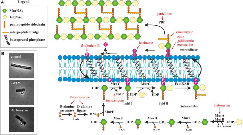
FIGURE 1 | Peptidoglycan synthesis as antibiotic target. (A) Overview of peptidoglycan synthesis in S. aureus and antibiotics targeting this pathway (modified from
Schneider and Sahl, 2010). Peptidoglycan synthesis is a common target of peptide antibiotics. With the exception of tunicamycin and fosfomycin, all antibiotics in this
figure are peptide-based. (B) Acetic acid/methanol fixation of B. subtilis. Inhibition of cell wall synthesis leads to extrusion of the protoplast through breaches in the
peptidoglycan layer.
2015). Bocillin binds to penicillin-binding proteins (PBPs) and in living bacteria. The amount of available techniques would go
can be used for visualizing the localization of these proteins or beyond the scope of this review, but we will describe a number
for competition experiments with other PBP inhibitors. Since of relatively easily accessible techniques that are well-suited for
bocillin does not recognize all PBPs, BODIPY fusions to other mode of action analysis of AMPs.
PBP inhibitors can be employed to distinguish between PBP
subpopulations (Stone et al., 2018). Membrane Composition
Bacterial membranes are complex mixtures of roughly equal
Cytoplasmic Membrane parts of lipids and proteins. The lipid composition of bacterial
The cytoplasmic membrane is the target of the majority of membranes is far from static and varies depending on a variety of
AMPs. A plethora of biophysical techniques are available to factors. Thus, bacteria readily adapt their membrane composition
assay the parameters of model membranes and such assays have under antibiotic stress (Fränzel et al., 2010; Saeloh et al., 2018).
been excessively used to characterize the membrane interactions The membrane composition of bacteria can be analyzed in
of AMPs in vitro. However, the true complexity of bacterial different ways. Head group composition can be easily analyzed
membranes cannot be mimicked, since not only the composition, using thin layer chromatography (Pogmore et al., 2018), a
but also the physicochemical properties vary drastically from technique that does not require expensive instrumentation or
species to species, between different growth conditions, between access to mass spectrometry facilities. Fatty acid composition
media, and growth phases. It has become apparent that model can be measured by gas chromatography (Saeloh et al., 2018).
membrane studies are not enough to truly describe the complex Lipidomics can also be performed by mass spectrometry allowing
nature of AMP-membrane interactions, the most prominent sensitive detection of lipid species, detection of head groups and
example being daptomycin (Pogliano et al., 2012; Müller et al., fatty acids, and fingerprinting [e.g., to identify bacterial species
2016b; Gray and Wenzel, 2020a; Grein et al., 2020). This according to their lipid profile (Fränzel et al., 2010; Rezanka et al.,
realization together with the growing interest in microbial 2015; Hewelt-Belka et al., 2016)].
membrane architecture (Jones et al., 2001; Lopez and Kolter, Antimicrobial compounds may bind to a specific lipid
2010; Barák and Muchová, 2013; Bramkamp and Lopez, 2015; species. One example for this is daptomycin, which binds
Strahl and Errington, 2017) has prompted the development of to phosphatidylglycerol lipids and prefers fluid membrane
a variety of in vivo techniques to analyze membrane physiology environments (Hachmann et al., 2011; Müller et al., 2016b).
Frontiers in Cellular and Infection Microbiology | www.frontiersin.org 10 October 2020 | Volume 10 | Article 540898You can also read


