Design of Optical Sensor Membrane Based on Polymer Poly(methyl methacrylate) for Paracetamol Detection in Traditional Herbal Medicine
←
→
Page content transcription
If your browser does not render page correctly, please read the page content below
Hindawi
International Journal of Analytical Chemistry
Volume 2018, Article ID 8918329, 8 pages
https://doi.org/10.1155/2018/8918329

Research Article
Design of Optical Sensor Membrane Based on Polymer
Poly(methyl methacrylate) for Paracetamol Detection in
Traditional Herbal Medicine

           Rimadani Pratiwi, Shelvy Elizabeth Suherman, Rajkannah A. L. Poongan, Mutakin
           Mutakin, and Aliya Nur Hasanah
           Department of Pharmaceutical Analysis and Medicinal Chemistry, Faculty of Pharmacy, Universitas Padjadjaran,
           Jatinangor 45363, Indonesia

           Correspondence should be addressed to Aliya Nur Hasanah; aliya.n.hasanah@unpad.ac.id

           Received 19 March 2018; Accepted 10 May 2018; Published 6 June 2018

           Academic Editor: Barbara Bojko

           Copyright © 2018 Rimadani Pratiwi et al. This is an open access article distributed under the Creative Commons Attribution
           License, which permits unrestricted use, distribution, and reproduction in any medium, provided the original work is properly
           cited.

           Generally, regulation states that herbal medicines are remedies containing plants or preparation of plants as active ingredients only.
           Paracetamol is one of the drugs that is frequently added in herbal medicine to enhance the effect as an analgesic. The government
           regulation disallows chemical drugs contained in herbal medicine due to the toxic effect of uncontrolled consumption. On this
           study, the optical sensor membrane from polymer poly(methyl methacrylate) (PMMA) was synthesized by phase inversion method
           and was used to detect paracetamol in herbal medicine. PMMA was made in three different concentrations 5%, 7.5%, and 10%
           and was mixed with ferric chloride (FeCl3), Folin-Ciocalteu, and Nessler reagent as specific colorimetric reagents for paracetamol
           detection, with a ratio of solvent:reagent was 6:4; 7:3; and 8:2. The result of the experiment shows that PMMA-FeCl3 7.5% (7:3),
           PMMA-Folin 5% (6:4), and PMMA-Nessler 5% (6:4) give the best performance for paracetamol detection. Real herbal medicine
           samples were analyzed to confirm the practical application of this sensor, and the result shows good agreement with UV-Vis data.
           The results show that optical sensor membrane which has been developed can be used as new detection method of paracetamol for
           community application.

1. Introduction                                                            herbal medicine should not contain any synthetic chemicals.
                                                                           Therefore, monitoring paracetamol in herbal medicine is
Paracetamol is a very common analgesic and antipyretic                     important to control the potential toxicity of paracetamol.
agent since its introduction in the mid-1950s. It has been                      Numerous analytical methods have been developed to
freely available without a prescription and has been com-                  determine paracetamol, including spectrophotometry [4],
monly used for pain relief [1]. At therapeutic doses, parac-               high-performance liquid chromatography (HPLC) [5], cap-
etamol is effective and safe for human health. However, the                illary electrophoresis [6], and liquid chromatography-mass
abnormal level of paracetamol may cause severe diseases                    spectrometry (LC-MS) [7]. While these methods are selective
such as liver disorder, nephrotoxicity, and hepatic necro-                 and sensitive, they also require expensive instrumentation,
sis [2]. Since paracetamol can be easily obtained, it can                  highly trained personnel, and limited applications for on-site
potentially be misused. In Indonesia, paracetamol is often                 analysis. Recently, an electrochemical sensor for paracetamol
added to herbal medicine to enhance the therapeutic effect.                recognition is growing [8–11]. Another interesting approach
Meanwhile, herbal medicines are containing plants as active                is based on the colorimetric method as a simple, rapid, and
ingredients only. The Indonesian Ministry of Health set the                reliable paracetamol determinant [12, 13].
regulation for herbal medicine that it should not contain                       Ferric chloride, Folin-Ciocalteu reagent, Nessler’s
synthetic chemicals or medicinal isolation results [3]. All                reagent, and Lieberman reagent are some of the colorimetric
other countries that used herbal medicine also state that                  reagents for paracetamol detection [14]. One of the potential
2 International Journal of Analytical Chemistry
substrate materials uses polymer. The polymer-based mixture solvent. The mixture solvent contains ethyl acetate
substrate has been widely utilized in the field of water that acts as a solvent and a specific reagent for paracetamol
treatment, pharmacology, and biology [15]. It has many (ferric chloride, Folin-Ciocalteu reagent, Nessler’s reagent,
advantages for a lab-on-chip device, since it is simple and and Lieberman reagent). This mixture solvent was made in
cost-effective, and it also has disposal material [16]. the different ratio of solvent and reagent, which are 6:4, 7:3,
In this experiment, an optical sensor membrane has been and 8:2, respectively. The mixture solvent was shaken at 100
developing for paracetamol determination based on colori- rpm speed for 5 minutes. Afterwards, the solution was coated
metric reaction. Poly(methyl methacrylate) (PMMA) is cho- on a glass plate and was allowed to dry at room temperature.
sen as membrane material due to its high thermal stability, After the fabrication was complete, the polymer membrane
mechanical strength, and chemical inertness [17]. The specific was cut into 1 cm2 . The polymer membrane was characterized
colorimetric reagent was mixed into a polymer to react with by three steps: characterization of the interaction between
paracetamol. A phase inversion method, as a straightforward optical sensor membrane and paracetamol standards, char-
and rapid fabrication method, was applied to synthesize acterization of optical sensor membrane using Scanning
polymer membrane [18]. The interfering ion and application Electron Microscope-Energy Dispersive X-Ray (SEM-EDX),
of this optical sensor membrane to herbal medicine were and performance of optical sensor membrane which consists
also investigated. The result shows that this system applies to of sensitivity test, stability test, and selectivity test.
paracetamol detection in herbal medicine and is comparable
to instrument method. 2.3. Application for Real Sample. A real herbal medicine
sample was collected from Bandung, West Java, and Sragen,
2. Materials and Methods Central Java, Indonesia. 20 samples of herbal medicine with
or without registration number were prepared to determine
All chemicals used were of analytical grade, and they paracetamol using spectrophotometry UV and optical sensor
were used without further purification. All solutions were membrane. The samples (20 g) were extracted using 150 ml
made using distilled water. Acetosal, ferric chloride (FeCl3 ), ethanol 96% by soxhlation method. The solvent was removed
folin, ibuprofen, potassium iodide (KI), mercury chlo- by evaporation, and the residue was dissolved in methanol.
ride (HgCl2 ), sodium hydroxide (NaOH), sodium nitrite The absorbance spectra were recorded at 200–350 nm. For the
(NaNO2 ), and paracetamol were purchased from Merck. optical sensor membrane analysis, 3 g of sample was dissolved
Sulphuric acid (H2 SO4 ) and ethanol 96% were obtained from in 10 ml ethanol 96% and then the sample was added to the
Emsure. Metampyrone was obtained from Medialabs. Ethyl membrane.
acetate was obtained from Bratacho Chemistry. Poly(methyl
methacrylate) (PMMA) was obtained from Aldrich Chem- 3. Results and Discussion
istry. The characteristic of polymer membrane was observed
by Scanning Electron Microscope-Energy Dispersive X-Ray 3.1. Reagent Preparation and Characterization of Interac-
(SEM-EDX) (Jeol JSM-651OLA). The absorbance measure- tion between Colorimetric Reagent and Paracetamol. The
ment was recorded by UV-Visible spectrophotometer (Ana- colorimetric sensing ability of reagent of paracetamol (fer-
lytic Jena Specord 200). ric chloride, Folin-Ciocalteu reagent, Nessler’s reagent, and
Lieberman reagent) was investigated by adding paracetamol
2.1. Reagent Preparation. The reagents used in this experi- to the reagent solution.
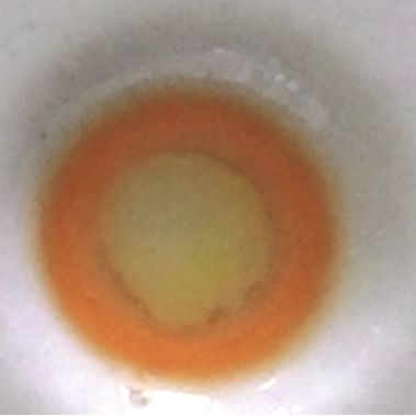
ment are ferric chloride, Folin-Ciocalteu reagent, Nessler’s Table 1 shows that the solution color changed after parac-
reagent, and Lieberman reagent. Ferric chloride 0.5 M was etamol was added to the FeCl3 , Folin-Ciocalteu, and Nessler's
made in water, whereas Folin-Ciocalteu reagent was made by reagent. FeCl3 gives blue and black colors, and the Folin-
dissolving 100 g sodium tungstate and 25 g sodium molybdate Ciocalteu gives a dark blue color when this reagent is reacted
in 500 ml water, then 50 ml phosphoric acid and 100 ml chlo- with phenol on paracetamol. Nessler reagent is aliphatic
ride acid were added and refluxed for 10 hours. The solution amide detection and provides an orange-brown colloid when
is cooled at room temperature, and 150 ml lithium sulfate, 50 paracetamol is added. Sodium hydroxide solution is used to
ml water, and four drop bromine solution were added and stabilize the complexity of the reaction between paracetamol
allowed to react for 2 hours. The solution is boiled for 15 and reagent. The color of Lieberman reagent solution will
minutes, allowed to cool, and then filtered and diluted up to change from colorless to violet if it is heated and is added to a
1000 ml. The reagent was used by diluting it with two portions single substitute benzene [14]. In this experiment, the heating
of water and by adding 4M sodium hydroxide for basic procedure is avoided, because it is not applicable to optical
condition. Nessler’s reagent was made by adding potassium sensor membrane. As a result, the color of Lieberman reagent
iodide to the mercury chloride saturated solution. Lieber- did not change, so this reagent did not continue to be used in
mann reagent was made by dissolving 5 g of sodium nitrite in optical membrane sensor.
50 ml sulphuric acid. These reagents were then tested against
paracetamol standards to know its color changes. 3.2. Characterization of Interaction between Optical Sen-
sor Membrane and Paracetamol. Poly(methyl methacrylate)
2.2. Fabrication of Optical Sensor Membrane Using PMMA. (PMMA) is a highly weather-resistant polymer and it is
PMMA was made in three different concentrations: 5%, 7.5%, optically transparent [19]. The chemical inertness of PMMA
and 10%. Each concentration was made by dissolving it in a makes it a good candidate to be applied in optical sensor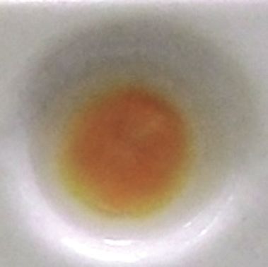
International Journal of Analytical Chemistry                                                                                          3

                                Table 1: Color change of the reagent in the presence of paracetamol (100 mg/ml).

Reagent                                      Reagent solution                        Blank                         Added paracetamol

FeCl3

Folin-ciocalteu

Nessler’s reagent

Lieberman

Blank: reagent solution + ethanol as a paracetamol solvent.

membrane. The critical point in this fabrication is choosing              complete, 100 mg/ml of paracetamol solution was added to
the right solvent that can dissolve the PMMA and the                      the membrane and was allowed to react with the reagent.
reagent. All of the specific reagents for paracetamol (ferric                 Table 2 shows that all of the variations of optical sensor
chloride, Folin-Ciocalteu reagent, and Nessler’s reagent) are             membrane give a positive result with different reaction time.
water soluble. The solubility of PMMA can be predicted by                 This reaction time is affected by the size of the pore on
Hildebrand solubility parameter. If the polymer and solvent               the membrane. The response time increased along with the
have similar Hildebrand solubility value, the polymer will be             increase of the concentration of the polymer. In this system,
easily soluble in that solvent [20]. The Hildebrand solubility            when the concentration of the polymer increases, the con-
                                                                          centration of the reagent decreases. The high concentration
value of PMMA is 9.3 kal1/2 cm−3/2 [21]. The solvent that
                                                                          of polymer makes the membrane more dense [23] and the
has similar value and that is possible to dissolve the reagent
                                                                          sample needs more time to adsorb and react with the small
is ethyl acetate with Hildebrand solubility value of 9.1 kal1/2           concentration of reagent in the membrane. The optical sensor
cm−3/2 [22]. Then, ethyl acetate is used as a solvent to                  membrane of PMMA-FeCl3 gives a positive result with the
dissolve the PMMA and the reagent. Based on a preliminary                 shortest time reaction. The best performance of this sensor is
study (data were not shown), the concentration of PMMA                    7.5% PMMA with mixture solvent ratio of 7:3.
that is below 5% produces a fragile membrane, whereas the                     As shown in Table 3, the color of this sensor changed
concentration that is above 10% produces a dense membrane                 from yellow to blue when it reacted with paracetamol.
and it makes it difficult to absorb the reagent. Therefore,               The color of sensor membrane of PMMA-Folin-Ciocalteu
the PMMA was prepared in the concentration of 5%, 7.5%,                   changed from green to dark blue in basic condition and from
and 10% of the ratio of the solvent and reagent is 6:4, 7:3,              white to brown for PMMA-Nessler, with the best result of
and 8:2 for each concentration. After the fabrication was                 5% PMMA in 6:4 mixture solvent for both. When all of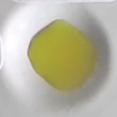
4                                                                                                   International Journal of Analytical Chemistry

                               Table 2: Characterization of optical sensor membrane in the presence of paracetamol.

                                                       Mixture Solvent                                                                Reaction
Reagent                       %PMMA                                                     Homogeneous polymer            Result
                                                        (EA:Reagent)                                                                    time
                                                               6:4                        Non-homogeneous                +                 1”
                                  5%                           7:3                        Non-homogeneous                +                 1”
                                                               8:2                     Non-homogeneous, fragile          +                 1”
                                                               6:4                      Homogeneous, fragile             +                 1”
FeCl3                            7.5%                          7:3                          Homogeneous                  +                 1”
                                                               8:2                          Homogeneous                  +                 1”
                                                               6:4                        Non-homogeneous                +                 1”
                                  10%                          7:3                          Homogeneous                  +                 1”
                                                               8:2                        Non-homogeneous                +                 1”
                                                               6:4                          Homogeneous                  +                 5”
                                  5%                           7:3                          Homogeneous                  +                11”
                                                               8:2                          Homogeneous                  +                10”
                                                               6:4                          Homogeneous                  +                 7”
Folin-Ciocalteu                  7.5%                          7:3                          Homogeneous                  +                10”
                                                               8:2                          Homogeneous                  +                41”
                                                               6:4                          Homogeneous                  +                45”
                                  10%                          7:3                          Homogeneous                  +                50”
                                                               8:2                          Homogeneous                  +               1’31”
                                                               6:4                         Homogeneous                   +                7”
                                  5%                           7:3                         Homogeneous                   +               12”
                                                               8:2                         Homogeneous                   +              1’37”
                                                               6:4                        Non-homogeneous                +               11”
Nessler’s                        7.5%                          7:3                        Non-homogeneous                +               32”
                                                               8:2                        Non-homogeneous                +              8’40”
                                                               6:4                         Homogeneous                   +               12”
                                  10%                          7:3                         Homogeneous                   +               55”
                                                               8:2                         Homogeneous                   +              6’30”
EA: ethyl acetate; (+): suitable color changes; (‘): minute; (“): second.

                                    Table 3: Color test of optical sensor membrane in the presence of paracetamol.

Optical sensor membrane                                                     Membrane                                   Added paracetamol

PMMA-FeCl3
7.5% 7:3

PMMA-Folin-Ciocalteu
5% 6:4

PMMA-Nessler’s reagent
5% 6:4
International Journal of Analytical Chemistry                                                                                       5

        Table 4: The elemental content of the membrane.              3.4. Performance of Optical Sensor Membrane
                                                  Element            3.4.1. Sensitivity Test. The sensitivity level of optical sensors
Membrane                      Element
                                              concentration (%)
                                                                     membrane was analyzed by determining the lowest measur-
                              Carbon                70.944           able concentration of paracetamol that can be detected. In
PMMA-FeCl3 7.5% 7:3           Oxygen                26.268           this experiment, different amounts of paracetamol (0 – 20
                              Chlorine               1.846           mg/ml) were added to the membrane. The time reaction of
                               Iron                  0.942           the color change in the presence of paracetamol was also
                                                                     observed. The design of optical sensor membrane for parac-
                              Carbon                59.462
                                                                     etamol detection will contain three strips of PMMA-FeCl3 ;
                              Oxygen                36.219           PMMA-Folin-Ciocalteu; PMMA-Nessler. Table 5 shows that
                              Sodium                 1.233           the result is in accordance with the minimum dosage form
PMMA-Folin-Ciocalteu
5% 6:4                       Phosphor                0.674           of paracetamol usually added to herbal medicine at 500 mg/
                              Sulphur                1.432           3000 mg. The different lowest concentration and reaction
                             Chlorine                0.651           time of the membrane were influenced by a particular
                                                                     reagent. This result corresponds to Table 2 that FeCl3 has the
                             Tungsten                0.330
                                                                     shortest response time. The intensity of the color change in-
                              Carbon                72.478           creased along with the increase of paracetamol concentration.
                              Oxygen                18.102
PMMA-Nessler’s reagent
5% 6:4                        Chlorine               5.741           3.4.2. Stability Test. In this experiment, the optical sensor
                             Potassium               1.074           membrane was stored in a zip-lock plastic at room temper-
                              Mercury               2.605            ature. The membranes were tested by reacting with paraceta-
                                                                     mol daily until the color ceased to change when paracetamol
                                                                     was added. The results are summarized in Table 6.
Table 5: The lowest measurable concentration of paracetamol on
the membrane.
                                                                     3.4.3. Selectivity Test. In addition to paracetamol, metampy-
Optical sensor membrane          Concentration     Reaction time     rone, acetosal, and ibuprofen were usually added to herbal
                                    mg/ml                            medicine because they have the same therapeutic effect.
PMMA- FeCl3 7.5% 7:3                  2.55             3’ 17”        Chemically, all of these drugs have a similar structure to
PMMA-Folin-Ciocalteu 5% 6:4           4.05               5’          paracetamol. The selectivity of the membrane was evalu-
PMMA-Nessler 5% 6:4                                    2’ 50”        ated by testing the membrane with these chemical drugs.
                                      4.01
                                                                     Metampyrone, acetosal, and ibuprofen were dissolved in
                                                                     ethanol (100 mg/ml) and were then added to the membrane,
     Table 6: The stability time of optical sensor membrane.         respectively. As shown in Table 7, the color of PMMA-
                                                                     FeCl3 changed to brown and purple when metampyrone
Optical sensor membrane                       Stability time (day)
                                                                     and acetosal were added, respectively. This result does not
PMMA-FeCl3 7.5% 7:3                                    122           affect the determination of paracetamol since the color
PMMA-Folin-Ciocalteu 5% 6:4                            125           of the membrane in the presence of paracetamol is blue.
PMMA-Nessler 5% 6:4                                    125           The PMMA-Folin-Ciocalteu membrane gives the same color
                                                                     when paracetamol and metampyrone were added. To show
                                                                     that paracetamol is a presence in the sample, the three
these membranes (PMMA-FeCl3 ; PMMA-Folin-Ciocalteu;                  membranes must give the positive result. Although the
PMMA-Nessler) give the positive result, the sample contains          PMMA-Folin-Ciocalteu membrane gives the same color
paracetamol.                                                         when metampyrone was added, a different color is obtained
                                                                     on PMMA-FeCl3 and no color change is observed on PMMA-
3.3. Characterization of Optical Sensor Membrane Using               Nessler. This result is technically applicable and it does not
Scanning Electron Microscope-Energy Dispersive X-Ray (SEM-           break government regulation since the regulation states that
EDX). The best result of optical sensor membrane was                 herbal medicine should not contain the chemical drug.
characterized by Scanning Electron Microscope-Energy Dis-
persive X-Ray (SEM-EDX) to observe the morphology of                 3.4.4. Application for Real Sample. To demonstrate the appli-
membrane and to confirm the element on the membrane.                 cability of optical sensor membrane for real sample analysis,
Visually, all of these membranes are homogeneous (Table 2).          20 herbal medicine samples were analyzed using spectropho-
As shown in Figure 1, the optical sensor membrane of                 tometry UV and optical sensor membrane. Paracetamol was
PMMA-FeCl3 has the smallest pore and is more homogenous              not detectable in these samples by using the developed optical
compared to the others. It causes this membrane to have the          sensor membrane and spectrophotometry UV. This can show
shortest time reaction. In the following, Table 4 shows that         that the sample which contains herbal medicine does not
the elements present in the membrane are the same as the             have an effect on the paracetamol detection. The sample
constituent element. This result confirms that the reagent is        was then spiked with 4.5 mg/ml of paracetamol. The results
absorbed into the membrane and it reacts with paracetamol.           show good agreement between optical sensor membrane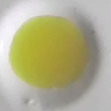
6                                                                                         International Journal of Analytical Chemistry

                                        Table 7: The robustness test of optical sensor membrane.

                                   Metampyrone                                Acetosal                              Ibuprofen
Optical sensor membrane           Color             Reaction            Color               Reaction          Color              Reaction
                                 change               time             change                 time           change                time
PMMA-FeCl3
                                 Brown                 1”              Purple                  1”               -                    -
7.5% 7:3
PMMA-Folin-Ciocalteu
                               Dark blue             3’ 57”               -                    -                -                    -
5% 6:4
PMMA-Nessler
                                    -                   -                 -                    -                -                    -
5% 6:4

              Table 8: Reaction time of optical sensor membrane on detection of paracetamol in herbal medicine sample.

                                                    Reaction time
Sample                          PMMA-FeCl3                                      PMMA-Folin                                PMMA-Nessler
                                  7,5% 7:3                                        5% 6:4                                    5% 6:4
1                                   5’45”                                           8’20”                                       11’25”
2                                       5’                                          8’10”                                       11’40”
3                                   5’25”                                           8’45”                                       11’05”
4                                   5’06”                                           8’30”                                       12’05”
5                                   5’37”                                           9’03”                                       11’45”
6                                   5’20”                                           8’50”                                       11’59”
7                                   6’01”                                           8’16”                                       11’45”
8                                   5’55”                                           8’34”                                       11’39”
9                                   5’05”                                           8’25”                                       11’46”
10                                  5’10”                                           8’43”                                       11’49”
11                                  5’30”                                            9’                                         12’07”
12                                  5’45”                                           8’12”                                       13’03”
13                                  6’06”                                           8’05”                                       11’55”
14                                  5’19”                                           8’56”                                       11’53”
15                                  5’05”                                           8’03”                                       11’57”
16                                  5’10”                                           7’58”                                       12’07”
17                                  5’58”                                           8’27”                                       11’55”
18                                  5’07”                                           8’50”                                       11’59”
19                                  5’29”                                           8’15”                                       11’45”
20                                  5’48”                                           8’28”                                       11’58”

method and spectrophotometry data. Both methods confirm               membrane depends on the reagent and the concentration
that the sample contains paracetamol. The reaction time               of polymer and solvent that are mixed into the membrane.
of the membrane to detect paracetamol is summarized in                The best results of optical sensor membrane are PMMA-
Table 8.                                                              FeCl3 7.5% (7:3), PMMA-Folin 5% (6:4), and PMMA-Nessler
                                                                      5% (6:4). In general, all of the optical sensor membranes
4. Conclusions                                                        are simple and applicable for on-site paracetamol analysis in
                                                                      herbal medicine.
The poly(methyl methacrylate) (PMMA) is a polymer that
can be developed as optical sensor membrane. This mem-                Data Availability
brane will absorb specific colorimetric reagent for paraceta-
mol detection, which makes it applicable for paracetamol              The data used to support the findings of this study are
detection in herbal medicine. The characteristic of this              included within the article.
International Journal of Analytical Chemistry                                                                                                            7

                          (a)                                             (b)                                              (c)

Figure 1: Morphology of the optical sensor membrane by SEM-EDX: (a) PMMA-FeCl3 7.5% 7:3; (b) PMMA-Folin-Ciocalteu 5% 6:4; (c)
PMMA-Nessler 5% 6:4.

Conflicts of Interest                                                                  based on CuNPs/fullerene-C60/MWCNTs: An electrochemical
                                                                                       nanosensor for trace recognition of paracetamol,” Analytica
The authors declare that there are no conflicts of interest                            Chimica Acta, vol. 917, pp. 107–116, 2016.
regarding the publication of this paper.                                         [9]   A. Mao, H. Li, D. Jin, L. Yu, and X. Hu, “Fabrication of electro-
                                                                                       chemical sensor for paracetamol based on multi-walled carbon
Acknowledgments                                                                        nanotubes and chitosan-copper complex by self-assembly tech-
                                                                                       nique,” Talanta, vol. 144, pp. 252–257, 2015.
This research was financially support by Faculty of Pharmacy,                   [10]   Y. Teng, L. Fan, Y. Dai, M. Zhong, X. Lu, and X. Kan, “Elec-
Universitas Padjadjaran, through Hibah Internal Fakultas                               trochemical sensor for paracetamol recognition and detection
Grant [223-2016].                                                                      based on catalytic and imprinted composite film,” Biosensors
                                                                                       and Bioelectronics, vol. 71, pp. 137–142, 2015.
References                                                                      [11]   L. Y. Shiroma, M. Santhiago, A. L. Gobbi, and L. T. Kubota,
                                                                                       “Separation and electrochemical detection of paracetamol and
[1] T. J. Meredith and R. Goulding, “Paracetamol,” Postgraduate                        4-aminophenol in a paper-based microfluidic device,” Analytica
    Medical Journal, vol. 56, no. 657, pp. 459–473, 1980.                              Chimica Acta, vol. 725, pp. 44–50, 2012.
[2] M. T. Olaleye and B. T. J. Rocha, “Acetaminophen-induced                    [12]   R. N. Gupta, R. Pickersgill, and M. Stefanec, “Colorimetric
    liver damage in mice: effects of some medicinal plants on the                      determination of acetaminophen,” Clinical Biochemistry, vol. 16,
    oxidative defense system,” Experimental and Toxicologic Patho-                     no. 4, pp. 220-221, 1983.
    logy, vol. 59, no. 5, pp. 319–327, 2008.
                                                                                [13]   M. E. Bosch, A. J. R. Sánchez, F. S. Rojas, and C. B. Ojeda,
[3] I. Dalli, D. Ramdhani, and A. N. Hasanah, “Design of indica-                       “Determination of paracetamol: Historical evolution,” Journal
    tor strip using polystyrene (PS) and polymethylmethacrylate                        of Pharmaceutical and Biomedical Analysis, vol. 42, no. 3, pp.
    (PMMA) for detection of diclofenac sodium in traditional pain                      291–321, 2006.
    relief herbal medicines,” Indonesian Journal of Chemistry, vol. 17,
                                                                                [14]   A. C. Moffat, Clarke’s Isolation and Identification of Drugs, The
    no. 1, pp. 71–78, 2017.
                                                                                       Pharmaceutical Press, London, UK, 2nd edition, 1986.
[4] Sirajuddin, A. R. Khaskheli, A. Shah, M. I. Bhanger, A. Niaz, and
    S. Mahesar, “Simpler spectrophotometric assay of paracetamol                [15]   L. Zhao, M. Li, M. Liu, Y. Zhang, C. Wu, and Y. Zhang, “Porphy-
    in tablets and urine samples,” Spectrochimica Acta Part A: Mole-                   rin-functionalized porous polysulfone membrane towards an
    cular and Biomolecular Spectroscopy, vol. 68, no. 3, pp. 747–751,                  optical sensor membrane for sorption and detection of cad-
    2007.                                                                              mium(II),” Journal of Hazardous Materials, vol. 301, pp. 233–241,
                                                                                       2016.
[5] E. A. Abdelaleem and N. S. Abdelwahab, “Validated stability
    indicating RP-HPLC method for determination of paracetamol,                 [16]   C.-W. Tsao, “Polymer microfluidics: Simple, low-cost fabrica-
    methocarbamol and their related substances,” Analytical Meth-                      tion process bridging academic lab research to commercialized
    ods, vol. 5, no. 2, pp. 541–545, 2013.                                             production,” Micromachines, vol. 7, no. 12, article no. 225, 2016.
[6] R. R. Cunha, S. C. Chaves, M. M. A. C. Ribeiro et al., “Simulta-            [17]   C.-W. Tsao and D. L. DeVoe, “Bonding of thermoplastic poly-
    neous determination of caffeine, paracetamol, and ibuprofen in                     mer microfluidics,” Microfluidics and Nanofluidics, vol. 6, no. 1,
    pharmaceutical formulations by high-performance liquid chro-                       pp. 1–16, 2009.
    matography with UV detection and by capillary electrophoresis               [18]   S. Mei, C. Xiao, and X. Hu, “Preparation of porous PVC mem-
    with conductivity detection,” Journal of Separation Science, vol.                  brane via a phase inversion method from PVC/DMAc/water/
    38, no. 10, pp. 1657–1662, 2015.                                                   additives,” Journal of Applied Polymer Science, vol. 120, no. 1, pp.
[7] H. Li, C. Zhang, J. Wang, Y. Jiang, J. P. Fawcett, and J. Gu,                      557–562, 2011.
    “Simultaneous quantitation of paracetamol, caffeine, pseudoe-               [19]   A. Nese, S. Sen, M. A. Tasdelen, N. Nugay, and Y. Yagci, “Clay-
    phedrine, chlorpheniramine and cloperastine in human plasma                        PMMA nanocomposites by photoinitiated radical polymer-
    by liquid chromatography-tandem mass spectrometry,” Journal                        ization using intercalated phenacyl pyridinium salt initiators,”
    of Pharmaceutical and Biomedical Analysis, vol. 51, no. 3, pp.                     Macromolecular Chemistry and Physics, vol. 207, no. 9, pp. 820–
    716–722, 2010.                                                                     826, 2006.
[8] P. K. Brahman, L. Suresh, V. Lokesh, and S. Nizamuddin, “Fab-               [20]   M. Belmares, M. Blanco, W. A. Goddard et al., “Hildebrand
    rication of highly sensitive and selective nanocomposite film                      and Hansen solubility parameters from Molecular Dynamics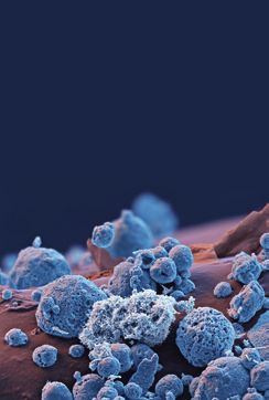
8                                                                        International Journal of Analytical Chemistry

     with applications to electronic nose polymer sensors,” Journal of
     Computational Chemistry, vol. 25, no. 15, pp. 1814–1826, 2004.
[21] A. Y. Kwok, G. G. Qiao, and D. H. Solomon, “Synthetic hydro-
     gels 3. Solvent effects on poly(2-hydroxyethyl methacrylate)
     networks,” Polymer Journal, vol. 45, no. 12, pp. 4017–4027, 2004.
[22] J. Burke, “Solubility parameters: theory and application,” 1984,
     http://cool.conservation-us.org/coolaic/sg/bpg/annual/v03/bp03-
     04.html.
[23] M. Yoo, S. Kim, J. Lim et al., “Facile synthesis of thermally
     stable core-shell gold nanoparticles via photo-cross-linkable
     polymeric ligands,” Macromolecules , vol. 43, no. 7, pp. 3570–
     3575, 2010.
Nanomaterial
Nanomaterials
         Journal of

                                          Journal of
                                                                             The Scientific
                                                                                                                                                                     Photoenergy
                                                                                                                                                                               International Journal of
                                          Analytical Methods                                                                Journal of

Hindawi
                                          in Chemistry
                                          Hindawi
                                                                             World Journal
                                                                             Hindawi Publishing Corporation
                                                                                                                            Applied Chemistry
                                                                                                                            Hindawi                                  Hindawi
www.hindawi.com            Volume 2018    www.hindawi.com      Volume 2018   http://www.hindawi.com
                                                                             www.hindawi.com                  Volume 2018
                                                                                                                     2013   www.hindawi.com            Volume 2018   www.hindawi.com                      Volume 2018

Advances in                                                                                                                                                          International Journal of
Physical Chemistry
Hindawi
                                                                                                                                                                     Medicinal Chemistry
                                                                                                                                                                     Hindawi
www.hindawi.com             Volume 2018                                                                                                                              www.hindawi.com                      Volume 2018

                                                            Submit your manuscripts at
                                                               www.hindawi.com

Bioinorganic Chemistry                                                                                                                                               Journal of
and Applications
Hindawi
                                                                                                                                                                     Materials
                                                                                                                                                                     Hindawi
www.hindawi.com             Volume 2018                                                                                                                              www.hindawi.com                      Volume 2018

Advances in                               Journal of                         BioMed                                         International Journal of                 International Journal of
Tribology
Hindawi
                                          Chemistry
                                          Hindawi
                                                                             Research International
                                                                             Hindawi
                                                                                                                            Spectroscopy
                                                                                                                            Hindawi
                                                                                                                                                                     Electrochemistry
                                                                                                                                                                     Hindawi
www.hindawi.com             Volume 2018   www.hindawi.com      Volume 2018   www.hindawi.com                  Volume 2018   www.hindawi.com            Volume 2018   www.hindawi.com                      Volume 2018

International Journal of                  Journal of                         Journal of                                     Enzyme                                   Biochemistry
Analytical Chemistry
Hindawi
                                          Spectroscopy
                                          Hindawi
                                                                             Nanotechnology
                                                                             Hindawi
                                                                                                                            Research
                                                                                                                            Hindawi
                                                                                                                                                                     Research International
                                                                                                                                                                     Hindawi
www.hindawi.com             Volume 2018   www.hindawi.com      Volume 2018   www.hindawi.com                  Volume 2018   www.hindawi.com            Volume 2018   www.hindawi.com                      Volume 2018
You can also read