The Implications for Cells of the Lipid Switches Driven by Protein-Membrane Interactions and the Development of Membrane Lipid Therapy - MDPI
←
→
Page content transcription
If your browser does not render page correctly, please read the page content below
International Journal of
            Molecular Sciences

Review
The Implications for Cells of the Lipid Switches
Driven by Protein–Membrane Interactions and the
Development of Membrane Lipid Therapy
Manuel Torres 1,2 , Catalina Ana Rosselló 1,2 , Paula Fernández-García 1,2 , Victoria Lladó 1,2 ,
Or Kakhlon 3 and Pablo Vicente Escribá 1, *
 1    Laboratory of Molecular Cell Biomedicine, Department of Biology, University of the Balearic Islands, Ctra.
      de Valldemossa km 7.5, E-07122 Palma, Spain; manuel.torres@luib.es (M.T.);
      ca.rossello@laminarpharma.com (C.A.R.); p.fernandez@laminarpharma.com (P.F.-G.);
      victoria.llado@laminarpharma.com (V.L.)
 2    Department of R&D, Laminar Pharmaceuticals SL. ParcBit, Ed. Naorte B, E-07121 Palma, Spain
 3    Department of Neurology, Hadassah-Hebrew University Medical Center, Ein Kerem, 91120 Jerusalem, Israel;
      ork@hadassah.org.il
 *    Correspondence: pablo.escriba@uib.es

 Received: 2 March 2020; Accepted: 19 March 2020; Published: 27 March 2020

 Abstract: The cell membrane contains a variety of receptors that interact with signaling
 molecules. However, agonist–receptor interactions not always activate a signaling cascade.
 Amphitropic membrane proteins are required for signal propagation upon ligand-induced receptor
 activation. These proteins localize to the plasma membrane or internal compartments; however,
 they are only activated by ligand-receptor complexes when both come into physical contact in
 membranes. These interactions enable signal propagation. Thus, signals may not propagate into
 the cell if peripheral proteins do not co-localize with receptors even in the presence of messengers.
 As the translocation of an amphitropic protein greatly depends on the membrane’s lipid composition,
 regulation of the lipid bilayer emerges as a novel therapeutic strategy. Some of the signals controlled
 by proteins non-permanently bound to membranes produce dramatic changes in the cell’s physiology.
 Indeed, changes in membrane lipids induce translocation of dozens of peripheral signaling proteins
 from or to the plasma membrane, which controls how cells behave. We called these changes “lipid
 switches”, as they alter the cell’s status (e.g., proliferation, differentiation, death, etc.) in response to
 the modulation of membrane lipids. Indeed, this discovery enables therapeutic interventions that
 modify the bilayer’s lipids, an approach known as membrane-lipid therapy (MLT) or melitherapy.

 Keywords: protein–membrane interactions; melitherapy; lipid bilayer; membrane lipid switch;
 peripheral amphitropic non-permanently bound membrane proteins

1. Introduction
      The fluid mosaic model of cell membranes [1] contemplates the incorporation of integral
transmembrane into their structure and their mobility in the bilayer as well as the association
of peripheral proteins. Later studies demonstrated that these proteins participate in transmembrane
communication in response to signals from neurotransmitters, hormones, cytokines, growth factors,
etc. [2,3]. These productive interactions activate intracellular signaling cascades in which second and
subsequent messengers regulate the expression of the genes that control the cell’s physiology [4].
In addition to this short-term messaging, mid- and long-term cytosolic and nuclear responses, the latter
mediated by the regulation of gene expression, can affect the cell’s behavior over several hours, days,
and even weeks [5]. In this regard, several issues must be considered. First, the physical interaction

Int. J. Mol. Sci. 2020, 21, 2322; doi:10.3390/ijms21072322                            www.mdpi.com/journal/ijms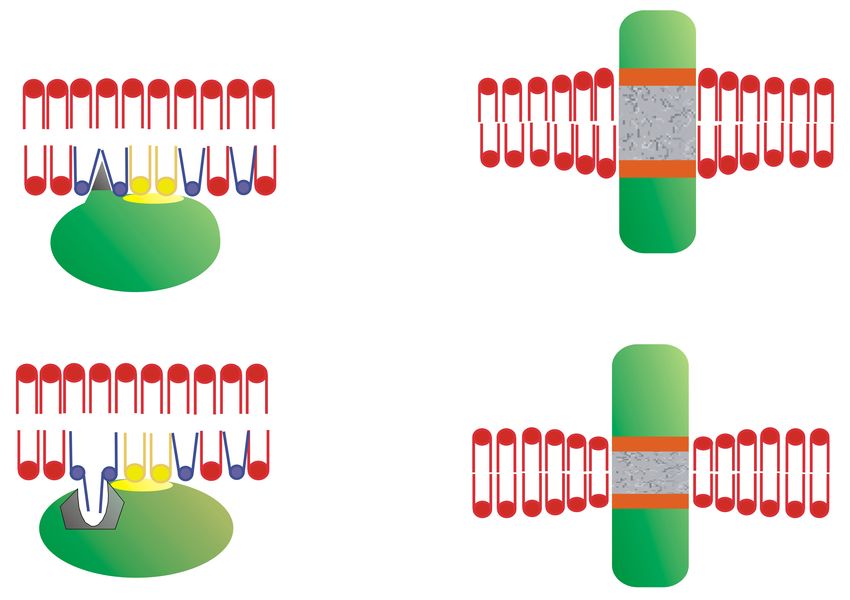
Int. J. Mol. Sci. 2020, 21, 2322                                                                     2 of 39

between membrane receptors and the amphitropic membrane proteins (either directly or through
adaptor or scaffolding proteins) is necessary for the transduction of most cell signals [6,7]. Second,
this interaction may not only depend on the expression of these proteins but also on the presence
of the peripheral proteins in the vicinity of the membrane receptor, which may be controlled by
membrane lipids [8,9]. Third, these interactions and the signals they produce are responsible for the
pathophysiological status of the cell, which may be influenced by external cues, genetic alterations,
alterations in membrane lipids, etc. [10,11]. Fourth, changes in the membrane lipid composition can
induce important changes in the cell that affect proliferation, differentiation, and/or cell death [12,13].
Last but not least, the regulation of membrane lipids controls the type and abundance of the proteins
in membranes, an approach that can be used to treat several conditions, including cancer, Alzheimer’s
disease (AD), cardiovascular diseases (CVDs), inflammation, etc. [14–17].
      In this context, the ability of membranes to generate microdomains due to the non-homogeneous
mixing of membrane lipids is critical [18,19]. A variety of microdomains have been described in
which either lamellar-prone or non-lamellar-prone lipids organize into different ordered or disordered
lipid platforms [8,20–22]. These membrane regions with varying size can be distinguished from their
adjacent microdomains in terms of their lipid and protein composition, bilayer thickness, lateral surface
pressure, acyl chain mobility, membrane morphology, etc. Microdomains with a high proportion of
hexagonal (HII ) phase-prone lipids, such as phosphatidylethanolamine (PE) or diacylglycerol (DAG),
are critical in the recruitment of peripheral amphitropic signaling proteins and thus, for cell growth and
differentiation [8,23]. Recent studies showed that the proportion of peripheral amphitropic signaling
proteins in membranes or aqueous compartments depends on both the membrane’s lipid composition,
and the amino acid sequence involved in protein–lipid interactions and co/post-translational lipid
modification of proteins [9,24]. Moreover, alterations to the peripheral signaling proteins at membranes
and in the cytosol have been associated with a variety of pathologies [25,26]. The ability to signal
proteins to translocate from the plasma membrane to intracellular compartments is a fundamental
means to regulate cell signaling. Indeed, the interactions of messengers with their membrane receptors
are nullified if the conformational change induced is not propagated from the receptor to its transducer.
      Protein–lipid interactions have a strong influence on the activity of cells, and, therefore, it is
important to investigate these interactions in basic biomedical and clinical studies. A number of
issues have to be considered, including how a protein’s structure defines its presence in membranes,
their abundance in different membrane regions or microdomains or in the cytoplasm, and how
protein structure regulates protein–lipid interactions. In addition, it is important to consider how
the membrane’s structure influences protein–lipid interactions, and how the pathophysiological and
pharmaceutical/nutraceutical regulation of membrane composition affects cell signaling. Studies with
specific proteins and lipids will be addressed in the first part of this review to explain how these
interactions occur. Subsequently, the coordinated effect of several proteins and lipids on general
pathophysiological processes will be addressed through what we define as “lipid switches”.

2. How Protein Structure Influences Protein–Lipid Interactions
      Peripheral amphitropic proteins can translocate from aqueous to membranous compartments
and they display a variety of structural features that define their interactions with membranes.
These proteins bear lipid or amino acid motifs that drive their interactions with specific lipid species
or lipid structures. In these interactions, the structure of both elements is crucial to understand how
they occur and how they can be modulated in relation to the cell’s physiology. We first address the
structural elements of proteins that influence their binding to cell membranes.
      Co-translational and post-translational lipid modifications in proteins are critical to influence
protein–lipid interactions, fulfilling roles above and beyond the mere binding of the protein to the
lipid bilayer [27]. These modifications contribute to the peripheral and transmembrane localization of
proteins at/in specific organelle membranes and membrane regions or microdomains [9,21,23,28–30].
In addition, certain amino acid domains in proteins that drive their binding to lipids influence their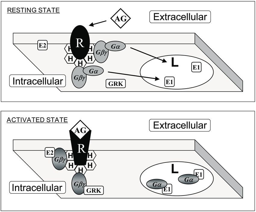
Int. J. Mol. Sci. 2020, 21, 2322 3 of 39
mobilization from the aqueous to particulate (membrane) compartments. Thus, there are proteins
that bind to membranes through post-translational lipid modifications (or covalent-lipid proteins,
CLPs) and others that bind through hydrophobic amino acid domains (or lipid-binding proteins, LBPs).
Interestingly, both types of interactions can be regulated, given that some covalent modifications
that influence the way proteins are sorted to different organelles or membrane microdomains are
reversible (e.g., S-palmitoylation). This fatty acylation regulates transmembrane protein transport
through the Golgi apparatus [31], protein–lipid raft interactions [32], the activity of G-protein coupled
receptors (GPCRs), and other membrane receptors [33]. Membrane lipid structure can also modify
these interactions as addressed below.
Palmitoylation also affects peripheral proteins, such as G proteins [34], and interestingly, most
G protein alpha subunits (Gα) are reversibly modified by palmitoylation [35]. Moreover, G protein
activation by GPCRs has been associated with increased palmitoylation, and the turnover and activation
of the Gαs subunit produces its reversible mobilization from the plasma membrane to the cytosol [36].
Inactive Gα subunits localize to non-lamellar-prone membrane microdomains that are rich in PE,
where they bind to Gβγ dimers to form Gαβγ heterotrimers [23]. In contrast to lipid rafts, these PE-rich
microdomains form liquid disordered (Ld) bilayer regions due to the low surface lipid packing and
high acyl chain mobility [8,37]. The strong affinity of the Gβγ dimer for these microdomains facilitates
the piggyback transport of the Gα subunit towards GPCRs, facilitating their productive interaction
with activated receptors [23]. The G protein activation induces a dissociation of the Gα subunit from
the Gβγ dimer. These Gα monomers have a preference for lipid raft (liquid ordered, Lo ) microdomains,
and they are mobilized to raft-like microdomains where they interact with signaling effectors, including
adenylyl cyclase, guanylyl cyclase, phospholipase C, ion channels, etc. [9,23,24]. Both electrostatic
and hydrophobic interactions participate in this peripheral protein mobilization from one membrane
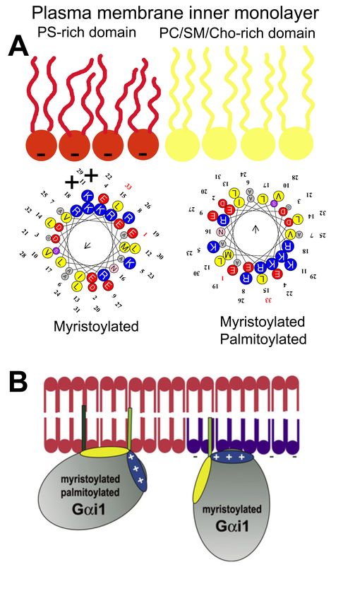
microdomain to another. In this scenario, Gαi1 protein palmitoylation is critical for its translocation
from phosphatidylserine (PS)-rich microdomains to microdomains with a high lamellar propensity,
mainly rich in phosphatidylcholine (PC) and/or sphingomyelin (SM) and cholesterol (Chol: [9]). This is
due to the fact that the N-terminal α-helix of the Gαi1 protein interacts with negatively charged
(PS-rich) membrane microdomains through a positively charged amino acid patch when the protein
is myristoylated but not palmitoylated [9]. However, the palmitoylated Gαi1 protein N-terminal
region interacts through an aliphatic or positively charged amino acid patch which induces monomeric
G protein mobilization from negatively charged membrane microdomains to neutral phospholipid
regions (Figure 1).
Thus, fatty acyl moieties in peripheral (amphitropic) membrane proteins not only serve as
anchors for their binding to membranes but also, they drive their mobilization from one membrane
microdomain to another. The relevance of membrane microdomains is that they form “clubs” with
a lipid composition that attracts different types of proteins. Proteins form nanoclusters in different
membrane lipid domains that exert physical interactions with signaling partners, resulting in productive
signaling under the correct circumstances. For signal amplification, a huge number of G proteins must
co-exist with GPCRs, a phenomenon mediated by protein–lipid interactions. Although protein–protein
interactions for GPCR-G protein coupling have been studied intensely (e.g., [38]), the crucial role of
lipids in this binding is not fully understood. In this context, GPCRs have G protein-subtype binding
preferences, although this coupling is not truly specific. One type of GPCR can bind to different
types of G proteins with similar or different affinities, and one G protein subtype can be activated
by different GPCRs [39]. Therefore, differences in expression in defined cells or variations in lipid
composition, which could regulate receptor-G protein interactions, have important consequences
for cell signaling. This is especially important during the pathophysiological and/or therapeutic
regulation of the plasma membrane lipid composition, as lipid modifications alter protein–membrane
interactions, the propagation of cell signals and the cell’s physiology (and even the regulation of gene
expression [20,21,40]).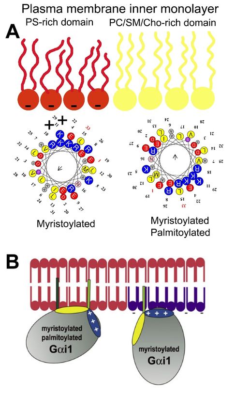
Int. J. Mol. Sci. 2020, 21, 2322                                                                          4 of 39

      Figure 1. Gαi1 protein–membrane interactions. (A) Geometry of the N-terminal α-helix with a myristoyl
      moiety interacting with PS-rich (negatively charged, red) membrane microdomains (left helix), and the
      α-helix with myristoyl and palmitoyl moieties (right helix) that interact with PC and/or SM and/or
      Chol microdomains (yellow). (B) Scheme of acylated Gαi1 protein–membrane interactions (Adapted
      from [9]).

     Other protein anchors frequently found in peripheral CLPs are isoprenyl moieties. This type of
modification involves adding farnesyl or geranylgeranyl residues to the C-terminal regions of signaling
proteins like Ras, Gγ protein, Cdc42, Rho, Rac, etc. Farnesyl (FTase) or geranylgeranyl transferase
(GGTase) catalyze the prenylation of the cysteine residue of the C-terminal CaaX box, where “a” refers
to an aliphatic amino acid and “X” to any amino acid [41]. When “X” is Leu, then the protein is
gerenylgeranylated, whereas the protein is farnesylated if “X” is Ser, Ala, Cys, Gly, Thr, His, Asn or
Gln, and “X” can be modified by both enzymes when it is Met, Val, Ile or Phe [41–44]. In addition,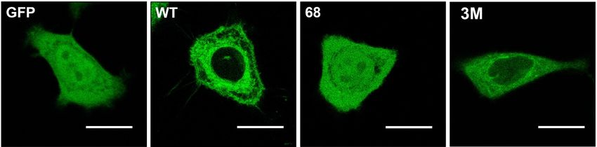
Int. J. Mol. Sci. 2020, 21, 2322                                                                             5 of 39

the CaaX box is also subject to proteolysis by the Ras converting enzyme 1 (RCE1) which removes
the last three amino acids (aaX: [45,46], and to methylation by isoprenyl carboxyl methyltransferase
(ICMT) that further increases hydrophobicity of the C-terminal region [47,48]. The processing of
peripheral signaling proteins with a CaaX motif influences their localization. Thus, C68 mutations in
Gγ2 protein:GFAP chimeras alter the localization of this fusion protein from the membrane to a more
homogeneous cell distribution (Figure 2) [24]. Interestingly, mutation of the polybasic cassette (R62,
K64, and K65) also causes mislocalization of the Gγ2 protein but with a different pattern to that of the
Cys mutations (Figure 2) [24].
 1111111111111

 2222222221
    Figure 2. Gγ2 protein–membrane interactions.         The green fluorescent protein (GFP), containing the
      wild-type C-terminal region of the Gγ2 protein (WT), shows a membrane localization that does not
      coincide with that of GFP alone (GFP). Mutations that alter the presence of the isoprenyl moiety
      (68: C68S) or the 3 C-terminal basic amino acids (3M: R62G, K64G, K65G) have a huge impact on the
      distribution of the protein in SF-767 cells. Bar = 15 µm (adapted from Reference [24]).

        In general, the formation of membrane microdomains with specific lipids favors the presence of
certain peripheral proteins, while hindering the interaction of other proteins. For example, caveolae
(“little caves”) form spatio-temporal platforms where EGFR, Ras, and Raf1 meet to propagate signals
promoting cell growth [49]. Similarly, Lo microdomains (e.g., lipid rafts) are preferred by Gαi1 proteins,
  Int. J. Environ.
whereas            Res. Public Health 2020,
             Ld microdomains           bind17,with
                                               x; doi: FOR affinity
                                                    high   PEER REVIEW                    www.mdpi.com/journal/ijerph
                                                                    to Gαβ and Gαβγ proteins [9,23]. Moreover, the
co-operative binding of Gαi1 proteins to lamellar-prone Lo membranes is in part due to the presence
of myristoyl or palmitoyl moieties [29]. The presence of fatty acyl moieties in GPCR nanoclusters
produced by the presence of G proteins regulates membrane lipid structure in a way that also
enhances the binding of Gαβ and Gαβγ proteins to non-lamellar-prone Ld microdomains but not to
Lo microdomains. By contrast, the presence of farnesyl or geranylgeranyl moieties in lipid bilayers
favors the co-operative binding of Gαβ and Gαβγ proteins to Ld membranes while inducing dramatic
reductions in Gαi1 protein binding to membranes [29,50].
        In addition to isoprenyl or acyl moieties that are found frequently at the C- or N-terminal domains
of amphitropic signaling proteins, a polybasic domain is also found flanking these lipidated amino
acids [9,24,51]. These positively charged amino acid clusters, mainly containing Arg or Lys residues,
define the preference of these proteins for lipid microdomains rich in PS or other negatively charged
lipids, with relaxed specificities, as well as participating in the mobilization of proteins between
membrane microdomains (Figures 1 and 3). In this context, the dynamics of proteins containing
polybasic domains and of membranes with negatively charged amino acids depends largely on
electrostatic interactions. Therefore, membrane areas with a high PS content attract polybasic amino
acid-containing proteins, yet they do not restrict their movement as tightly as membranes with
PtdIns(4,5)P2 [52]. This phenomenon suggests that proteins which prefer membrane microdomains
rich in monovalently charged PS may be more able to move among membrane microdomains than
those proteins that interact with polyvalent anionic lipids like PIP2.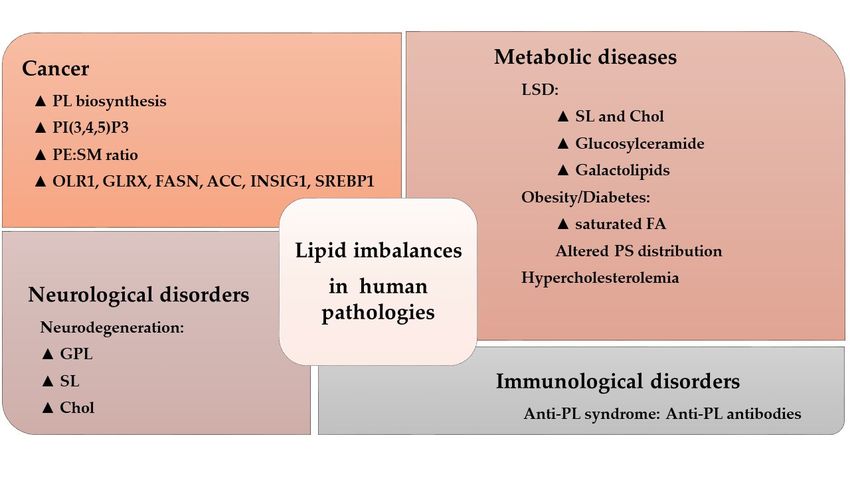
Int. J. Mol. Sci. 2020, 21, 2322                                                                         6 of 39

      Figure 3. Interaction of G proteins with membrane microdomains. Upper panel: G protein-coupled
      receptor (GPCR, R) and heterotrimeric G (Gαβγ) proteins prefer membrane microdomains rich
      in non-lamellar-prone (H) lipids. This effect is driven by prenylated Gαβ dimers. Lower panel:
      Upon receptor-induced activation, acylated Gα subunits are mobilized to lamellar-prone membrane
      microdomains (L). In addition, localization to phosphatidylserine-rich or -poor domains is controled
      by a polybasic domain exposed to the membrane, and not by the presence or absence of a palmitoyl
      moiety (see Figure 1). AG: agonist; E1 and E2: effector protein 1 and 2: GRK: GPCR Receptor Kinase.
      Adapted from Reference [23].

     Other amino acids also participate in CLP–membrane interactions. For example, hydrophobic
amino acids and motifs are involved in the reversible or permanent interaction of proteins with
membranes. One such interaction occurs with the transmembrane domains of integral membrane
proteins. In this context, many structures provide permanent protein anchorage to membranes.
Proteins with only one transmembrane domain have been found, such as Notch [53], or with multiple
domains like the proton-coupled folate transporter [54]. These transmembrane regions frequently
form α-helices, as the GPCRs [55], although some β-barrel structures have also been found like those
found in porins [56].
     However, this work focuses on the interactions of non-permanently bound membrane proteins,
a scenario in which a number of amino acid sequences are known to be involved in reversible
interactions of amphitropic signaling LBPs with membranes. For example, the C1 and C2 protein
domains have been thoroughly studied, the former members of the Cys-rich protein superfamily that
are classified as typical if they bind to DAG and phorbol esters or atypical if they do not [57–59].
This amino acid domain is especially interesting in relation to protein kinase C (PKC), as it may be a dual
C1A/C1B domain in classic and novel PKC isozymes (α, β, α, δ, θ, ε, and η) or a single domain (C1) in
the atypical PKCs (ι/λ, ζ: [58–60]). Interestingly, DAG induces negative curvature strain in membranes
and it favors the appearance of non-lamellar (HII ) lipid structures in vitro [61], a biophysical property
Int. J. Mol. Sci. 2020, 21, 2322 7 of 39
of membranes that regulates PKC binding [8,14]. In biological membranes, DAG reduces the surface
packing strain and the polar head lateral pressure, which allows specific protein and lipid insertion.
When non-lamellar phase propensity is altered by pathophysiological processes or therapeutic agents,
the interaction of PKC (and of other peripheral proteins) is affected, inducing relevant changes in
its interaction with the plasma membrane [8,14]. The C1 domain also recognizes tumor promotor
phorbol esters, which induce non-lamellar phases but in a different manner to that caused by DAG or
PE, such that PKC was associated with conditions related to cell growth, as in cancer [62,63], AD [64],
CVDs [65], and immunological diseases [66], etc. Thus, not only do PKC–membrane interactions
define relevant signaling events, but they also may be involved in pathophysiological and therapeutic
processes [8,14]. Initially identified in PKC, the DAG and phorbol ester responsive C1 domain was
subsequently described in other protein families: the Unc-13 scaffolding proteins, MRCKs, RasGRP
proteins, protein kinase D, chimaerins, and the β and γ DAG kinase isoforms [60]. Protein–membrane
interactions are crucial to therapies and the potential use of the C1 domain as a drug target has already
been proposed [67]. Like the C1 motif, the C2 motif is found in numerous eukaryotic proteins involved
in cell signaling and its conserved sequence serves as a membrane docking motif [68]. It has been
found in many relevant proteins, including PKCα [69], synaptotagmin [70], phospholipase Cα [71],
and cytosolic phospholipase A2 [72]. The C2 domain is involved in the binding of PKCα to PS, an
important phospholipid in the inner leaflet of the plasma membrane [69]. In classic but not novel
PKC isozymes, Ca2+ ions are involved in this binding, which may also occur with other negatively
charged phospholipids [73]. In addition, this domain can also bind PIP2 due to the fact of its lysine
residues [59,73]. The phosphoinositide binding site is located in the β3-β4 strands of the C2 domain
in PKCα [74]. Over one hundred C2 domains have been reported in proteins with a wide range of
signaling functions, covering protein phosphorylation, vesicular transport, lipid modifications, GTPase
regulation, etc. [74]. Despite the homology among these C2 domains, particular structural differences
drive their diverse protein–lipid interactions, suggesting that specific therapies could be targeted to
C2 domains.
Other motifs and domains have been described that favor the interaction of LBPs with membranes.
For example, myelin basic protein (MBP) exerts electrostatic interactions between positively charged
amino acids and negatively charged phospholipids at the plasma membrane [75]. Many peripheral
proteins containing poly-lysine clusters, and like the aforementioned G proteins, they display
electrostatic interactions with negatively charged phospholipids that are modulated by other membrane
lipids [76]. In the case of MBP, it binds around Chol-rich membrane microdomains in which the
presence of SM is required for C1 variants of the MBP but not for C8 variants. This fact appears to be
related to multiple sclerosis (MS) in which more MBPs are found in the brains of patients where SM
levels are lower relative to healthy adults.
In addition to these lipid-binding domains, other protein motifs mediate interactions with
membrane lipids. For example, the spectrin homology 3 (SH3) domain is common to signaling
proteins that mediate protein–protein interactions through binding to proline-rich sequences. However,
SH3 domains may also be involved in protein–lipid interactions, such as in caskin1, which is
involved in binding to lysophosphatidic acid (LPA) and sphingosine-1-phosphate lipids [77]. Similarly,
the pleckstrin homology (PH) domain of phosphoinositide-dependent kinase 1 (PDK1) is involved in
protein–lipid interactions and it was suggested that it might constitute an alternative drug target rather
than the catalytic site [78]. In addition, sterile alpha motifs (SAMs) are amino acid regions usually
involved in protein–protein interactions. Nevertheless, they have also been seen to participate in
protein–membrane interactions in the p73α protein due to the fact of its capacity to bind to membrane
phospholipids [79]. All these protein regions are involved in the interactions with biological membranes
that regulate cell signaling and that represent potential druggable motifs [80]. This approach is one of
the potential means to develop therapies based on the control of the membrane lipid bilayer and the
signals they regulate (i.e., MLT [21]).Int. J. Mol. Sci. 2020, 21, 2322 8 of 39
3. How Membrane Lipid Structure Influences Protein–Lipid Interactions
In general, proteins can interact with membranes via interactions with specific lipid species,
with membrane lipid structures, electrostatic, hydrophobic interactions, etc. Peripheral membrane
proteins can use more than one of these strategies to bind to membranes, which favors their versatile
localization to membrane microdomains or cell compartments, which depends on the membrane lipid
composition. Numerous studies have defined the interactions between peripheral proteins and specific
membrane lipids, some of which were described above. A wide range of proteins have been shown
to interact with Chol, phosphoinositides, PS, SM, free fatty acids (FFAs), etc. However, this section
aims to review the interaction of amphitropic signaling proteins with membrane structures rather
than with specific lipid species. This type of interaction deserves further attention because: (i) the
plasma membrane is a critical hub for signaling proteins; (ii) cells can regulate their lipid composition
according to a range of pathophysiological situations; (iii) membrane lipids organize into different
microdomains rich in specific lipid species, which attract different types of proteins; and (iv) proteins
that prefer certain types of lipid structures can drive productive interactions involving the reception
and propagation of cell signals [8,9,20,23,37]. The interaction of the non-permanently bound membrane
proteins, G proteins and PKC, with non-lamellar-prone (HII ) membrane structures was first described
some years ago [8]. In this context, one of the mechanisms of action by which anthracyclines exert their
antitumor action was through the inhibition of HII -phase propensity and the subsequent mislocalization
of these signaling proteins. This phenomenon explained why anthracyclines could kill cancer cells
solely by interacting with the plasma membrane but not entering the cells [81]. Subsequently, important
modifications of the plasma membrane’s lipid composition by anthracyclines was seen to be relevant
to their mechanism of action [12].
The plasma membrane is a critical element in cell signaling, as almost all incoming or outgoing
messages must cross this barrier. Changes in the plasma membrane lipid composition and structure are
involved in numerous physiological and pathological phenomena. For example, the body temperature
of fish that live in rivers undergoes important variations since the water temperatures can range from 4
to 20 ◦ C. In these fish, important temperature-dependent changes in phospholipid species and fatty
acyl chains can be seen [82,83]. Membrane lipid structure is temperature dependent, changing in
accordance with the temperature of the water in fishes [84]. The changes in lipid species in the brain of
cold-water fishes between summer and winter maintain membrane fluidity, and other biophysical
properties of membranes, constant, which in turn ensures correct protein function and cell signaling in
these animals [82,83]. If temperature and lipid composition can alter membrane lipid structure and cell
signaling, pathophysiological changes and pharmaceutical/nutraceutical interventions that regulate
membrane lipid composition may also influence the health of cells.
Heterotrimeric G proteins were thought to prefer Ld membrane microdomains when extracted
from the rat brain [37]. Using purified G protein monomers (Gα), dimers (Gβγ), and trimers (Gαβγ),
the Gαβ heterodimer was seen to drive the interaction of Gαβγ heterotrimers with membranes [23].
Thus, one of the roles of Gαβ dimers is to bring Gα monomers into contact with GPCRs. Using G protein
mutants, we described the molecular basis of these interactions for different G protein subunits [9,24].
Membrane microdomains rich in PE form liquid disordered (Ld ) membrane microdomains, which differ
in their lipid composition and structure from lipid rafts, caveolae, synaptosomes, and other types or
membrane lipid microdomains. The localization and activity of important peripheral signaling proteins
is very sensitive to changes in membrane structure [8]. Therefore, natural or synthetic molecules
that regulate lipid polymorphism in vitro and membrane microdomains in vivo [85] can regulate the
localization and activity of peripheral membrane proteins, and thereby modulate cell signaling.
Cell membranes favor the formation of multiprotein complexes, in which certain proteins receive
signals, other proteins act as scaffolds, and others participate in signal propagation or second messenger
production. The protein complexes involved in signal propagation are frequently called signalosomes,
and their arrangement and activity is very dependent on membrane lipid composition, altering the
protein–lipid interactions for example those involved in AD [86]. Moreover, sphingolipids appear to beInt. J. Mol. Sci. 2020, 21, 2322 9 of 39
critical in the prognosis of anaplastic lymphoma. Thus, ALK+ lymphomas may express an ALK fusion
protein involved in cancer cell survival, or the Cbp/PAG adaptor protein and the Lyn kinase signalosome
that recruits other transcription factors and signaling enzymes. Lyn is not particularly active in ALK+
lymphoma membranes that contain sphingolipid-rich domains (i.e.: raft-like membrane microdomains)
which impairs the productive signaling of the Lyn-Cbp/PAG signalosome [87]. Similarly, the lipid
composition of membranes is critical for the binding of isoprenyl-bearing proteins to membranes and
for the subsequent propagation of messages from receptors to effectors as well as for the propagation to
further signaling proteins like transcription factors [14,23,24,40,88]. Therefore, the plasma membrane
appears to act as a switch, and alterations in its composition cause dramatic translocations of proteins
to or from the plasma membrane (see below). Such signals appear to be especially relevant in the
context of cell proliferation. Thus, either the increase in cell proliferation caused by tumor alterations
or decreased proliferation related to neurodegeneration (e.g., AD or Parkinson’s disease (PD)) have
been related to membrane lipid modifications [8,12,15,88,89].
As described above, membrane microdomains act as sites where signaling partners exert productive
interactions. As such, signaling proteins can interact with downstream signal transducers, sharing their
affinity for certain membrane lipids or lipid structures. Lamellar-prone Lo membrane microdomains
(e.g., lipid rafts or caveolae) contain specific lipids that define their membrane lipid structure and that are
involved in selecting the proteins that bind to them [9,90]. The ability of lipids to organize into different
structures (lipid mesomorphism) depends on the lipid composition and external physical factors,
such as temperature. The mosaic of lipid structures that defines different membrane microdomains
facilitates a number of different protein–lipid interactions [20,21,91]. Moreover, peripheral proteins
modulate the membrane lipid organization, and, thus, they can regulate the interaction of amphitropic
signaling proteins with membranes [29]. A very interesting case is the effect of myristic acid, palmitic
acid, and isoprenyl moieties covalently bound to proteins. These post-translational modifications
regulate the membrane structure in a way that facilitates the co-operative binding of protein molecules
that bear these modifications. Thus, myristoyl and palmitoyl moieties favor the binding of G proteins
to lamellar-prone (Lo ) membrane microdomains, and isoprenyl moieties favor their binding to
non-lamellar-prone (Ld ) membrane microdomains [29]. In addition, myristoyl and palmitoyl moieties
favor Lo lipid structures while isoprenyl moieties favor Ld lipid structures [29]. Non-lamellar-prone
lipids also regulate the membrane shape, and negative Gaussian curvature enhances the activity of
DGKe and controls the PtdIns-cycle at endoplasmic reticulum-plasma membrane contact sites [92,93].
Given the relevance of PtdIns in cellular functions, the membrane shape appears to be a critical
parameter in the cell’s physiology. In fact, membrane structures with special shapes, such as membrane
contact sites, synaptosomes, caveolae, etc., host important function that require bilayers with differential
biophysical properties.
Another well-studied protein–lipid interaction is the Ca2+ -mediated fusion of synaptic vesicles to
membranes in order to release neurotransmitters into the synaptic cleft. In this process, Ca2+ binding to
the C2 domain of synaptotagmin mediates vesicle exocytosis, assisting fusion to the plasma membrane
via its interaction with a SNARE/complexin complex in presynaptic terminals [94]. In general,
non-lamellar-prone membrane microdomains rich in PE or DAG are necessary for interactions with
the C2 domain in certain proteins. Moreover, they are necessary for membrane fusion and fission
phenomena, such as exocytosis and endocytosis, which require the formation of inverted curvature
non-lamellar (HII ) intermediates [95–97].
An important feature of biological membranes is transbilayer lipid asymmetry, which influences
both membrane physical properties and protein lipid interactions [98]. Thus, higher levels of SM and
PC have been found in the outer plasma membrane leaflet, whereas PE and PS are more abundant in
the inner leaflet [99]. This asymmetry has a relevant impact on protein–membrane lipid interactions
and, indeed, the number of peripheral proteins bound to the inner leaflet is higher than that bound
to the outer leaflet [100]. Transmembrane asymmetry is also observed in other cell membranes,Int. J. Mol. Sci. 2020, 21, 2322 10 of 39
such as the mitochondrial outer membrane, which causes differential biophysical properties that affect
permeabilization and protein–membrane interactions [101].
In summary, membrane lipid composition plays a crucial role in the interaction of peripheral
membrane proteins with the lipid bilayer which is mediated by the binding of these signaling proteins to
specific lipid species and to supramolecular membrane structures, known as membrane microdomains.
Microdomains like synaptosomes, caveolae, lipid rafts, liquid disordered domains, etc., act as signal
propagation platforms where signaling partners have a higher probability of physically interacting.
4. Altered Membrane Lipid and Amphitropic Protein Interactions in Human Diseases
The activity of many amphitropic proteins depends on their membrane interactions, which are
modulated by the lipid composition of the membrane. The activity of several important signaling
proteins is regulated by protein–lipid interactions, including Src kinase, RAS-guanine nucleotide
exchange factor, cytidylyltransferase, PKC, phospholipase C, vinculin, and DnaA protein. Therefore,
alterations to membrane lipids can have an important influence on several diseases. For example,
cystic fibrosis causes lipid imbalances that affect surfactant function, producing a negative effect
on breathing [102,103]. In mouse models of cystic fibrosis, a similar lipid imbalance was found in
affected organs, although administration of docosahexaenoic acid (DHA) normalized both these lipid
changes and the animal’s health status [104]. In brain injury, a significant increase in SM, PE, PC,
and the derivatives lysoPE and lysoPC have been described at acute and/or sub-acute time points [105].
Likewise, distinct lipid imbalances have been associated with other pathologies, as recently reviewed
33333333
(Figure 4) [30].
Figure 4. Lipid imbalances and human pathologies. Alterations to the lipidome in a variety of conditions.
The triangle indicates increased levels or pathway activity: PL, phospholipid; PtdIns(3,4,5)P3,
44444444
phosphatidylinositol 3,4,5-trisphosphate; PE, phosphatidylethanolamine; SM, sphingomyelin; OLR1,
oxidized low-density lipoprotein receptor 1; GLRX, glutaredoxin; FASN, FA synthase; ACC, acetyl-CoA
carboxylase; INSIG1, insulin induced gene 1; SREBP1, sterol regulatory element-binding protein 1;
LSD, lysosomal disorder; SL, sphingolipid; Chol, cholesterol; FA, fatty acid; PS, phosphatidylserine
(Adapted from [30]).
These alterations could modify the activation of amphitropic proteins. In diabetes, DAG levels are
chronically elevated in various tissues, such as the retina, aorta, heart and renal glomeruli, liver and
skeletal muscles, leading to abnormal PKC activation [106]. The PKC exists in a cytosolic auto-inhibited
latent form or in a membrane-associated active form. Its membrane recruitment is accompanied by
a conformational rearrangement that relieves auto-inhibitory interactions, enabling PKC to bind toInt. J. Mol. Sci. 2020, 21, 2322 11 of 39
membranes through its C1 and/or C2 domains, and allowing it to phosphorylate its targets [107,108].
A large number of human diseases are related to alterations that affect PKC isoenzymes. For example,
PKCα is a major regulator of heart contractility [109], platelet aggregation in thrombosis [110], and it has
been implicated in virtually every stage of the development of atherosclerotic disease [111]. In ischemia
and reperfusion injury, PKCδ and PKCε have been attributed opposing roles [112], and PKCα is altered
in hypertensives subjects [25]. Moreover, altered phospholipid metabolism has been well documented
in several neurological and psychiatric diseases [113], including AD, in which PKC signaling is critical
for the non-toxic degradation of the amyloid precursor protein (APP) and the inhibition of GSK3β,
which controls tau phosphorylation. In addition, misregulation of PKC signaling may be involved in
the origin of AD [114]. Moreover, alterations to PKC have also been described in the brain of heroin
addicts [115]. PKC activity also plays an important role in cancer, having been described as both a tumor
promoter and tumor suppressor, as reviewed in depth elsewhere [116]. Interestingly, PKC-membrane
interactions are also involved in the mechanism of action of certain antitumor drugs [8,14]. The next
sections review protein–lipid interactions in important pathologies and therapeutic approaches based
on the regulation of such interactions.
5. Protein–Lipid Interactions in Cancer
The RAS family of amphitropic proteins are mutated (especially K-RAS) in 95% of pancreatic,
45% of colorectal, and 35% of lung cancers [117]. Guanine nucleotide exchange factors (GEFs) and
GTPase activating proteins (GAPs) control RAS activation by inducing GDP exchange for GTP or GTP
hydrolysis to GDP, respectively. To regulate RAS activation, GEFs and GAPs are recruited to plasma
membrane microdomains close to RAS. The activity of K-RAS has been directly related to membrane
regions rich in PS, which interact with a polybasic amino acid region in the C-terminal region of this
protein [118].
The lipids, SM and Chol, participate in the formation of membrane rafts that attract specific proteins
which regulate relevant cellular events, such as cell death or cancer cell differentiation [119]. In this
context, palmitoylation mediates the affinity of a protein for lipid rafts. For example, S-palmitoylation
of the Gαi1 protein regulates its interaction with lipid rafts and affects its membrane localization [9].
In the case of H-RAS and K-RAS, farnesylation, and subsequent palmitoylation regulate their trafficking
between the Golgi complex and the plasma membrane [120], and their ensuing signaling efficiency [121].
Palmitoylation of RAS occurs at Golgi membranes and it drives RAS to the plasma membrane via
vesicle trafficking [122]. The presence of RAS at the plasma membrane is necessary for its activity as a
tumor promoter, which depends on its covalent acylation. But palmitoylation is not only important for
RAS activity, it is also essential for the function of other oncogenes (e.g., EGFR) and tumor suppressors
(e.g., SCRIB, melanocortin 1 receptor).
The Wnt signaling pathway regulates a variety of cellular processes, including differentiation,
proliferation and stem cell pluripotency. Hence, aberrant activation of the Wnt-FZD signaling leads to
tumorigenesis in many tissues [123], including the breast [124], prostate [125], colon, brain [126], and
pancreas [127,128]. In this context, Wnt proteins are modified by lipidation, a post-translational addition
essential for their activity in both normal and cancer cells [129]. Members of the Wnt family undergo
two types of post-translational modification that influence their interactions with lipid bilayers and
that are essential for Wnt signaling: serine acylation and the subsequent S-palmitoylation of cysteine.
Wnt signaling involves crosstalk with other important cell signaling pathways including the Notch,
Hedgehog, and EGFR cascades [130], all of them altered to some degree in different cancers [131–133].
All these key signaling regulators are controlled by lipid–protein interactions, which highlights the
relevance of these interactions in cancer. Accordingly, modulation of these lipid–protein interactions
may produce potential therapeutic benefits in the treatment of cancer [8,20,134,135]. This approach has
been termed MLT or melitherapy, and it has been demonstrated to combine high efficacy and safety in
clinical trials (e.g., ClinicalTrials.gov identifiers NCT01792310 and NCT03366480).Int. J. Mol. Sci. 2020, 21, 2322 12 of 39
6. Protein–Lipid Interactions in Neuroregeneration
Neurodegenerative diseases are a public-health issue worldwide and these are conditions with
relevant unmet clinical needs. Classic therapies focus on preventing or delaying neuronal degeneration,
whereas more recent interest has also focused on neuroregenerative therapies. For a long time, there
has been a strong movement towards understanding the requirements of Neural Stem/Progenitor
Cells (NSPCs) for therapeutic goals. The finding that NSPCs persist in adults, and the discovery of
relevant transcription factors and signaling pathways, including signaling lipids that influence NSPC
behavior and of neurogenesis, raised hope in therapies based on NSPC regulation and the potentiation
of neurogenesis [136]. In this section, we will show that lipid alterations and protein–lipid interactions
are involved in the development of neurodegenerative diseases and their therapy [15,137].
Lipid metabolism regulates the proliferation of NSPCs. Cholesterol is one of the most abundant
lipids in the central nervous system (CNS), and as Chol deficits can alter brain development, its effects
during this period have been studied widely [138]. However, how Chol affects NSPCs is poorly
understood. Interestingly, decreased Chol biosynthesis has little effect on NSPC proliferation when
compared to its effect on newborn neurons, which undergo massive death by apoptosis when Chol is
limited. Similarly, the radial glial network that supports the migration of newborn neurons also seems
to be affected by changes in Chol [139,140]. Consequently, reduced Chol levels at cell membranes
seems to compromise brain neurogenesis and NSPC migration. Weaker Chol biosynthesis affects the
mitotic behavior of NSPCs and it induces premature differentiation into neurons, which could explain
why newborn neurons undergo apoptosis in these conditions [140]. Interestingly, these defects can at
least be partially prevented by feeding pregnant animals Chol supplemented diets [140]. In addition,
Chol biosynthesis compromised NSPCs contain more intracellular lipid droplets and increased VEGF
(Vascular Endothelial Growth Factor), the latter promoting angiogenesis [139]. Both events suggest
that impaired Chol biosynthesis in NSPCs activates compensatory mechanisms for Chol-lipoprotein
uptake from the blood. Therefore, the widely described defects in brain development could be a
consequence of the NSPC dysregulation induced by Chol depletion in NSPCs, newborn neurons and
the glial fiber network. This evidence indicates that cell membrane Chol regulates neurogenesis and
neuronal cell migration.
Polyunsaturated fatty acids (PUFAs), like DHA (C22:6, n-3) and AA (arachidonic acid, C20:4, n-6),
are abundant in the CNS. Their conversion from essential precursors is very poor in humans and they
are mostly obtained from dietary sources. Lipids are indeed abundant in the CNS and the brain is
the organ with the highest DHA levels [141]. Botth DHA and AA are involved in many signaling
cascades, both acting as precursors for docosanoids and eicosanoids that fulfil different roles in the
cell [142,143]. Studies reviewing the effect of these PUFAs on NSPC regulation support a role for both
of them in neurogenesis during brain development and adulthood. Specifically, AA increases NSPC
proliferation, and it probably influences the maintenance of the NSPC pool, whereas DHA promotes
neuronal differentiation [144,145]. However, not only do the individual levels of these two PUFAs in
cell membranes play a role in neurogenesis but also, the ratio between them is determinant as a lipid
switch. Interestingly, the experimental combination of both PUFAs did not outperform individual
enrichment which suggests that an optimal ratio of omega-3 and omega-6 PUFAs (i.e., DHA:AA) must
be established to enhance neurogenesis [146]. Therefore, modulation of these PUFA levels and the
omega-3 to omega-6 ratio may constitute an adjustable lipid switch that can turn-off pathological
neurodegeneration [147]. Due to the higher proportion of omega-6 PUFAs in western diets, low dietary
omega-6/omega-3 ratios have been widely described as beneficial on diverse pathologies.
In general, PUFAs have unique biophysical properties in membranes, regulating their interactions
with proteins. They favor the occurrence of Ld membrane microdomains [85,148], which are associated
with changes in protein–lipid interactions. In this context, a decline in DHA biosynthesis correlates
with cognitive impairment in AD patients [149]. Alterations to membrane lipids in neurons have been
proposed as upstream events that later generate molecular changes implicated in neurodegeneration,
such as Aβ production and tau phosphorylation. These lipid alterations might affect protein–lipidInt. J. Mol. Sci. 2020, 21, 2322 13 of 39
interactions that would activate the neurodegenerative cascade, as well as modulating neuroprotection
and neuroregeneration [15]. Indeed, treatment with the PUFA 2-hydroxydocosahexaenoic acid inhibits
amyloid production, tau phosphorylation, and it induces an increase in PUFAs and the recovery of
cognitive scores in a mouse model of human AD (5XFAD mice: [15]).
Lipids regulate NSPC proliferation, migration and differentiation through a variety of mechanisms,
one of which involves lipid raft modulation. These membrane microdomains are implicated in the cell
signaling pathways that regulate stem cell maintenance, such as the EGF (endothelial growth factor),
insulin, and Wnt/β-catenin pathways [150,151]. In this context, both Chol and PUFAs can modulate
lipid-raft-mediated signaling by regulating the composition of these structures [21,147]. For instance,
increased levels of cell membrane PUFAs are associated with increased NSPC proliferation due to the
disruption of protein localization to lipid rafts [152]. Membrane lipids can also regulate signaling in
NSPCs through FA binding to specific receptors, such as FABPs (fatty acid binding proteins). Three
members of this family are expressed in the brain: FABP3, FABP5 and FABP7 [153]. The protein
FABP3, is related with neuritogenesis and synaptogenesis, whereas FABPs 5 and 7 are involved in
NSPC differentiation and migration [154]. Interestingly, FABP7 may regulate the Pax6 and Notch
signaling cascades, which are highly relevant in NSPCs [155,156]. Other receptors influenced by DHA
and other PUFAs and that are involved in neurogenesis have also been described. Thus, DHA has
been shown to bind (directly or via FABPs) to PPARγ (peroxisome proliferator-activated receptor γ),
a nuclear receptor that mediates the expression of transcription factors that enhance neurogenesis [157].
The PUFA omega-3, DHA, also binds to GPR40 (G-protein coupled receptor 40), the activation of
which generates DAG and IP3, a signaling molecule produced from the lipid PtdIns that mediates
Ca2+ release from the ER, leading to the neuronal differentiation of NSPCs [158]. Although many
activities mediated by PUFAs can be attributed to their influence on membrane structure and the
ensuing regulation of protein–lipid interactions, PUFAs like DHA and AA may also exert their
physiological roles via bioactive metabolites. These metabolites may also bind to cellular receptors to
modulate neurogenesis. For instance, the DHA endocannabinoid-like metabolite synaptamide and the
hydroxylated DHA-derivative NPD1 (neuroprotetin D1) both promote the neuronal differentiation of
NSPCs [143].
7. Lipid–Protein Interactions in Diabetes
Insulin resistance has been widely associated with an altered cell membrane composition,
particularly in Type-2 diabetes mellitus (T2DM). Insulin resistance is characterized by a restriction in
the ability of insulin to exert its physiological functions in tissues, leading to insulin hypersecretion
by the pancreas as a compensatory mechanism to maintain glucose homeostasis. Unfortunately,
this hyperinsulinemia induced by insulin resistance contributes to pancreatic β-cell failure and the
further development of diabetes [159].
Cell lipids are essential regulators of insulin sensitivity since changes in the dynamic properties
of the cell membranes (e.g., membrane fluidity) in part lead to insulin resistance. For example,
membrane viscosity is enhanced, and insulin resistance increased in the liver of insulin-resistant
animals. A similar relationship was also found in humans, where an increase in PUFAs augments
membrane fluidity, which has been associated with upregulated insulin sensitivity in skeletal muscle
membranes [160]. Cell membranes enriched in saturated fatty acids and Chol (common dietary
components in industrialized countries) are prone to form Lo membrane microdomains, such as lipid
rafts, which promote membrane rigidity and viscosity. By contrast, enrichment in monounsaturated
fatty acids (MUFAs) and PUFAs promotes the formation of Ld membrane microdomains, favoring
membrane fluidity [148]. In this context, one variant of the raft domains are caveolae structures.
Caveolin is an insulin receptor (IR) activator, the latter requiring the scaffolding activity of the former
to become activated in caveolae microdomains [161].
IR activation and its affinity for insulin depends on the cell membrane composition and structure.
Decreased membrane fluidity caused by a high saturated FA content leads to less IR in the plasmaInt. J. Mol. Sci. 2020, 21, 2322 14 of 39
membrane and reduced insulin affinity. However, the presence of PUFAs (particularly omega-3
PUFAs like DHA) increases membrane fluidity and insulin sensitivity [162]. Moreover, increases
in GM3 (a ganglioside strongly associated with lipid rafts) promote IR depletion from lipid rafts,
disrupting insulin signaling [163,164]. Accordingly, insulin signaling is enhanced in mice lacking
GM3 synthase [165]. In addition, IR can form complexes independently with Caveolin 1 and GM3,
and insulin signaling depends on IR localization to caveolae in adipocytes [161,166]. In this scenario,
a hypothesis for pathological insulin resistance in adipocytes proposes that the dissociation of IR from
Caveolin 1 occurs as a result of IR–GM3 interactions in lipid rafts [167]. In fact, this is a plausible
mechanism to explain how certain Lo membrane microdomains impair insulin signaling. PKC is
also modulated by omega-3 PUFAs in diabetic patients and in diabetes, DAG levels are chronically
elevated in many peripheral tissues, leading to abnormal PKC activation. In this context, DAG favors
the occurrence of Ld membrane microdomains which induce the recruitment of PKC to the plasma
membrane and its ensuing activation [8,14,88]. Activated PKC enhances IRS (insulin receptor substrate)
phosphorylation at Ser/Thr residues, which inhibits a conformational change in IRS that is necessary
for IR-mediated Tyr phosphorylation and insulin signaling via PI3K [168]. However, omega-3 PUFAs
inhibit PKC to favor insulin signaling [169]. The lipid composition of the plasma membrane also
influences glucose transport via GLUT. Indeed, epidemiological studies indicate that dietary changes
from unsaturated towards saturated lipids inhibit the insertion of GLUT4 into the plasma membrane,
thereby altering glucose uptake from the blood and insulin sensitivity [170]. By contrast, experimental
Chol depletion increases the density of GLUT4 receptors at the plasma membrane [171]. Interestingly,
GLUT4 translocation to the plasma membrane is, in part, controlled by activation of the IR–IRS–PI3K
axis which means that an increase in membrane fluidity (mediated by PUFA enrichment) in the
presence of insulin may activate GLUT4 translocation to the plasma membrane [172]. Finally, GLUT4
expression is under the control of PPARγ, such that the presence of DHA in cell membranes and an
optimal omega-3 to omega-6 ratio may promote GLUT4 expression [169]. Together, this evidence
suggests that the membrane lipid composition acts as a switch that regulates the cell’s sensitivity to
insulin, whereby lipids that promote membrane fluidity like omega-3 PUFAs potentiate the insulin
response and activate the enzymatic machinery for glucose uptake.
There is abundant evidence demonstrating the association between dietary fats and diabetes,
which supports the use of dietary fat interventions and melitherapy as therapeutic strategies in
diabetic patients. A meta-analysis of studies involving T2DM patients concluded that diets with high
MUFA content (33% of the total energy in the form of fat) resulted in lower insulin requirements
and decreased glycaemia than low-fat diets (25% of the total energy in the form of fat: [173]). In this
context, high oleic acid (OA) intake improves the glycemic status and reduces the saturated FA levels in
diabetic patients, while increasing the unsaturated FA content [21]. Moreover, high OA consumption
ameliorates the health status of diabetic patients while regulating the lipid content in membranes,
which also regulates the membrane association of relevant peripheral proteins [174]. In this scenario,
therapy with unsaturated FA derivatives has been shown to reduce glycemia in rats, while other
analogues that regulate lipid metabolism prevent T2DM [175]. Therefore, membrane lipid composition
is regulated by MUFA intake and that of other fatty acids, controlling insulin sensitivity by modifying
the membrane structure [160]. The effect of omega-3 PUFAs in preventing insulin resistance in animals
appears to be more robust [176]. A growing body of evidence shows that increases in the unsaturation
index in the cell membrane, and particularly in omega-3 PUFAs, is associated with stronger insulin
sensitivity [177]. In rats, diets that differ in their FA profile induce marked differences in FA levels in
muscle and the liver. Indeed, diets rich in α-linolenic acid or fish oil increase omega-3 PUFAs and
lower omega-6 PUFAs [178,179]. In general, improved insulin sensitivity has been associated with the
enrichment of omega-3 PUFAs in cell membranes, and although the exact mechanism mediating this
effect is not yet fully understood, protein–lipid interactions probably play a relevant role in the control
of glycemia [162]. Therefore, the biophysical properties of lipid bilayers and structural membraneYou can also read


