AGRICULTURE AND NATURAL RESOURCES - ThaiJO
←
→
Page content transcription
If your browser does not render page correctly, please read the page content below
Agr. Nat. Resour. 55 (2021) 569–578

             AGRICULTURE AND
             NATURAL RESOURCES
                                                                                                          Journal homepage: http://anres.kasetsart.org

Research article

Effect of sugar and CaCl2 concentration on fluorescence quenching
characteristics of whey proteins by delphinidin derivatives from butterfly
pea flower
Tung Thanh Vuonga, Subin Srivichaia, Parichat Hongsprabhasa,b,*
a
    Department of Food Science and Technology, Faculty of Agro-Industry, Kasetsart University, Bangkok 10900, Thailand
b
    Center of Excellence on Agricultural Biotechnology (AG-BIO/MHESI), Bangkok 10900, Thailand

Article Info                                          Abstract
Article history:                                      The interactions were investigated between the flavonoid delphinidins from butterfly
Received 24 February 2021
                                                      pea (Clitoria ternatea) dried flower and commercial whey protein isolate (WPI)
Revised 2 August 2021
Accepted 13 August 2021                               in the presence of osmolytes commonly used in food products, namely sucrose,
Available online 31 August 2021                       lactose and CaCl2. Ternatin-B2 (T-B2) was used as a representative anthocyanin quencher
                                                      since it was the major delphinidin derivative in the water extract of butterfly pea as
Keywords:
                                                      determined using liquid-chromatography mass spectrometry-ion trap-time of flight.
Butterfly pea,
Clitoria ternatea,                                    Fluorescence-quenching studies of whey proteins in the WPI revealed the underlying
Fluorescence,                                         thermodynamics parameters, indicating that the binding of whey proteins to anthocyanins
Protein,
                                                      was spontaneous and exothermic within the temperature range 35–65°C, with 0–150 mM
Whey
                                                      added sucrose or lactose or 0–500 mM added CaCl2. H-bonds were the main force
                                                      responsible for the formation of the whey protein-anthocyanin complexes. Increased
                                                      temperature lowered the binding constant (Ka) and the number of binding sites (n) on the
                                                      protein molecules for anthocyanins (p < 0.05). Increasing the sucrose concentration did
                                                      not significantly affect the Ka and n values (p > 0.05). However, the n values significantly
                                                      increased when the lactose concentration was above 75 mM. Furthermore, increasing
                                                      the CaCl2 concentration above 375 mM increased the Ka and n values (p < 0.05).
                                                      Understanding the binding mechanisms in the formation of whey protein-anthocyanin
                                                      complexes under different temperatures, osmotic pressures and ionic strengths could help
                                                      rationalize the selection of ingredients for biomolecular encapsulation of delphinidin
                                                      derivatives using whey protein isolate as a carrier.

    * Corresponding author.
      E-mail address: parichat.h@ku.th (P. Hongsprabhas)

     online 2452-316X print 2468-1458/Copyright © 2021. This is an open access article under the CC BY-NC-ND license (http://creativecommons.org/licenses/by-nc-nd/4.0/),
     production and hosting by Kasetsart University of Research and Development Institute on behalf of Kasetsart University.

     https://doi.org/10.34044/j.anres.2021.55.4.07
570 T.T. Vuong et al. / Agr. Nat. Resour. 55 (2021) 569–578
Introduction It is apparent that different osmolytes could influence
protein conformations and their susceptibility for further
Bovine whey proteins are soluble proteins separated from interactions with other compounds. However, there has
casein during cheese and casein manufacturing. The major been limited study on milk minerals such as calcium in the
whey proteins are β-lactoglobulin (β-LG), α-lactalbumin interactions between whey proteins and anthocyanins and
(α-LA), bovine serum albumin (BSA) and immunoglobulins this merits further investigation.
and about 80% of whey proteins are composed of β-LG and The butterfly pea flower (Clitoria ternatea) is consumed
α-LA (Schmidt et al. 1984). Whey proteins are an excellent as a fresh flower or a beverage in Asia (Jeyaraj et al., 2020).
source of branched-chain amino acids compared to caseins and The polyacylated delphinidins (ternatins) are responsible for
may affect glucose metabolism and muscle protein synthesis the blue-color (Kazuma et al., 2003) and for the health-promoting
(Graf et al., 2011). bioactive compounds of butterfly pea flower petal, namely
The stability of whey protein globular structure is influenced antioxidative, antibacterial, antifungal, anti-inflammatory,
by sugars and salts solubilized in a water solvent as the addition antiproliferative, antidiabetic activities (Jeyaraj et al., 2020).
of both osmolytes causes changes in the water medium, such Although butterfly pea flower extract has potential use as
as a change in osmotic pressure and surface tension, which a natural blue colorant for the food industry, little is known
subsequently change the water structure and its solvent on the interactions of butterfly pea flower anthocyanins in
properties (Arakawa and Timasheff, 1982a, b). Sugar and salt the presence of nutrients such as proteins, carbohydrates and
osmolytes can also affect the molecular structure of proteins by minerals. Such interactions could influence the biological
reacting with proteins directly. The changes in the water solvent activity and bioavailability levels of both proteins and
and protein properties may enhance or limit protein stability anthocyanins due to the changes in the conformation and
and solubility, depending on the types and the concentrations reversibility of the interactions (Acharya et al., 2013; Casanova
of sugar and salt used (Arakawa and Timasheff, 1982a, b). et al., 2018; Naczk et al., 2006).
The conformation of globular whey proteins such as Fluorescence-quenching measurement is one of the most
β-LG and BSA can be stabilized using glucose, lactose versatile approaches to investigate small ligands binding to
and sucrose (Arakawa and Timasheff 1982a). Arakawa and proteins as fluorescence emission occurs in protein fluorophores,
Timasheff (1982a) demonstrated that sugars increased the mainly originating from tryptophan (Trp), tyrosine (Tyr) and
water surface tension. From the thermodynamic point of view, phenylalanine (Phe) residues (Lakowicz, 2006). When these
subsequent surface free energy perturbation plays a leading residues interact with small ligands like anthocyanin, the
role in preferential protein interaction with sugar molecules. intrinsic fluorescence is often lowered, with the decrease in
The stabilizing effect of sugars arises from preferential H-bonds the quantum yield of fluorophore in the presence of ligand
between protein and sugar rather than the H-bonds between being divided into two classes of quenching: static quenching
protein and water (Arakawa and Timasheff, 1982a). Therefore, and dynamic quenching (Lakowicz, 2006). Static quenching
the replacement of H-bonds helps to maintain the native results from complex formation between protein and ligand,
conformation of globular whey proteins when sugar is present. determined by the decrease in the quenching constant when
CaCl2 is also known as a salting-in or destabilizing salt since the temperature increases; in contrast, dynamic quenching
it binds to proteins and increases the surface tension of water results from the collision of fluorophore and quencher and
(Arakawa and Timasheff, 1982b). Ca-induced aggregation of increasing the temperature thus increases the quenching
whey proteins in whey protein isolate (WPI) was found to be constant (Lakowicz, 2006).
a slow and time-dependent process (Zhu and Damodaran, 1994). Recently, Vuong and Hongsprabhas (2021) reported
The rate and the extent of whey protein aggregation were the influence of pH levels between 2.5 and 11.0 on the
maximum at 20–40 mM of calcium ions (Zhu and Damodaran, binding mechanisms of delphinidin derivatives extracted from
1994) due to both charge dispersion and Ca-crosslinking. C. ternatea flower on the whey proteins in whey powder
Subsequent interactions such as protein-ligand binding may (WP) and whey protein isolate (WPI) in the buffered systems.
be enhanced for molecular encapsulation of ligands in the They found that protein-anthocyanin complex formation
presence of Ca2+ ion. For example, bovine α-LA can be used involved hydrophobic interactions and electrostatic interactions
as a carrier for Ca 2+ and flavonoids such as genistein and over such a pH range in the buffered systems. In addition to
kaempferol (Mohammadi and Moeeni, 2015). pH, the high contents of indigenous lactose and minerals inT.T. Vuong et al. / Agr. Nat. Resour. 55 (2021) 569–578 571
WP, compared to those in WPI, played significant roles in recorded using a TECAN microplate reader (Infinite 200 PRO;
determining the strength of the binding forces and the number Grodig, Austria). The anthocyanin content was expressed
of binding sites on the whey protein molecules accessible as ternatin B2 (T-B2) equivalent, as shown in Equation 1:
by the delphinidin derivative.
The current study further explored the influences of sugar (1)
and calcium osmolytes on the binding characteristics of whey
proteins in WPI to the anthocyanins in the extract of C. ternatea where A = [(A520nm – A700nm)pH 1.0 – (A520nm – A700nm)pH 4.5];
dried flower. It was hypothesized that the protein-stabilizing MW = 1,638 g/mol for T-B2; DF is the dilution factor;
effect of sugar and the protein-destabilizing effect of CaCl2 l = pathlength (measured in centimeters); ε is the molar
could further influence protein conformation and subsequent extinction coefficient = 29,000 L/mol.cm; and 103 is the factor
interactions with anthocyanins. The current study attempted for conversion from grams to milligrams. The calculated
to characterize the leading forces involved in the binding concentration of monomeric anthocyanin was expressed
mechanisms in the presence of osmolytes commonly used in as T-B2, the major blue anthocyanin in dried butterfly pea
food products in distilled water instead of a buffered system flower determined using liquid-chromatography mass
to minimize the influences of the ionic strength of the buffer. spectrometry-ion trap-time of flight (LCMS-IT-TOF) described
Understanding the influences of osmolytes in the interactions below.
of whey protein-anthocyanin complex could help design
the affinity strength between protein and anthocyanin and Profiling of anthocyanins in water extract of butterfly pea
process parameters for novel delivery systems of delphinidin dried flower
derivatives using whey protein isolate as a carrier. The anthocyanins in the water extract of C. ternatea
were analyzed using the method described by Srivichai and
Materials and Methods Hongsprabhas (2020). Briefly, the extract was passed through
a C-18 Sep-Pak cartridge (VertiPak TM C18-LP; Vertical
Raw materials Chromatography Co., Nonthaburi, Thailand) previously
activated with MeOH, followed by 0.1% HCl (volume per
Dried butterfly pea (C. ternatea) flowers were purchased volume; v/v) in deionized water. The flavonoid anthocyanins
from a local market in Bangkok, Thailand and kept in a sealed adsorbed onto the Sep-Pak column while sugars, acids and
aluminum bag and at -20°C until used. The whey protein isolate other water-soluble compounds were removed by washing the
(WPI) powder was purchased from Milk Specialties (Eden mini-column with 5 mL 0.01% HCl.
Prairie, MN, USA). It contained 85.7% protein (Lowry et al., Anthocyanins were recovered using 50% EtOH (v/v)
1951), 2.65% ash and 7.23% moisture content (Association containing 0.1% HCl (v/v) and stored at -20°C. Samples
of Official Analytical Chemists, 2000). The reducing sugar were passed through a 0.45 μm filter before analysis.
was determined using the dinitrosalicylic method described Chromatographic analyses were performed on an LCMS-
by Miller (1959) after the protein had been precipitated using IT-TOF (Shimadzu; Tokyo, Japan) coupled with a Prevail™
absolute EtOH (Richmond et al., 1982) using lactose for the C18 column (5 µm, 150 × 4.6 mm). The linear gradient
calibration curve. The WPI contained 2.92% lactose equivalent. elution program used 0.5% (v/v) aqueous trifluoroacetic acid
(phase A) and 80% (v/v) acetonitrile in MeOH (phase B) at
Profiling anthocyanins in water extract of butterfly pea flower a flow rate of 0.05 mL/min. The wavelength of the ultraviolet-
visible detector was set at 520 nm.
Extraction of anthocyanins from butterfly pea dried flower The mass spectrometry (MS) parameters were: capillary
The water extract of butterfly pea flower was prepared voltage, 4.5 kV; interface temperature, 200°C; heat block
using a dried flower-to-distilled water ratio of 1:100 (weight temperature, 200°C; and gas flow (N 2 ), 1.5 L/min.
per weight basis) at 80°C for 30 min and passing through The instrument was operated in positive ion mode, scanning
a Whatman no. 1 filter paper (Vuong and Hongsprabhas, 2021). in the range m/z 100–2500, using a collision gas (argon)
The monomeric anthocyanin in the extract was determined pressure of 50% and a collision energy of 50% (Srivichai and
using the method described by Lee et al. (2005). The absorbance Hongsprabhas, 2020).
of samples at the wavelengths of 520 nm and 700 nm was572 T.T. Vuong et al. / Agr. Nat. Resour. 55 (2021) 569–578
Fluorescence quenching study of whey proteins using extract Determination of the dominant force involved in the binding
of butterfly pea flower was based on thermodynamic parameter signs and values.
For example, a negative ΔG and a negative ΔH indicate
Effect of T-B2 equivalent concentration on fluorescence that the binding process is spontaneous and exothermic.
quenching characteristics of whey proteins When ΔH is positive and ΔS is also positive, the binding
WPI solution containing 10 mg/mL protein was prepared process is endothermic and hydrophobic interactions are
in distilled water. The pH of the WPI solution was 5.5. the main force. However, when ΔH is negative and ΔS
The maximum fluorescence wavelength of protein is negative, H-bonds are the leading force. The electrostatic
fluorophore was determined using the TECAN microplate interactions play an important role in the binding when ΔH
reader to scan the fluorescence emission intensity in the range is negative and ΔS is positive (Fisicaro et al., 2004; Acharya
310–850 nm at 25°C when the excitation wavelength was et al., 2013; Davidov-Pardo and McClements, 2014; Joye
280 nm. The maximum fluorescence intensity of whey et al., 2015).
proteins was recorded at 332 nm. The Gibbs free energy change can be calculated from
The binding characteristics between whey proteins and Ka as described in Equation 3:
anthocyanins in the water extract in the range 35–65°C were
determined by mixing 0.5 mL of protein solution (10 mg ΔG = –RT ln Ka (3)
protein/mL) with 0.5 mL of extracts containing different
concentrations of anthocyanin, based on the molecular where R is the gas constant (8.14 J/mole K), T is the absolute
weight (MW) of T-B2. The final quencher concentrations temperature (K), and Ka is the binding constant obtained from
[Q] after mixing with protein solution were 0 µM T-B2, Equation 2 at each temperature.
2.65 µM T-B2 equivalent, 5.3 µM T-B2 equivalent, 10.6 µM The change in enthalpy (ΔH) when there is an increase
T-B2 equivalent, 15.89 µM T-B2 equivalent, 21.19 µM T-B2 or decrease in temperature from T1 to T2 was calculated by
equivalent and 26.48 µM T-B2 equivalent. The final protein applying the van’t Hoff equation shown in Equation 4:
concentration was 5 mg/mL.
The reaction was allowed to proceed at 35°C (308.15 K), (4)
45°C (318.15 K) and 65°C (338.15 K) in a water-bath for
30 min. The Tecan microplate reader was used to evaluate where T1 and T2 are the temperatures at which Ka1 and Ka2
the fluorescence intensity using the excitation wavelength are measured.
at 280 nm and the fluorescence emission wavelength at The entropy change was calculated as described in Equation 5:
332 nm. The binding constant (Ka), the number of binding
sites (n) and thermodynamic parameters were calculated ΔG = ΔH – TΔS (5)
based on a double logarithmic plot as shown in Equation 2
(Lakowicz, 2006): Effect of osmolyte concentrations on formation of whey
protein-anthocyanin complexes
(2) WPI solution containing 10 mg/mL protein was prepared
in distilled water. An amount (250 µL) of protein solution
where F 0 and F are the fluorescence intensities in the was mixed with 250 µL of sugar (sucrose or lactose) solutions
absence and presence of anthocyanin quencher, respectively, containing 30 mM, 150 mM, 300 mM, 450 mM and 600 mM
Ka is the binding constant, n is the number of binding sites sugar to generate different osmotic pressures (π) in the protein
and [Q] is the concentration of anthocyanin calculated as solutions before mixing with C. ternatea extract. The osmotic
T-B2 equivalent. pressure of the osmolytes was calculated using Equation 6:
Non-covalent forces involved in complex formation of whey π = iMRT (6)
proteins and butterfly pea anthocyanins
Four major non-covalent forces are involved in small where π is the osmotic pressure (atm), i is the van’t
molecules and protein binding: H-bonds, van der Waals Hoff factor of osmolyte (1 for sugar and 3 for CaCl 2 due
force, hydrophobic interactions and electrostatic interactions. to dissociation), M is the molar concentration of the soluteT.T. Vuong et al. / Agr. Nat. Resour. 55 (2021) 569–578 573
(mol/L), R is the universal gas constant (0.08206 L⋅atm/mol⋅K), Results and Discussion
and T is the absolute temperature (K).
The 5 mg protein/mL mixed solution (500 µL) was further Profiling anthocyanins in water extract of butterfly pea flower
mixed with 500 µL of C. ternatea water extract containing
5.3 µM T-B2 equivalent, 10.6 µM T-B2 equivalent, 21.2 Fig. 1 shows the flavonoid anthocyanin chromatograms of
µM T-B2 equivalent, 31.8 µM T-B2 equivalent, 42.4 µM the C. ternatea flower water extract. LCMS-IT-TOF identified
T-B2 equivalent and 53.0 µM T-B2 equivalent. After mixing, the anthocyanins in the water extract as ternatin C2 (peak 3),
the final concentration of protein was 2.5 mg/mL and the ternatin B4 (peak 4), ternatin B3 (peak 6), ternatin D3 (peak 7),
final concentration of added sugar was in the range 0–150 mM. ternatin B2 (peak 8) and ternatin D1 (peak 9), as summarized
The binding characteristics of the whey proteins with in Table 1. All these anthocyanins had a delphinidin-based
C. ternatea extract in the presence of CaCl2 were investigated structure and were polyacylated. These results were in good
by preparing a WPI solution containing 10 mg/mL protein agreement with those recently reviewed by Jeyaraj et al.
in distilled water. An amount (250 µL) of protein solution (2020).
was mixed with 250 µL of salt solutions containing 100 mM Peak 3 had a molecular ion precursor [M] + at 1491 and
CaCl2, 500 mM CaCl2, 1,000 mM CaCl2, 1,500 mM CaCl2 five fragment ions MS2 m/z at 1243 ([M–malonoylglucoside]+),
or 2,000 mM CaCl 2 to alter the osmotic pressure (π) and 1 0 8 0 ( [ M – m a l o n o y l g l u c o s i d e – g l u c o s e ] +) , 7 7 3
the ionic strength (I) of the protein-CaCl2 solutions. The 5 mg ([M–malonoylglucoside–glucose–p-coumaroylglucoside]+)
protein/mL mixed solution (500 µL) was further mixed with a n d 6 11 ( [ M – m a l o n o y l g l u c o s i d e – d i - g l u c o s e – p -
500 µL of C. ternatea water extract containing 5.3 µM T-B2 coumaroylglucoside] +). The MS data suggested that this
equivalent, 10.6 µM T-B2 equivalent, 21.2 µM T-B2 equivalent, anthocyanin had one delphinidin unit, five glucose units,
31.8 µM T-B2 equivalent, 42.4 µM T-B2 equivalent and one malonic acid unit and two p-coumaric acid units.
53.0 µM T-B2 equivalent. After mixing, the final concentration Peak 3 was tentatively identified as ternatin C2, as reported
of protein was 2.5 mg/mL and the final concentration of by Terahara et al. (1998).
added CaCl2 was in the range 0–500 mM. Peak 4 had a molecular ion precursor [M]+ at 1329 and five
The protein-sugar mixed solutions or the protein-CaCl2 fragment ions MS2 m/z at 1081 ([M–malonoylglucoside]+),
mixed solutions containing final concentrations of anthocyanin 919 ([M–malonoylglucoside–glucose] + ), 773 ([M–
of 2.65 µM T-B2 equivalent, 5.3 µM T-B2 equivalent, 10.6 malonoylglucoside –glucose–p-coumaric acid] +), 611 ([M
µM T-B2 equivalent, 15.89 µM T-B2 equivalent, 21.19 µM –malonoylglucoside–di-glucose–p-coumaric acid]+) and 465
T-B2 equivalent and 26.48 µM T-B2 equivalent were incubated ([M–malonoylglucoside–di-glucose–di-p-coumaric acid]+).
at 35°C (308.15 K), 45°C (318.15 K) and 65°C (338.15 K) The MS data suggested that this anthocyanin contained one
in a water-bath for 30 min before reading the fluorescence delphinidin unit, four glucose units, one malonic acid unit and
intensity at an excitation wavelength of 280 nm and two p-coumaric acid units. Peak 4 was tentatively identified as
an emission wavelength of 332 nm. The fluorescence ternatin B4, as reported by Terahara et al. (1996).
intensity was recorded to calculate the binding constant (Ka),
number of binding sites (n) and thermodynamic parameters,
as described above.
Statistical analysis
The experiments were carried out in three separate trials.
The data were analyzed using analysis of variance at the
95% significance level. Tukey’s test was used to determine
significant differences among mean values. All statistical
analyses were performed using the Graphpad Prizm
8.4.2 software (GraphPad Software Inc.; San Diego, CA, Fig. 1 Liquid chromatography profile of anthocyanins in water extract
USA). of Clitoria ternatea dried flower, where peak numbers correspond
to anthocyanins in Table 1574 T.T. Vuong et al. / Agr. Nat. Resour. 55 (2021) 569–578
Table 1 Mass spectrometric data and tentative identification of anthocyanins in water extract of Clitoria ternatea dried flower
Peak Retention Area (%) ± SD Molecular MS2 fragment ion Tentative anthocyanins Reference
number time ion precursor (m/z) in water extract of
(tR, min) [M]+ (m/z) C. ternatea dried flower
1 37.24 1.02±0.10 627 Unknown Unknown
2 39.32 3.49±0.35 950 Unknown Unknown
3 39.95 4.92±0.49 1491 1491[M]+→1243, 1081, 773, 611 Ternatin C2 Terahara et al. (1998)
4 42.20 18.50±1.89 1329 1329[M] → 1081, 919, 773, 611, 465
+
Ternatin B4 Terahara et al. (1996)
5 43.06 1.89±0.19 465 Unknown Unknown
6 43.72 5.11±0.39 1638 1638[M]+→ 1389, 1227, 1081, 919, 611 Ternatin B3 Terahara et al. (1996)
7 44.73 3.26±0.38 1167 1167[M] → 919, 773, 611, 465
+
Ternatin D3 Terahara et al. (1998)
8 45.54 30.57±7.20 1638 1638[M]+→1551, 1389, 1227, 1081, 919, 611 Ternatin B2 Terahara et al. (1996)
9 47.02 16.87±1.90 Unknown Unknown Unknown
10 48.06 14.38±1.52 1783 1783[M] →1535, 1227, 919, 611
+
Ternatin D1 Terahara et al. (1998)
Peak 6 had a molecular ion precursor [M]+ at 1638 and five 1227 ([M–malonoylglucoside–p-coumaroylglucoside] +),
fragment ions MS2 m/z at 1389 ([M–malonoylglucoside]+), 919 ([M–malonoylglucoside–di-p-coumaroylglucoside] +)
1 2 2 7 ( [ M – m a l o n o y l g l u c o s i d e – g l u c o s e ] +) , 1 0 8 1 and 611 ([M–malonoylglucoside–tri-p-coumaroylglucoside]+).
([M– malonoylglucoside–glucose–p-coumaric acid]+), 919 The MS data suggested that this anthocyanin had one
([M–malonoylglucoside–di-glucose–p-coumaric acid] + ) delphinidin unit, five glucose units, one malonic acid unit and
and 611 ([M–malonoylglucoside–di-glucose–di-p-coumaric four p-coumaric acid units. Peak 10 was tentatively identified
acid]+). The MS data suggested that this anthocyanin contained as ternatin D1, as reported by Terahara et al. (1998).
one delphinidin unit, five glucose units, one malonic acid The major anthocyanin in the C. ternatea dried flower in
unit and three p-coumaric acid units. Peak 6 was tentatively the current study was ternatin B2 (T-B2). Further calculation
identified as ternatin B3, as reported by Terahara et al. (1996). on fluorescence quenching of whey proteins by anthocyanin
Peak 7 had a molecular ion precursor [M]+ at 1167 and quencher was calculated based on the T-B2 equivalent
four fragment ions MS2 m/z at 919 ([M–malonoylglucoside]+), concentration.
733 ([M–malonoylglucoside–p-coumaric acid] + ), 611
([M– malonoylglucoside–p-coumaric acid–glucose] +) and Fluorescence quenching study of whey proteins using water
465 ([M–malonoylglucoside–di-p-coumaric acid–glucose]+). extract of butterfly pea flower
The MS data suggested that this anthocyanin contained one
delphinidin unit, three glucose units, one malonic acid unit and Fluorescence-quenching measurements elucidated
two p-coumaric acid units. Peak 7 was tentatively identified as the underlying forces involved in the formation of the whey
ternatin D3, as reported by Terahara et al. (1998). protein-anthocyanin complex in distilled water. The results
Peak 8 had a molecular ion precursor [M]+ at 1638 and six showed that the fluorescence quantum yield of protein
fragment ions MS2 m/z at 1551 ([M–malonic acid]+), 1389 fluorophores was quenched by C. ternatea extract (Fig. 2).
([M–malonic acid–glucose]+), 1227 ([M–malonic acid–di- In the absence of C. ternatea extract, WPI solution had the
glucose]+), 1081 ([M–malonic acid–di-glucose–p-coumaric highest fluorescence intensity at 332 nm (Fig. 2A). The addition
acid] +), 919 ([M–malonic acid– tri-glucose–p-coumaric of C. ternatea extracts containing different anthocyanin
acid]+) and 611 ([M–malonic acid–di-p-coumaric acid–tri- concentrations, calculated as T-B2 equivalent, reduced the
glucose– di-p-coumaric acid]+). The MS data suggested that fluorescence intensity of whey proteins in a concentration-
this anthocyanin contained one delphinidin unit, five glucose dependent manner (Fig. 2B).
units, one malonic acid unit and three p-coumaric acid units. Fig. 3 shows that increasing the temperature from 35°C
Peak 8 was tentatively identified as ternatin B2, as reported by (308.15 K) to 65°C (338.15 K) significantly decreased the
Terahara et al. (1996). binding constant Ka and the number of binding sites n.
Peak 10 had a molecular ion precursor [M]+ at 1783 and The magnitudes of the binding constant Ka were relatively
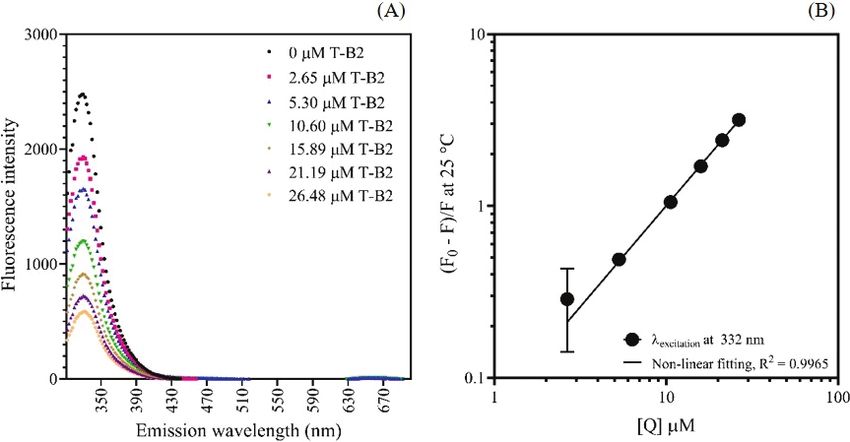
four fragment ions MS2 m/z at 1535 ([M–malonoylglucoside]+), high, indicating that the anthocyanins had a strong affinityT.T. Vuong et al. / Agr. Nat. Resour. 55 (2021) 569–578 575 with proteins (Acharya et al., 2013). The value of n indicated Table 2 reveals that the protein-anthocyanin complex a single or more than one binding site on the protein molecules. formed simultaneously (negative ΔG). The ΔG values at this The n value was slightly greater than 1.0 when binding occurred magnitude (30 kJ/mol) are considered weak, compared to the in the range 35–45°C, possibly because of the involvement of energy required for covalent bond formation (approximately both dynamic and static quenching; or more binding sites being 400 kJ/mol), according to Koolman and Roehm (2005). available with slightly different binding constants (Acharya Therefore, it was likely that non-covalent binding was et al., 2013). Whey proteins are mixed proteins composed of responsible for the formation of the protein–anthocyanin β-LG, α-LA and BSA. The available Trp residue for quenchers complexes in the distilled water. The bindings were exothermic could be different as the temperature increased from 35°C (negative ΔH). The negative ΔH and negative ΔS suggested to 45°C. However, when the reaction temperature increased H-bond involvement as the primary force (Acharya et al., 2013; to 65°C, the number of binding sites decreased to less than Chung et al., 2015, He et al., 2016) in the complex formation 1.0 (Fig. 2C), probably due to more collisions of proteins and of delphinidin-based anthocyanins with whey proteins in quenchers when the temperature was raised. distilled water. Fig. 2 Fluorescence quenching of whey proteins in WPI by water extract of Clitoria ternatea: (A) fluorescence spectra at different anthocyanin concentrations calculated based on ternatin-B2 (T-B2); (B) log-log plot of ([F0-F]/F) as a function of anthocyanin quencher concentration ([Q]) at 25°C, where R2 = goodness of fit on non-linear curve regression Fig. 3 Effect of absolute temperature on: (A) binding constant; (B) the number of the binding site on whey protein molecule for anthocyanin binding, where error bars indicate + SD and different lowercase letters above columns indicate significant (p < 0.05) differences
576                                                  T.T. Vuong et al. / Agr. Nat. Resour. 55 (2021) 569–578

Table 2 Thermodynamic parameters of whey protein-anthocyanin interaction in distilled water calculated based on ternatin-B2
                        Temperature                                     ∆G (kJ/mol)ns                           ∆H (kJ/mol)        ∆S (J/mol K)
             °C                             K                                                                    T1 = 35°C          T2 = 65°C
             35                           308.15                         -30.77±0.23                           -32.948±4.728       -7.415±4.349
             45                           318.15                         -30.59±0.09
             65                           338.15                         -30.44±0.00
ΔG = Gibbs free energy change; ΔH = enthalpic change; ΔS = entropic change; T1 and T2 = temperature difference at which the ΔH was calculated.
Value = means ± SD (n = 3 separate trials)

    Whey protein solutions containing different sugar                               differently. This study further illustrated that the conformational
osmolyte concentrations were investigated for their binding                         changes of whey proteins induced by different sugars could
characteristics with C. ternatea extract at 35 °C, to determine                     lead to different binding sites on the whey protein molecules
the influences of the medium properties on complex formation                        accessible by anthocyanins.
(Fig. 4). The Ka values were within the range 11.3×10 4                                 The influences were further investigated of a divalent
–16.8×10⁴ M-1 in the lactose solution and 14.4×10⁴–15.7× 10⁴                        cation, ionic strength and osmotic pressure on the binding
M-1 in the sucrose solution (Fig. 4A; p ≥ 0.05). However, the                       characteristics and complex formation of proteins and
sugar types significantly affected the number of binding sites                      anthocyanins. Table 3 shows that increasing the concentration
on the whey protein molecules accessible by anthocyanins to                         of CaCl2 to above 375 mM (which provided an ionic strength
form a complex (Fig. 4B). At a lactose concentration lower                          (I) of 1.125 M and osmotic pressure (π) of 28.448 atm)
than 37.5 mM, the value of n on whey protein molecules was                          increased the binding constants and the number of binding sites
lower than that at a high lactose concentration and that of                         in the complex formation (p < 0.05). A high concentration of
sucrose solutions (p < 0.05).                                                       CaCl2 could increase the affinity and strengthen the interactions
    The microenvironment surrounding the protein molecules                          of whey proteins and delphinidin-based anthocyanins. It was
induced by sugar osmolytes could alter the protein conformation                     also possible that the formation of a Ca-flavonoid complex
differently when different sugars were used. Arakawa and                            occurred at high CaCl2 concentration as reported by Castilho
Timasheff (1982a) demonstrated that lactose had a stronger                          et al. (2018) on quercetin and Ca2+. However, more work is
cohesive force than sucrose and increased the water surface                         needed to elucidate the complexion sites in delphinidin-based
tension to a greater extent than sucrose. In turn, this resulted                    anthocyanins.
in each sugar influencing the conformation of whey proteins

Fig. 4 Effect of sugar type and concentration on: (A) binding constant; (B) number of binding sites of whey proteins with Clitoria ternatea anthocyanins
at 35°C calculated based on ternatin-B2T-B2. The osmotic pressure was calculated based on respective added sugar concentrations were 0.190 atm,
0.948 atm, 1.897 atm, 2.845 atm and 3.793 atm. Error bars indicate + SD and different lowercase letters above columns indicate significant (p < 0.05)
differences.
T.T. Vuong et al. / Agr. Nat. Resour. 55 (2021) 569–578 577
Table 3 Effect of CaCl2 concentration on binding constant and number of binding sites of protein with Clitoria ternatea anthocyanin at 35°C calculated
based on ternatin-B2
[CaCl2] Ionic strength Calculated osmotic pressure of Binding constant × 104 (/M1) Number of binding sites
(M) (M) CaCl2 solution (atm)
0 0.000 13.56±4.73c 0.966±0.027b
0.025 0.075 1.897 14.76±2.11 c
0.976±0.013b
0.125 0.375 9.483 14.04±2.28 c
0.970±0.011b
0.250 0.750 18.965 14.23±3.26 c
0.972±0.017b
0.375 1.125 28.448 20.63±1.36b 1.005±0.006a
0.500 1.500 37.930 26.32±1.94 a
1.025±0.006a
Means ± SD (n = 3 separate trials) superscripted by different lowercase letters are significantly (p < 0.05) different.
Fig. 5 shows that the complex formation was a simultaneous Timasheff, 1982b) and subsequently result in different binding
and exothermic process (negative ΔG and ΔH values). mechanisms of proteins with anthocyanins.
The negative values of ΔH and ΔS also suggested that In summary, this study further demonstrated that different
despite the increase in osmotic pressure and ionic strength, types and concentrations of the osmolytes commonly used in
the H-bonds remained the leading force in the formation of food product could determine the leading force responsible
the whey protein-anthocyanin complex in CaCl2 solution. for the formation of a protein-anthocyanin complex in distilled
The result from this study were different from Vuong and water, where the proteins solely governed the buffering
Hongsprabhas (2021) that was carried out in buffered systems capacity of the mixed solutions. Understanding the binding
using 0.1 M citrate buffer, 0.2 M phosphate buffer or 0.1 M mechanisms of complex formation between whey proteins
carbonate buffer, in which hydrophobic interactions were and delphinidin derivatives via H-bonds under different
the main forces involved in the formation of the whey temperatures, osmotic pressures and ionic strengths could help
protein-delphinidin complexes. This was probably because in the design and rationalization of the types and concentrations
the Ca2+ ion could likely bind directly to whey proteins and of osmolyte to manipulate protein conformation and its further
alter their conformations (Arakawa and Timasheff, 1982b). interactions with polyacylated delphinidin derivative via
Further investigation is still necessary into the influences reversible H-bonds in the future.
of anion types and concentrations on the conformation of
whey proteins and subsequent binding mechanisms during Conflict of Interest
whey protein-anthocyanin complex formation. Different
anions could perturb the surface free energy of the protein The authors declare that there are no conflicts of interest.
molecules and their conformations differently (Arakawa and
Fig. 5 Effect of added CaCl2 concentration on: (A) Gibbs free energy change; (B) enthalpic change; (C) entropic changes in binding of whey proteins
with Clitoria ternatea anthocyanins at 35°C calculated based on ternatin-B2, where error bars = + SD578 T.T. Vuong et al. / Agr. Nat. Resour. 55 (2021) 569–578
Acknowledgements Joye, I.J., Davidov-Pardo, G., Ludescher, R.D., McClements, D.J. 2015.
Fluorescence quenching study of resveratrol binding to zein and
gliadin: Towards a more rational approach to resveratrol encapsulation
The Faculty of Agro-Industry, Kasetsart University,
using water-insoluble proteins. Food Chem. 185: 261–267. doi.org/
Bangkok, Thailand and the Kasetsart University Financial 10.1016/j.foodchem.2015.03.128
Support Fund provided a graduate student fellowship to author Kazuma, K., Noda, N., Suzuki, M. 2003. Flavonoid composition related
Vuong. The Kasetsart University Research and Development to petal color in different lines of Clitoria ternatea. Phytochemistry
Institute (KURDI) provided funding and the Center of 64: 1133–1139. doi.org/10.1016/S0031-9422(03)00504-1
Excellence on Agricultural Biotechnology (AG-BIO/MHESI) Koolman, J., Roehm, K.H. 2005. Color Atlas of Biochemistry, 2nd ed.
Georg Thieme Verlag. Stuttgart, Germany.
provided use of the HPLC-ELSD during the preliminary
Lakowicz, J.R. 2006. Principles of Fluorescence Spectroscopy, 3rd ed.
characterization of the anthocyanins. Springer. New York, NY, USA.
Lee, J., Durst, R.W., Wrolstad, R.E. 2005. Determination of
References total monomeric anthocyanin pigment content of fruit juices,
beverages, natural colorants, and wines by the pH differential method:
Association of Official Analytical Chemists. 2000. Official Methods of collaborative study. J. AOAC Int. 88: 1269–1278. doi.org/10.1093/
Analysis, 17th ed. The Association of Official Analytical Chemists. jaoac/88.5.1269
Gaithersburg, MD, USA. Lowry, O.H., Rosebrough, N.J., Farr, A.L., Randall, R.J. 1951. Protein
Arakawa, T., Timasheff, S.N. 1982a. Stabilization of protein structure measurement with the Folin phenol reagent. J. Biol. Chem. 193:
by sugars. Biochemistry 21: 6536–6544. doi.org/10.1021/bi00268a033 265–275. doi.org/10.1016/S0021-9258(19)52451-6
Arakawa, T., Timasheff, S.N. 1982b. Preferential interactions of Miller, G. 1959. Use of dinitrosalicylic acid reagent for determination
proteins with salts in concentrated solutions. Biochemistry 21: of reducing sugar. Anal. Chem. 31: 426–428. doi.org/10.1021/
6545–6552. doi.org/10.1021/bi00268a034 ac60147a030
Acharya, D.P., Sanguansri, L., Augustin, M.A. 2013. Binding of Mohammadi, F., Moeeni, M. 2015. Analysis of binding interaction of
resveratrol with sodium caseinate in aqueous solutions. Food Chem. genistein and kaempferol with bovine α-lactalbumin. J. Funct. Foods.
141: 1050–1054. doi.org/10.1016/j.foodchem.2013.03.037 12: 458–467. doi.org/10.1016/j.jff.2014.12.012
Casanova, F., Chapeau, A.L., Hamon, P., Carvalho, A.F., Bouhallab, Naczk, M., Grant, S., Zadernowski, R., Barre, E. 2006. Protein precipitating
C.S. 2018. pH- and ionic strength-dependent interaction between capacity of phenolic of wild blueberry leaves and fruits. Food Chem.
cyanidin-3-O-glucoside and sodium caseinate. Food Chem. 267: 96: 640–647. doi.org/10.1016/j.foodchem.2005.03.017
52–59. doi.org/10.1016/j.foodchem.2017.06.081 Richmond, M.L., Barfuss, D.L., Harte, B.R., Gray, J.I., Stine, C.M. 1982.
Chung, C., Rojanasasithara, T., Mutilangi, W., McClements, D.J. 2015. Separation of carbohydrates in dairy products by high performance
Enhanced stability of anthocyanin-based color in model beverage liquid chromatography. J. Dairy Sci. 65: 1394–1400. doi.org/10.3168/
systems through whey protein isolate complexation. Food Res. Intern. jds.S0022-0302(82)82360-6
76: 761–768. doi.org/10.1016/j.foodres.2015.07.003 Schmidt, R.H., Packard, V.S., Morris, H.A. 1984. Effect of processing
Castilho, T.S.D., Matias, T.B., Nicolini, K.P., Nicolini, J. 2018. Study on whey protein functionality. J. Dairy Sci. 67: 2723–2733. doi.
of interaction between metal ions and quercetin. Food Sci. Hum. org/10.3168/jds.S0022-0302(84)81630-6
Wellness. 7: 215–219. doi.org/10.1016/j.fshw.2018.08.001 Srivichai, S., Hongsprabhas, P. 2020. Profiling anthocyanins in
Davidov-Pardo, G., McClements, D.J. 2014. Resveratrol encapsulation: Thai purple yams (Dioscorea alata L.). Int. J. Food Sci. 2020: 1594291.
Designing delivery systems to overcome solubility, stability and doi.org/10.1155/2020/1594291
bioavailability issues. Trends Food Sci. Technol. 38: 88–103. doi.org/ Terahara, N., Oda, M., Matsui, T., Osajima, Y., Saito, N., Toki, K.,
10.1016/j.tifs.2014.05.003 Honda, T. 1996. Five new anthocyanins, ternatins A3, B4, B3, B2,
Fisicaro, E., Compari, C., Braibanti, A. 2004. Entropy/enthalpy and D2, from Clitoria ternatea flowers. J. Nat. Prod. 59: 139–144.
compensation: Hydrophobic effect, micelles and protein complexes. doi.org/10.1021/np960050a
Phys. Chem. Chem. Phys. 6: 4156–4166. doi.org/10.1039/B404327H Terahara, N., Toki, K., Saito, N., Honda, T., Matsui, T., Osajima, Y. 1998.
Graf, S., Egert, S., Heer, M. 2011. Effects of whey protein supplements Eight new anthocyanins, ternatin C1-C5 and D3 and pre-ternatins
on metabolism: Evidence from human intervention studies. Curr. A3 and C4 from young Clitoria ternatea flowers. J. Nat. Prod. 61:
Opin. Clin. Nutr. Metab. Care. 14: 569–580. doi: 10.1097/MCO. 1361–1367. doi.org/10.1021/np980160c
0b013e32834b89da Vuong, T.T., Hongsprabhas, P. 2021. Influences of pH on binding
He, Z., Xu, M., Zeng, M., Qin, F., Chen, J. 2016. Interactions of milk mechanisms of anthocyanins from butterfly pea flower (Clitoria
α- and β-casein with malvidin-3-O-glucoside and on the stability of ternatea) with whey powder and whey protein isolate. Cogent Food
grape skin anthocyanin extracts. Food Chem. 199: 314–322. doi.org/ Agric. 7: 1889098, doi.org/10.1080/23311932.2021.1889098
10.1016/j.foodchem.2015.12.035 Zhu, H., Damodaran, S. 1994. Effects of calcium and magnesium ions
Jeyaraj, E.J., Lim, Y.Y., Choo, W.S. 2020. Extraction methods of on aggregation of whey protein isolate and its effect on foaming
butterfly pea (Clitoria ternatea) flower and biological activities of properties. J. Agric. Food Chem. 42: 856–862. doi.org/10.1021/
its phytochemicals. J. Food Sci. Technol. 58: 2054–2067. doi.org/ jf00040a003
10.1007/s13197-020-04745-3You can also read