Molecular Mechanism for Lipid Flip-Flops
←
→
Page content transcription
If your browser does not render page correctly, please read the page content below
13554                                         J. Phys. Chem. B 2007, 111, 13554-13559

Molecular Mechanism for Lipid Flip-Flops

                 Andrey A. Gurtovenko*,† and Ilpo Vattulainen‡,§,⊥
                 Computational Laboratory, Institute of Pharmaceutical InnoVation, UniVersity of Bradford, Bradford, West
                 Yorkshire, BD7 1DP, United Kingdom, Institute of Physics, Tampere UniVersity of Technology, P.O. Box 692,
                 FI-33101 Tampere, Finland, Helsinki UniVersity of Technology, P.O. Box 1100, FI-02015 HUT, Finland, and
                 MEMPHYS - Center for Biomembrane Physics, UniVersity of Southern Denmark, Odense, Denmark
                 ReceiVed: September 4, 2007

                 Transmembrane lipid translocation (flip-flop) processes are involved in a variety of properties and functions
                 of cell membranes, such as membrane asymmetry and programmed cell death. Yet, flip-flops are one of the
                 least understood dynamical processes in membranes. In this work, we elucidate the molecular mechanism of
                 pore-mediated transmembrane lipid translocation (flip-flop) acquired from extensive atomistic molecular
                 dynamics simulations. On the basis of 50 successful flip-flop events resolved in atomic detail, we demonstrate
                 that lipid flip-flops may spontaneously occur in protein-free phospholipid membranes under physiological
                 conditions through transient water pores on a time scale of tens of nanoseconds. While the formation of a
                 water pore is induced here by a transmembrane ion density gradient, the particular way by which the pore is
                 formed is irrelevant for the reported flip-flop mechanism: the appearance of a transient pore (defect) in the
                 membrane inevitably leads to diffusiVe translocation of lipids through the pore, which is driven by thermal
                 fluctuations. Our findings strongly support the idea that the formation of membrane defects in terms of water
                 pores is the rate-limiting step in the process of transmembrane lipid flip-flop, which, on average, requires
                 several hours. The findings are consistent with available experimental and computational data and provide a
                 view to interpret experimental observations. For example, the simulation results provide a molecular-level
                 explanation in terms of pores for the experimentally observed fact that the exposure of lipid membranes to
                 electric field pulses considerably reduces the time required for lipid flip-flops.

Introduction                                                        processes, it has remained unclear whether this view is indeed
                                                                    the most plausible one. Yet the understanding of the molecular
   Membranes of most animal cells are known to be asymmetric        mechanisms of lipid flip-flops is of substantial biological
with regard to transmembrane distribution of lipids across a        importance since a loss of transmembrane lipid asymmetry can
membrane.1,2 This asymmetry is crucial for an array of cellular     have severe consequences.10 For instance, while negatively
functions and plays an important role, e.g., in membrane            charged phosphatidylserine lipids are usually located in the inner
mechanical stability,3 modulation of the activity of membrane       leaflet of the plasma membrane, their appearance in the outer
proteins,4 and programmed cell death.5 Therefore, to maintain       leaflet is known to correlate with programmed cell death.5 In
an asymmetric transmembrane lipid distribution, cellular mem-       more general terms, understanding how membrane lipids
branes have means to translocate (flip-flop) lipid molecules from   maintain the asymmetric transmembrane distribution and achieve
one membrane leaflet to another.                                    their nonrandom distribution in cells is one of the key challenges
   One way cells make this happen is to employ special              in cell biology.11
mechanisms to actively transport lipids across a lipid bilayer         The reason lipid flip-flops and their detailed molecular
using specific membrane proteins, flippases, whose biological       mechanisms in the absence of flippases are so difficult to
functions and relevance are widely recognized.6,7 To comple-        characterize is rather obvious. From an experimental point of
ment active translocation, cells also use passive transport         view, it is a matter of resolution since these processes take place
mechanisms that facilitate the migration of lipids from one         over molecular scales. Meanwhile, from a computational
leaflet to another. For this purpose, the translocation may take    viewpoint, the atomistic modeling of flip-flops has been
place with the help of proteins or without them.8 The mecha-        considered to be a prohibitive task for decades to come, since,
nisms associated with transmembrane transport in protein-free       in general, lipid flip-flops are very slow processes; they are
membranes are exceptionally poorly understood, however. It is       characterized by an average life time from several hours to
commonly assumed that the transbilayer movement of lipids in        several days.12,13
protein-free membranes takes place as a single-molecule                However, there are strong indications that lipid translocation
process,4,8 where the cooperative motion of nearby lipids allows    across a lipid membrane is a pore-mediated process. It has been
the migrating lipid to more easily cross the transition state of    demonstrated14 that the experimentally determined activation
the flip-flop event.9 However, since flip-flops are very rare       energy for radioactive chloride flux across lipid membranes is
                                                                    close to the activation energy of lipid flip-flop.12 Further, brief
  * Corresponding author. E-mail: A.Gurtovenko@bradford.ac.uk.      electric pulses (electroporation) have been shown to enhance
  † University of Bradford.
  ‡ Tampere University of Technology.                               the transbilayer mobility of phospholipids.15 All together, the
  § Helsinki University of Technology.                              above findings suggest that a major fraction of flip-flops takes
  ⊥ University of Southern Denmark.                                 place through water defects in lipid membranes.
                              10.1021/jp077094k CCC: $37.00 © 2007 American Chemical Society
                                                 Published on Web 11/08/2007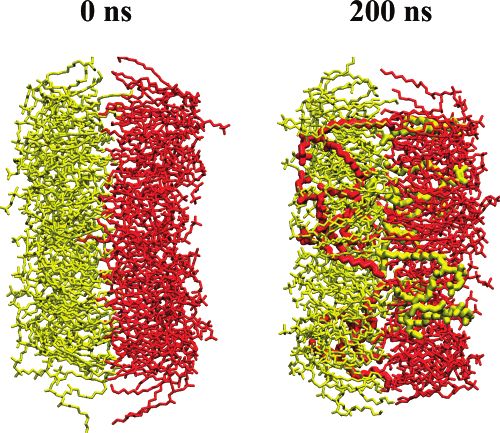
Molecular Mechanism for Lipid Flip-Flops J. Phys. Chem. B, Vol. 111, No. 48, 2007 13555
On the computational side, several observations of defect-
mediated lipid flip-flops have been reported; most of them were
related to flip-flop events in lipid membranes far from physi-
ological conditions, though. In particular, very recent lipid
membrane simulations indicated that, when a lipid was dragged
by an external force through the membrane interior, the
formation of a small water pore was observed; the energies
required for lipid flip-flop and for the formation of the pore
were found to be identical.16 On the basis of these findings, the
authors concluded that lipid flip-flop could be a defect-mediated
process. Meanwhile, de Vries et al. have reported on the pore-
mediated equilibration of lipids between two leaflets in the early
phases of vesicle self-assembly;17 the results are appealing, but
the far from equilibrium conditions under self-assembly are
markedly different from conditions where the dynamics in stable
cell membranes takes place. Later, a defect-mediated lipid flip-
flop was reported in molecular dynamics (MD) simulations of
membranes under the influence of other external factors such
as antimicrobial peptides18 and butanol.19 Finally, a recent
computational study20 reported a single pore-mediated lipid flip-
flop that was driven by an extremely high transmembrane
voltage, i.e., again under conditions far from those that can be
observed in a cell. Furthermore, the authors observed that
sodium cation flux can be a factor that facilitates the dragging
of lipids across a water pore. Overall, despite their limitations
(membrane exposure to external factors or nonequilibrium
conditions), all the above studies support the view that flip-
flops in protein-free membranes may take place through defects
in membranes.
In this paper, we show through extensive atomic-scale MD
simulations that the computational approach can actually provide
a great deal of insight into the mechanism (or one of the
mechanisms) associated with lipid flip-flops. The simulations
conducted under physiological conditions provide compelling
evidence that, initially, the key event leading to flip-flop is the
spontaneous formation of a water pore. Having formed, the
water pore facilitates the spontaneous migration of lipids across
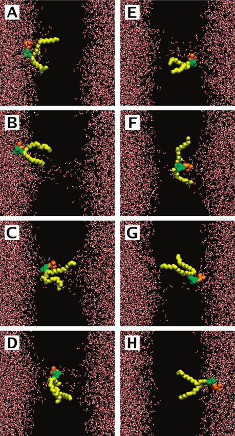
the membrane (see Figure 1). On the basis of 50 flip-flop events
resolved in atomic detail, we provide a detailed molecular
picture for lipid translocation across a membrane and discuss
the forces driving these processes. Remarkably, the average time
required for a lipid to translocate through a preformed water Figure 1. Pore-mediated lipid flip-flop: (A) 0 ps, (B) 43.85 ns, (C)
pore was found to be around 60 ns. Together with the large 118.9 ns, (D) 122.4 ns, (E) 152.7 ns, (F) 204.65 ns, (G) 208.9 ns, and
(H) 215 ns. Lipids (except for the flip-flopped one) are not shown;
number of flip-flops observed here, this strongly suggests that
water is shown in red and white, acyl chains of the flip-flopped lipid
the actual flip-flop event is a rapid process, while the spontane- are shown in yellow, and its choline and phosphate groups are shown
ous formation of a water defect in lipid membranes is the rate- in orange and green, respectively.
limiting step in the process of lipid flip-flop. The experimental
data that is available for comparison and discussed at the end mentally measured area and volume per lipid (see, e.g., ref 22).
of this article is consistent with the simulation results. Water was modeled using the simple point charge model.23 For
The most notable contribution of this work is the unprec- sodium, potassium, and chloride ions, we employed the default
edented view for a large number of diffusiVe (driven by thermal set of parameters supplied within the GROMACS force field.24
fluctuations) lipid flip-flops observed in atomistic detail, thus The Lennard-Jones interactions were cut off at 1 nm. For the
clarifying the mechanism of flip-flops under conditions close electrostatic interactions, the particle-mesh Ewald method25,26
to physiological ones. There is indeed ample reason to stress was used. The simulations were performed in the NpT ensemble
that, in the present study, the simulation conditions really match with temperature and pressure kept constant by the Berendsen
closely those observed in a cell close to equilibrium, without scheme.27 Pressure was set to 1 bar. Temperature was set to
artificial external fields or far from equilibrium conditions. 323 K, which is well above the main phase transition temper-
ature of a DMPC bilayer (297 K).
Methods Water pores in lipid membranes were induced by adding salt
(NaCl or KCl) and by creating an imbalance of cations (sodium
The atomic-scale MD simulations were performed on lipid or potassium) across the membrane.22,28 To model the trans-
membranes comprised of zwitterionic dimyristoylphosphatidyl- membrane ionic charge imbalance explicitly, a double-bilayer
choline (DMPC) lipids. Force-field parameters for lipids were setup (i.e., two lipid bilayers of 128 lipids each in a simulation
taken from the united atom force-field of Berger et al.;21 the box) was employed,29,30 amounting to about 42 000 atoms in
force-field has been shown to correctly reproduce the experi- the system. The time step used in the integration of equations13556 J. Phys. Chem. B, Vol. 111, No. 48, 2007 Gurtovenko and Vattulainen
TABLE 1: Summary of Lipid Flip-Flop Events
system salta tsim [ns]b Nflip-flopc tflip-flop [ns]d
1 NaCl 215 8 66 ( 13
2 NaCl 95 2 78 ( 18
3 NaCl 125 5 78 ( 9
4 NaCl 50 3 38 ( 5
5 KCl 200e 10 70 ( 10
6 KCl 35 5 27 ( 3
7 KCl 200e 13 66 ( 10
8 KCl 80 4 58 ( 13
a
Type of salt used in a simulation. b Total time of a simulation.
Simulations were extended until a pore was closed, except for systems
5 and 7. c Number of flip-flop events. d Average duration of a lipid
flip-flop process. e Pore did not close over the course of the simulation.
of motion was 2 fs. In all, we considered eight different bilayer
systems with pores; most systems were simulated until a pore
was closed, with the exception of systems 5 and 7, which had
pores open even after 200 ns (see Table 1). The total simulated
time amounted to 1 µs. All simulations were performed using
the GROMACS suite.24
Results
To study pore-mediated lipid flip-flops, water pores were first
preformed in membranes composed of DMPC lipids. This was
accomplished by creating a transmembrane imbalance of cations
(either sodium or potassium ions), which induces a spatially Figure 2. (Top) Time evolution of positions of the CMs of head groups
for four flip-flopped lipids of system 1. The z ) 0 corresponds to the
and time-dependent electric field across the membrane. To this
center of the membrane; solid black lines show the average positions
end, we employed a double-bilayer setup where the system of lipid head groups in the two opposite leaflets, extracted from the
included two lipid bilayers in the lamellar fluid phase. The ion intact membrane before pore formation. (Bottom) Time evolution of
concentration imbalance employed in this study was intention- the tail-to-head orientation for the same four flip-flopped lipids. The
ally chosen to be rather large (six cations per bilayer of 128 tail-to-head orientation was characterized as the angle between the
lipids), so that the formation of water pores occurred on a bilayer normal and the vector directed from the CMs of the acyl chains
nanosecond time scale. After a pore has been formed, one to the CMs of head groups. Solid black lines again show typical values
of the angle for lipids in the opposite leaflets.
observes the transport of ions through the pore, which quickly
discharges the transmembrane ionic charge imbalance and makes
1D). This eventually leads to the appearance of the lipid in the
the pore metastable. A detailed discussion of the overall process
opposite membrane leaflet, accompanied by the subsequent
of pore formation and subsequent ion leakage can be found
reorientation of the lipid (Figure 1E). The irreversible accom-
elsewhere.22,28
modation of a lipid in the opposite leaflet (if successful) turns
There is reason to emphasize that the transmembrane leakage
out to be a rather slow process (more than 50 ns is required for
of ions is a much faster process compared to lipid flip-flop:
the particular lipid considered here; see Figure 1E-G) since it
the ionic charge imbalance across a membrane discharges almost
involves spontaneous detachment of a head group out of pore
fully within 5-6 ns after pore formation. Therefore, the leakage
of ions can affect lipid flip-flops at a very early stage only. “walls” and lateral diffusion of a lipid away from the pore. This,
The remaining ionic charge imbalance after the first few however, can be greatly facilitated by pore closure, which occurs
nanoseconds of pore formation is essentially negligible (usually at t = 210 ns for this particular system (Figure 1H).
one cation per bilayer) or even zero, which corresponds to a To further characterize lipid flip-flops, in Figure 2 we show
complete discharging of the transmembrane potential. Water the time evolution of the positions of centers of mass (CMs) of
pores, being in a metastable state, stay open from about 35 to several lipid head groups. Also depicted in the same figure is
200 ns. the lipids’ overall orientation within a membrane (shown here
After the formation of a water pore, we witness spontaneous are the trajectories of four typical flip-flopped lipids of system
pore-mediated translocation of lipid molecules from one leaflet 1). As seen, one can distinguish two somewhat different types
to another. Table 1 summarizes the lipid flip-flop events of lipid flip-flops: (i) very fast flip-flops of lipids directly
observed. The overall process of lipid flip-flop is visualized in involved in the initial formation of a water pore (these are
Figure 1 for one particular lipid of the simulation system 1. characterized by a rapid onset of translocation, which coincides
Starting from an intact lipid membrane (Figure 1A), a water with a pore formation event, and by a rather short time (10-20
pore spanning the entire membrane is first formed; the pore is ns) required for successful accommodation in the opposite leaflet
laterally located far away from the lipid in question and does (orange curve in Figure 2)); and (ii) flip-flops of lipids that are
not affect it (Figure 1B). After about 100 ns, the lipid diffuses either involved in the initial pore formation but require
laterally to the pore site and becomes part of the pore, lining considerably longer time to accomplish translocation (blue curve
the pore by its head group (Figure 1C). At this moment, the in Figure 2) or are initially remote from a pore but diffuse to
spontaneous diffusive translocation of a lipid through the pore the pore site with time (red and green curves; note that the red
initiates: it involves the progressive diffusion of a lipid head curve corresponds to the translocation of the lipid exemplified
group in the pore coupled with the simultaneous desorption of above in a series of snapshots shown in Figure 1). The number
lipid hydrocarbon chains out from the membrane leaflet (Figure of flip-flops in category (i) is 9 out of 50 events observed (threeMolecular Mechanism for Lipid Flip-Flops J. Phys. Chem. B, Vol. 111, No. 48, 2007 13557
for NaCl and six for KCl), thus the majority of flip-flops (about
80%) belong to category (ii).
Remarkably, the translocation of a lipid across a membrane
closely correlates with the overall reorientation of the lipid
measured through the angle between the bilayer normal and
the vector directed from the CM of a lipid’s hydrocarbon chains
to the CM of its head groups (see Figure 2 (Bottom)). When a
lipid is accommodated in a membrane leaflet, this tail-to-head
vector makes an average angle of 22° (or 158° depending on
the leaflet) with the bilayer normal. During flip-flop, a lipid
changes its orientation, with the corresponding angle lying
between the two values being typical for opposite leaflets. Close
to the center of the membrane, lipids tend to adopt an orientation
perpendicular to the bilayer normal, which is most clearly seen
in Figure 2 for a lipid shown by the green curve. It is also
noteworthy that lipids’ tail-to-head orientation is subject to much
larger fluctuations when a lipid is in the middle of a membrane
compared to the situation where a lipid is localized in a leaflet;
this is due to the fact that lipids in leaflets are more densely
packed than lipids in the membrane interior. Figure 3. A membrane with the maximal number of lipid flip-flops
observed (system 7 in Table 1): intact membrane at t ) 0 ns (left) and
Discussion membrane with an opened pore at t ) 200 ns (right). Lipids in opposite
leaflets are shown in yellow and red; for clarity’s sake, the size of
Overall, we observed 50 spontaneous flip-flop events (see flip-flopped lipids has been enlarged.
Table 1). The fact that needs to be emphasized here is that these
flip-flop events were coupled to the spontaneous formation of fast reorientation of some lipid head groups toward the
a water pore discussed in refs 28 and 22. For comparison, in membrane interior,28,32 so that the lipids participating in pore
numerous atomistic simulation studies of related intact single- formation are moved out of their equilibrium state. If desorption
component lipid bilayers, we have not identified any flip-flops, of lipid chains occurs simultaneously with the entering of head
despite the long simulation time scales of several microseconds groups in the pore, one can observe a very fast lipid flip-flop.
(data not shown). This highlights the importance of water pore In most cases, however, the above scenario takes place only
formation as a key step in lipid flip-flop. occasionally, since, while a lipid could translocate through half
The flip-flop mechanism observed here takes place in two of the membrane, the time required for lipid accommodation
stages. First, starting from conditions that model the situation in the opposite leaflet can be considerable (see Figure 2). Pore
in the vicinity of the plasma membrane, our model system closure, in turn, is able to considerably speed up the flip-flops
includes an initial ionic concentration imbalance across the of partly translocated lipids: Irreversible membrane resealing
membrane. Such a transmembrane ion concentration difference makes it impossible for such lipids to move back to their original
is an inherent feature of plasma membranes of eukaryotic cells.31 leaflet and effectively pushes them to the opposite one (see,
Local fluctuations in ion densities then give rise to a strong e.g., the translocation of a lipid shown by the red curve in Figure
local electric field across the bilayer, which in turn initiates the 2, in which case pore closure occurs at t = 210 ns). Therefore,
formation of a water pore22,28 coupled to the subsequent rapid early pore closure can decrease the duration of flip-flop
ion leakage that dramatically reduces the local field strength significantly, as seen for simulation systems 4 and 6, which
and hence the pore size, yet the pore remains open for a have the shortest pore lifetimes (Table 1).
considerably long period of time, which ranges from about ∆t The findings also suggest that pore-mediated translocation
) 35 ns to several hundred nanoseconds. We stress that, in the of one lipid can promote the flip-flops of others. If a lipid lining
present case, we employed a rather large ion concentration a pore translocates to the opposite side of a membrane, it has
imbalance to promote very fast pore formation. Alternatively, to be substituted by another lipid from the same leaflet to keep
the formation of a water pore could be induced by an external the water pore stable. If this “new” lipid also translocates
electric field,32-34 mechanical stress,32,35 or thermal fluctua- successfully, the process of substitution needs to be repeated.
tions.36,37 Second, at the same time, the lipids in the bilayer Indeed, for bilayer system 7 with KCl salt (Table 1), we
diffuse laterally in the bilayer plane over a distance of lD ) observed a sequence of almost concerted flip-flops of three
x4D∆t, where D ≈ 1 × 10⁻⁷ cm2/s is a typical lateral lipids. Remarkably, this particular system is characterized by
diffusion coefficient in fluid lipid bilayers. For a typical lifetime the largest number of flip-flop events (13 flip-flops in this
of the pore, ∆t ≈ 100 ns, the diffusion length of a lipid in the particular system), which leads to a considerable mixing of lipids
plane of the membrane is lD ≈ 2 nm, that is, about 3 times the from the opposite membrane leaflets over the time span of 200
size of the lipid molecule. This implies that there are about a ns (Figure 3).
few tens of lipids near the pore that could access it via lateral The role of salt type was elucidated by considering pores
diffusion before pore closure. Once they do so, many of them induced by an imbalance of either sodium or potassium cations.
undergo flip-flop by translocating through the pore. In the case of NaCl salt, one finds 18 successful lipid flip-flops
Although the average duration of lipid flip-flops through a with an average duration of 64 ( 7 ns. In turn, for systems
preformed pore was found to be around 60 ns, flip-flop times with KCl, 32 pore-mediated lipid translocations were found to
for individual lipids scatter considerably and range from 10 to occur with an average duration of 60 ( 6 ns. Hence, the average
130 ns. Among factors that influence the rate of lipid translo- times required for lipid flip-flops turn out to be very similar in
cation, there is reason to mention the significance of pore both cases. However, the type of salt used does affect the
opening and membrane resealing. Formation of a pore implies probability of lipid flip-flop in the system: bilayers with NaCl13558 J. Phys. Chem. B, Vol. 111, No. 48, 2007 Gurtovenko and Vattulainen and KCl salt were simulated for 485 and 515 ns, respectively, without salt ions, lending further support to the above conclu- but the overall number of lipid flip-flops was almost twice as sion. Overall, the formation of a water pore spanning the large in the case of KCl. Thus, the probability for a spontaneous membrane is most likely the only required prerequisite for flip-flop event to occur is higher by a factor of 1.7 in a bilayer transmembrane lipid translocation. system with potassium cations. This finding correlates well with The main result of the present study is the observation that the fact that sodium ions demonstrate much stronger interactions the rate of lipid flip-flops is significantly enhanced by the with zwitterionic phosphatidylcholine lipids than do potassium spontaneous formation of water pores. Evidently, one should ions.22 In particular, Na+ ions are known to bind to carbonyl then ask whether the rate of pore formation is comparable to oxygens of lipids, leading to the formation of tight complexes the lipid flip-flop rate in the absence of proteins or pores. To between neighboring lipid molecules.22,30,38 The formation of consider this issue, let us first note that the average time of such complexes most likely hinders considerably the desorption pore-mediated lipid translocations has here been found to be of lipid chains out of the leaflets toward the membrane interior, ∼60 ns. For comparison, the flip-flop rate is available from whereby decreasing the probability of flip-flops. experimental measurements. In particular, on the basis of What, then, is the force that drives lipids residing close to exchange experiments13 performed at T ) 323 K on large the pore to flip-flop through it? Is it due to the electrostatic unilamellar vesicles composed of DMPC lipids, the upper limit coupling between the dipolar head group and the spatially for flip-flop half-time was estimated to be 0.7 h. Assuming that varying electric field (dielectrophoresis), or related to the the formation of a water pore in DMPC membranes is the rate- spontaneous sealing of the hydrophobic membrane region limiting step in the process of flip-flop, we can estimate the through hydrophobic interactions, or is it simply due to thermal probability of pore formation or the pore density F in fluctuations? First, let us mention that the lipids considered in DMPC membranes at T ) 323K, which is given by16 this study are neutral, thus the electrostatic potential is not F ) (tflip-flop jpore Alipid)-1. Here, tflip-flop ) 1 h is the flip-flop expected to play the main role. We have confirmed this by time taken from the experiment, jpore ) (1/60) ns-1 ) 1.67 × considering the membrane-normal component of the total 107 s-1 is the lipid flux through the pore obtained from our electrostatic force exerted on the lipids involved in flip-flops. MD simulations, and Alipid ) 0.66 nm2 ) 0.66 × 10⁻¹⁴ cm2 is That is found to fluctuate around zero, without component to the average area per DMPC lipid. This gives us the equilibrium drive translocation. Second, however, the spatially inhomoge- pore density F equal to 2.5 × 10³ pores/cm2. neous electric field defined by the instantaneous positions of The obtained value for F is in reasonable agreement with ions could contribute to lipid flip-flops via dielectrophoretic values available from other sources. In particular, the equilibrium motion, especially at the early stages of pore formation before pore density employed in models of electroporation39,40 ranges ion leakage through a pore reduces the local field strength. from 0.8 × 10² to 1.5 × 10⁵ pores/cm2. Tieleman and Marrink Therefore, one can expect that the rapid flip-flops observed in reported the density of ∼100 pores/cm 2 for a dipalmitoylphos- the beginning of the simulations could in part be driven by the phatidylcholine (DPPC) bilayer.16 Moving on, from spatially varying transmembrane electric field due to ion the pore density F we can estimate the free energy imbalance. Indeed, the contribution of the dielectrophoretic force ∆Gpore required for pore formation; ∆Gpore is coupled to exerted on the dipole of a lipid at such a high ionic charge F as16 F ) exp(-∆Gpore/kBT)/Alipid. With F ) 2.5 × 103 cm-2 imbalance was found to be comparable with thermal fluctua- and T ) 323 K, one has ∆Gpore = 67 kJ/mol. This value is tions. However, the flip-flops taking place right after pore somewhat smaller than the value of 80 kJ/mol reported recently formation (which constitute only 20% out of all flip-flop events) for a DPPC membrane at the same temperature. This difference can be considered as being artificially promoted since they are is not, however, surprising since chains of DMPCs are two coupled to the high ionic charge imbalance employed in this hydrocarbons shorter than those of DPPC. This means that a study, that is, to the method used to create water pores. DMPC membrane has a smaller hydrophobic core compared Furthermore, at longer times, after around 10-20 ns from the to a DPPC bilayer, and therefore less energy is required for the moment of pore formation, the transmembrane electric field is formation of a pore in the DMPC membrane. almost fully discharged by ion leakage. Under these conditions, Summarizing, our atomic-scale MD simulations demonstrate the dielectrophoretic force exerted on lipids was found to be that the appearance of a water pore spanning a phospholipid about an order of magnitude smaller than the force due to membrane inevitably leads to diffusive transmembrane trans- thermal fluctuations. This supports the view that dielectrophore- location of lipids through the pore; this translocation occurs sis is not driving flip-flops. This is particularly true when the spontaneously on a time scale of about 60 ns and is mainly system has lost its memory of how the pore was formed. For driven by thermal fluctuations. This strongly supports the idea instance, the ionic charge imbalance for system 7 is essentially that the formation of a water pore in a membrane is the rate- zero already at t ) 10 ns, implying that lipid flip-flops during limiting step in lipid flip-flop, which typically takes hours. Once the subsequent 190 ns are hardly affected by the spatially a pore has been formed, the subsequent actual pore-mediated varying local electric field. Third, what we find from the lipid translocation occurs extremely fast. Combining our simula- simulations is that the flips and flops are essentially symmetric. tion results and available experimental data, we found that the The number of flips (events that initiated from the leaflet facing free energy required for pore formation in DMPC membranes an initial excess of cations) was found to be 22, while the at T ) 323 K is about 67 kJ/mol, in line with previously reported number of flops (in the opposite direction) was 28. Considering studies. Furthermore, while pore formation can be induced the total number of events, the flip-flops are symmetric within through various ways using, for example, an external electric statistical fluctuations. field, our results show that the formation of pores and the Thus, one can conclude that the translocation of lipids through subsequent flip-flops can take place under physiological condi- transient water pores is mainly diffusive, i.e., driven by thermal tions because of salt ions in the vicinity of the membrane. fluctuations. Furthermore, very preliminary studies (data not In addition to their biological relevance, our findings also shown) have confirmed that lipid flip-flops can also be observed offer a molecular-level framework for the development of ways in bilayer systems where pores are formed by an alternate means to promote lipid transmembrane translocation and make it
Molecular Mechanism for Lipid Flip-Flops                                                   J. Phys. Chem. B, Vol. 111, No. 48, 2007 13559

possible to manipulate lipid distribution across cell membranes.                 (9) Marti, J.; Csajka, F. S. Phys. ReV. E 2004, 69, 061918.
As the present results indicate that the formation of water pores               (10) Boon, J. M.; Smith, B. D. Med. Res. ReV. 2002, 22, 251-281.
                                                                                (11) Holthuis, J. C.; Levine, T. P. Nat. ReV. Mol. Cell Biol. 2005, 6,
is the only prerequisite for lipid flip-flops, novel techniques              209-220.
should introduce transient defects (pores) into the membranes.                  (12) Kornberg, R. D.; McConnell, H. M. Biochemistry 1971, 10, 1111-
In particular, our study provides a molecular-level explanation              1120.
                                                                                (13) Wimley, W. C.; Thompson, T. E. Biochemistry 1990, 29, 1296-
for the experimentally observed fact that the exposure of lipid              1303.
membranes to electric field pulses considerably reduces the time                (14) Toyoshima, Y.; Thompson, T. E. Biochemistry 1975, 14, 1525-
required for lipid flip-flops:41 External electric field induces             1531.
electroporation of a membrane, drastically increasing the number                (15) Schwarz, S.; Haest, C. W. M.; Deuticke, B. Biochim. Biophys. Acta
                                                                             1999, 1421, 361-379.
of pathways (pores) available for pore-mediated lipid flip-flops.               (16) Tieleman, D. P.; Marrink, S.-J. J. Am. Chem. Soc. 2006, 128,
Similarly, the presented pore-mediated mechanism for lipid flip-             12462-12467.
flops can also be invoked to better understand lipid transmem-                  (17) de Vries, A. H.; Mark, A. E.; Marrink, S. J. J. Am. Chem. Soc.
brane asymmetry and to develop means to alter that: by forming               2004, 126, 4488-4489.
                                                                                (18) Leontiadou, H.; Mark, A. E.; Marrink, S.-J. J. Am. Chem. Soc. 2006,
defects in membranes, at least local anesthetics, amphiphilic                128, 12156-12161.
compounds, and pore-forming peptides are expected to facilitate                 (19) Dickey, A. N.; Faller, R. Biophys. J. 2007, 92, 2366-2376.
the occurrence of flip-flops.10 The view and mechanism                          (20) Kandasamy, S. K.; Larson, R. G. J. Chem. Phys. 2006, 125, 074901.
                                                                                (21) Berger, O.; Edholm, O.; Jahnig, F. Biophys. J. 1997, 72, 2002-
presented here is particularly interesting in terms of providing             2013.
insight into the action of antibacterial agents, which are known                (22) Gurtovenko, A. A.; Vattulainen, I. Biophys. J. 2007, 92, 1878-
as pore-forming peptides and killers of cells.42                             1890.
   Overall, the present results provide strong support for previous             (23) Berendsen, H. J. C.; Postma, J. P. M.; van Gunsteren, W. F.;
                                                                             Hermans, J. In Intermolecular Forces; Pullman, B., Ed.; Reidel: Dordrecht,
suggestions16-20 and put forward a new mechanism for lipid                   The Netherlands, 1981; pp 331-342.
flip-flop in protein-free membranes under physiological-like                    (24) Lindahl, E.; Hess, B.; van der Spoel, D. J. Mol. Model. 2001, 7,
conditions, thus updating the present view. It would be of                   306-317.
                                                                                (25) Darden, T.; York, D.; Pedersen, L. J. Chem. Phys. 1993, 98, 10089-
particular interest to elaborate on this matter further by consider-         10092.
ing how flip-flops take place in asymmetric lipid membranes43                   (26) Essman, U.; Perera, L.; Berkowitz, M. L.; T. Darden, H. L.;
characterized by a nonzero membrane potential, which likely                  Pedersen, L. G. J. Chem. Phys. 1995, 103, 8577-8592.
plays a role in pore formation together with an ion concentration               (27) Berendsen, H. J. C.; Postma, J. P. M.; van Gunsteren, W. F.; DiNola,
                                                                             A.; Haak, J. R. J. Chem. Phys. 1984, 81, 3684-3690.
imbalance across the membrane. That would also clarify the                      (28) Gurtovenko, A. A.; Vattulainen, I. J. Am. Chem. Soc. 2005, 127,
specific role of flippases in plasma membrane-type membranes                 17570-17571.
rich in phosphatidylserines, phosphatidylcholines, and choles-                  (29) Sachs, J. N.; Crozier, P. S.; Woolf, T. B. J. Chem. Phys. 2004,
terol.                                                                       121, 10847-10851.
                                                                                (30) Gurtovenko, A. A. J. Chem. Phys. 2005, 122, 244902.
                                                                                (31) Alberts, B.; Bray, D.; Lewis, J.; Raff, M.; Roberts, K.; Watson, J.
   Acknowledgment. Funding from the Academy of Finland                       D. Molecular Biology of the Cell, 3rd ed.; Garland Publishing: New York,
(I.V.) is gratefully acknowledged. The simulations were per-                 1994.
formed at the Finnish IT Center for Science and on the                          (32) Tieleman, D. P.; Leotiadou, H.; Mark, A. E.; Marrink, S.-J. J. Am.
                                                                             Chem. Soc. 2003, 125, 6382-6383.
HorseShoe (DCSC) supercluster at the University of Southern                     (33) Tieleman, D. P. BMC Biochem. 2004, 5, 10.
Denmark.                                                                        (34) Vernier, P. T.; Ziegler, M. J.; Sun, Y.; Chang, W. V.; Gundersen,
                                                                             M. A.; Tieleman, D. P. J. Am. Chem. Soc. 2006, 128, 6288-6289.
References and Notes                                                            (35) Leontiadou, H.; Mark, A. E.; Marrink, S.-J. Biophys. J. 2004, 86,
                                                                             2156-2164.
    (1) Gennis, R. B. Biomembranes: Molecular Structure and Function;           (36) Deamer, D. W.; Bramhall, J. Chem. Phys. Lipids 1986, 40, 167-
Springer-Verlag: New York, 1989.                                             188.
    (2) Zachowski, A. Biochem. J. 1993, 294, 1-14.                              (37) Paula, S.; Volkov, A. G.; VanHoek, A. N.; Haines, T. H.; Deamer,
    (3) Manno, S.; Takakuwa, Y.; Mohandas, N. Proc. Natl. Acad. Sci.         D. W. Biophys. J. 1996, 70, 339-348.
U.S.A. 2002, 99, 1943.                                                          (38) Böckmann, R. A.; Hac, A.; Heimburg, T.; Grubmüller, H. Biophys.
    (4) Pomorski, T.; Hrafnsdottir, S.; Devaux, P. F.; van Meer, G. Semin.   J. 2003, 85, 1647-1655.
Cell DeV. Biol. 2001, 12, 139-148.                                              (39) Gowrishankar, T. R.; Esser, A. T.; Vasilkoski, Z.; Smith, K. C.;
    (5) Balasubramanian, K.; Schroit, A. J. Annu. ReV. Physiol. 2003, 65,    Weaver, J. C. Biochem. Biophys. Res. Commun. 2006, 341, 1266-1276.
701-734.                                                                        (40) Smith, K. C.; Neu, J. C.; Krassowska, W. Biophys. J. 2004, 86,
    (6) Bevers, E. M.; Comfurius, P.; Dekkers, D. W. C.; Zwaal, R. F. A.     2813-2826.
Biochim. Biophys. Acta 1999, 1439, 317-330.                                     (41) Dressler, V.; Schwister, K.; Haest, C. W. M.; Deuticke, B. Biochim.
    (7) Pomorski, T.; Menon, A. K. Cell. Mol. Life Sci. 2006, 63, 2908-      Biophys. Acta 1983, 732, 304-307.
2921.                                                                           (42) Brogden, K. A. Nat. ReV. Microbiol. 2005, 3, 238-250.
    (8) Raggers, R. J.; Pomorski, T.; Holthuis, J. C. M.; Kälin, N.; van       (43) Gurtovenko, A. A.; Vattulainen, I. J. Am. Chem. Soc. 2007, 129,
Meer, G. Traffic 2000, 1, 226-234.                                           5358-5359.
You can also read