Mechanisms of Cisplatin-Induced Acute Kidney Injury: Pathological Mechanisms, Pharmacological Interventions, and Genetic Mitigations - MDPI
←
→
Page content transcription
If your browser does not render page correctly, please read the page content below
cancers
Review
Mechanisms of Cisplatin-Induced Acute Kidney Injury:
Pathological Mechanisms, Pharmacological Interventions,
and Genetic Mitigations
Kristen Renee McSweeney , Laura Kate Gadanec                           , Tawar Qaradakhi, Benazir Ashiana Ali              , Anthony Zulli *,†
and Vasso Apostolopoulos *,†

                                          Institute for Health and Sport, Victoria University, Werribee, VIC 3030, Australia;
                                          kristen.mcsweeney@live.vu.edu.au (K.R.M.); laura.gadanec@live.vu.edu.au (L.K.G.);
                                          tawar.qaradakhi@live.vu.edu.au (T.Q.); benazir.ali@live.vu.edu.au (B.A.A.)
                                          * Correspondence: anthony.zulli@vu.edu.au (A.Z.); vasso.apostolopoulos@vu.edu.au (V.A.)
                                          † These authors contributed equally.

                                          Simple Summary: Nephrotoxicity is the dose-limiting factor of cisplatin treatment. Nephrotoxicity
                                          is characterized by reduced kidney function. Although an often-reversible condition, effects are
                                          notably seen years after treatment with cisplatin has ceased. It has an extensive pathophysiological
                                          map. The purpose of this article is to consolidate cisplatin-induced acute kidney injury literature
                                          and present it in one collective paper. It explores each individual mechanism linked to the disease,
                                          the pharmacological options that have been tested to target each of them, and the results obtained
                                by each study. The paper also describes genetic modification studies and their effectiveness in

                                          preventing disease development.
Citation: McSweeney, K.R.; Gadanec,
L.K.; Qaradakhi, T.; Ali, B.A.; Zulli,    Abstract: Administration of the chemotherapeutic agent cisplatin leads to acute kidney injury (AKI).
A.; Apostolopoulos, V. Mechanisms
                                          Cisplatin-induced AKI (CIAKI) has a complex pathophysiological map, which has been linked to
of Cisplatin-Induced Acute Kidney
                                          cellular uptake and efflux, apoptosis, vascular injury, oxidative and endoplasmic reticulum stress,
Injury: Pathological Mechanisms,
                                          and inflammation. Despite research efforts, pharmaceutical interventions, and clinical trials spanning
Pharmacological Interventions, and
                                          over several decades, a consistent and stable pharmacological treatment option to reduce AKI
Genetic Mitigations. Cancers 2021, 13,
1572. https://doi.org/10.3390/
                                          in patients receiving cisplatin remains unavailable. This has been predominately linked to the
cancers13071572                           incomplete understanding of CIAKI pathophysiology and molecular mechanisms involved. Herein,
                                          we detail the extensively known pathophysiology of cisplatin-induced nephrotoxicity that manifests
Academic Editor: Takeo Nakanishi          and the variety of pharmacological and genetic alteration studies that target them.

Received: 29 January 2021                 Keywords: cisplatin; acute kidney injury; AKI; cisplatin-induced acute kidney injury; nephrotoxicity
Accepted: 25 March 2021
Published: 29 March 2021

Publisher’s Note: MDPI stays neutral      1. Introduction
with regard to jurisdictional claims in   1.1. Cisplatin
published maps and institutional affil-
                                               Cisplatin (cis-diamminedichloroplatinum II) is a platinum-containing antineoplastic
iations.
                                          drug first approved for clinical use in 1978 [1]. It is used extensively to treat a repertoire
                                          of malignancies per se or as a tailored combination in treatment [1]. Cisplatin is used to
                                          treat breast [2], cervical [2], oesophageal [3], bladder [4], small cell lung [5], and testicular
                                          cancers [6]. Cisplatin is also used as a combination therapy to treat high grade cancers
Copyright: © 2021 by the authors.
                                          such as osteosarcoma [7] and soft-tissue cancers including squamous cell carcinoma [8].
Licensee MDPI, Basel, Switzerland.
                                          Cisplatin is one of the most potent and effective chemotherapies used to date [9], and its
This article is an open access article
                                          antitumor effects are well established [2,9,10]. However, the exact mechanism of cisplatin-
distributed under the terms and
                                          induced cell death remains largely unknown. It is widely accepted that cisplatin causes
conditions of the Creative Commons
Attribution (CC BY) license (https://
                                          1–2 intrastrand or 1–3 interstrand crosslinks with purine bases on the deoxyribonucleic
creativecommons.org/licenses/by/
                                          acid (DNA) strand [9,11]. This crosslinking impairs DNA repair mechanisms, inhibiting
4.0/).
                                          the production of a viable DNA replication template, stimulating cell-cycle arrest leading

Cancers 2021, 13, 1572. https://doi.org/10.3390/cancers13071572                                            https://www.mdpi.com/journal/cancers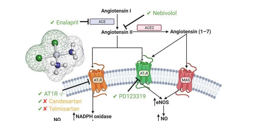
Cancers 2021, 13, 1572                                                                                              2 of 42

                         to cell death [12]. Irrespective of its potent anticancer properties and efficacy, the clinical
                         usage of cisplatin is limited due to the severity of adverse side effects including ototoxicity
                         and neurotoxicity [13,14] and its dose-limiting factor nephrotoxicity [12,15–21].

                         1.2. Nephrotoxicity
                              Nephrotoxicity results from a rapid decline of excretory mechanisms within the
                         kidney [22], enhancing the accretion of waste products produced by protein metabolism
                         (including urea, nitrogen, and creatinine) [22–24]. Acute kidney injury (AKI) is commonly
                         caused by nephrotoxic injury to kidney tissue, resulting in acute tubular necrosis [25].
                         It can also result from inadequate urinal drainage [26]. Decreased drainage causes an
                         increase in intratubular pressure and decreases glomerular filtration rate (GFR). Decreased
                         GFR can additionally be stimulated by afferent arteriole vasoconstriction [27]. Despite
                         improved prognosis following the removal of diuretics to promote volume expansion and
                         hydration, the prevalence of cisplatin-induced AKI (CIAKI) remains high [28]. Although
                         cisplatin-induced nephrotoxicity can manifest in a variety of ways, acute tubular necrosis
                         (ATN) is the most prevalent [29]. In the clinical setting, AKI frequently occurs despite
                         low-dose cisplatin administration [30]. The uptake of cisplatin into proximal tubular
                         epithelial cells (PTEC) is the initiator of the toxic effects of cisplatin [31]. To date, despite
                         burgeoned research, there is no intervention that adequately treats or prevents CIAKI in
                         cancer patients [32]. Therefore, further understanding the molecular pathways and their
                         interactions is essential in finding or developing a suitable pharmacological treatment to
                         be used in conjunction with cisplatin.

                         1.3. Pathophysiology of Cisplatin-Induced AKI
                              A variety of molecular pathways and mechanisms have been investigated to deter-
                         mine the unknown pathological events caused by CIAKI. The key molecular mechanisms
                         involved in cisplatin-induced nephrotoxic adverse effects include cellular uptake and
                         accumulation, inflammation, oxidative stress, vascular injury, endoplasmic reticulum (ER)
                         stress, and necrosis and apoptosis (Figure 1). A plethora of pharmacological agents (Table 1)
                         and genetic alterations (Table 2) have been investigated in experimental preclinical studies
                         of CIAKI. Despite the prevalence of nephrotoxicity in cisplatin-treated patients, its clinical
                         application must be accompanied by other treatments to counteract its harmful effects
                         while allowing it to exert its potent anticancer properties. Cisplatin cellular uptake is the ini-
                         tiator of the nephrotoxic effects, with several studies investigating the various therapeutic
                         options that promote renoprotection (Figure 2). The purpose of this review is to collectively
                         present the magnitude of preclinical studies in addition to presenting the clinical studies
                         recently completed and currently being conducted for the treatment of CIAKI. The data
                         from these studies illustrates the broad pathophysiological mechanisms involved and the
                         potential for their inter-relationships. This review sheds light on the current failure in
                         preclinic to clinic translatability given the lack of studies currently moving from animal
                         models to human clinical trials. Despite the frequent protective therapies evaluated in
                         models of CIAKI, there is no evidence of treatment progression with almost all therapies
                         evaluated, posing a highly concerning issue for cisplatin patients.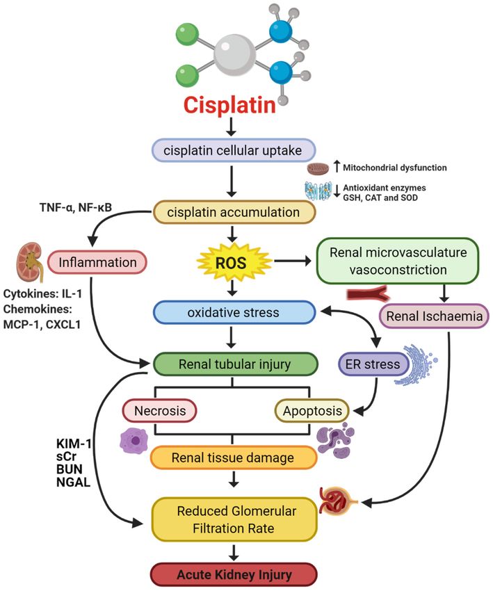
Cancers 2021, 13, 1572                                                                                                           3 of 42

     Figure 1. Pathophysiological map of the key molecular pathways demonstrated to play a role in the pathogenesis of
     cisplatin-induced acute kidney injury (AKI). The mechanisms associated with cisplatin-induced AKI (CIAKI) are complex,
     and the relationship between the key pathways remains unknown. However, it is believed that the detrimental nephrotoxic
     effect of cisplatin in renal tissue is due to platinum accumulation. Cisplatin accumulation triggers increased production of
     tumor necrosis factor alpha (TNF-α) [33,34] and reactive oxygen species (ROS), stimulating inflammation [35], oxidative
     stress [36], vascular injury [31], and apoptotic pathways [37]. The apoptotic mechanisms then promote renal tissue damage
     leading to the key clinical manifestation of nephrotoxicity (a reduction in glomerular filtration rate (GFR)) resulting in
     CIAKI. Abbreviations: GSH, glutathione; CAT, catalase; SOD, superoxide dismutase; TNF-α, tumor necrosis factor alpha;
     ROS, reactive oxygen species; ER stress, endoplasmic reticulum stress; and GFR, glomerular filtration rate. IL-1, Interleukin
     1; MCP-1, monocyte chemoattractant protein 1; CXCL1, C-X-C Motif Chemokine Ligand 1; KIM-1, Kidney Injury Molecule 1;
     sCr, Serum Creatinine; BUN, Blood Urea Nitrogen; NGAL, Neutrophil gelatinase-associated lipocalin. Figure adapted from
     “Cisplatin nephrotoxicity: mechanisms and renoprotective strategies” by N. Pabla and Z. Dong, 2008, Kidney International,
     Volume 73, P994-1007, Copyright [2008] by the Elsevier.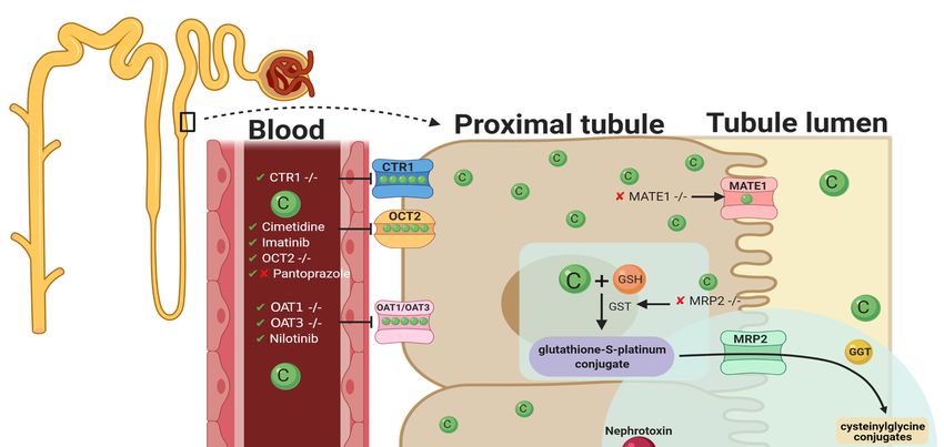
Cancers 2021, 13, 1572                                                                                                                          4 of 42

      Table 1. Pharmacological interventions assessed for renoprotective effects against cisplatin-induced AKI in vitro and in vivo,
      papers published in 2020.

 Drug                    Mechanism of Action           Findings                            In Vitro             In Vivo              Reference
                                                      ↓ Markers of oxidative stress
                                                      (HO-1 and 4-HNE)
 Aucubin                 Anti-inflammatory                                                 –                    BALB/c mice          [38]
                                                      ↓ Apoptosis (caspase-3,
                                                      caspase-9 and PARP)
                         Anti-inflammatory,
                         Antioxidative,
                                                       ↓ Tubular Injury
                         oxygen-free radical                                                                    C57BL/6J
 Curcumin                                              ↓ BUN                               –                                         [39,40]
                         scavenging, antifibrotic,                                                              mice/rats
                                                       ↓ sCr (rats)
                         and anticancer
                         activities
                                                       ↑ Body weight and
                                                       renal index
                 Antiapoptotic via
                                                       ↓ Tubular epithelial                                     Sprague
 Dexmedetomidine α2AR/PI3K/AKT                                                             –                                         [41]
                                                       cell apoptosis                                           Dawley Rats
                 pathway
                                                       ↓ Expression of GRP78,
                                                       CHOP and Caspase-12
                                                       ↓ Inflammation (iNOS)
                                                       ↓ Apoptosis (BAX)
 Etoricoxib              Anti-inflammatory                                                 –                    Rats                 [40]
                                                       No changes to creatinine,
                                                       BUN, GSH, and MDA
                                                       ↓ sCr and BUN
                         Antioxidant and
                                                       ↓ PAS tubular injury score
 Eugenol                 anti-inflammatory                                                 –                    BALB/c mice          [42]
                                                       ↓ cytoplasmic vacuolization
                         properties
                                                       of proximal tubular cells
                                                       ↓ sCr and BUN
                         Inhibits Ferroptotic cell     ↓ apoptosis (TUNEL stain)
 Ferrostatin-1                                                                             –                    C57BL/6J mice        [43]
                         death                         ↓ Tubular injury score (H&E)
                                                       ↓ Lipid peroxidation
                                                       ↓ ROS generation
                         Anti-inflammatory,
 Isoorientin                                           ↓ Apoptosis                         mTECs                Nrf2−/−              [44]
                         antioxidant
                                                       ↓ Inflammation
                                                       ↓ Tubular injury
                         Antioxidant,
                                                       ↓ markers of oxidative stress
 Monotropein             anti-inflammatory and                                             –                    BALB/c mice          [45]
                                                       ↓ markers of apoptosis
                         antiapoptotic
                                                       ↓ BUN, no reduction in sCr
                                                       ↓ MDA (HK-2 cells)
                         Synthetic vitamin D           ↓ Cell death (HK-2 cells)
 Paricalcitol                                                                              HK-2 cells           WT mice              [43]
                         deficiency                    ↓ sCr and BUN (WT mouse)
                                                       ↓ Tissue Injury (WT mouse)
                                                       ↓ sCr and BUN
                                                       ↓ mRNA expression of IL-1β,
                                                       IL-6, TNF-α
 Quercetin               Anti-inflammatory                                                 –                    C57BL/6J mice        [46]
                                                       ↓ reduced tubular
                                                       necrosis score
                                                       ↓ activity of Syk/NF-κB
      Abbreviations: sCr, SCr; BUN, blood urea nitrogen; PAS, periodic acid Schiff; KIM-1, kidney injury molecule-1; ROS, reactive oxygen
      species; mTECs, medullary thymic epithelial cells; iNOS, inducible nitric oxide synthase; BAX, BCL2-associated X protein; GSH, glutathione;
      MDA, malondialdehyde; TUNEL, Terminal deoxynucleotidyl transferase dUTP nick end labelling; H&E, haematoxylin and eosin; HK-2,
      human kidney 2; VDR, Vitamin D receptor; mRNA, messenger ribonucleic acid; −/− , knockout; IL-1β, Interleukin-1 beta; IL-6, Interleukin-
      6; TNF-α, tumor necrosis factor-alpha; Syk, spleen tyrosine kinase; NF-κB, nuclear factor kappa B; CHOP, CCAAT-enhancer-binding
      protein homologous protein; GRP78, glucose-regulated protein 78; HO-1, heme oxygenase-1; 4-HNE, 4-hydroxynonenal; and PARP, poly
      (ADP-ribose) polymerase, ↓; Decreased, ↑; Increased, −/−; genetic deletion, −/−.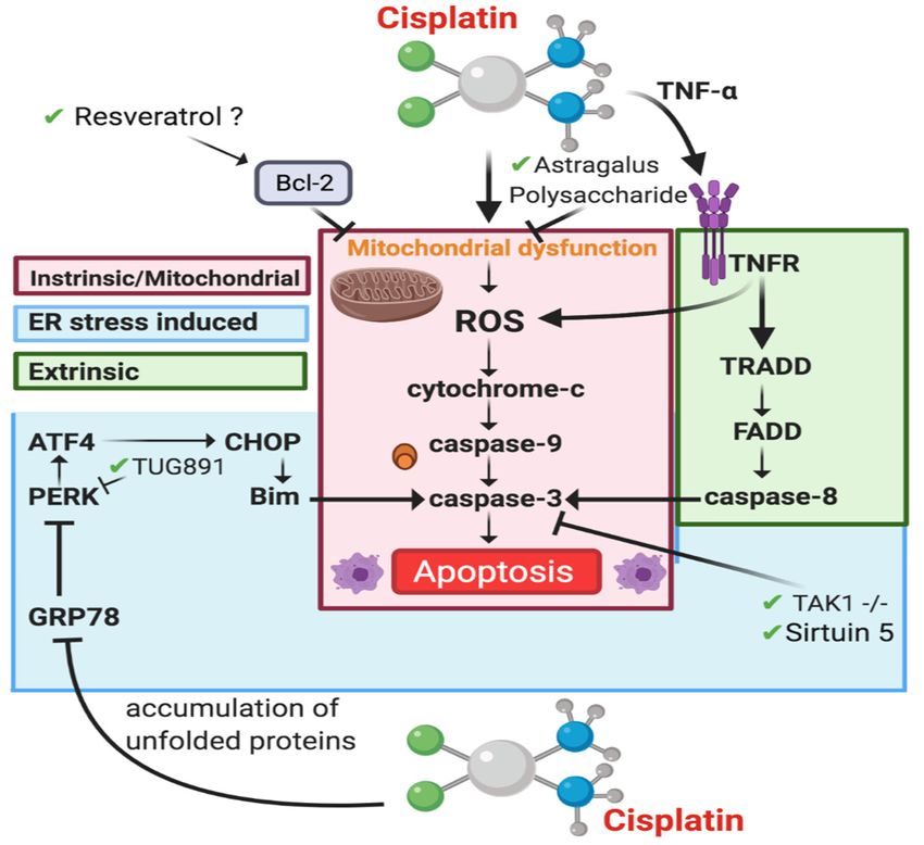
Cancers 2021, 13, 1572 5 of 42
2. Pharmacological Approaches Targeting Cisplatin Cellular Uptake
2.1. Cellular Uptake Transporters of Cisplatin
The cellular uptake of cisplatin has been implicated in the pathogenesis of CIAKI.
Organic cation transporter 2 (OCT2), copper transporter 1 (CTR1), and the less explored
volume-regulated anion channels (VRAC) are involved in cisplatin transportation into
kidney cells [57] by enabling platinum accumulation, which has been linked to kidney
dysfunction [28] (Figure 2). Kidney tissue following cisplatin treatment showed a five-fold
Cancers 2021, 13, x FOR PEER REVIEW 6 of 42
increase in cisplatin concentration compared to serum, indicative of PTEC accumulation [9].
Organic cation transporter 2 is one of the transporters affiliated with cisplatin cellular uptake.
Figure 2.
Figure 2. Graphical
Graphical representation
representationofof key molecules
key andand
molecules pathways involved
pathways in cisplatin
involved transportation
in cisplatin initiating
transportation nephrotoxic
initiating ne-
effects. Key transporters responsible for cellular uptake of cisplatin from the blood into PTECs resulting
phrotoxic effects. Key transporters responsible for cellular uptake of cisplatin from the blood into PTECs resulting in a much greater
in a
platinum
much concentration
greater compared to compared
platinum concentration the blood. toThe
thekey interventions
blood. trialed to date
The key interventions and their
trialed effectiveness
to date in targeting
and their effectiveness
CIAKI
in are also
targeting illustrated.
CIAKI are also Diagram
illustrated.details the cellular
Diagram details processes
the cellularinvolved
processes in involved
the cellularin uptake [57–64],
the cellular [57–64]
efflux
uptake [65,66],
,
[65,66], and
and metabolism
efflux of metabolism
cisplatin intoofa cisplatin
highly reactive
into a thiol
highly(nephrotoxin)
reactive thiol[67] [67] and
and the treatment
(nephrotoxin) targeted to prevent
the treatment them. to
targeted
prevent them.
2.2. Organic Cation Transporter 2 (OCT2)
2.2. Organic Cation Transporter 2 (OCT2)
OCT2 is expressed on the basolateral membrane of PTEC [57,59,60] and plays a central
OCT2 is expressed
role in cisplatin uptake on
intothe basolateral
tubular membraneAmongst
cells [57,59,60]. of PTECthe [57,59,60] and plays
transporters a cen-
responsible
tral role in cisplatin uptake into tubular cells [57,59,60]. Amongst the transporters
for CIAKI, it has been shown that 30% of nephrotoxic effects caused by cisplatin is directly respon-
sible for CIAKI,
mediated by OCT2it has been [68].
uptake shown that
In in 30%studies
vitro of nephrotoxic effects
investigating caused by cisplatin
OCT2-mediated is
cisplatin
directly
cellularmediated by OCT2 uptakeinhibition
uptake, pharmacological [68]. In inwas
vitronoted
studies investigating
of OCT2 OCT2-mediated
by cimetidine-inhibited
cisplatin cellular uptake,
cisplatin-induced pharmacological
apoptosis. inhibition was
In addition, nephrotoxicity noted of by
stimulated OCT2 by cimetidine-
cisplatin transporta-
inhibited
tion into cisplatin-induced
renal tubular cellsapoptosis. In addition,
and subsequent nephrotoxicity
platinum accumulation stimulated
could be bydecreased
cisplatin
transportation into renal tubular
with orally administered imatinibcells and subsequent
(a tyrosine platinum
kinase inhibitor) in accumulation
rats. Histologicalcould be
inves-
decreased with orally administered imatinib (a tyrosine kinase inhibitor)
tigations of kidney tissue confirmed that there was no evidence of severe renal damage in rats. Histolog-
ical investigations of kidney tissue confirmed that there was no evidence of severe renal
damage in mice co-treated with cisplatin and imatinib. However, tubular degeneration
was observed in cisplatin-treated groups [61]. The results of blood analysis (plasma urea,
nitrogen, creatinine, and creatinine clearance) were indicative of improved kidney func-
tion and platinum accumulation following imatinib adjunct therapy. In OCT2-expressed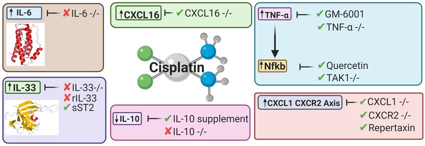
Cancers 2021, 13, 1572                                                                                           6 of 42

                         in mice co-treated with cisplatin and imatinib. However, tubular degeneration was ob-
                         served in cisplatin-treated groups [61]. The results of blood analysis (plasma urea, nitrogen,
                         creatinine, and creatinine clearance) were indicative of improved kidney function and
                         platinum accumulation following imatinib adjunct therapy. In OCT2-expressed HEK293
                         cell studies, adjunct administration of cisplatin and imatinib showed decreased accumu-
                         lation of platinum in PTECs and decreased cisplatin-induced cytotoxicity [61]. However,
                         despite the renoprotective effects observed in preclinical animal models, imatinib has not
                         provided positive toxicology results. According to the US Food and Drugs Administration
                         adverse reporting system, 44 imatinib-treated cases cited renal-related toxicity. Of these
                         44 cases, 25 manifested as AKI [69]. As such, this may not be an adequate clinical treatment.
                         Potentially irreversible acute kidney injury was also observed in a nonclinical trial in
                         imatinib-treated chronic myeloid leukemia patients [70].
                               An experimental study using OCT2-deficient mice showed impairment of cisplatin
                         uptake in renal cells, evident by reduced platinum accumulation [60]. Cairimboli et al.
                         confirmed the importance of OCT2 in cisplatin uptake [58]. The authors associated the
                         overexpression in HEK293 cells with increased cisplatin uptake causing cisplatin toxicity,
                         because of increased cellular sensitivity [58]. To date, many pharmacological approaches
                         targeting molecules responsible for cisplatin uptake or transportation into PTECs have been
                         explored [57,61,71–74]. A murine model of CIAKI demonstrated downregulation of OCT2
                         expression by formononetin inhibited the development of AKI associated with cisplatin
                         treatment through stimulation of renal tubular cell proliferation, survival, and apoptosis
                         inhibition [71]. Despite the ameliorating effects of in vivo OCT2 inhibition in murine mod-
                         els of CIAKI, human studies failed to display the same renoprotective effects. Fox and
                         colleagues used a randomized crossover experimental design to assess the prevention of
                         cisplatin-induced nephrotoxicity using the OCT2 inhibitor pantoprazole, in young patients
                         with osteosarcoma. To assess the effects, novel biomarkers were tested to investigate
                         glomerular and tubular function. Measurement of serum cystatin c was used as an indirect
                         indicator of GFR, and urinary biomarkers N-acetyl-β-glucosaminidase (NAG), kidney
                         injury molecule-1 (KIM-1), and neutrophil gelatinase-associated lipocalin (NGAL) were
                         used to quantify the degree of renal injury caused by cisplatin. The results of this study
                         showed that concurrent administration of cisplatin with pantoprazole provided no protec-
                         tion against renal injury or function in young cancer patients [75]. Interestingly, a more
                         recent study showed that pantoprazole can ameliorate CIAKI in mice [62]. Given the
                         contradictory results of OCT2 inhibition on CIAKI in animal-versus-human studies further
                         research needs to be conducted. It is important to note that OCT2 murine models were
                         nontumor bearing, whilst the human studies were conducted in cancer patients, which
                         could be a contributing factor to the failed clinical study.

                         2.3. Copper Transporter 1
                              CTR1 is located on the basolateral membrane of proximal tubules and is highly ex-
                         pressed in human kidneys [57,59]. The exact role of CTR1 in cellular uptake of cisplatin into
                         renal proximal tubules resulting in nephrotoxicity is incompletely understood. However,
                         studies have shown that CTR1 downregulation is protective against platinum accumu-
                         lation [57]. The knockdown of CTR1 reduces cisplatin nephrotoxicity by up to 80% in
                         both mouse embryonic fibroblasts and yeast [72,74]. In vivo studies indicated elevated
                         levels of CTR1 expression was associated with increased cisplatin accumulation in tumors,
                         a process also observed in PTEC [57,76]. Pabla and colleagues investigated the relation-
                         ship between cisplatin and CTR1 expression to further define the role that CTR1 plays
                         in nephrotoxicity. Interestingly, in mice, there were no significant differences in CTR1
                         expression 1–3 days following cisplatin treatment. They also demonstrated that incubation
                         of HEK293 cells with copper generated both monomeric and trimeric CTR1 knockdown, re-
                         sulting in approximately 50% diminution in cisplatin accumulation and a 30% reduction in
                         apoptosis. Furthermore, CTR1 knockdown cells incubated with the OCT/MATE inhibitor
                         cimetidine further inhibited both cellular uptake and apoptosis following treatment with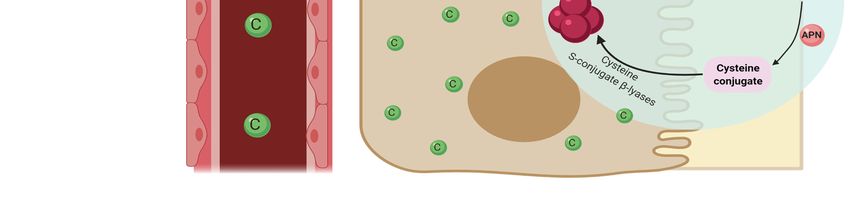
Cancers 2021, 13, 1572                                                                                             7 of 42

                         cisplatin [57,58]. The results of this study have shown that although both CTR1 inhibition
                         and OCT2 inhibition alone are options to prevent nephrotoxicity, the combination of CTR1
                         and OCT2 inhibition together has better therapeutic potential. Interestingly, the majority of
                         cellular uptake research regarding cisplatin into renal cells has focused on the two major
                         cisplatin transporters OCT2 and CTR1. However, there have also been suggestions that
                         there are other entry points for cisplatin into renal cells that are yet to be explored in
                         models of CIAKI such as VRAC channels. Reduced VRAC channel activity is associated
                         with cisplatin resistance [77], and the presence of VRAC channels in kidney cells [78]
                         highlights a potential avenue for CIAKI research. Additionally, impaired cisplatin efflux
                         has been shown to contribute to cisplatin accumulation and the nephrotoxic effects that
                         follow [65,79].

                         2.4. OAT1/OAT3
                               In addition to OCT2 and CTR1, the organic anion transporter (OAT) family(OAT1
                         and OAT3 have also shown to transport cisplatin and potentially a nephrotoxic metabo-
                         lite into PTEC resulting in nephrotoxic injury to renal cells [63]. OAT transporters are
                         largely concentrated in the basolateral membrane of PTEC and facilitate transportation of
                         hydrophilic anions into cells. This intake is via secondary/active transportation respon-
                         sible for regulating anion balance in the body [80]. To investigate the influence of OAT
                         transporters on cisplatin-induced nephrotoxicity, C57BL/6J mice with genetic deletion
                         of OAT1 and OAT3 were injected with 30 mg/kg cisplatin. In cisplatin-treated wildtype
                         mice, there were increases in biomarkers of CIAKI, in addition to histological indica-
                         tion of kidney damage such as tubule dilation and necrosis. There was no evidence of
                         kidney dysfunction in OAT1- and OAT3-deficient mice treated with cisplatin. Further
                         studies are needed to further understand the role of each individually and the interaction
                         they have together on CIAKI nephrotoxicity. A different model was used to investigate
                         OAT-stimulated CIAKI using nilotinib. Nilotinib is a tyrosine kinase inhibitor shown to
                         noncompetitively inhibit OCT2 and both OAT1 and OAT3 [63]. Nilotinib was given to
                         OCT1/2 −/− mice simultaneously with cisplatin, with results indicating no loss of kidney
                         function in the adjuvant cisplatin- and nilotinib-treated group as confirmed by reduced
                         BUN levels. This indicates that OAT1/inhibition by nilotinib provides some evidence
                         of amelioration of CIAKI; however, as the paper elucidates, further investigations in the
                         mechanisms of mitigation are required [63]. A separate study investigated the effects of
                         nilotinib in a rodent model of CIAKI. Male Wister albino rats were treated with 25 mg/kg
                         Nilotinib 4 days prior to a single intraperitoneal injection of 6 mg/kg cisplatin and 6 days
                         following the cisplatin injection. Results of their study showed that nilotinib improved
                         creatinine clearance compared to cisplatin-treated rats; however, it had no influence on
                         increased BUN [64]. It was also observed that nilotinib attenuated cisplatin increase in
                         MDA, a biomarker of oxidative stress [81]. Morphological changes showed amelioration
                         of CIAKI by nilotinib. Although this study did not look specifically at nilotinib influence
                         on OAT1 and OAT3, it does confirm its ability to prevent CIAKI. Given this information,
                         there is a clear correlation and link between the transporters, and therefore, further inves-
                         tigations need to be undertaken to understand their interactions and the influence that
                         has on mediating cisplatin uptake. Given cisplatin uptake is the initial step mediating its
                         nephrotoxic effects, potentially inhibiting all three synergistically may be an ideal strategy
                         for CIAKI prevention.

                         3. Pharmacological Approaches Targeting Cisplatin Cellular Efflux
                               Apically localized efflux transporters P-type copper transporting ATPases (ATP7A
                         and ATP7B), multi-antimicrobial extrusion protein transporter-1 (MATE 1), and multidrug-
                         resistance-associated protein (MRPs) mediate excretion of cisplatin into the urine [57,59,65,82].
                         These transporters are highly expressed in the proximal and distal tubules [57]. Tubular
                         injury is a key pathology associated with the nephrotoxic effects of cisplatin. Tubular injury
                         promotes reduced GFR and therefore delayed urinary excretion of cisplatin, leading to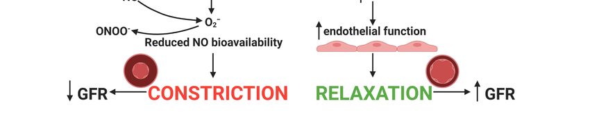
Cancers 2021, 13, 1572                                                                                          8 of 42

                         platinum accumulation within the tubules [83]. Given the pathogenesis linked to platinum
                         accumulation in PTEC, increasing the expression of cisplatin efflux transporters has been a
                         molecular target against CIAKI.

                         3.1. Apically Localized Efflux Transporters P-Type Copper Transporting ATPases7A/B
                               Although there is little research available determining the effects of ATP7A and ATP7B
                         on cisplatin nephrotoxicity, they have both been extensively investigated in the setting of
                         cisplatin drug resistance. Studies investigating overexpression of both ATP7A and ATP7B
                         in cancer models have been shown to be independently linked to poor survival in ovarian
                         cancer patients and cisplatin resistance in prostate carcinoma cells, respectively [84–86].
                         Therefore, overexpression of ATPases could prevent CIAKI; however, its therapeutic poten-
                         tial may not exceed the possible negative outcomes to cancer cells.

                         3.2. Multidrug-Resistance-Associated Protein 2
                               MRPs are associated with mediating the efflux of cisplatin and its nephrotoxic conju-
                         gates from kidney cells [87]. Previously, research investigating the role of MRP expression
                         in cisplatin accumulation demonstrated that increased MRP expression resulted in reduced
                         cisplatin accumulation and therefore has been suggested to play a critical role in cisplatin-
                         induced nephrotoxicity [87]. Given its role in nephroprotection, you would expect its
                         expression to be downregulated in response to cisplatin treatment, given that platinum
                         accumulation is a well-established complication. However, acute renal failure induced in
                         rats showed a significant upregulation of MRP2 72 h after cisplatin treatment; however,
                         there were only minor increases in MRP4 expression compared to controls [88].
                               The glutathione-s-platinum conjugate, whose metabolism is responsible for the pro-
                         duction of the reactive thiol nephrotoxin, is suggested to be eliminated by MRPs [89].
                         To determine the role of MRP2 on cisplatin efflux, MRP2-deficient mice were treated with
                         20 mg/kg of cisplatin, resulting in enhanced platinum accumulation and proximal tubular
                         injury. MRP2-deficient mice showed increased mRNA expression of GST, the enzyme
                         which catalyzes the formation of the cisplatin–glutathione conjugate. Platinum accumu-
                         lation was reduced in transgenic knock-in Mrp2-knockout mice, indicating that MRP2
                         plays a role in the accumulation of platinum [66]. However, the mechanisms involved in
                         this accumulation and the effect they have on the production of the reactive thiol remain
                         unclear. It is possible that an increase in MRP expression might be seen in models of
                         nephrotoxicity; however, no studies are yet to present data investigating this. In addition
                         to MRPs, multi-antimicrobial extrusion protein 1 (MATE1/SLC47A1) is suggested to be
                         involved in platinum accumulation associated with cisplatin treatment [83].

                         3.3. Multi-Antimicrobial Extrusion Protein 1
                               MATE1 expression is largely concentrated in the brush-border membrane of PTECs
                         and assists epithelial cell elimination of cationic molecules into urine [83]. Cisplatin has
                         been identified as a substrate for MATE1 [90]. MATE1 has shown to mediate cisplatin
                         efflux and prevent cisplatin accumulation in tubular cells preventing cisplatin nephrotoxic
                         effects [65]. However, following cisplatin treatment, downregulation of MATE1 expression
                         in human tubular epithelial cells is observed [83]. Given the ability for MATE1 to facilitate
                         cisplatin urinary excretion, further investigations have been undertaken to isolate its role
                         in cisplatin-induced AKI. A model of cisplatin-induced nephrotoxicity was undertaken in
                         MATE1 −/− mice [65]. Blood urea nitrogen (BUN) and plasma creatinine concentration
                         were significantly elevated in cisplatin-treated MATE1 −/− mice, compared with cisplatin-
                         treated wildtype mice. It was observed that renal concentrations of platinum were at a
                         20-fold increase compared to plasma concentration in MATE1-deficient mice [65]. Increased
                         serum creatinine (sCr) and BUN were also observed in MATE1 pharmacological inhibition
                         studies [65,91]. Therefore, it has been concluded that MATE1 plays a key role in cisplatin
                         accumulation and is thus a contributor to CIAKI [65]. A study investigating MATE1
Cancers 2021, 13, 1572 9 of 42
upregulation in models of CIAKI may be a good therapeutic avenue in the prevention
of nephrotoxicity.
4. Interventions Targeting Molecular Mechanisms CIAKI
4.1. Oxidative Stress
Despite the collection of research that has focused specifically on cisplatin transporta-
tion and accumulation, other models of CIAKI have targeted key molecules involved
in ROS formation [92]. A balance occurs between ROS production and the antioxidant
defense system to maintain homeostasis [2,3]. Cisplatin disrupts this equilibrium through
overproduction of ROS and impaired antioxidant defense systems. This triggering re-
duced production of key antioxidants, including superoxide dismutase (SOD), glutathione
(GSH) [2], and catalase (CAT) [93]. The reduction in the functionality of the antioxi-
dant defense system leads to overexpression of key markers of oxidative stress following
cisplatin treatment [36]. Elevated levels of cisplatin in PTEC also increase cisplatin accu-
mulation in mitochondria, stimulating mitochondrial dysfunction, mitochondrial damage,
and ROS production [94,95]. Following cisplatin infiltration into renal epithelial cells,
it becomes a potent nephrotoxin via gamma-glutamyl transpeptidase (GGT)-dependent
metabolic activation (Figure 2) [96]. Glutathione-S-transferase (GST) mediates the forma-
tion of glutathione-S-platinum conjugates, which passes through the kidneys [34]. It is
cleaved into cysteine–glycine conjugate by Gama-glutamyl-transpeptidase [67]. Amino-
dipeptidases further metabolizes this into cysteine conjugates, which are then transported
into proximal tubules. Cysteine-S-conjugate β-lyase (CSCβL) is metabolized to form the
cysteine conjugate into a highly reactive thiol, which is the initiator of cisplatin’s cell toxic-
ity [67,96]. Elevated levels of the highly reactive thiol molecule produced after cisplatin
uptake is metabolized by CSCβL catalyze the enzymatic activation of glucose-6-phosphate
dehydrogenase and hexokinase, increasing ROS [1].
Studies have determined the use of natural antioxidants including, vitamin C [97],
vitamin E [98], and activation of the vitamin D receptor [43] to target ROS formation,
which have all shown nephroprotective properties against renal toxicity [15]. The beneficial
co-therapy of cisplatin and vitamin C in C57BL/6 mice has been previously demonstrated.
Mice were inoculated with Lewis lung carcinoma followed by treatment with cisplatin.
Levels of sBUN and sCr presented cisplatin-treated mice demonstrated higher levels of
oxidative damage. Decreased levels of kidney dysfunction were also observed in adjunct
vitamin C and cisplatin-treated mice, without compromising cisplatin cytotoxicity [97].
Furthermore, there have been extensive pharmaceutical interventions assessed for their
antioxidant effects on CIAKI [94]. Oxidative stress, evident by reduced MDA/MPO
expression, was observed in necrostatin-1- and cisplatin-treated mice [99]. This was also
observed in hesperetin- and cisplatin-treated HK-2 cells. Both drugs, showed reduced levels
of apoptosis [100], which could interfere with the cytotoxicity of cisplatin. Experimental
and clinical studies have focused on cisplatin nephrotoxicity, specifically through targeting
mechanisms and molecular pathways associated with pharmacological inhibition ROS
production or stimulation of antioxidant pathways [36,44,49,92,93,97,101]. A pathway of
interest recently targeted in CIAKI research is the nuclear factor erythroid 2-related factor
2/heme oxygenase-1 (Nrf2/HO-1) signaling pathway [100].
Monotropein (Nrf2/HO-1 Antioxidant Pathway)
Activation of Nrf2 has shown promise in multiple experimental models as a key
modulator in the suppression of oxidative stress and inflammation to preserve kidney
function [102,103]. Nrf2 is responsible for maintenance of the cellular redox balance,
antioxidant response, and phase II detoxification process [104]. Nrf2 expression has shown
to be downregulated in rats following cisplatin treatment [105]. Renal expression of
Nrf2 and HO-1 was downregulated compared to control rats, however rats pretreated
with Sinapic acid (SA) followed by cisplatin resulted in marked increase in Nrf2 and
Ho-1 expression. Cisplatin treatment resulted in a significant downregulation of keyCancers 2021, 13, 1572 10 of 42
antioxidant enzymes SOD, CAT, and GSH. SA and cisplatin-treated rats showed elevation
in these key antioxidants indicating an enhanced antioxidant defense system following
SA treatment [103]. Pharmacologically, it has been demonstrated that activation of the
Nrf2 signaling pathway by N,N-dimethylformamide (DMF) following cisplatin treatment
attenuated AKI [102], as well as tubulointerstitial lesions [106]. Both ameliorating effects
are associated with stimulation of antioxidants such as HO-1 and NAD(P)H quinone
oxidoreductase 1 (NQO1) [102,106]. Stimulation of these two antioxidants was observed in
mice treated with Isoorientin a flavone, suggested to activate the Nrf2 signaling cascade [44].
This was confirmed in Isoorientin-treated Nrf2-deficient mice, where renoprotection was
abolished [44]. Taken together, these studies provide an insight to the promising effects
of the Nrf2 pathway as a target for CIAKI. However, studies into the interaction between
Nrf2 signaling activators and the apoptotic pathways that mediate cisplatin’s cytotoxicity
have yet to be determined.
4.2. Vascular Injury
Interestingly, a focus on anti-inflammatory and antioxidant pathways has been high-
lighted in most pharmaceutical and genetic modification studies published recently. Re-
duced GFR caused by reduced renal blood flow is a key pathology of CIAKI [107]. Little
research specifically targeting the vasoconstriction properties of cisplatin to promote renal
perfusion has been undertaken. Vasoconstriction stimulated through activation of adeno-
sine A1 receptors (AT1 s) by cisplatin is a suggested mechanism contributing to CIAKI.
Additionally, CIAKI has been linked to vascular injury via endothelial dysfunction [31,107].
Reduced renal blood flow to kidney tissue through elevated vasoconstriction and impaired
vascular autoregulation stimulated by damage to the endothelium is implicated in the
pathogenesis of CIAKI [12]. Cisplatin has been suggested to cause damage to the vas-
culature within renal tubules [12]. It results in vascular resistance and constriction of
vascular smooth muscle cells (VSMC) leading to reduced renal blood flow, decreased GFR,
and hypoxia of renal tubular cells, leading to kidney damage [31,108]. Cisplatin has shown
to alter the response of renal vascular endothelium to vasoactive substances [15]. Kidney
vasculature and tubules are known to have extensive sympathetic nerve innervation, releas-
ing catecholamines from their terminals, and triggering G-coupled adrenoceptors on the
cell surface [109]. Adrenoceptors increase calcium and trigger contractions of vasculature
muscle resulting in vasoconstriction of smooth muscle cells [110,111]. Vascular injury is
linked to elevations in oxidative stress, resulting in a sequence of metabolic disturbances.
Cisplatin treatment leads to oxidative stress induced by ROS and impaired function of
the antioxidant defense system [79]. Excessively produced ROS include superoxide (O2 − ),
hydroxyl radical (HO•), hydrogen peroxide (H2 O2 ), peroxynitrite (ONOO− ), nitrogen
oxide (NO•), and hypochlorous acid (HOCl). Increased oxidative stress leads to endothelial
damage, resulting in impaired endothelial dependent vascular relaxation. Endothelial
dependent VSMC relaxation is NO dependent and alterations in its production or bioavail-
ability disrupt relaxation. Additionally, eNOS function is impaired when ROS production
or function is not repressed [112]. Amino acids such as L-arginine synthesize the produc-
tion of eNOS to produce NO [9]. Excess production of NO by inducible NO synthase is
increased following cisplatin treatment [64]. NO reacts with the NO scavenger O2 − to
produce peroxynitrite (ONOO− ) [4,10–12], resulting in reduced NO bioavailability and
endothelial dysfunction [113].
Recently, levosimendan, a calcium sensitizing vasodilator, has been used in a model of
CIAKI [114]. It attenuated renal damage, improved renal blood flow, and enhanced kidney
morphology. Despite all those factors, there was only slight alleviation in biomarkers
associated with kidney dysfunction (sCr and BUN), indicating only partial prevention of
CIAKI. Levosimendan significantly reduced TNF-α expression in cisplatin-treated rats,
indicating levosimendan also displays anti-inflammatory properties [114]. Interestingly,
despite levosimendan renoprotective effects, earlier studies demonstrated it also plays
a role in the prevention of H2 O2− induced apoptosis of cardiomyocytes [115], indicatingCancers 2021, 13, 1572 11 of 42
antiapoptotic properties taken together with levosimendan may potentially interfere with
Cancers 2021, 13, x the
FORcytotoxic properties
PEER REVIEW of cisplatin. The vasculature plays a role in the pathophysiology of
CIAKI; to further elucidate these effects, research into the renin angiotensin system (RAS)
has been studied.
The RAS has been investigated in models of CIAKI for at least two decades to f
The Renin Angiotensin System in Cisplatin-Induced Acute Kidney Injury
identify the direct effects of cisplatin on the vasculature and renal hemodynamics
The RAS has
Thebeen
RASinvestigated
is responsiblein for
models of CIAKI forbalance
the homeostatic at leastof
two decades
blood to further
pressure. Angiotensin
identify the direct
I) is converted to angiotensin II, which stimulates angiotensin II type [116].
effects of cisplatin on the vasculature and renal hemodynamics I receptor (AT
The RAS is responsible for the homeostatic balance of blood pressure. Angiotensin I (Ang
angiotensin II type 2 receptor (AT2), stimulating vasoconstriction and vasodilati
I) is converted to angiotensin II, which stimulates angiotensin II type I receptor (AT1 )
spectively [117,118]. Stimulation of AT1 has been associated with renal injury [119],
and angiotensin II type 2 receptor (AT2 ), stimulating vasoconstriction and vasodilation,
AT2 activation has been correlated with renoprotection, through IL-10 stimulation a
respectively [117,118]. Stimulation of AT1 has been associated with renal injury [119], whilst
6 downregulation [118,120–122]. Interestingly both receptors have been investigate
AT2 activation has been correlated with renoprotection, through IL-10 stimulation and IL-6
CIAKI model to assess the role of both angiotensin II receptors in nephrotoxicity (
downregulation [118,120–122]. Interestingly both receptors have been investigated in a
3).
CIAKI model to assess the role of both angiotensin II receptors in nephrotoxicity (Figure 3).
Figure 3. Cisplatin-induced acute kidney injury effects on the renin angiotensin system. A large focus
has been on inhibition or genetic deletion of AT1 and AT2 receptors, both of which show amelioration
Figure 3. Cisplatin-induced acute kidney
in CIAKI [64,120,123–125]. injury effects
Additionally, onACE
both the renin angiotensin system.
and Angiotensin A large
II inhibition focus
have alsohas been on inhib
shown
tion or genetic deletion of AT1 and AT2 receptors, both of which show amelioration in CIAKI [64,120,123–125]. Additio
ameliorating qualities. Targeting the renin angiotensin system (RAS) system to prevent CIAKI is a
ally, both ACE and Angiotensin II inhibition have also shown ameliorating qualities. Targeting−the renin angiotensin sy
promising pathway in potential treatments. Abbreviations: NO, nitric oxide; ONOO , peroxynitrite;
tem (RAS) system to prevent CIAKI is a promising pathway in potential treatments. Abbreviations: NO, nitric oxid
O2 − , superoxide;
ONOO−, peroxynitrite; eNOS, endothelial
O2−, superoxide; nitric oxidenitric
eNOS, endothelial synthase;
oxideACE, angiotensin
synthase; converting converting
ACE, angiotensin enzyme; enzym
GFR, glomerular
GFR, glomerular filtration rate.
filtration rate.
The role of the AT1 receptor in response to nephrotoxicity is conflicting amongst the
The role of the AT1 receptor in response to nephrotoxicity is conflicting amon
literature, with evidence linking it to renoprotection in AT1 lymphocyte knockout models,
literature, with evidence linking it to renoprotection in AT1 lymphocyte knockout m
whilst renal epithelial AT1 knockout worsened AKI pathogenesis [126]. Interestingly, it has
whilst renal epithelial AT1 knockout worsened AKI pathogenesis [126]. Interestin
been reported that AT1 stimulation worsened CIAKI through increased TNF-alpha [126].
has been reported that AT1 stimulation worsened CIAKI through increased TNF
Renal dysfunction and TNF-alpha expression was reduced in mice deficient in PTEC
[126]. Renal dysfunction and TNF-alpha expression was reduced in mice defic
AT1 receptor expression compared to WT, highlighting the protective effects of the AT1
PTEC AT1 receptor expression compared to WT, highlighting the protective effects
AT1 receptor in CIAKI [126]. Confirming these results, treatment of rats with the se
AT1 antagonist telmisartan almost restored BUN and sCr back to control levels ind
of restored renal function[124]. Interestingly telmisartan has shown in a mouse mo
CIAKI to exacerbates cisplatin-induced nephrotoxicity[123].Cancers 2021, 13, 1572 12 of 42
receptor in CIAKI [126]. Confirming these results, treatment of rats with the selective AT1
antagonist telmisartan almost restored BUN and sCr back to control levels indicative of
restored renal function [124]. Interestingly telmisartan has shown in a mouse model of
CIAKI to exacerbates cisplatin-induced nephrotoxicity [123].
These results show that there is both ameliorating effects between AT1 deficiency and
stimulation in CIAKI. Additionally, this was supported by treatment with another AT1
receptor antagonist candesartan [127], with one study reporting no impact on reduction
in BUN and creatinine following cisplatin treatment [125], whilst another showed pro-
tection. Given this information, further understanding of the role of the AT1 receptor in
the pathogenesis of cisplatin-induced AKI is a critical component of understanding the
pathophysiological map of the disease.
Administration of telmisartan could be causing off-target effects given the renoprotec-
tive effects were attributed to antioxidant and anti-inflammatory properties rather than its
involvement in the RAS [124]. The protective effects of AT1 inhibition in cisplatin-treated
mice may be through increased activation of AT2 or the mitochondrial assembly recep-
tor (MAS) receptor, resulting in enhanced vascular relaxation and promoting medullary
blood flow. However, antagonism of the AT2 receptor by PD123319 improved renal func-
tion, indicated by reduced BUN and sCr when concurrently treated with cisplatin [125],
suggesting that although stimulation of AT2 is vasoprotective, it has exhibited both reno-
protective and renotoxic properties [125]. Given this information, a cisplatin model using
a potent and highly selective AT2 receptor agonist may elucidate further as to its role in
nephroprotection, specifically in CIAKI.
Vascular dysfunction is worsened by the overactivation of AngII production and the
depletion of Angiotensin converting enzyme 2 (ACE2) [128–131]. Angiotensin convert-
ing enzyme 2 (ACE2) activation stimulates production of angiotensin (1-7) from AngII
metabolism [132], increasing vascular relaxation through activation of the MAS recep-
tor [133]. Morsi et al. (2015) confirmed AngII plays a role in cisplatin-induced nephrotoxic-
ity; however, the direct effects it has on vascular relaxation remains unclear. In cisplatin-
treated rats, there was increased protein expression of AngII, iNOS, TNF-α, and caspase-3
and decreased expression of eNOS. Nebivolol itself had no impact on protein expression;
however, adjunct treatment of nebivolol with cisplatin improved eNOS expression and
reduced expression of AngII, iNOS, TNF-α, and caspase-3 compared to cisplatin. Nebivolol
is a selective β1-adrenoreceptor antagonist, shown to have microvasculature vasodila-
tory [134], antioxidant [112,135], anti-inflammatory [136], and antiapoptotic properties.
To determine the exact effects of nebivolol has on vascular function, further examinations
into AT1 and AT2 with its use should be conducted. It is also unclear as to the exact
mechanisms mediating the renoprotective effects of nebivolol, given that it may be having
effects on the vasculature, inflammatory responses, and apoptotic pathways [64]. Further
research specifically investigating the direct effects of vasodilation on cisplatin accumula-
tion and resulting nephrotoxicity needs to be conducted. Cisplatin reduces expression of
key molecules responsible for cisplatin efflux whilst increasing cellular uptake transporters.
As such, specifically increasing blood flow without attenuating other pathologies such as
inflammation and ROS production may in fact worsen CIAKI through increased platinum
accumulation, potentiating worsened PTEC death.
4.3. Cell Death
There are two mechanisms whereby cisplatin induces cell death, necrosis, and apop-
tosis. Initial research showed necrosis to be the only mechanism responsible for renal
damage caused by cisplatin. However, Lieberthalet et al. (1996) were some of the first to
show that apoptosis also played a part in cisplatin-induced cell death. The study showed
that high-dose cisplatin treatment induced cell death via necrosis, whilst a low dose stimu-
lated apoptotic cell death. Clinically cisplatin is administered through frequent low-dose
infusion in attempts to prevent nephrotoxic effects, which differs from the previous high-
dose method [137]. However, studies investigating the relationship between necrosis andCancers 2021, 13, 1572 13 of 42
apoptosis continues to be explored as death mechanisms caused by cisplatin-induced
nephrotoxicity remain elusive [15].
4.3.1. Necrosis
Cisplatin induces cell death phenotypes in a concentration dependent manner. High
concentrations of cisplatin cause a type of death independent of the classical features of
apoptosis which resembles necrosis [138–140]. Necrotic damage is localized to the PTEC
rather than the distal tubule as it reabsorbs filtered molecules including glucose, proteins,
electrolytes, and drugs [141]. Sancho-Martinez et al. (2011) conducted a study in vitro
using HK2 and Jurkat T cells, which showed activation of the apoptotic program with
high necrotic concentrations of cisplatin [141]. The apoptotic program is further aborted
at the level of effector caspases which emanates into necrotic-like death phenotype [141].
Necrosis is a passive mode of cell death which activates an inflammatory and immune re-
sponse [141,142]. It is characterized by cell swelling, plasma membrane rupture, and loss of
organelle structure [143]. Necrosis was considered as accidental cell death, which is unreg-
ulated [144] until genetic programmed necrosis was discovered in vivo [145,146]. A type
of receptor-interacting protein kinase-based cell death that has similar signaling pathways
with apoptosis and plays a major role in CI-AKI is namely necroptosis [142,147]. Xu et al.
(2015) used RIP3-KO and mixed-linage kinase domain-like protein (MLKL) knockout mice
to investigate the role of necroptosis in CIAKI [142]. Necroptotic cell death induces inflam-
matory cytokines including, TNF-α, TNF-related weak inducer of apoptosis, and IFN-γ
in cisplatin-treated kidneys [142]. Expression of these cytokines further contributes to the
induction of receptor-interacting protein 1 (RIP1), RIP3, and MLKL expression in vivo that
enhances the necroptotic signaling pathway by positive feedback [142]. Thus, necrotic
cell death in renal tubules is dependent of the RIP1/RIP3 and MLKL which is stimulated
by cisplatin [142]. Necrosis is caused by high-dose concentrations of cisplatin, whereas
apoptosis is stimulated by lower doses [141]. There have been multiple signaling pathways
linked to cisplatin-induced apoptosis. These pathways include the intrinsic (mitochondrial),
extrinsic, and endoplasmic reticulum stress apoptosis pathways.
4.3.2. Apoptosis-Intrinsic/Mitochondrial Pathway
Cisplatin has been shown to induce the mitochondrial apoptotic pathway through a
variety of physiological processes including ROS production and the release of cytochrome
c through stimulation of proapoptotic proteins [89] (Figure 4). Mitochondrial dysfunction
is associated with CIAKI [94,148]. A variety of pharmaceutical interventions that have
targeted the mitochondrial apoptotic pathway have shown that nephroprotection is linked
with a decrease in caspase-3 activity; however, cisplatin increases it [54,64,149]. The an-
tiapoptotic protein Bcl-2 is established to prevent mitochondrial dysfunction-induced
apoptosis [150]. Following cisplatin treatment, western blot analysis showed Bcl-2 expres-
sion is dose dependently downregulated in HK2 cells, leading to cytochrome c release,
caspase-3 cleavage, and apoptosis. Cisplatin-treated cells showed a dose dependent re-
duction in Sirtuin 5 (Sirt5) expression. Sirt5 overexpression increased bcl-2 expression and
reduced apoptosis as determined via Annexin V/PI staining. Results also showed that
adjunct Sirt5 and cisplatin therapy reduced bcl-2 expression, a pathway known to induce
apoptosis, indicating that Sirt5 exhibits its renoprotection in a bcl-2 independent manner,
through activation of caspase-3. Interestingly resveratrol a Sirt5 activator in humans [151]
attenuated CIAKI; however, there was no evaluation on Sirt5 expression [152]. It is possible
that Sirt5 was activated by resveratrol and inhibited mitochondrial apoptosis by stimulat-
ing bcl-2 to inhibit mitochondrial dysfunction. Overexpression of bcl-2 is associated with
reduced cisplatin cytotoxicity [153] and therefore may not be a suitable treatment option.Cancers 2021, 13, 1572 14 of 42
Cancers 2021, 13, x FOR PEER REVIEW 15 of 42
Figure 4. ApoptoticFigure 4. Apoptotic
pathways involvedpathways
in CIAKI.involved instimulation
Cisplatin CIAKI. Cisplatin
of ROSstimulation of ROS leads
leads to cytochrome to cyto-
release and effector
chrome release and effector caspase activation in the Intrinsic/mitochondrial pathway
caspase activation in the Intrinsic/mitochondrial pathway (red) [54,94,154], ER stress is involved in two (red)
mechanisms,
through stimulation[54,94,154]
of PERK to, ER stress ER
promote is involved in twocaspase-3
stress induced mechanisms, through
activation stimulation
(blue) of PERKapoptosis
[155]. Extrinsic to promoteis caused
ER stress induced
by TNFR1 signaling caspase-8 (green). caspase-3 activation (blue) [155] . Extrinsic apoptosis is caused by TNFR1 signal-
ing caspase-8 (green).
4.3.3. Apoptosis-Extrinsic Pathway
4.3.3. Apoptosis-Extrinsic Pathway
The extrinsic apoptotic pathway is triggered when a ligand binds to a death receptor
The extrinsic apoptotic pathway is triggered when a ligand binds to a death receptor
located on the cytoplasmic membrane of cells (Figure 4). Activation of the extrinsic path-
located on the cytoplasmic membrane of cells (Figure 4). Activation of the extrinsic path-
way contributes to the loss of tubular cells in AKI [156]. The extrinsic apoptotic pathway
way contributes to the loss of tubular cells in AKI [156]. The extrinsic apoptotic pathway
activates the caspase-8 molecules, which further activates the downstream effector caspase-
activates the caspase-8 molecules, which further activates the downstream effector
3 [15,157]. Cisplatin induces the extrinsic pathway through an increase in proinflammatory
caspase-3 [15,157]. Cisplatin induces the extrinsic pathway through an increase in proin-
cytokine TNF-α expression via its death receptor 1 (TNFR1). It is well established in the
flammatory cytokine TNF-α expression via its death receptor 1 (TNFR1). It is well estab-
literature that TNFR1 is a major component of the TNF family, which plays an important
lished inrole
thein
literature that TNFR1
the extrinsic apoptotic is apathway
major component
as shown in ofstudies
the TNF family,
where whichknockout
TNFR1 plays mice
an important role in the extrinsic apoptotic pathway as shown in studies
have been suggested to be resistant to CIAKI [157–159]. The pathways of apoptosis are where TNFR1
knockout mice have been
inter-related. There suggested
are threeto be resistant
ways to CIAKI
the extrinsic pathway [157–159]. Theto
is linked pathways of
the mitochondrial
apoptosis are inter-related.
pathway that result There are three
in caspase-8 ways the[160].
activation extrinsic
ROSpathway
generation is linked to the
in the mitochondria
mitochondrial
results in a direct link to the fas gene and Fas-ligand (Fas-1), which results the
pathway that result in caspase-8 activation [160]. ROS generation in in apopto-
mitochondria results
sis [157]. In ininvitro
a direct link using
studies, to the NRK-53E
fas gene and Fas-ligand
cells (Fas-1), which
and α(E)-catenin resultscell line
knockdown
in apoptosis
(C2) to[157]. In in vitro
identify studies,apoptotic
the specific using NRK-53E
pathway cells and α(E)-catenin
stimulated knockdown of α(E)-
by downregulation
cell line catenin
(C2) to (linking
identify protein)
the specific apoptotic pathway stimulated by downregulation
[157]. As a result, it was reported that a reduction in a(E)-catenin of
α(E)-catenin (linking
expression protein)the
increased [157]. As a result,of
susceptibility it AKI
was via
reported that a reduction
the Fas-mediated in a(E)-
apoptotic pathway,
catenin which
expression
confirmsincreased
the rolethe susceptibility
of Fas [157]. ROS of AKI via also
generation the results
Fas-mediated apoptotic
in the phosphorylation of
pathway, which confirms the role of Fas [157]. ROS generation also results
p38 and mitogen-activated protein kinase (MAPK) resulting in increased TNF-α produc- in the phos-
phorylation of p38Ramesh
tion [161]. and mitogen-activated
and Reeves (2005) protein
usedkinase (MAPK)inhibitor
a p38-MAPK resultingcommonly
in increased known as
TNF-α production [161]. Ramesh and Reeves (2005) used a p38-MAPK
SKF-86002 in vivo and reported that TNF-α levels were significantly decreased inhibitor com-as well as
monly known as SKF-86002
the inhibition of p38 in vivo andagainst
protected reported that [162].
CIAKI TNF-αItlevels were
was also significantly
suggested that de-
a hydroxyl
creased radical
as wellscavenger,
as the inhibition
dimethyl of thiourea
p38 protected against
prevented the CIAKI
activation[162]. It was
of the also sug-pathway
p38-MAPK
gested that
which a hydroxyl
completely radical scavenger,
ameliorated dimethyl
CIAKI thioureastudy
[162]. Another prevented
by Wang the and
activation of (2018)
colleagues
the p38-MAPK pathway which completely ameliorated CIAKI [162]. Another study byYou can also read