MODELS FOR ALS DISEASE MECHANISM
←
→
Page content transcription
If your browser does not render page correctly, please read the page content below
DISEASE MECHANISM AND
     MODELS FOR ALS
N EW CONCEPT : AGING AS MAJOR REGULATOR OF ALS PATHOLOGY

A REVIEW ON THE CURRENT KNOWLEDGE OF ALS IS GIVEN BY DISCUSSING DISEASE
 MECHANISMS AND MODELS THAT ARE USED TO COMBAT THE BATTLE AGAINST ITS
       PATHOLOGY .   FURTHERMORE , A NEW CONCEPT WILL BE PROPOSED .

                            Susanne Elise Baars

                              December 2012
Image on front page:
Researchers suggest that stimulation of cell signaling delays the onset of ALS, by causing motor
                                                                             neurons ‘humming’.
                                  Image: © ISTOCKPHOTO/KIYOSHI TAKAHASE SEGUNDO
        Image reference: www.scientificamerican.com/article.cfm?id=potential-new-weapon-against-als
HUBRECHT INSTITUTE

 DISEASE MECHANISM AND
     MODELS FOR ALS
       New concept: aging as major regulator of ALS pathology

A REVIEW ON THE CURRENT KNOWLEDGE OF ALS IS GIVEN BY DISCUSSING DISEASE
 MECHANISMS AND MODELS THAT ARE USED TO COMBAT THE BATTLE AGAINST ITS
       PATHOLOGY .   FURTHERMORE , A NEW CONCEPT WILL BE PROPOSED .

                                Final report:
                         Period: 12-11-2012 – 15-01-2013

                               Susanne Elise Baars
                                 Nassaustraat 6D
                                     3583 XE
                                      Utrecht
                              Tel: 0031-652048615
                         email: s.e.baars@students.uu.nl

                Master Experimental and Clinical Neuroscience
                           University of Utrecht

                         Supervisor: Professor N. Geijsen
                               Hubrecht Institute

                        Examiner: Professor J. Pastorkamp
                            Rudolf Magnus Institute
                        University Medical Centre Utrecht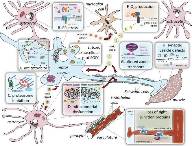
TABLE OF CONTENTS
Acknowledgement .................................................................................................... 1
Abstract .................................................................................................................... 2
Abbreviations ........................................................................................................... 3
Introduction ............................................................................................................. 4
   Aim of this thesis ............................................................................................................. 4
   ALS pathology and diagnosis .......................................................................................... 4
   Causes of ALS .................................................................................................................. 5
     Genetics .................................................................................................................................................5
ALS disease mechanism and models ...................................................................... 6
   Disease Mechanisms ....................................................................................................... 6
     Proteotoxicity ........................................................................................................................................6
     Glutamate excitotoxicity .....................................................................................................................9
     Oxidative Stress ................................................................................................................................. 10
     Compromised axonal transport ...................................................................................................... 11
     Astrocytes and Glial: non-cell autonomous death of motor neurons ...................................... 12
     Inflammation: viral infection and immune imbalance ................................................................ 13
     Neuro-vascular system: a neurovascular disease .......................................................................... 14
     RNA metabolism............................................................................................................................... 15
   Disease models .............................................................................................................. 17
   Discussion...................................................................................................................... 19
A new concept: Aging ............................................................................................. 21
   The IIS pathway ............................................................................................................ 22
   TDP-43/FUS as link between aging and the cellular stress response .......................... 24
   Athletes .......................................................................................................................... 26
   Association between Ataxin-2 and TDP-43/FUS.......................................................... 28
   Discussion...................................................................................................................... 31
Aging and Stem cells ..............................................................................................34
   Intrinsic and Extrinsic Stem cell properties .................................................................. 34
     Extrinsic Properties in general ........................................................................................................ 34
     Stem cell nice: .................................................................................................................................... 35
     Intrinsic aging..................................................................................................................................... 36
     Cell fate ............................................................................................................................................... 36
   Embryonic Stem Cells ................................................................................................... 38
   Link between stem cell function, aging and ALS. ......................................................... 38
   Discussion...................................................................................................................... 40
In vitro based therapeutic strategies ......................................................................42
   Human embryonic stem cells (hESCs) ......................................................................... 42
   Human induced pluripotent stem Cells (human iPSCs) ............................................... 42
   iPSC disease modelling of ALS pathology .................................................................... 43
   Drug screening and development.................................................................................. 45
   Cell Therapy................................................................................................................... 45
   Discussion...................................................................................................................... 46
Proposal for further research ..................................................................................48
Bibliography ........................................................................................................... 51
Supplementary Figures ...........................................................................................70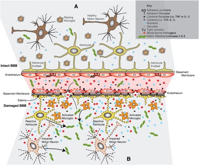
DISEASE MECHANISM AND MODELS FOR ALS

ACKNOWLEDGEMENT
I would like to express my very great appreciation to Professor N. Geijsen, my supervisor, for his
enthusiastic and patient guidance and useful assessment of this thesis. I would also like to extend
my thanks to the technician of his laboratory of the department Pluripotent stem cells in
development and disease, for her help in learning me the basics of working with induced
pluripotent stem cells (iPSCs). My grateful thanks are also extended to Professor R.J. Pasterkamp,
for willing to be second reviewer.

1 | ACKNOWLEDGEMENT

S.E. Baars

ABSTRACT
Amyothrophic lateral sclerosis (ALS, Lou Gehrig’s disease) is a severe adult-onset
neurodegenerative disease, characterized by progressive premature loss of upper and lower motor
neurons. Eventually the disease leads to death due to respiratory failure1. ALS is the third most
common neurodegenerative disease2 and cause of adult mortality3. It appears to be caused by a
complex interaction among genetics and environmental factors2. While approximately 10% of
ALS cases are familial, the remaining cases are believed to be sporadic. However, the exact
working mechanism underlying the selective motor neuron death in ALS is not resolved yet.
Since a wide variety of cellular processes are involved in ALS, a general, systemic principle might
underlie it.

Recent data provides compelling evidence for a major role of aging in the development of
ALS4,5,6,7. This hypothesis was enhanced by the discovery that reduced Insulin/IGF signalling
(IIS) correlates with lifespan extension8 and a decreased risk to develop ALS9. Its pathway is not
only regulating lifespan, metabolism and stress resistance4, TDP-43 has also showed to be part of
it, a well known ALS gene9. TDP-43 is primarily a nuclear protein that participates in common
heteromultimeric complexes, which is involved into diverse RNA processes and stress granule
formation16,10. Furthermore, TDP-43 is identified as the major component of insoluble
cytoplastic inclusions in both sALS as fALS, and associated with the oxidative stress response. It
seems likely that this TDP-43 protein mediates longevity through activation of the IIS pathway
under stressfull conditions11. Besides the activation of various cellular cascades, there is a vicious
circle in which TDP-43 expression becomes upregulated. Interestingly, it has been found that
during stress, the threshold for TDP-43 phosphorylation is lowered. This lead to increased
proteotoxicity12. This in turn seems to cause and/or to accelerate motor neuron degeneration.

However, besides aging, also heavy exercise is in part mediated by the IIS pathway and involved
in ALS development13. During heavy exercise, enormous amount of ROS are generated which
causes oxidative stress and disturb the epigenetic codes14.

Interestingly, age-related changes seemed to be determined by the age of the systemic
environment, rather than by the cell-autonomous age15. Therefore, it has been suggested that
signals from the systemic environment drive age-related, intrinsic changes, mediated by the IIS
pathway. This activates a cascade of events that eventually can lead to ALS pathology. However,
current animal models are not suitable to further investigate this postulation. Not only did the
obtained results fail when translated to the human situation, also are fundamental genetic and
anatomical differences between both species in age-related pathways found16,17. Therefore, new
and improved models are needed. In vitro cell based therapies seems to be the first feasible
model to investigate the disease mechanism for both sALS and fALS by utilizing induced
pluripotent stem cells (iPSCs)18. iPSCs can give us insights into disease mechanisms, drug
discovery, cell therapy and a potential new diagnostic method for patients with ALS19,20,21.
Furthermore, will this technique enable the identification of a systemic regulation of aging on
ALS pathology in patient derived iPSCs.

                                                                                        Abstract   |2
DISEASE MECHANISM AND MODELS FOR ALS

ABBREVIATIONS
ALS                            Amytrophic lateral sclerosis
MN                             Motor Neurons
UMN                            Upper Motor Neurons
LMN                            Lower Motor Neurons
FTLD                           frontotemporal dementia
sALS                           Sporadic ALS
fALS                           fALS
ER stress                      endoplasmic reticulum stress
UPR                            unfolded protein response
IGF1                            insulin-like growth factor 1
TNFα                           tumour necrosis factor
BBB                            blood brain barrier
BSCB                           blood spinal cord barrier
MP2                            myelin protein 2
MMp9                           matric metallopeptidase 9
iPSC                           induced pluripotent stem cells
ESCs                           Embryonic stem cells
Human iPSC                     Human Induced Pluripotent Stem Cell

3 | INTRODUCTION
S.E. Baars

INTRODUCTION
Amyothrophic lateral sclerosis (ALS, Lou Gehrig’s disease) is a severe adult-onset
neurodegenerative disease, characterized by progressive premature loss of upper and lower motor
neurons (MNs) in the motor cortex, the anterior horn of the brainstem and spinal cord which
eventually leads to death due to respiratory failure1. Next to being third most common
neurodegenerative disease with a worldwide prevalence of 4-6 per 100.00022, ALS is also one of
the most common causes of adult mortality in this disease group. The peak age of onset is
between 50 and 70 years2.

The broad variety of disease manifestations in ALS appears to be caused by a complex
interaction among genetics and environmental factors. Approximately 10% of ALS cases are
familial, while the remaining cases are believed to be sporadic23. Although the enormous efforts
put into unravelling the underlying disease mechanism, no efficacious drug or therapy has been
developed yet24. Numerous hypothesises have been proposed to underlie ALS, but none of them
is able to fully explain the mechanism of pathogenesis.

AIM OF THIS THESIS
Within this thesis I will review the proposed disease mechanisms of ALS by discussing the
current hypothesizes and models that are used to investigate it. Furthermore, I will propose a
new concept with respect to ALS pathology and outline a research proposal for future
investigation.

ALS PATHOLOGY AND DIAGNOSIS
ALS pathology is characterized by loss of motor neurons in the anterior horns of the spinal cord,
motor nuclei of the brain stem and motor cortex, which together make up the corticospinal tract
(with a relative sparing of sensory neurons)25,23. Signals are transmitted from motor neurons in
the brain (upper motor neurons, UMN) to motor neurons in the spinal cord (lower motor
neurons, LMN). From there they are passed through to particular muscles. Degeneration of these
motor neurons ceases signal transmission, which result in muscle weakness, spasticity,
fasciculation’s and atrophy throughout the body26. Eventually progressively loss of voluntary
movement is observed27. In the contrary to other anterior horn diseases and motor
neuropathology’s, such as spinal muscular atrophy or primary lateral sclerosis, ALS shows
degeneration of both upper and lower motor tracts28. The primary clinical symptoms and signs of
ALS include impairment affecting limb, bulbar, axial and respiratory function. Specific UMN
signs are hyperreflexia, spasticity, extensor plantar response (up going toes), and positive jaw jerk,
while LMN signs include weakness, muscle atrophy, cramps and fasciculation’s. However,
differences in site of onset, pattern and speed of spread as well as the degree of upper motor
neuron (UMN) and/or lower motor neuron (LMN) dysfunction produce a remarkably variable
disorder29. Notably, also frontotemporal dementia (FTLD) is seen in 5% of the cases30.

Although, the clinical diagnosis is primarily based on clinical manifestations, it is supported by
laboratory studies, electrodiagnostics and imaging (Supplementary Table.1). At molecular and
cellular level, a wide range of processes are associated with ALS as well, such as protein
misfolding and aggregation (ubiquitinated and neurofilamental inclusions) and mitochondrial
damage. Also energy deficit, ER stress31, changes in calcium homeostasis32 and excitotoxicity are
seen33. Furthermore, specific vulnerability of motor neuron degeneration is determined by its
characteristics (for a overview about motor neuron vulnerability features, see supplementary
table.2). Additionally, degeneration of motor neurons is often accompanied by reactive gliosis34.

                                                                                   Introduction   |4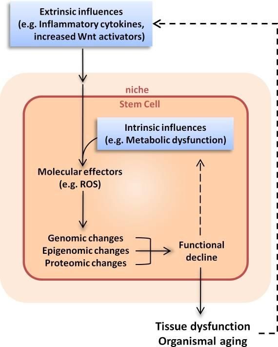
DISEASE MECHANISM AND MODELS FOR ALS

These processes have all been intensively investigated and will be shortly reviewed in the next
sections.

CAUSES OF ALS
ALS seems to be a multifactorial disease, which appears in a broad variety of disease
manifestations caused by a complex interaction among genetics and environmental factors.
People of all races and backgrounds are affected35, but there appears to be a slight male
predominance36. Although the exact working mechanism remains unclear, ALS is most often
seen in elderly individuals, which indicates that aging might be an important risk factor5..
Furthermore, multiple external risk factors have been associated with ALS, such as: ingestion of
high concentrations of β-methylamino- L-alanine37, use of cholesterol-lowering medication38,
intensive physical exercise39 including football playing13,40 and service in the USA Army41. The
latter possibly links to intermittent work-related hypoxia42 or to head injury43, 44. Other
environmental factors include exposure to heavy metals45, cigarette smoking46, and pesticides or
herbicides47. Although exposure to toxics initially seemed to be important risk factors, no
additional evidence has been found in the last 25 years48. Lastly, associations are found between
ALS, viral infections (HIV)49 and prion disease50.

GENETICS
Besides environmental factors, several genes have been associated with ALS as well.
Approximately 90% of patients51 suffering from ALS don’t have a family history, which is
defined as sporadic ALS (sALS)23. The residual cases are inherited termed familial ALS (fALS).
Whereas fALS is known to be caused by mutations in at least 10 different genes 52, sALS do not
show a defined genetic profile. Genes which are associated with ALS include a wide range of
cellular processes, namely: involvement into antioxidant response53, axonal and vesicular
transport54, angiogenesis, endoplasmic reticulum (ER) stress and unfolded protein response
(UPR) and RNA metabolism33.

fALS is inherited as a dominant trait with different penetrance and expressivities among ALS
patients23. Mutations in the SOD-1 gene are the most common cause of fALS (20%), followed by
mutations in TDP-43 (5%), FUS (5%), and ANG (

S.E. Baars

ALS DISEASE MECHANISM AND MODELS
This section will discuss the disease mechanism of ALS and the most commonly used models
with their application.

DISEASE MECHANISMS
The exact working mechanism underlying the selective motor neuron death in ALS is not
resolved yet, although numerous hypothesizes have been proposed. These hypothesizes include
proteotoxicity, glutamate excitotoxicity, oxidative stress (mitochondrial dysfunction), proteasome
inhibition (aggregate formation and ER stress), non-cell autonomous function mediated by
astrocytes and glial cells, compromised axonal transport, inflammation, neurovascular and RNA
metabolism. The SOD-1 transgenic animal model has been most widely used in ALS research,
since this gene was first identified to be associated with significant subpopulation of both fALS
as sALS.

This section will briefly outline the core principles of each hypothesizes. Interestingly, although
they all seem plausible, either one of them can be the primary cause of the development of ALS,
but also be a consequence of an earlier underlying event. People have always considered ALS as
a pure motor disorder. However, subsets of patients have emphasizes the involvement of other
cell types and working mechanisms as well. The multitude of contributing factors does underline
the complexity of the disease. Nowadays, ALS is more seen as a multisystem disorder in which
motor neurons appear to be the most severely affected59.

PROTEOTOXICITY
Accumulation of aggregated proteins is the major phenotypical hallmark of ALS pathology 60, 61.
Interestingly, this is seen in almost all neurodegenerative diseases, including Alzheimer’s disease62,
Parkinson’s disease62, frontotemporal dementia (FTLD) and Huntington’s disease63, but also in
prion disease64. These protein aggregates consist of damaged and misfolded proteins that cannot
be removed by the normal protein degradation mechanisms65. TDP-43 was identified as the
major component of these insoluble inclusions in both sALS as fALS. After this discovery, FUS
has also been identified to be components of protein aggregations. TDP-43 and FUS are both
primarily nuclear proteins that participate in common heteromultimeric complexes which are
enrolled in RNA transcription, translation, splicing, nucleo-cytoplasmic shuttling, transport for
local translation, and stress granule formation16,10. Research suggests that they interact with other
proteins by binding glycine-rich domains. Under pathological conditions, they are redistributed to
the cytosol where they form abnormal protein-protein associations66. Notably, SOD-1, another
well known ALS causing gene, has also been identified in protein aggregations 50. However,
toxicity caused by this gene seems not correlated to its aggregation potential. Therefore, further
research proceeded on the function of aggregate prone proteins and their proteotoxicty in
relation to ALS.

Proper protein folding has shown to be crucial for cellular function and viability67. During cell
division, damaged and oxidized proteins are sequestered and incorporated by the mother cells.
This mechanism enables daughter cells to consist of an undamaged proteome68. However,
neurons, which are post-mitotic cells, do need another mechanism to ensure protein quality67.
Many proteins do fold spontaneously but they do need assistance to correct their structures by a
specialized set of chaparones, which perform a quality-control process as well. Nonetheless,
aggregation-prone proteins challenge these systems, by making them fail to handle them. Also,
protein homeostatic system becomes less efficient and less effective during aging69.

                                                           ALS disease mechanism and models      |6
DISEASE MECHANISM AND MODELS FOR ALS
Fig.1: Supposed mechanism of protein
aggregation. Aggregation prone proteins
causes co-aggregation of other proteins and
impairment of proteasome activity. This lead
to distortion of other cellular proteins, such as
for chaperones and mitochondrial dysfunction.
(Image adapted from Boillée et al, 2006).
Unfortunately, a failure to properly fold one protein may result in protein aggregations, which
can destabilize the proteome even further. This may lead to uncontrolled aggregation of other
proteins as well and causes proteotoxicity70. Moreover, when misfolded or damaged proteins are
not removed, this disturbs cellular process and can trigger mitochondria dysfunction and the
unfolded protein response (UPR), which lead to endoplasmatic reticulum (ER) stress. Beside,
studies have shown that mutant SOD-1 activates components which mediate ER stress31.
Eventually ER stress can cause cell death through caspases-mediated apoptosis. Furthermore,
protein aggregation have also shown to cause mitochondrial dysfunction (Fig.1)71. Nonetheless,
there is no universal agreement as to what function they have in disease pathology.
Moreover it is still under debate if protein aggregates could either directly cause neuronal
dysfunction and death, or that they arise as a harmless by-product, or if they arise to protect cells
by sequestering of toxic proteins. Major evidence for protein aggregation as primary cause is
given by the fact that almost all ALS models feature prominent, cytoplasmic, intracellular
inclusions in motor neurons as well as in the astrocytes surrounding them72. They are highly
immunoreactive and develop often before onset of clinical symptoms, as an early sign of ALS 73.
Aggregative proteins have showed to behave in a dose dependent manner, with enhanced
aggregations during disease progression, and to generate motor neuron degeneration11.
Several mechanisms have been suggested to cause toxicity in motor neurons. On the one hand,
mutant SOD-1 aggregates may assemble other proteins, which are normally required for
neuronal function74. On the other hand, aggregations are caused by a reduction of protein-
folding chaperones, which are needed to catalyze folding of other proteins as well75. Therefore,
mutant SOD-1 aggregates causes proteosomal dysfunction, which lead to disrupted protein
turnover. Furthermore, imbalance of protein synthesis and degradation compromises its ability to
degrade other critical components as well. Finally, almost all aggregates seems to co-express
ubiquitin. This was an interestingly finding, since ubiquitin targets proteins to the proteasome
degradation pathway for clearance71. This implies that accumulation of ubiquitinated, misfolded
proteins, may affect the proteasome and impair normal protein degradation of proteins, such as
SOD-1.
7 | ALS DISEASE MECHANISM AND MODELSS.E. Baars
However, recent research has provided evidence consistent with each of the above-mentioned
mechanisms. While it was previously assumed that high molecular mass aggregates are the cause
of ALS, recent data in this field implies that small oligomeric aggregative structures, which are
highly toxic to the cell, may underlie pathology76,77. Therefore the production of less-toxic large
aggregates seems to be a protective, compensatory mechanism to detoxify the cell as much as
possible78,79. It has now been suggested that during lifespan, the cell’s capacity to remove these
small toxic aggregates become reduced. This could be either the result of an increased amount of
small toxic aggregations during lifetime or to a diminished clearance capacity of the cell during
disease progression. In either way, more toxic aggregations appear, which causes damage that
may lead to neuronal death. This mechanism seems especially important in low turnover cells (i.g.
motorneurons), since protein aggregates are not diluted as in high turnover cells due to cell
divisions. Moreover, mother cells retain aggregates during early divisions and buddying, which
protect new cells from damage.
Taken together, aggregates are seen in almost all neurodegenerative diseases. This accumulation
of misfolded proteins appears to disrupt the proteasome degradation pathways and induces ER-
stress. The selective vulnerability of aggregates on motor neurons in ALS has to be elucidated.
Current literature has linked multiple protein aggregation to ALS pathology9,62,80. However, until
now, only SOD-1 and TDP-43/FUS protein aggregations have been found to contribute to ALS
pathology. Although proteotoxicity shows clear indications for the development of ALS
pathogenesis, it could be argued if this is the primary cause of ALS, or that these proteins form
aggregates upon stimulation of another mechanism. Initiation of protein misfolding includes
random processes, caused by a reduction of protein folding chaperones, proteosomal
dysfunction or by the formation of abnormal protein-protein interactions80, 81. Therefore,
alterations in other proteins are expected to be involved into ALS pathology as well. Questions
arise about the working mechanism of proteotoxicity in ALS, since protein aggregations of only
certain proteins has been observed to cause ALS82.
Protein aggregations have been associated in numerous neurodegenerative diseases. For instance,
the accumulation of hyperphosphorylated tau protein, which is involved into the pathogenesis of
Alzheimer Diseases (AD)83. Interestingly, certain tau isoforms have been reported in other
neurodegenerative diseases as well, such as in ALS84. However, not all patients that expresses tau
protein aggregates develop ALS pathology85. This creates doubts about the specificity of the
aggregate protein and may suggests that proteotoxicity is mediated by another mechanism rather
than causing ALS itself. In this case, protein inclusions and aggregates may represent an end stage
of a molecular cascade, while earlier steps in this cascade may be more directly involved into
pathogenesis.
Nevertheless, studies support the idea that at least one aspect of toxicity may arise by processes
mediated by misfolded aggregated proteins or by loss of essential components as consequence of
protein aggregate formation72.
ALS disease mechanism and models |8DISEASE MECHANISM AND MODELS FOR ALS
GLUTAMATE EXCITOTOXICITY
Glutamate is the main excitotoxin in the brain, albeit being major excitatory neurotransmitter in
the mammalian CNS57. During normal homeostasis, glutamate can be increased in the synaptic
cleft, but this level will rapidly decrease. In ALS pathology, high glutamate levels are maintained
due to increased glutamate release or an impaired re-uptake mechanisms (Fig.2)32. These high
levels of glutamate stimulate presynaptic glutamate receptors, which subsequently enhance the
release of additional glutamate via a positive feed forward mechanism. This causes increased
intracellular Ca2+ levels, which lead to excitotoxicity and eventually cell death. Interestingly,
excitatory amino-acid transporters (EAAT2) are often down regulated in the motor cortex and
spinal cord of ALS patients71. These EAAT2 receptors are the major terminator of excitatory
signals through re-uptake of glutamate from the synapse into glial cells and neurons. Eventually
opening of mitochondrial permeability pores will occur due to high calcium concentrations86.
Consequently mitochondria swell and release reactive oxygen species (ROS) along with other
proteins that stimulate apoptosis.
Fig.2: Excitotoxicity in ALS. Excessive
glutamate lead to enhanced receptor
stimulation. This causes increased glutamate
release and inadequate removal and re-uptake.
Therefore, electrochemical gradients change
and the receptor function converse. Increased
levels of calcium activated enzymes that are
toxic to the cell. Subsequently, reactive oxygen
species (ROS) is produced, which impairs
mitochondrial function. This viscious circle
eventually lead to excitotoxicity and
degeneration of motor neurons in ALS. (Image
adapted from Van dan Bosch, et al., 2006)
The pore can also stimulate the mitochondria to release even more calcium, to inhibit the
production of adenosine triphosphate (ATP) and to activate ATP synthases, which hydrolyses
ATP instead of producing it. Elimination of energy metabolism may distores the electrochemical
gradients of certain ions. These gradients are needed to remove glutamate from the extracellular
space by activation of glutamate transporters. However, when the electrochemical gradient
changes, this causes a reversal in transporter function: instead of removing glutamate they now
act as active secreting glutamate channels, which enhance excitotoxicity even more(fig.2)87.
Moreover, besides the influence of calcium influx on excitotoxicity, it has recently been noted
that extra-synaptic NMDA receptor activation stimulates CREB protein inactivation. This results
in loss of mitochondrial membrane potential and apoptosis. Additional research on selective
motor neuron vulnerability, showed extremely sensibility of motor neurons to excitotoxicity. This
selective sensibility is caused by a low Ca2+ buffering capacity and a high number of Ca2+
permeable AMPA receptors32.
All in all, this hypothesis shows one of the most robust mechanisms and is supported by much
evidence. Moreover, the only drug available on the market to slow disease progression is riluzole,
which has anti-excitotoxic properties. However, how this excitotoxicity arises remains to be
questioned.
9 | ALS DISEASE MECHANISM AND MODELSS.E. Baars
It could be argued that if protein aggregates, formed by mutant gene products (as discussed in
the previous paragraph), might assemble in motor neurons and/or decrease glutamate uptake in
the surrounding glial cells57. Interestingly, recent research has found that glutamate
hyperstimulation leads to the formation of microaggregates in iPSC-derived neurons. However,
further research is needed to investigate if excitotoxicity may underly proteotoxicity as well. Also
in this context, it remains unclear why this process would especially involve SOD-1, TDP-
43/FUS protein aggregation.
OXIDATIVE STRESS
Oxidative stress derives from an accumulation of reactive oxygen species (ROS), which arise as
by-products of aerobic metabolism88,87. With aging, oxidative stress accumulates within the
neuron and may cause a reduction in the ability of the biological system to eliminate or to repair
ROS-induced damage.
Cellular ROS is caused by a leakage of electrons from the mitochondrial respiratory chain, due to
an imbalance between the production and the removal of reactive oxygen. Particularly, the early
components from the electron transport chain, complexes I and III, have showed to be prone to
leak electrons to molecular oxygen89. Therefore mitochondria are the major source of ROS
production, followed by the endoplasmic reticulum.
Under normal conditions, the cell has multiple anti-oxidant defence mechanisms to remove ROS
and to prevent oxidative stress. One of these mechanisms includes the catalytic removal of ROS
by superoxide dismutase, which is mainly regulated by SOD-1. However, mutations in SOD-1
disrupt enzyme function and leads to increased ROS production, which results in enhanced
oxidative stress (Fig.3)90,73. Moreover, research showed reduced proteasomal activity in both,
mutant SOD1 and TDP43/FUS expressing mice, which is caused by aggregations that disrupt
the ubiquitin-proteasome system91. Under normal conditions this degradation pathway protects
the cell by taking up damaged, misfolded and unfunctional proteins. Dysfunction of the
proteasome and lysosome systems eventually lead to mitochondrial dysfunction with increased
oxidative stress92. The process by which ROS creates oxidative stress is as followed: Firstly,
reactive oxygen causes incomplete reduction of molecular oxygen during oxidative
phosphorylation and lead to the production of hydrogen peroxide (H2O2) and the superoxide
radical anion (O2-). Subsequently, ROS will react with radical nitric oxide (NO) and will form the
oxidant peroxynitrite (ONNOO-)93,94, and H2O2 will eventually form the highly reactive
hydroxyl radical, OH. These radicals causes damage to proteins, lipids and DNA53. This can alter
protein conformations and change the active site of the enzymes, change membrane properties
by oxidation, and introduce mutations into DNA/RNA. Notably, some components of the
electron transport chain are encoded by mitochondrial DNA. Mitochondrial DNA is already
exposed to a higher mutation rate compared to nuclear DNA92. However, once oxidative stress
is present, other mechanisms become involved as well, such as excitotoxicity. Excitotoxicity
causes increased intracellular calcium levels, which enhance ROS production. Nonetheless, ROS
seems to inhibit glutamate uptake through loss of EAAT2 transporter expression and enhanced
expression of calcium permeable AMPA receptors in glial cells. Furthermore, activation of
microglial results in secretion of cytokines and further ROS. Lastly, mutant protein aggregates
inhibit neurofilament assembly and cytoskeletal transport.
Thereby, the central nervous system appears to be more susceptible to oxidative damage than
other tissues. This is caused by the accumulation of metal ions throughout the CNS, which favor
ROS production33. The selective vulnerability of motor neurons may be caused by their high
ALS disease mechanism and models | 10DISEASE MECHANISM AND MODELS FOR ALS
metabolic input. Motor neurons have a relative big size and long axon length. This high-energy
demand goes along with many mitochondria, with the side effect of increased oxidative stress.
Taken together, oxidative stress regulates multiple mechanisms, but in light of ALS, neuronal
degeneration may result from a complex interaction of ROS, excitotoxic stimulation, genetics and
dysfunction of proteins and organelles (e.g. mitochondria, ER) 53. Furthermore, the neurotoxic
effects of ROS are not only limited to neurons, but could also affect glial cells. However, further
research has to be performed to determine the causative role of oxidative stress in ALS
manifestation.
Fig.3: Molecular pathways involved in oxidative stress. Oxidative stress is caused by the
enhancement of ROS, excitotoxicity and protein aggregates. (Image adapted from Ferraiuolo, L., et al.
57
2011) .
COMPROMISED AXONAL TRANSPORT
While previous stated mechanisms share many aspects and seem to be related to each other,
compromised axonal transport includes a distinct mechanism.
Since motor neurons have extending axons that can be more than a meter in length, efficient
transportation systems are needed. In both, patients and animal models of ALS, alternations in
large (myelinated) axonal structure by misaccumulation of neurofilements have been reported.
Also the two forms of axonal transport, kinesin-mediated (anterograde) and dynein-mediated
(retrograde), are decreased 90. Notably, transgenic mice which express a mutation in the NF-L
subunit of neurofilament, which is similar in human, develop motor neuron degeneration 95.
However, deletion of this mutation reversed the phenotype partly and caused prolonged
survival96,97. Consistently, presymptomatic deficits of slow axonal transport has been described in
mutant SOD1 mice98, which showed similar misaccumulation of neurofilement.
Furthermore, genetic evidence implicates axonal transport defects as an important mechanism in
the development of motor neuron degeneration. Altered gene and protein expressions have been
found in multiple genes associated with transport, like: loss of kinesin motor proteins KIF1Bβ99,
KIF5a100, KIF21A101, VAPB54, Rab7102, and Vps54103. Also, mutant SOD-1 mice showed
significant inhibition of retrograde axonal transport before onset of the disease90.
Although many of these features are similar to human ALS pathology, abnormalities in
neurofilaments could be either causal or just being a harmless byproduct of neuronal
degeneration of mutant SOD1 protein. Moreover, similar as with other protein aggregations,
11 | ALS DISEASE MECHANISM AND MODELSS.E. Baars
accumulation of neurofilaments in motor neuron cells could also arise from a protective view of
perspective: to protect the neuron against SOD1-mediated damage by buffering diverse
components. Nonetheless, it has been shown that particular motor neurons, are highly sensitive
to defects in axonal transport90. This may be due to their extreme polarization. So while protein
aggregation, glutamate hyperstimulation and oxidative stress are linked, axonal transport seems to
be a separate potential cause.
ASTROCYTES AND GLIAL: NON-CELL AUTONOMOUS DEATH OF MOTOR
NEURONS
Although most research is performed on previous outlined hypothesizes, there is growing
evidence for a non-cell autonomous death of motor neurons. This means that the degeneration is
not caused by the motor neurons themselves but rather through extracellular influences.
Research showed that glial cells play a particular important role in this. On the one hand,
astrocytes support neurons by providing them with nutritional factors and regulate
neurotransmitter concentrations, metabolic or ionic homeostatis104. While on the other hand
microglial cells clear damaged and dead cells from the environment. Gliosis is an early process
seen in pathogenesis in both the brain and the spinal cord105. Although ALS is caused by motor
neuron degeneration, neighbouring cells may mediate this in a non-cell autonomous manner.
Consistently, all known familial forms are caused by mutations in genes that are ubiquitously
expressed (i.g.SOD1 and VAPB) throughout the body or at least expressed in multiple cell types
(i.g. VEGF and ANG). Therefore, researchers started to examine the role of glial cells.
To investigate this in detail, chimeric animals were developed that express mutant SOD-1
selectively in motor neurons, microglial cells or astrocytes 106. Results showed that expression in
all three cell types were needed to cause ALS. After this, they removed 30-50% of the mutant
SOD-1 gene in motor neurons and found a delayed onset while disease progression remained the
same107. Animals who expressed a combination of mutant and wild type SOD-1 showed a
positive correlation with the proportion of surviving mutant SOD-1 expressing motor neurons
and lifespan of the mice. In addition, research with C.elegans has confirmed this. Glial cell
activation also increases after motor neuron injury. After damage, motor neurons seem to release
mutant porteins, fragments or distressed signals to the extracellular environment. It has been
proposed that glial cells then start to secrete protective components. However, if the stimulation
becomes chronically, toxic substances are released instead. These released proteins are involved
in the production of nitric oxide or inflammation, to activate other glial cells. However, it is not
known how they become activated in the first place, neither how this especially involves motor
neurons. It could be that this results from specific signals released from the motor neurons after
injury as stated above. Nonetheless, it seems that this active dialogue between motor neurons and
glial cells contributes to disease progression.
Since fALS and sALS show similar disease manifestation, it is suggested that a more general
principle is causing the disease. A non-cell autonomous mechanism might underlie this
principle107. However, existing disease models do not give the possibility to research a general
mechanism for fALS and sALS. Therefore, induced Pluripotent Stem Cells (iPSCs), an upcoming
in vitro technique, used more and more to further decipher the influence of astrocytes and glia
on disease progression107. Evidence deriving from this novel technique will be dicussed in more
detail in a later chapter.
ALS disease mechanism and models | 12DISEASE MECHANISM AND MODELS FOR ALS INFLAMMATION : VIRAL INFECTION AND IMMUNE IMBALANCE A common feature of ALS is the presence of neuroinflammatory reactions consisting of activated glial and T cells. Until recently, this feature has been viewed as a consequence rather than a possible cause of motor neuron degeneration108. However, focus has now been put on inflammation as a target to treat ALS109. Thus, instead of non-cell mediated motor neuron degeneration caused by intrinsic properties of glial cells, a primary role of inflammation has also been proposed. Upon injury, microglial accumulate. Investigators have discovered a positive correlation between this microgliosis and disease progression110. Although it was already known that microglia becomes increasingly activated after motor neuron injury, research now also showed activation of microglia before motor neuron degeneration and thus before clinical disease-onset109. Animal models were used to further investigate the potential link between inflammation and motor neuron degeneration. Microglia are the macrophages in the nervous system. Next to monitor the extracellular environment, they closely interact with astrocytes and neurons. This is mediated by the M1 activation pathway. They can be identified as CD11b expressing cells and are activated by a range of different signals. Futhermore, they act as the first line of defence against infection or injury. When activated, they acquire an amoeboid like appearance and start to secrete proinflammatory molecules, like: tumour necrosis factor TNFα, interferon-γ, and interleukin 1-β. Furthermore they also upregulate NO and O2. This mechanism protect against invading organisms and clears hazards. Besides this, activated microglia also mediate T-cells infiltration into the CNS and modulate the neuroinflammatory reactions. To regulate the extant of the immune response neighbouring astrocytes and inflammatory T-cells release anti-inflammatory molecules via the M2 activation pathway, e.g. insulin-like growth factor 1 (IGF1), interleukin 4 and 10. In more detail, T-cells seemed to be the critical components in mediating disease progression. They are protective prior to onset of disease, but when the disease proceeds, T-cell number increase and their cytotoxic influence outweigh their neuroprotective effect. It has been suggested that the absence of this neuroprotective effect, mediated by the immune system, determines disease onset111. Research have found aberrant proinflammatory cytokines and an increased number of proinflammatory mediators in patients with ALS. Moreover, anti- inflammatory drugs have showed to be efficacious on several aspects of ALS in mutant SOD-1 mice. Thereby, inflammation also mediates exitotoxicity, which causes increased Ca2+ into motor neurons, which lead to neuronal death. In healthy conditions, astrocytes reduces the Ca2+ permeability of AMPA-type glutamate receptors by enhanced expression of Glur2 in motor neurons112. Although there seems to be a concededly effect of gliosis on ALS onset and disease progression, the influence could be mediated by other processes as well. So are oligodendrocytes, ependymal and subependymal cells proposed to involved in neuroinflammation as well. However, their function in ALS is poorly understood113 13 | ALS DISEASE MECHANISM AND MODELS
S.E. Baars
NEURO-VASCULAR SYSTEM: A NEUROVASCULAR DISEASE
After the discovery of the influence of local inflammation, researchers started to investigate
systemic regulated inflammation114. As discussed above, glial cells seem to be activated in patients
with ALS. Recent research has discovered that neuroinflammation is regulated by the peripheral
immune system, rather than through local responses. Particularly T-cells seem to be prone to be
transferred from the systemic regulation into the brain and spinal cord parenchyma.
However, this field is still standing in its infancy. Therefore, it is important to further investigate
the systemic influence on changes that occur in the brain during disease progression and to
unravel the exact interaction between the peripheral and the local immune responses. Up-
regulation of 32 cellular adhesion molecules has already been found. Adhesion molecules may
responsible for the arrest of circulating leukocytes and diapedesis into the brain parenchyma114.
Controversially, tight junction proteins emerge to be down-regulated, which leads to increased
vascular permeability115. From this point of view, it is interesting to further focus on the role of
the blood brain barrier (BBB) and the blood spinal cord barrier (BSCB), as these are the
separations between systemic and local immune responses connected by the vascular system.
Various structural and functional alternations of the BBB/BSCB have been found in ALS patiens
(Fig.4)114. Major hallmarks of BBB/BSCB impairment are endothelial cell and astrocyte end-feet
degeneration, modified basement membrane composition, tight junction and transporter system
impairment, vascular protein leakage, extensive extracellular edema, as well as myelin protein
(MP2) and matric metallopeptidase 9 (MMp9) activation116. These processes eventually allows
entry of blood burne substances into the brain, which might set up a cascade which may lead to
direct and/or indirect motor neuron degeneration. Furthermore, high levels of IgG, albumin and
complement C3a deposits have been found in the spinal cord and motor cortex, which implies
vascular ruptures. These deficits appear to occur before expression of inflammatory molecules
(i.e. before macrophage activation) in the brain and before motor neuron degeneration117.
Interestingly, pre- and post- symptomatic mutant SOD-1 mice have showed decreased capillary
length, capillary diameter and capillary115. This may imply ischemia or a similar event, that
underlie this pathology. However, inconsistencies have been found between studies in both
human as in mice.
In conclusion, motor neuron degeneration does not appear to be intrinsic. Systemic regulation
seems to mediate neuronal survival and death, by regulation of various cell types (i.e. glial cells)
and components within the CNS. Impairment of the BBB/BSCB may be the primary event prior
to ALS onset. Furthermore, local BBB/BSCB impairment at the spinal cord or brainstem may
cause the selective motor neuron degeneration in ALS. Nonetheless, it remains unclear how big
the systemic influences are with respect to the severity of changes in glial cells (M1 and M2
activity) and their influence on disease progression. However, some controversial data has been
found. Therefore, additional research is needed to unravel these mechanisms further. Though,
this field of research showed promising results and may lead to future therapy on maintaining,
repairing, strengthening the BBB/BSCB and reducing inflammation114.
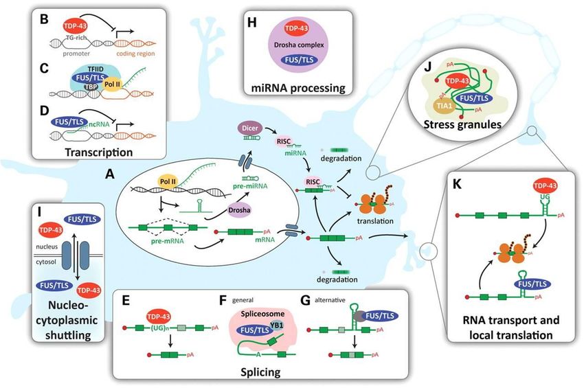
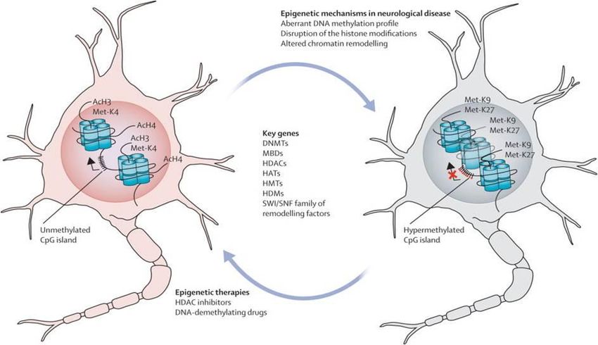
ALS disease mechanism and models | 14DISEASE MECHANISM AND MODELS FOR ALS Fig.4: ALS a neurovascular mechanism. A. Represent a healthy condition; the BBB/BSCB maintains homeostasis through selective regulation of the blood circulation within the spinal cord and brain. This process includes endothelial cells and their tight/adherens junctions, astrocyte end-feet, perivascular macrophages, pericytes, and the basement membrane. They are all needed for providing a barrier. B Sturctural and functional impairment of the BBB/BSCB allows entry of blood- borne substances, by endothelial cell and astrocyte end-feet degeneration, modified basement membrane composition, tight junction and transporter system impairment, vascular protein leakage, extensive extracellular edema, and MMP-2 and MMP- 9 activation Additionally, cytokines (i.e. TNF and IL-1) are released from activated microglia and reactive astrocytes. (Image is adapted from Garbuzova-Davisa, S., et al,. 2011) 107. RNA METABOLISM Recently, another disease mechanism has been suggested to play an important role in ALS as well. Currently, an increasing number of mutations, which affect proteins involved in RNA processing, have been identified in ALS. These mutations imply disrupted RNA dependent mechanisms (Fig.5). Evidence for this modifications in epigenetics, transcriptomics, and proteomics have been found in both animal models as in humans suffering from ALS33. On the one hand modified expression of genes involved in cytoskeletal dynamics, protein degradation system and mitochondrial dysfunction has been found in motor neurons118. Similar altered gene expression have found in muscle. On the other hand alternations in the insulin-like growth factor-1 and the RNA-binding protein ROD1119 occur in glial cells. Therefore, altered control of gene expression might be the overarching mechanism of ALS24., Epigenetic modifications showed to underlie altered gene expression, due to a disparity between histone acetyl transferases (HATs) and histone deacetylases (HDACs). The function of HATs is to modify core histone tails, which enhance DNA accessibility to transcription factors (TFs). In addition, HDACs activity results in transcriptional repression and gene silencing. However, many of the transcription initiation complex TF are themselves substrates to HATs and HDACs. Interestingly, ALS linked proteins, TDP-43 and FUS/TLS, control the expression of certain HDACs 120. 15 | ALS DISEASE MECHANISM AND MODELS
S.E. Baars

Another way in which epigenetic control of transcription may be modified is by methylation of
DNA methyltransferases (DNMTs) or histone methyltransferases (HMTs). Both use S-adenosyl-
methionine (SAM) as methyl donor. In eukaryotes, DNA methylation occurs through covalent
modifications of cytosine residues in CpG dinucleotides leading to gene silencing. Histones or
TF can be methylated by lysine or guanidinyl residues according to their function (i.g. activate or
repress transcription). These processes may have enormous impact on ALS.

 Fig.5: Epigenetic mechanisms. Healthy neurons or glial cells (left) express mRNA of a gene in occupancy from
 an unmethylated promoter, CpG island and a set of histone modifications. During ALS development altered DNA
 methylation, histone modifications and chromatin remodeling appear. (Image adapted from Urdinguio, R.G, et al.
 2009)114.

Another hypothesis is that RNA dysmetabolism could underlie the altered gene expression as
well (Supplementary fig.1)120. Genes, which have already been identified in patients with ALS,
appeared to be involved in the following processes: pre-mRNA splicing, mRNA transport,
translational regulation and mRNA decay. Nonetheless, additional research has to be performed
to investigate the specific motor neuron vulnerability to mutations in RNA-binding proteins.

In general, the function of a RNA protein is dependent on its associated proteins (i.e. RNA
binding proteins, RBP) that together forms the ribonuclear particle (RNP). These complexes
regulate RNA processes, such as alternative splicing. Alternative splicing is responsible for the
generation of multiple mRNAs from a single transcript. Changes in the secondary structure of a
protein subsequently lead to functional alterations. Moreover, various genes have already been
found to express multiple splice variants.

Although the protein is still unknown, the C90RF72 gene is most likely involved in RNA
processing. Recently it has been associated with an extension of a noncoding GGGGCC
hexanucleotide repeat in fALS patients. When transcribed, this repeat forms intracellular
accumulations of RNA fragments in cells121. This sequence may represent a potential binding site
for RNA-binding proteins as hnRNP A2/B1 (TDP-43 interactor). To date, it has identified as
most common genetic defect found in ALS patients122.

Another interesting player in RNA processing is TDP-43. Next to the fact that it has already
been identified as a major component in ubiquitinated inclusions11, it showed to be an

                                                               ALS disease mechanism and models           | 16
DISEASE MECHANISM AND MODELS FOR ALS ubiquitously expressed RNA-DNA binding protein. Research found that TDP-43 is implicated in multiple aspects of RNA processing, including: transcriptional regulation, alternative splicing and microRNA processing. Pathologic cytoplasmic TDP-43 inclusion are seen in both motor neuron and glial cells in various animal and human ALS patients59. This could imply different cascades in these genetic differ phenotypes. Most mutations in TARDBP are located on a glycine-rich domain involved in protein-protein interactions and nuclear transport. Additional evidence that dysregulated RNA processing may contribute to motor neuron injury in ALS arises from the detection of biomarkers of RNA oxidation34. Furthermore transcriptional repression also occurs within motor neurons in the presence of mSOD1123. This may provide a possible link between epigenetic modifications with mitochondrial damage and oxidative stress in ALS. Oxidative stress and mitochondrial dysfunction often occur together. Excessive ROS production negatively influences the functionality of the organelles that in turn would produce more ROS. Presumably, an interaction between oxidative stress, redox signalling and epigenetic modulations may cause ALS outcome. In this vicious circle, oxidative stress modulates gene expression by alternations of DNA accessibility. Additionally it is also a modulator of TFs. The latter implies that ROS and HDACs may coincide within the same mechanism. Next to being involved in regulation of proteins in redox reactions, HDACs also modulate alternative splicing and the activity of TFs. Furthermore, mitochondrial damage causes also alternations in selected splicing variants and RNA metabolism. This is associated with SOD-1 linked ALS as a consequence of mitochondrial stress123. Eventually, this may causes proinflammatory responses. Overall, ALS may arise from RNA dysmetabolism, which explains the broad diversity of modified processing within the brain and muscle. Furthermore, altered expression of genes involved into RNA metabolism has been found in motor neurons. But besides mutations, which affect RNA transcription or processing, RNA proteins, also needs to be correctly transported along the axon to the neuromuscular junction (NMJ). Axonal inclusions may block axonal transport, which may underlie ALS disease onset. Therefore, the primary site of damage in an ALS patient might determine the severity, age of onset and progression33. Moreover, different alternations of gene and/or RNA expression may cause the different outcomes on disease pathology. However, RNA dysmetabolism may also be caused by oxidative stress. For the future, this field of research should definitely be further investigated. DISEASE MODELS Since the wide variety of disease manifestations, research is performed on many different aspects of ALS. To investigate the cellular and molecular basis of ALS, a wide variety of model organisms have been used, including nematodes (Caenorhabditis elegans), arthropods (Drosophila melanogaster), Wsh (zebraWsh, Danio rerio), rodents (mouse, Mus musculus and rat, Rattus norvegicus) as well as non-human primates (rhesus monkey, Macaca mulatta)(See the supplementary table.3 for an overview)16. These animal models are particular important for certain pathological and therapeutic studies which are impossible to perform in human ALS patients. Nonetheless, postmortem tissue remains also very important for investigation of the disease. To establish a good working model, much research has been performed on human genetics. To mimic the disease in model organisms, a pattern of inheritance is important. Linkage studies have provided knowledge of chromosomal loci and sequencing of putative loci has identified multiple 17 | ALS DISEASE MECHANISM AND MODELS
You can also read