Role and regulation of the heat shock proteins Hsp90α
←
→
Page content transcription
If your browser does not render page correctly, please read the page content below
Role and regulation of the heat shock proteins Hsp90α
                       and β in Multiple Myeloma

                                       Dissertation

                     zur Erlangung des akademischen Grades
                              doctor rerum naturalium
                                   (Dr. rer. nat.)

                                     im Fach Biologie

                                 eingereicht an der

                  Mathematisch-Naturwissenschaftlichen Fakultät I
                        der Humboldt-Universität zu Berlin

                                           von

                            Sarika Jain, B.Sc., M.Sc.
                              24.01.1979, New Delhi
                                     INDIA

                    Präsident der Humboldt-Universität zu Berlin
                          Prof. Dr. Christoph Markschies

             Dekan der Mathematisch-Naturwissenschaftlichen Fakultät I
                          Prof. Dr. Christian Limberg

Gutachter:   1. Prof. Harald Saumweber
             2. Prof. Wolfgang Uckert
             3. Dr. Ralf C. Bargou

Tag der mündlichen Prüfung: 06. 12. 2007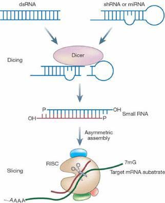
DECLARATION

I hereby certify that the thesis entitled ‘Role and regulation of the heat shock proteins
Hsp90D and E in multiple myeloma’ submitted to MDC/HUB for the fullfillment of the
Ph.D. degree, embodies the original research work. The microarray gene chip experiment
was performed in cooperation with Dr. Ute Ungethüm and Dr. Ralf-Jürgen Kuban from the
Laboratory of Functional Genomics, Charité, University Medicine Berlin, Germany and
immunohistochemical analyses was performed in cooperation with Dr. Mindaugas Andrulis
from the Institute of Pathology, University Hospital of Würzburg, Germany (current address
Institute of Pathology, University Hospital of Heidelberg, Germany). Apart from these two
experiments, the work was carried out by me by exclusively using the indicated resources.

SARIKA JAIN
ACKNOWLEDGEMENT

I would like to express my deep gratitude towards the Max Delbrück Center for Molecular
Medicine and the Humboldt University of Berlin for accepting me as a Ph.D. student in the
International Ph.D. Program. I can definitely say that working at the MDC was an experience
of a lifetime for me.

With profound veneration I wish to thank Dr. Ralf C. Bargou for giving me an opportunity to
work in his group at the MDC and also for his support and encouragement throughout the
course of this work.

I, from the depths of my heart sympathize with all the myeloma patients. I hope new
therapeutic strategies will soon emerge and will help combat this disease in a much better and
successful way.

A special thanks goes to all my labmates for helping me out at difficult times and making my
stay in Berlin and Würzburg wonderful and fun-filled. I want to thank Dr. Thorsten Stühmer
and Dr. Manik Chatterjee for their invaluable advice and critical reading of this thesis.

I wish to thank my friends Dr. Valentina Margania and Andreas Wülf for being there for me
in good and bad times. The memorable times I have spent with them and with Pia, Brigitte
and Tam and their families were some of the best in my life.

I would like to extend special thanks to my parents, family members and friends who were
always right beside me during all the phases of life, showered me with their love, support and
blessings and helped me making my dreams come true.

SARIKA JAIN
To nani...
INDEX

1      INTRODUCTION                                                 9
1.1    Multiple Myeloma                                             9
1.2    Clinical features                                            9
1.2.1 Symptoms                                                      9
1.2.2 Therapy                                                       10

1.3    Pathogenesis of Multiple Myeloma                             11
1.3.1 Multistep transformation process                              11
1.3.2 Bone marrow microenvironment                                  13
1.3.3 Signal transduction pathways in Multiple Myeloma              15

1.4    Heat shock protein 90                                        15
1.5    Heat shock protein 90 inhibition                             18
1.5.1 Anti-Heat shock protein 90 drugs                              18
1.5.2 siRNA-mediated gene expression knockdown                      20

2      AIM OF THE STUDY                                             22

3      MATERIALS                                                    23
3.1    Myeloma cell lines and primary cells                         23
3.2    Bacterial strains                                            23
3.3    Chemicals and reagents                                       23
3.4    Kits                                                         24
3.5    Antibodies                                                   25
3.6    Antibiotics                                                  25
3.7    Media, buffers and solutions                                 25
3.8    Drugs                                                        26
3.9    Oligonucleotides                                             26
       For Hsp90D and Hsp90E siRNA expression vector construction
3.10   Radioactive nucleotide                                       27
3.11   Plasmids                                                     28
3.12   Appliances                                                   28

                                                                         5
3.13 Consumables 28
3.14 Software 28
4 METHODS 29
4.1 Cell culture 29
4.1.1 Multiple Myeloma cell lines 29
4.1.2 Primary Multiple Myeloma cells 29
4.1.3 Primary bone marrow stromal cells 30
4.1.4 Primary human umbilical vein endothelial cells 30
4.1.5 Primary osteoclasts 31
4.2 Co-culture of myeloma cells with bone marrow stromal cells, 31
human umbilical vein endothelial cells or osteoclasts
4.3 Transient transfection of INA-6/MM.1s myeloma cells 32
4.3.1 Electroporation of INA-6/MM.1s myeloma cells 32
4.3.2 Enrichment of electroporated INA-6/MM.1s myeloma cells 32
4.4 Apoptosis detection assay 33
4.5 Sant7 preparation 34
4.6 Microarray analysis 35
4.7 Northern-blot analysis 38
4.7.1 RNA preparation 38
4.7.2 RNA agarose gel electrophoresis 38
4.7.3 RNA transfer by capillary blot 39
4.7.4 Radioactively labeled probe preparation 39
4.7.5 Northern-blot hybridization 40
4.8 Western-blot analysis 40
4.8.1 Whole cell protein extract preparation 40
4.8.2 SDS-PAGE electrophoresis 40
4.8.3 Immunoblotting 41
4.9 Construction of siRNA expression vectors 41
4.9.1 Selection of target sequences 42
4.9.2 pSUPER restriction digest 42
4.9.3 Oligonucleotide annealing 42
4.9.4 Ligation 44
4.9.5 Transformation and sequence confirmation 44
4.10 Immunohistochemical analysis 44
65 RESULTS 46
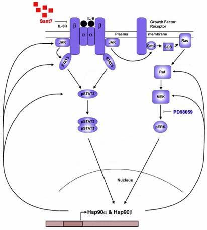
5.1 A positive feedback loop exists between the IL-6R/STAT3 pathway, the 46
Ras/MAPK pathway and the Hsp90D/E proteins in Multiple Myeloma
5.1.1 Gene expression profiling of INA-6 cells after combined blockade of the 46
IL-6R/STAT3 and Ras/MAPK pathways
5.1.2 Heat shock proteins Hsp90D and Hsp90E are downregulated by combined 48
disruption of the IL-6R/STAT3 and Ras/MAPK pathways
5.1.3 Heat shock protein 90 inhibition by 17-DMAG attenuates levels of pSTAT3, 50
STAT3 and pERK in INA-6 and ANBL-6 cells
5.2 Heat shock proteins Hsp90D and Hsp90E are overexpressed in primary 51
Multiple Myeloma cells and are upregulated by co-culture of myeloma
cell lines with bone marrow stromal cells
5.2.1 Heat shock proteins Hsp90D and Hsp90E are strongly overexpressed in 51
Multiple Myeloma cells but not in MGUS or normal plasma cells
5.2.2 Heat shock proteins Hsp90D and Hsp90E are upregulated in myeloma cells 52
by co-culture with bone marrow stromal cells
5.3 Heat shock proteins Hsp90D and Hsp90E are essential for the survival 53
of Multiple Myeloma cells
5.3.1 siRNA-mediated combined knockdown of heat shock proteins Hsp90D and 53
Hsp90E is required to induce apoptosis in INA-6/MM.1s myeloma cells
5.3.2 Pharmacological inhibition of heat shock protein 90 activity by17-DMAG 56
induces apoptosis in Multiple Myeloma cell lines
5.3.3 The fatal effects of siRNA-mediated knockdown of heat shock proteins 57
Hsp90Dand Hsp90E could not be mitigated by the presence of bone
marrow stromal cells
5.3.4 Pharmacological inhibition of heat shock protein 90 activity by 17-DMAG 58
induces apoptosis in Multiple Myeloma cell lines and primary myeloma cells
even in the presence of cells from the bone marrow microenvironment
6 DISCUSSION 61
7 SUMMARY 70
8 ZUSAMMENFASSUNG 72
79 REFERENCES 74
10 APPENDIX 87
10.1 Abbreviations and acronyms 87
10.2 Differentially expressed gene list 89
10.3 Lebenslauf 94
10.4 Publikationen 95
10.5 Curriculum vitae 96
10.6 Publication 97
81. INTRODUCTION
1.1 Multiple Myeloma
Malignant plasma cells (PCs) called as myeloma cells grow in the form of localised tumors
or plasmacytomas which can be single or multiple and can be confined within the bone
marrow and bone (medullary) or develop outside the bone in soft tissues (extramedullary).
The condition of having multiple plasmacytomas inside or outside the bone is called Multiple
Myeloma (MM). It is characterized by the accumulation of terminally differentiated B cells
which have a low proliferative index and extended life span in the bone marrow (BM). It is
presumed that MM evolves through a multistep transformation process involving complex
chromosomal abnormalities, genetic lesions and other oncogenic events. This haematological
malignancy is incurable to date with an incidence of 20% among all blood related cancers
and 1% among all cancers. 1,2
1.2 Clinical features
1.2.1 Symptoms
The characteristic symptoms of MM include skeleton destruction, impaired haematopoiesis,
and renal failure. Uncontrolled proliferation of MM cells leads to bone destruction through
the activation of osteoclasts and inhibition of osteoblast activity. Disruption of the balance
between osteoclasts and osteoblasts, in favour of the former, leads to progression of MM and
destruction of bone. The affected bone may fracture or crush, causing extensive pain and
sometimes leading to spinal cord compressions.3,4 Bone marrow failure causes anemia,
because specific inhibition of erythropoiesis is another feature of MM.5 Excessive amount of
monoclonal antibody produced by the myeloma cells is the main reason for the renal
dysfunction. In particular, the monoclonal immunoglobulin light chains also called as Bence
Jones proteins, might deposit in the fine tubular network of kidney as casts and disrupt the
normal function of the organ.6 Hypercalcemia (due to bone damage), infections and
nephrotoxic drugs also contribute to the compromised kidney function. Myeloma patients are
also more prone to infections because of lack of normal levels of functional antibodies in the
9blood and aberrant monocyte/macrophage functions.7-9 Increased concentration of circulating
antibodies in the blood leads to increase in the viscosity and volume of plasma. Thus, blood
flow is impaired which can cause bruising, bleeding, hazy vision, headaches and cardiac
problems.10,11
1.2.2 Therapy
Asymptomatic myeloma stage, Monoclonal Gammopathy of Undetermined Significance
(MGUS), is not treated because there is no evidence of any benefit from the treatment for the
patient so far. Rather, this stage is closely monitored for disease progression. Treatment
regimens differ depending on the age and the eligibility of the patient for stem cell
transplantation. Oral melphalan was first used for the treatment of myeloma nearly 50 years
ago and in combination with prednisolone has been the standard treatment for many years.
The response rate to this treatment is 40-60% without any complete remission.12
Combination drug strategies like vincristine, adriamycin and dexamethasone (VAD), or
cyclophosphamide, vincristine, adriamycin and methylprednisolone (CVAMP) are
commonly used as induction therapies for newly diagnosed patients eligible for transplant.
These drug combinations are not toxic for the stem cell population and induce rapid
remissions.13,14 Once a maximum response to the initial treatment is achieved, high-dose
melphalan chemotherapy is employed along with stem cell rescue. The response to
melphalan treatment along with stem cell transplantation is dramatic with 50% of patients
achieving a complete remission.14 Radiation therapy can be effective in patients with severe
local problems like bone destruction, pressure on nerves or spinal cord, but the main
disadvantage is the destruction of normal bone marrow stem cells in the area of exposure.
Though these therapeutic strategies show positive results and are of high potential, none of
these is curative. Initial trials with interferon-D to be used as maintenance therapy have
produced conflicting results though some benefit in remission prolongation has been
observed.13,14 Bisphosphonates are potent inhibitors of bone resorption and are given
throughout the course of treatment and maintenance.3 Thalidomide and its analogues like
lenalidomide (CC-5013) and actimid (CC-4047) have been shown to inhibit angiogenesis,
promote cytotoxicity mediated by natural killer cells and induce apoptosis in MM cells.15
10Bortezomib, a proteasome inhibitor, inhibits NF-NB activation, blocks IL-6 and VEGF
upregulation and might induce apoptosis in drug resistant myeloma cells.14 Other novel
agents such as histone deacetylase inhibitors, farnesyltransferase inhibitors, arsenic trioxide
and Hsp90 inhibitors are in early phase I/II clinical trials.12,14
1.3 Pathogenesis of Multiple Myeloma
1.3.1 Multistep transformation process
Myeloma cells arise probably from the activated post-germinal center B cells which have
encountered an antigen and are undergoing proliferation. During this phase B cells are
genetically unstable and undergo a number of important modifications like somatic
hypermutation, isotype switching and affinity maturation which together equip the cells to
target their antigen in a successful manner. However, sometimes aberrations in these normal
recombination and selection procedures give rise to abnormal plasma cells. These abnormal
plasma cells with enhanced proliferation rate, extravasate to the bone marrow where they
expand and set the stage for the development of MM. Based on the severity of the disease,
different intermediate stages have been defined to account for the pathogenesis and
progression of MM. MM arises via progression from a abnormal plasma cell through a
premalignant, hypothesised state termed as Monoclonal Gammopathy of Undetermined
Significance (MGUS) to the clinically overt myeloma.1
MGUS is the earliest identified asymptomatic, premalignant stage of myeloma
tumorigenesis. It is characterised by monoclonal plasma cell proliferation in the bone marrow
and the absence of end-organ damage.16 Numerical (hyperdiploid and hypodiploid) and
structural (deletions and translocations) chromosomal abnormalities are often present in
MGUS PCs and help to prevent the differentiation and normal death of these premalignant
cells. Primary translocations involving mainly the chromosomal locus 14q32 and one of five
partner loci: 11q13 (CCND1, cyclin D1 gene), 4p16 (FGFR-3 and MMSET), 6p21 (CCND3,
cyclin D3 gene), 16q23 (c-maf) and 20q11 (mafB), are considered as early pathogenetic
events.17,18 Trisomies of chromosomes 3, 5, 7, 9, 11, 15, 19 and 21 and monosomy of
chromosome 13 or 13q14 deletion are the most common karyotypic abnormalities detected
(Fig. 1).19,20 The likelihood of progression from MGUS to MM increases with the increase in
11serum monoclonal immunoglobulin concentration and is accompanied with an increase in
microvessel density (MVD).21,22
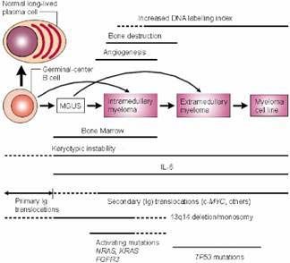
Figure 1.1 Molecular pathogenesis of myeloma. Myeloma arises from a germinal center B cell through a
multistep transformation process. Numerous karyotypic abnormalities, activating mutations participate in the
generation and progression of the disease.17
Complex genetic events occurring in the neoplastic plasma cells lead to the progression of
MGUS to MM.12,23 Intramedullary myeloma is considered as clinically active myeloma
which is characterised by the typical symptoms like osteolytic bone destruction, bone marrow
failure, impaired haematopoiesis and renal failure.14 During this stage, myeloma cells
specifically reside in the bone marrow (BM) which offers them a unique local
microenvironment, supporting their growth and survival.24 They harbour complex secondary
mutations and translocations involving NRAS and KRAS mutations, FGFR-3 mutations, c-
MYC dysregulation and P16 methylation (Fig. 1).17,25 The result of all these genetic
alterations is the disruption of normal cell cycle regulation and pro-apoptotic pathways. Apart
from these genetic alterations, adhesion of MM cells to cells of the bone marrow
microenvironment (BMM) like osteoclasts (OCs), bone marrow endothelial cells (BMECs)
and bone marrow stromal cells (BMSCs) has important clinical and pathological relevance.26-
28
12Extramedullary myeloma is one possible aggressive end-stage of the disease where myeloma
cells gain bone marrow microenvironment independence and localise at different
extramedullary sites like lung, liver, pleural fluid and ascites.29,30 An increasing number of
genetic abnormalities like NRAS, c-MYC and BCL-2 mutations and deletions in TP53 and Rb
tumor suppressor genes are frequently associated with extramedullary stages of MM.17,25
Mutations or deletions of TP53 gene are rare events in MM and are more frequent in the
extramedullary phase of the disease (Fig. 1.1).159 Stroma-independence and enhanced
extravasation out of the bone marrow suggest that the myeloma cells have developed
molecular mechanisms to prevent apoptosis and support growth in an autocrine fashion.
1.3.2 Bone marrow microenvironment
The characteristic feature of MM is the inherent capacity of neoplastic plasma cells to reside
in the bone marrow.30 The BMM is composed of several cell types that are intimately
involved in the evolution and progression of MM, including BMSCs, OCs, BMECs,
Osteoblasts, erythrocytes, haematopoietic stem cells, progenitor and precursor cells.28 A
variety of adhesion molecules like CD44, very late antigen 4 (VLA-4), CD54, syndecan-1
(CD138) and others mediate both homotypic and heterotypic adhesion of MM cells to the
cells of the BMM and extracellular matrix (ECM) proteins.31-33 This intricate interaction of
MM cells with the BM milieu not only localises the tumor cells to the BM but also leads to
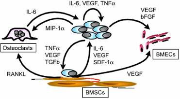
the secretion of a variety of cytokines and a plethora of growth factors (IL-6, IGF-1, VEGF,
SDF-1DbFGF, MIP-1D, SCF, TGF-E) (Fig. 1.2).28,34 IL-6, the best characterised myeloma
growth factor is primarily produced by BMSCs after adhesion of MM cells, though reports
about autocrine IL-6 production by MM cells also exist.35,36 MM cells become independent
of IL-6 stimulated, IL-6R/JAK/STAT3 pathway in the presence of BMSCs and adhesion-
mediated induction of other survival-promoting cascades like the Ras/MAPK pathway has
been observed.37 Both, the IL-6R/JAK/STAT3 and the Ras/MAPK pathways, are important
for myeloma growth and are stimulated independently of each other by BMSCs. Selective
blockade of either the IL-6R/JAK/STAT3 or the Ras/MAPK pathway in the presence of
BMSCs only partially affects MM cell viability. In contrast, combined targeting of both
pathways leads to massive induction of apoptosis.38 Apart from IL-6, attachment of MM cells
to BMSCs also upregulates the expression of angiogenic factors (VEGF, bFGF) and
13osteoclastogenic factors (RANKL, MIP-1D) which further facilitate MM cell growth.39-41
MM was the first haematological malignancy in which a significant correlation between
angiogenesis, prognosis and survival could be identified.42,43 VEGF is an established key
player in sustaining angiogenesis in MM and other malignancies. VEGF promotes BMEC
growth, mobilisation of BMEC precursors and vascular development and is secreted by
BMSCs, MM cells and BMECs.39,44,45 A paracrine loop between BMSCs and myeloma cells
has been identified where MM cell-derived VEGF stimulates IL-6 and VEGF production in
BMSCs and IL-6 derived from BMSCs stimulates VEGF secretion from MM cells (Fig.
1.2).46 BMECs also promote myeloma cell growth by secreting CXC-chemokines and by
direct cell-to-cell contact which mediates myeloma cell proliferation and modulates MM cell
homing.47,48
Figure 1.2 Cytokines and growth factors in the multiple myeloma bone marrow milieu. Attachment of MM
cells to BMSCs upregulates cytokine production both from MM cells and BMSCs. These cytokines also
promote the growth of osteoclasts and BMECs which further supports myeloma growth and progression.28
MM is characterised by devastating bone destruction due to osteoclast activation and
inhibition of osteoblast activity. OCs can support long term myeloma cell survival by direct
cell-to-cell contact and have been shown to be even more efficient than BMSCs in promoting
myeloma growth.49 MM cells disrupt the delicate balance between osteoclasts and
osteoblasts, which is maintained by the receptor activator of nuclear factor kappa B
(RANK)/RANK ligand (RANKL) system.26 Osteoprotegerin, the decoy receptor for
RANKL, is secreted by BMSCs and inhibits OC differentiation and function.50 Adhesion of
MM cells to BMSCs upregulates RANKL expression and downregulates osteoprotegerin
production thus leading to osteoclastogenesis and osteolysis.51 Myeloma cells express
14dickkopf 1 (DKK1) protein and secreted frizzled-related protein 2 (sFRP-2), which inhibit
the WNT signaling pathway which is crucial for osteoblast differentiation.52,53 Inhibition of
OC activity in a SCID-hu mouse model has shown that the growth of intramedullary MM
cells is dependent on OCs, indicating the importance of OCs in MM bone disease.26,54
1.3.3 Survival signaling pathways in Multiple Myeloma
Apart from the secretion of growth factors, attachment of MM cells to the cells of the BMM
also activates a number of survival signaling cascades. Heterogeneity of the BM milieu and
the interactions of MM cells with the components of the BMM trigger pleiotropic cascades of
proliferative/anti-apoptotic signaling which leads to MM growth and survival. IL-6R/STAT3,
Ras/MAPK and PI3K/Akt are the major signal transduction pathways associated with
survival and expansion of the malignant myeloma clone.55,56 IL-6 binding to the IL-6R
present on the MM cell surface leads to receptor dimerisation and phosphorylation and
activation of the Janus kinase (JAK)/signal transducer and activator of transcription 3
(STAT3) pathway. Ras/Raf/mitogen-activated protein kinase (MAPK) kinase
(MEK)/extracellular signal-regulated kinase (ERK) and phosphatidylinositol-3 kinase (PI3-
K)/Akt (PKB) signaling pathways can also be activated by IL-6, apart from other means like
cell adhesion and growth factor mediated induction.56-58 Blockade of the PI3K/Akt pathway
has been shown to inhibit proliferation and enhance apoptosis in MM cell lines and primary
MM (pMM) cells.59 Among other pathways, the WNT/E-catenin and NF-NB pathways are
the central mediators which support myeloma growth. MM cells express high levels of E–
catenin. WNT signaling has been shown to enhance MM growth while its blockade leads to
inhibition of myeloma cell survival.60 Attachment of MM cells to BMSCs induces NF-NB
activation and secretion of IL-6 and other growth factors.35 NF-NB activity is also required
for osteoclastogenesis which further supports myeloma growth.61
1.4 Heat shock protein 90
Heat shock proteins (Hsps) were first described in Drosophila larvae in 1962 by Ritossa as
dramatic alterations in gene activity due to changes in the puffing patterns in polytene
chromosomes in response to elevated temperatures.62 Hsps function as molecular chaperones
by interacting with other proteins called ‘client proteins’ to help them attain their
15thermodynamically stable conformation by assisting their proper folding, refolding and
translocation processes.63,64 Despite the name ‘heat shock proteins’ or ‘stress proteins’ Hsps
are ubiquitously and constitutively expressed under normal conditions and are among the
most primordial and highly conserved proteins during evolution.65-67 One of the most
abundant proteins present in eukaryotic cells is heat shock protein 90 kDa (Hsp90) which
interacts with and stabilises a plethora of polypeptides. Hsp90 is primarily a cytosolic protein
but different paralogues have been reported from other organelles like endoplasmic reticulum
(glucose related protein 94–Grp94) and mitochondria (tumor necrosis factor receptor-
associated protein1-TRAP1). The two major known cytosolic isoforms of Hsp90,
Hsp90D(inducible form) and Hsp90E(constitutive form) are 85% identical on protein level
and presumed to have arisen as a result of gene duplication approximately 500 million years
ago. The functional genomic locations of the human Hsp90Dand Hsp90Egenes have been
mapped on to the 14q32-33 and 6p21 chromosomal regions respectively. 68 (Fig. 1.3) Hsp90
mainly exists in homodimeric form but monomers, heterodimers and higher oligomers have
also been reported. Structurally, Hsp90 consists of a 25 kDa ATP binding N-terminal domain
and a 55 kDa C-terminal domain linked by a 35 kDa charged linker region. The N-terminal
domain of Hsp90 contains a unique ATP binding site including a Bergerat fold which is
characteristic of bacterial gyrases, topoisomerases and histidine kinases (Fig. 1.3).68-70
Figure 1.3 Schematic representation of the Hsp90 isoforms. Amino acid sequence is indicated as 1 to 800.
Hsp90 has three domains and a highly charged hinge region connecting the middle domain with the N-terminal
domain. The functional significance of each domain and the drug binding sites are also listed.68
Hsp90 is an ATP dependent chaperone which functions as part of a multi-chaperone complex
in association with a variety of co-chaperones.70,71 The Hsp90 genes are regulated at
transcriptional level by a highly conserved basic mechanism. The transcriptional induction
requires the activation and binding of special proteins called heat shock transcription
16factors/heat shock factors (HSTFs/HSFs) to the specific DNA sequence elements called heat
shock elements (HSEs).69 Hsp90 is required for essential physiological processes associated
with the cell cycle, cell differentiation and proliferation, normal growth and development and
inevitable housekeeping functions for example normal protein turnover. Hsp90’s clientele
breadth extends from several key signaling proteins to kinases, transcription factors, cell
cycle regulators, steroid receptors, telomerase, oncoproteins and others.69 (see also
www.picard.ch/DP/downloads/Hsp90interactors.pdf). Apart from its classic chaperone role,
Hsp90 is important for cellular differentiation as it has been observed that mouse embryos
lacking Hsp90E are unable to develop the placental labyrinth.72 Specific inhibition of Hsp90
activity leads to ubiquitinylation of its clients and causes enhanced proteasome-dependent
proteolysis.69 Hsp90 has been documented as a key player in antigen processing and
presentation during immune responses and is currently the focus of clinical trials with a
specific tumor immunogenicity vaccine.73,74 Multiple defects in B cell receptor signaling
have been observed by depletion of Hsp90E75 Hsp90 interacts with a number of signaling
molecules (Akt, PDK1, STAT3, MEK), serine/threonine and tyrosine kinases (ErbB2, v-Src,
Wee1, Raf-1), cell cycle (Cdk4/Cdk6, Cdc37) and cell death (Apaf-1, RIP-1)
regulators.69,71,76,77 The cellular ability to know whether to grow, divide, differentiate or die
depends upon the response towards a number of different extracellular signals. Hsp90 helps
the cells to orchestrate these responses by providing a platform for different pathways to
cross talk and by stabilizing and holding the proteins and signaling receptors and molecules
in their active states. Hsp90 has been shown to be overexpressed in cancer cells and its
clients include proteins that contribute to all hallmarks of cancer.78-80 The molecular
mechanism of overexpression of Hsps in cancer is still a matter of debate and has different
etiologies. It has been proposed that the sub-optimal cellular environment in cancers (low
glucose levels, pH, hypoxia, oncoprotein activation, genetic and epigenetic alterations) and
alterations in p53, contribute to elevated Hsp levels.78,81 The need to have high levels of Hsps
is not well understood but speculation is that chaperones can buffer mutations which
accumulate during the transformation process and thus promote cell growth and viability of
otherwise unstable cells.80,82 The most compelling evidence supporting the involvement of
Hsp90 in cancer is the fact that a large proportion of its client proteins are bona fide proto-
oncogene products or proteins that participate in oncogenic pathways and important cellular
processes like cell cycle control and apoptosis, which are normally impaired during
17transformation.71 Hsp90Ehas been shown to be associated with the Bcl-2 protein in mast
cells. This association is important for the anti-apoptotic function of Bcl-2 as dissociation of
these two proteins leads to the release of cytochrome C from mitochondria and activation of
caspase 3 and caspase 7, leading to mast cell apoptosis.157 Hsp90 is regarded essential for
telomerase stability and thus permits unlimited growth.83 It has also been shown to stabilise
VEGF and nitric oxide synthase which are important for the development of new capillaries
in tumors.84 Considering all this, a particularly attractive thought is to target Hsp90 and
disrupt multiple oncogenic signal transduction pathways simultaneously, which then should
translate into broad-spectrum activity against different tumor types and advanced cancers.
1.5 Heat shock protein 90 inhibition
1.5.1 Anti-Heat shock protein 90 drugs
Many efforts have focused on anti-Hsp90 drug development and this field is evolving rapidly
now that new drugs have yielded positive results in clinical trials. These include natural
product antibiotics such as benzoquinone ansamycins and their derivatives which bind to the
ATP binding site present in the N-terminal domain of Hsp90. Other molecules like purine-
based small molecule inhibitors, and several unrelated compounds that bind to a second ATP
binding site present in the C-terminal region of Hsp90 are also under development. Hsp90
inhibition alters its chaperone function leading to trapping of client proteins in their immature
conformation. These unstable proteins are degraded in a proteasome dependent manner
which leads to cell cycle arrest and cell death.71,85,86 Hsp90 drugs specifically target cancer
cells. The exact mechanism of the tumor cell selectivity of these natural products has yet to
be elucidated, but it is supposed that in tumor cells, Hsp90 is present in a conformation which
is more susceptible to inhibition.87,88 The first Hsp90 inhibitors identified, such as herbimycin
and geldanamycin (GA) belong to the benzoquinone ansamycin class, and were initially
discovered as naturally occurring antibiotics in the fermentation broth of Streptomyces
hygroscopicus (Fig. 1.4A).89 The benzoquinone moiety present in these molecules
distinguishes them from other ansamycins and confers selectivity for Hsp90. Tight binding of
GA to the N-terminal ATP binding pocket of Hsp90 prevents the formation of a mature and
active Hsp90 complex and leads to client protein degradation.90 Despite their promising anti-
18tumor profile, these drugs exhibited intolerable hepatotoxicity and/or cellular instability in
animals.91 Subsequent screening among GA derivatives led to the identification of 17-
allylamino-17-demethoxy-geldanamycin (17-AAG), which had a similar mode of action and
effects as GA but lower toxicity (Fig. 1.4B).92,93 Phase I/II clinical trials with 17-AAG
demonstrated that this drug is well tolerated, though with schedule dependent and dose
limiting toxicity.94,95 Despite these early promising results, 17-AAG has several limitations
regarding solubility and cumbersome formulations (DMSO as solvent is required). In
addition, 17-AAG appears to be extensively metabolised, which leads to rapid clearance of
the drug from the body and generation of toxic products like free radicals.94,96 Efforts to
improve the solubility and bioavailability of 17-AAG lead to the development of 17-
(dimethylaminoethylamino)-17-demethoxygeldanamycin (17-DMAG). 17-DMAG was
developed as a second-generation derivative of GA which has a similar mode of action and
the same in vivo and in vitro activity like 17-AAG (Fig. 1.4C). It is the first derivative of the
ansamycin class of Hsp90 inhibitors which is water soluble and orally bioavailable. 17-
DMAG is more potent and less toxic than its parent compound 17-AAG. It also does not
undergo extensive metabolism like 17-AAG and shows a wide range of tissue
distribution.97,98 17-DMAG is currently under phase I clinical trials in patients with advanced
cancers.
Figure 1.4 Benzoquinone ansamycin derivatives as Hsp90 inhibitors. These natural product Hsp90
inhibitors bind to the ATP binding site of the Hsp90 protein present in the N-terminal domain and inhibit its
function leading to proteasome-dependent client protein degradation. Geldanamycin (A) and its derivatives 17-
AAG (B) and 17-DMAG (C) (Figures from www.invivogen.com)
191.5.2 siRNA-mediated gene expression knockdown
One of the most recent tools in the field of sequence-specific inhibition of gene expression is
the phenomenon of RNA interference (RNAi). First discovered in 1998 in the nematode C.
elegans, RNAi is currently the most widely used technique to inhibit gene expression in
functional genomics.99 It is a naturally occuring endogenous phenomenon which evolved to
protect the genome against invasion by viruses and transposons, and also to orchestrate the
functioning of developmental programs. Genetic and biochemical investigations revealed a
conserved cellular machinery that cleaves long double stranded RNA (dsRNA) molecules to
generate 21-22 nucleotide long small interfering RNAs (siRNAs), which direct the sequence
specific degradation of their target mRNA.100 RNAi can be triggered by a variety of
molecules such as dsRNA, short hairpin RNAs (shRNAs) or endogenous hairpin micro
RNAs (miRNAs).
Figure 1.5 RNAi-mediated post-transcriptional gene silencing mechanism. Processing of long dsRNAs,
miRNAs or shRNAs by Dicer leads to the formation of siRNAs, which are 21-22 nucleotide long RNA
duplexes with symmetric 2 nucleotide 3’ overhangs and 5’ phosphate groups. siRNAs associate with proteins to
form an RNA-induced silencing complex (RISC), which unwinds the siRNA duplex. The antisense strand
guides the RISC to the mRNA target for endonucleolytic cleavage. (Figure from www.calandopharma.com)
20These different types of RNA molecules are processed into siRNAs by an RNAse III type
enzyme called Dicer. Dicer cleaves these RNAs to produce 21-22 nucleotide long siRNA
duplex molecules, that contain a 2 nucleotide 3’ overhang, a 5’ phosphate group and 3’
hydroxy termini. The RNA-induced silencing complex (RISC) is a protein complex which
incorporates siRNAs and unwinds the duplex siRNA into single stranded siRNA using an
ATP dependent RNA-helicase activity. The antisense strand of the siRNAs guides the RISC
to the target mRNA, where RISC-associated endoribonuclease cleaves the target at a single
site in the middle, thus resulting in degradation of the specific mRNA causing post-
transcriptional gene silencing (Fig 1.5).100-102
This specific target gene expression knockdown tool, provides an opportunity to study the
effects of loss of gene function. The large amounts of cDNA microarray data generated to
identify differentially regulated genes in diseased tissues, makes it almost impossible to
identify a potential drug target. siRNAs designed to specifically target differentially
expressed genes in diseased tissue have proved useful in validating suitable drug targets for a
particular disease.103 Considerable efforts have been made to improve delivery of siRNAs in
in vitro and in vivo systems. Many adenoviral, adeno-associated viral, retroviral and lentiviral
vectors and siRNA-producing plasmids have been developed.102 Electroporation, use of
lipophilic agents and both local and systemic injections are the current siRNA delivery
methods of choice, yielding the most efficient post-transcriptional gene silencing
results.101,102 Several proof-of-principle experiments have been performed in animal models
which underpin the therapeutic potential of siRNAs. However, the lack of truly efficient
siRNA delivery systems, currently limits the use of siRNA-based therapies in humans.102
212 AIM OF THE STUDY
Multiple signaling pathways contribute to the survival and proliferation of MM cells. In
particular, the IL-6R/STAT3 and Ras/MAPK signaling pathways have been shown to play a
major role in the pathogenesis and malignant growth of MM. Selective individual inhibition
of these pathways using specific inhibitors like Sant7 (an IL-6 receptor antagonist) and
PD98059 (a MEK inhibitor) had only modest effects on MM cell survival. However, the
combined inhibition of both these pathways led to massive induction of apoptosis even in the
presence of BMSCs. The aim of this study was to investigate the molecular mechanism
underlying the fatal effects of combined pathway blockade as compared to single pathway
disruption, using myeloma cell lines and primary myeloma cells obtained from patients. To
pinpoint the genes that were differentially expressed after combined disruption of IL-
6R/STAT3 and Ras/MAPK pathways a microarray gene chip expression analysis was
performed. Owing to their contribution in tumorigenesis, out of many potential target genes
obtained, the investigation was narrowed down to explore the role of Hsp90 (Hsp90Dand
Hsp90E in MM biology. Although some researchers have tried to differentiate between
Hsp90 isoforms, the knowledge about the independent functional aspects of these proteins is
still limited. In this MM study, the expression of both Hsp90 isoforms was analysed in situ
by immunohistochemistry and the individual role of Hsp90Dand Hsp90Eproteins was
evaluated in greater detail, by si-RNA mediated knockdown of each isoform. To explore the
functional consequences of Hsp90 blockade in the presence of cells from the supportive
BMM three different co-culture systems - BMSCs + MM, HUVECs + MM and OCs + MM -
representing the major cellular populations present in the BMM were used. In addition, the
effects of 17-DMAG, a second generation geldanamycin-derived pharmacological inhibitor
of Hsp90, were evaluated in the context of the BMM. It was anticipated that differential gene
expression analysis together with siRNA-mediated knockdown, would provide a better
insight into the molecular mechanisms that support MM cell survival. This work is an effort
towards a better understanding of the cooperation between two different signaling pathways
and the regulation of Hsp90 proteins in MM cells, which might open new doors for the
treatment of this deadly disease.
223 MATERIALS
3.1 Myeloma cell lines and primary cells
INA-6 Human Multiple Myeloma cell line
(from Prof. Gramatzki, Kiel, Germany)
ANBL-6 Human Multiple Myeloma cell line
(from Prof. Jelinek, Rochester, USA)
MM.1s Human Multiple Myeloma cell line
(from Prof. Steve Rosen, Chicago, USA)
Primary Multiple Myeloma cells From the Robert Rössle Clinic Berlin-
Buch and from Würzburg University
Clinic, Würzburg
Primary bone marrow stromal cells Please refer to 4.1.3
Primary human umbilical Promocell, Heidelberg, Germany
vein endothelial cells
Primary Osteoclasts Please refer to 4.1.5
3.2 Bacterial strains
Escherichia coli XL-1 blue Invitrogen, Karlsruhe, Germany
3.3 Chemicals and reagents
Acrylamide Roth, Karlsruhe, Germany
Agarose BMA, Rockland, USA
Ammoniumpersulfate Sigma-Aldrich, St. Louis, USA
Bacto-agar Difco/BD, Sparks, USA
Bacto-tryptone Difco/BD, Sparks, USA
Bacto-yeast extract Difco/BD, Sparks, USA
dNTP mix Clontech, Palo Alto, USA
Ethidium bromide Sigma-Aldrich, St. Louis, USA
23Fetal bovine serum Biochrom, Berlin, Germany
Glutamine (glutamax) Gibco, Karlsruhe, Germany
Non-fat dry milk powder TSI, Zeven, Germany
Sodium pyruvate Gibco, Karlsruhe, Germany
Trypsin/EDTA solution Biochrom, Berlin, Germany
Tween 20 Sigma-Aldrich, St. Louis, USA
Ficoll Biochrom, Berlin, Germany
OptiPrep Axis-Shield, Oslo, Norway
Sodium dodecyl sulfate Roth, Karlsruhe, Germany
PMSF Sigma-Aldrich, St. Louis, MO, USA
Aprotinin Sigma-Aldrich, St. Louis, MO, USA
NP40 Sigma-Aldrich, St. Louis, MO, USA
BSA Sigma-Aldrich, St. Louis, MO, USA
Tris Roth, Karlsruhe, Germany
Dithiothreitol Sigma-Aldrich, St. Louis, MO, USA
Anti-CD138 paramagnetic beads Miltenyi Biotec (Gladbach, Germany)
Cell separation columns Miltenyi Biotec (Gladbach, Germany)
RPMI 1640 Biochrom, Berlin, Germany
DMEM Gibco, Karlsruhe, Germany
D-MEM Gibco, Karlsruhe, Germany
Endothelial cell growth medium Promocell, Heidelberg, Germany
3.4 Kits
HexaLabel Kit MBI Fermentas, St. Leon-Rot, Germany
Nucleobond Midi and Maxi Kits Clontech, Palo Alto, CA, USA
pGEMT cloning Kit Promega, Madison, WI, USA
QIAEXII Agarose Gel Extraction Kit Qiagen, Hilden, Germany
QIAprep Spin Miniprep Kit Qiagen, Hilden, Germany
RNeasy Mini Kit Qiagen, Hilden, Germany
HUVECs Detach Kit-30 Promocell, Heidelberg, Germany
ECL Western blotting Kit Amersham, Freiburg, Germany
24Bradford assay Kit Bio-Rad, München, Germany
Human annexin V-FITC/PI staining Kit Bender MedSystems, Vienna, Austria
3.5 Antibodies
Rabbit anti-human pan Hsp90 Santa Cruz Biotechnology, Heidelberg,
Germany
Mouse anti-human Hsp90D Stressgen Bioreagents, Ann Arbor, USA
Rat anti-human Hsp90E Stressgen Bioreagents, Ann Arbor, USA
Rabbit anti-human Hsp90D Chemicon, Temecula, USA
Rabbit anti-human Hsp90E Chemicon, Temecula, USA
Mouse anti-human E-actin Stressgen Bioreagents, Ann Arbor, USA
Rabbit anti-mouse IgM-HRP Stressgen Bioreagents, Ann Arbor, USA
Anti-human STAT3 and pSTAT3 Cell Signaling Technologies
Anti-human ERK Calbiochem, Bad Soden, Germany
Anti-human pERK Cell Signaling Technologies
3.6 Antibiotics
Penicillin PAN Biotech, Aidenbach, Germany
Streptomycin PAN Biotech, Aidenbach, Germany
Ampicillin Sigma-Aldrich, St. Louis, MO, USA
3.7 Media, buffers and solutions
Immunoblot transfer buffer 25 mM Tris-base, 192 mM glycine, 20%
methanol, pH 8.3
LB medium 10 g bacto-tryptone, 5 g yeast extract, 5 g
NaCl in 1l H2O
1X Phosphate buffered saline (PBS) 0.8 mM Na2HPO4, 0.2 mM NaH2PO4, 14
mM NaCl
252X Laemmli sample buffer 100 mM Tris-base, 20% glycerol, 8%
SDS, 10% E-mercaptoethanol, 0.01%
bromophenolblue, pH 6.8
10X MOPS 200 mM MOPS, 50 mM sodium acetate,
10 mM EDTA, pH 7.0
20X SSC 3 M NaCl, 300 mM Tri-sodium citrate,
pH 7.0
TBE 100 mM Tris-base, 100 mM boric acid,
2.5 mM EDTA
3.8 Drugs
Sant7 Please refer to 4.5
PD98059 Calbiochem, Bad Soden, Germany
17-DMAG InvivoGen, San Diego, California, USA
3.9 Oligonucleotides
For Hsp90D and Hsp90E siRNA expression vector construction
(sequences derived from the actual genes are represented in bold)
Hsp90D1 (position 634-652)
Forward: 5´-GATCCCCCAGTTTATTGGATATCCCATTCAAG
AGATGGGATATCCAATAAACTGTTTTTGGAAA-3´
Reverse: 5´-AGCTTTTCCAAAAACAGTTTATTGGATATCCCA
TCTCTTGAATGGGATATCCAATAAACTGGGG-3´
Hsp90D2 (position 218-236)
Forward: 5´-GATCCCCGGAAAGAGCTGCATATTAATTCAAG
AGATTAATATGCAGCTCTTTCCTTTTTGGAAA-3´
Reverse: 5´-AGCTTTTCCAAAAAGGAAAGAGCTGCATATTAA
TCTCTTGAATTAATATGCAGCTCTTTCCGGG-3´
26Hsp90D3 (position 1196-1214)
Forward: 5´-GATCCCCCCCGTGAGATGTTGCAACATTCAAG
AGATGTTGCAACATCTCACGGGTTTTTGGAAA-3´
Reverse: 5´-AGCTTTTCCAAAAACCCGTGAGATGTTGCAACA
TCTCTTGAATGTTGCAACATCTCACGGGGGG-3´
Hsp90E1 (position 1266-1284)
Forward: 5´-GATCCCCCAAGGAGAATTACAAGAAATTCAAG
AGATTCTTGTAATTCTCCTTGTTTTTGGAAA-3´
Reverse: 5´-AGCTTTTCCAAAAACAAGGAGAATTACAAGAAA
TCTCTTGAATTTCTTGTAATTCTCCTTGGGG-3´
Hsp90E2 (position 1823-1841)
Forward: 5´-GATCCCCCCCAGGCACTTCGGGACAATTCAAG
AGATTGTCCCGAAGTGCCTGGGTTTTTGGAAA-3´
Reverse: 5´-AGCTTTTCCAAAAACCCAGGCACTTCGGGACAA
TCTCTTGAATTGTCCCGAAGTGCCTGGGGGG-3´
Hsp90E3 (position 1569-1587)
Forward: 5´-GATCCCCGCAGCTCAAGGAATTTGATTTCAAG
AGAATCAAATTCCTTGAGCTGCTTTTTGGAAA-3´
Reverse: 5´-AGCTTTTCCAAAAAGCAGCTCAAGGAATTTGAT
TCTCTTGAAATCAAATTCCTTGAGCTGCGGG-3´
Hsp90E4 (position 1923-1941)
Forward: 5´-GATCCCCGGCTGAGGCCGACAAGAATTTCAAG
AGAATTCTTGTCGGCCTCAGCCTTTTTGGAAA-3´
Reverse: 5´-AGCTTTTCCAAAAAGGCTGAGGCCGACAAGAAT
TCTCTTGAAATTCTTGTCGGCCTCAGCCGGG-3´
3.10 Radioactive nucleotide
[D-32P]dCTP (3000 Ci/mmol, 10 mCi/mL) NEN, Boston, MA, USA
273.11 Plasmids
pEGFP-N3 Clontech, Heidelberg, Germany
pcDNA3.1-CD4' Please refer to 4.3.1
pGEMT Easy Promega, Madison, WI, USA
pSUPER From Dr. Agami (The Netherlands)
3.12 Appliances
Gene Pulser BIO-RAD, München, Germany
FACSCalibur flow cyctometer BD Biosciences, Heidelberg, Germany
Western blotting apparatus Biometra, Göttingen, Germany
3.13 Consumables
Electroporation cuvettes Invitrogen, Karlsruhe, Germany
Nitrocellulose membrane Whatman, Dassel, Germany
Zeta probe membrane NEN Life Sciences, Boston, USA
X-ray film Kodak, Rochester, USA
3.14 Software
Leica 2.5 Leica, Heidelberg, Germany
CellQuest BD Biosciences, Heidelberg, Germany
Microarray Suite 5.0 Affymetrix, Santa Clara, CA, USA
284 METHODS
4.1 Cell culture
All cell types were cultured at 37qC and 5% CO2
4.1.1 Multiple Myeloma cell lines
The human IL-6-dependent MM cell lines (INA-6 and ANBL-6) and IL-6-independent cell
line (MM.1s) were maintained in RPMI 1640, supplemented with 20% and 10% fetal bovine
serum (FBS) respectively, and with 100 U/ml penicillin, 100 Pg/ml streptomycin, 2 mM
glutamine, 1 mM sodium pyruvate. 2 ng/ml recombinant IL-6 was added to the INA-6 and
ANBL-6 cultures.104-106
4.1.2 Primary Multiple Myeloma cells
Mononuclear cells from the bone marrow aspirates of 24 different MM patients were
separated by Ficoll-Hypaque density gradient centrifugation. The cell fraction obtained after
Ficoll separation was first washed with PBS and then with separation buffer (1X PBS, 0.5%
FBS, 2.5 mM EDTA). Cells were resuspended in 200 Pl of separation buffer and incubated
with 20 Pl of anti-CD138 paramagnetic microbeads for 15 minutes at 4qC. 1 ml of cold
separation buffer was added and CD138 positive MM cells were purified over magnetic-
activated cell sorting (MACS) large cell columns. Purified MM cells (Fig. 4.1) were further
cultured in RPMI 1640, supplemented with 20% FBS, 100 U/ml penicillin, 100 Pg/ml
streptomycin, 2 mM glutamine, 1 mM sodium pyruvate and 10 ng/ml recombinant IL-6.37 All
samples were taken from routine diagnostic specimens after informed consent of the patients.
Figure 4.1 Multiple Myeloma cells. Bone marrow aspirate with malignant plasma cells stained with
Pappenheim stain. (Figure taken from www.upci.upmc.edu)
294.1.3 Primary bone marrow stromal cells
Mononuclear cells from the bone marrow aspirates of MM patients were separated by Ficoll-
Hypaque density gradient centrifugation. Adherent cell populations left after enrichment of
pMM cells were long-term cultured and expanded in Dulbecco’s modified Eagle medium
(DMEM), supplemented with 20% FBS, 100 U/ml penicillin, 100 Pg/ml streptomycin, 2 mM
glutamine, 1 mM sodium pyruvate.37 pBMSCs (Fig. 4.2) obtained from 3 different MM
patients were used in this study.
Figure 4.2 Primary bone marrow stromal cells. (Figure taken from www.mpg.de)
4.1.4 Primary human umbilical vein endothelial cells
Primary human umbilical vein endothelial cells (HUVECs) (Fig. 4.3) were cultured
according to the manufacturer’s protocol (Promocell, Heidelberg, Germany). De novo
HUVECs were cultured in endothelial cell growth medium supplemented with 0.4%
endothelial cell growth supplement with heparin from bovine hypothalamic tissue, 2% FBS,
0.1 ng/ml epidermal growth factor (EGF), 1Pg/ml hydrocortisone, 1 ng/ml basic fibroblast
growth factor (bFGF). HUVECs were subcultured at 70% - 80% confluency using the
Promocell detach kit. HUVECs were used between their 3rd and 8th passage.
Figure 4.3 Primary human umbilical vein endothelial cells. (Figure taken from www.pharmaceutical-
int.com)
304.1.5 Primary osteoclasts
Mononuclear cells from buffy coats derived from healthy donors were seperated by Ficoll
density gradient centrifugation and 1 X 10⁶ cells were seeded per well in a 96 well plate in D-
MEM, supplemented with 10% FBS, 100 U/ml penicillin, 100 Pg/ml streptomycin, 2 mM
glutamine, 1 mM sodium pyruvate and with 25 ng/ml macrophage colony stimulating factor
(MCSF) and 30 ng/ml receptor activator of nuclear factor kappa B ligand (RANKL). The
adherent cell fraction was cultured for about 3 weeks and then the cells were stained for
tartarate-resistant acid phosphatase (TRAP) positivity to detect mature osteoclasts (Fig. 4.4).
An azo dye with purplish red colour is generated in the presence of the enzyme when the
TRAP substrate, supplemented with tartarate is used.107
Figure 4.4 Primary osteoclasts. Human adherent peripheral blood mononuclear cells were cultured in
supplemented D-MEM containing MCSF and RANKL for 3 weeks before they were stained for TRAP
positivity (dark red).164
4.2 Co-culture of myeloma cells with bone marrow stromal cells, human
umbilical vein endothelial cells or osteoclasts
MM cells (INA-6, ANBL-6 or pMM cells) were co-cultured with different cellular
components of the BMM. 1 X 10³ BMSCs, 2 X 10³ HUVECs or 1 X 10² OCs were cultured
in 96 well plates in 200 Pl of their respective culture medium and were left for 1 day to attach
prior to the addition of MM cells. 1 X 10⁴ MM cells were added onto the BMSC, HUVEC or
OC layer and were left overnight to interact and adhere.37,40,108 Once MM cells were adhered,
pathway inhibitors (Sant7 or PD98059 or both) or Hsp90 inhibitor 17-DMAG were added
and cells were assayed for viability using annexin V-FITC/propidium iodide (PI) staining.
For Western blot analysis 1 X 10⁵ MM cells were co-cultured with 1 X 10⁴ BMSCs.
314.3 Transient transfection of the INA-6/MM.1s myeloma cells
4.3.1 Electroporation of INA-6/MM.1s myeloma cells
To electroporate, INA-6/MM.1s cells were collected at a density of 2 X 10⁵/ml – 3 X 10⁵/ml
at 200xg. Cells were resuspended at 1 X 10⁷ cells/ml in fresh RPMI 1640 without any
additives but containing the desired plasmid constructs. 20 Pg/ml of pEGFP, 15 Pg/ml of
pCD4' and 10 Pg/ml of each siRNA-pSUPER construct were used. For the construction of
pCD4'the CD4'cDNA from pMACS 4.1 (Miltenyi Biotech) was subcloned into the
EcoRI/HindIII sites of pcDNA3.1+ (Invitrogen).38 Empty pSUPER vector was used as
control. An electroporator and electroporation cuvettes with electrode distance of 0.4 cm
were employed at settings of 960 PF and 280 V. 5 X 10⁶ INA-6 or MM.1s cells in 500 Pl
unsupplemented RPMI 1640 medium were used for each electroporation. Immediately after
electroporation, cells were transferred to an equal volume of fresh medium without additives
and kept at 37qC until all the samples were addressed. Electroporated cells were transferred
to the prewarmed fully supplemented medium with 2 ng/ml of IL-6 and cultured overnight.38
4.3.2 Enrichment of electroporated INA-6/MM.1s myeloma cells
Transfection efficiency was checked by GFP/PI FACS analysis. Electroporated cells were
collected at 200xg after overnight culture and washed with wash buffer (1X PBS, 5 mM
EDTA). After washing, cells were resuspended in 320 Pl of cold separation buffer and were
incubated with 80 Pl of anti-CD4 MACSelect 4 paramagnetic microbeads at 4qC for 15
minutes. Following incubation, 1 ml of cold separation buffer was added and CD4 positive
transfected MM cells were purified by magnetic-activated cell sorting (MACS) columns.
Large cell columns were used for INA-6 cells and small cell columns for MM.1s cells. The
columns were washed twice with 1 ml of cold separation buffer and the retained cells were
eluted using 3 ml of complete RPMI 1640 medium. Cells were collected at 200xg and
resuspended in a 3.3:1 mixture of complete RPMI 1640 medium/optiprep to remove any dead
cells. Following density gradient centrifugation at 1400xg for 10 minutes, the enriched live
cell fraction was collected, washed and resuspended in fresh RPMI 1640 medium and was
used for further experiments.38
324.4 Apoptosis assay
1X Binding buffer 10 mM HEPES/NaOH, pH 7.4, 140 mM NaCl, 2.5 mM CaCl2
Cells were assessed for apoptosis by using a human annexin V-fluorescein isothiocyanate
(FITC)/PI staining kit according to the manufacturer’s instructions. This kit is based on the
simple principle of high affinity binding of annexin V to phosphatidylserine (PS). PS is
present in the cytoplasmic side of the plasma membrane. During execution of apoptosis PS is
translocated from the inner side of the plasma membrane to the outer leaflet and is exposed at
the cell surface. Exposure of PS on the cell surface is regarded as an early stage apoptosis
marker. Annexin V-FITC binds to the exposed PS and thus labels the cells which can then be
easily detected by FACS. Propidium iodide (PI) positivity is a late stage apoptosis marker
which points towards the significant changes in the plasma membrane permeability and
allows discrimination between apoptotic cells (Fig. 4.5).109,110 To detect the apoptotic cell
fraction, cells were washed with PBS and collected at 200xg. Cell pellets were resuspended
in 100 Pl of binding buffer, 2.5 Pl of annexin V-FITC mix and 5 Pl of 1 mg/ml PI and
incubated for 15 minutes at RT in dark. The percentage of apoptotic and viable cell fractions
was analysed by flow cytometric analysis after subsequent dilution with 300 Pl of binding
buffer.
Figure 4.5 Annexin V-FITC/PI staining. Annexin V-FITC/PI staining is performed to distinguish live cells
from the apoptotic cell population. Live cells (annexin V-FITC/PI negative) occupy the lower left corner while
early stage apoptotic cells (annexin V-FITC positive but PI negative) represent the cellular population on the
lower right side. Necrotic (annexin V-FITC negative but PI positive) and late stage apototic cells (annexin V-
FITC positive and PI positive) are represented by the upper left and upper right corners, respectively.
334.5 Sant7 preparation
Lysis buffer B 8 M urea, 100 mM NaH2PO4, 10 mM Tris-base,
pH 8.0.
Wash buffer C 8 M urea, 100 mM NaH2PO4, 10 mM Tris-base,
pH 6.3.
Elution buffer E 8 M urea, 100 mM NaH2PO4, 10 mM Tris-base,
10% glycerol, pH 4.5.
Dialysis buffer I 1 M urea, 50 mM Tris-base pH 8.0, 2 mM
reduced glutathione, 0.2 mM oxidized
glutathione, 50 mM Glycine, 5 mM EDTA, 150
mM NaCl, 2 mM MgCl2, pH 8.0
Dialysis buffer II 50 mM Tris, 10% glycerol, 150 mM NaCl, 2 mM
MgCl2, pH 8.0.
ZelluTrans Roth 12,0 membrane (Roth, Karlsruhe, Germany) was cut according to the
desired size and was sterilised by heating for 10 minutes in 2% NaHCO3 solution with 1 mM
EDTA, pH 8.0. The membrane was washed with distilled water and was heated again in 1
mM EDTA, pH 8.0 for 10 minutes. After washing with water, the membrane was allowed to
cool down and was stored in water at 4qC until further use. Recombinant Sant7 protein was
expressed in E. coli strain BL-21, which is transformed with an expression plasmid for Sant7
and was purified. Briefly, LB medium containing 100 Pg ampicillin /ml was inoculated with
20 ml of overnight preculture of E. coli (BL-21). IPTG was added at a final concentration of
1.5 mM to the culture when the optical density reached about 0.7 and then the culture was
incubated for another 4 hours with continuous shaking. Cells were harvested at 5000 rpm for
10 minutes at RT. The pellet was resuspended with 60 ml lysis buffer B and was incubated at
RT for 1 hour with continuous shaking. The supernatant was collected by centrifugation at
8000 rpm for 30 minutes at RT. Ni-NTA agarose (Qiagen, Hilden, Germany) was washed
three times with lysis buffer B at 1400 rpm for 6 minutes at RT. The washed Ni-NTA
agarose pellet was resuspended with the supernatant obtained after bacterial lysis and was
incubated at RT for 2 hours with continuous shaking. The pellet was collected and later
34washed with 50 ml of wash buffer C at 1400 rpm for 6 minutes at RT. The washed pellet was
resuspended in 30 ml elution buffer E and was incubated for 15 minutes at RT with
continuous shaking. The supernatant was collected at 6000 rpm for 15 minutes at RT.
Dialysis was performed at 4qC. For this, the prepared ZelluTrans membrane was rinsed with
distilled water. The ends of the membrane were clamped to form a bag which was then filled
with the eluate. The bag was suspended in 200 ml of dialysis buffer 1 (DB 1) for 2 hours
after which 300 ml of the same buffer was added and dialysis continued for another 2 hours.
Old buffer was replaced with 250 ml of fresh DB 1 for 2 hours. 250 ml of DB 1 was further
added which was replaced after 2 hours with 1 litre of DB 1. The dialysis bag was left
suspended overnight in this buffer. Next day, DB 1 was replaced with DB 2 and the
procedure was repeated as it was performed for DB 1. The content of the bag was emptied
into a Centricon plus 20 filter device (Millipore, Schwalbach, Germany) which was
centrifuged at 8000 rpm for 15 minutes at 4qC. the flow through was discarded and
recombinant Sant7 was collected in the retentate cup by inverting the filter device and
centrifuging at 8000 rpm for 15 minutes at 4qC. Final protein concentration was measured by
using Bradford’s assay and recombinant Sant7 was stored at -20qC until further use.38
4.6 Microarray analysis
First strand synthesis buffer 50 mM Tris-base (pH 8.3), 75 mM KCl, 3 mM
MgCl2.
Second strand synthesis buffer 20 mM Tris-base (pH 6.9), 4.6 mM NaCl, 90
mM KCl, 0.25 PM NAD+, 10 mM (NH4)2SO4.
MES buffer 1.22 M MES, 890 mM NaCl (pH 6.6).
Hybridization buffer 100 mM MES, 1 M NaCl, 20 mM EDTA, 0.01%
Tween 20.
Non stringent buffer 900 mM NaCl, 60 mM NaH2PO4, 6 mM EDTA,
0.01% Tween 20.
Stringent buffer 100 mM MES, 100 mM NaCl, 0.01% Tween 20.
Streptavidin-phycoerythrin solution 10 Pg/ml streptavidin-phycoerythrin (Molecular
Probes, Leiden, The Netherlands), 2 Pg/Pl
35You can also read


