Abstract book Venue: KARL STORZ - Besucherund Schulungszentrum Berlin Scharnhorststraße 3 10115 Berlin Germany - DRFZ
←
→
Page content transcription
If your browser does not render page correctly, please read the page content below
Abstract book

                Venue:
               KARL STORZ
  Besucher- und Schulungszentrum Berlin
           Scharnhorststraße 3
          10115 Berlin Germany

                     3rd German Mass Cytometry User Forum, Berlin   1
We are moste grateful to our Sponsors and Exhibitors

                                                   funded by

2   3rd German Mass Cytometry User Forum, Berlin
Dear friends and collegues,
On behalf of the steering committee of the           On Thursday evening, we invite you to discuss
German Mass cytometry Network, I am pleased to       the posters, and to relax and mingle with your
welcome you to our 3rd German Mass Cytometry         colleagues at the evening event generously
User Forum in Berlin. After very successful          sponsored by Fluidigm. I also would like to thank
meetings in 2018 and 2019 I am looking forward       all commercial exhibitors for their generous
to continue the series in 2020.                      financial support to make this meeting possible.

The organizing committee has put together an         I am looking forward to an inspiring Mass
exciting 2-day program around different aspects      Cytometry User Forum in Berlin and wish you
of mass cytometry covering technology basics &       all an exciting meeting and a pleasant stay in
reagents, computational data analysis, imaging       Berlin.
mass cytometry and several applications in
different fields such as immunology, oncology, and   Yours,
personalised medicine. Workshops on technology
& reagents, and computational data analysis will
provide the opportunity to ask questions and
receive expert answers, and to discuss cutting-
edge developments in the field. Plenty of time       Henrik Mei
will be devoted to abstract presentations and
poster discussions.

                                                     3rd German Mass Cytometry User Forum, Berlin   3
Thursday, January 23rd
9:00           Welcome (Henrk Mei)

                                                Session 1
Chairs: Bertram Bengsch & Henrik Mei
09:15          Antonio Cosma, Luxembourg: Business intelligence, number theory and mass cytometry
               Henrik Mei, Berlin: Chronic inflammation
10:45          Coffee break

                                        Session 2 News from ...
Chairs: Bertram Bengsch & Henrik Mei
11:15	... Freiburg: Bertram Bengsch - Understanding T cell exhaustion in different human
       disease entities
	... Ulm: Fabian Gärtner - Phenotyping of immune cells in adipose and lean mice after
       thorax muscle trauma
       Selected abstract:
        Camila Fernandez-Zapata - Diversity of human myeloid compartment in active lesions
        of late stage multiple sclerosis determined by mass cytometry

12:45          Lunch break

                           Session 3 - Presentation of selected abstracts
Chair: Axel Schulz

14:15
      Selected abstracts:
  Marie Urbicht - Dissecting immune cell modulation during therapeutic targeting of
  CD38 in Systemic Lupus Erythematosus using mass cytometry
	Jessica Suwandi - 1 Multidimensional analyses of proinsulin peptide-specific regulatory
  T cells induced by tolerogenic dendritic cells
	Marilena Letizia - Mass cytometry reveals effects of Store-operated Calcium Entry pa-
  thway in human intestinal inflammation
15:45          Coffee break

                    Session 4 Workshop: Mass Cytometry Basics and Reagents
Chairs: Henrik Mei & Axel Schulz
16:30 	ISAC Lecture:
         Michael Leipold, Stanford: CyTOF in large studies: Challenges and lessons learned
         Selected abstract:
          Heidi Ødegaard Notø - Single cell gating by cell cycle analysis in mass cytometry
          Pannel discussion: Moderator Axel Schulz

                           Poster presentation and get together
Chairs: Désirée Kunkel & Sarah Warth
18:30-23:00 Poster Session and Dinner: generously supported by Fluidigm Corporation

4       3rd German Mass Cytometry User Forum, Berlin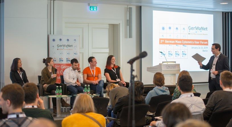
Friday, January 24th

                                           Session 5
Chair: Henrik Mei
9:00        Burkhard Becher, Zurich: High-dimensional cytometry for immunophenotyping
            Thomas Höllt, Leiden: Explorative visual analytics for large single-cell data
10:30       Coffee break

                              Session 6 Workshop Data Analysis
Chair: Marie Urbicht
11:15	Tyler Burns, Berlin: A visual interrogation of dimension reduction tools for single-cell
       analysis
       Selected abstract:
       Ina Stelzer - A third trimester multi-omic clock predicts the spontaneous onset of labor
       Panel discussion Moderator Marie Urbicht, Berlin
13:15       Lunch break

                                    Session 7 News from...
Chair: Désirée Kunkel
14:45	... Berlin (DRFZ): Andreas Grützkau - How many roads must a biomarker walk down,
       before you call it a BIOMARKER? Hope and Hype for new cellular biomarkers in
       rheumatology
	... Berlin (BCRT): Thomas Sell - Modelling cell-type specific signalling networks by mass
       cytometry in intestinal organoids
       Selected abstract:
        Marieke Ijsselsteijn - Characterisation of granzyme b positive myeloid cells with immu-
        notherapeutic potential for colorectal cancer patients using imaging mass cytometry.
16:15       Coffee break

                                  Session 8 Fluidigm Session
Chair: Henrik Mei
16:45       Invited talk sponsored by Fluidigm:
            Marie-Laure Yaspo, Berlin: Systems omics for exploring melanoma subtypes

                              Poster prize award and farewell
Chair: Henrik Mei
17:30       Poster prize (sponsored by Beckman Coulter Life Sciences) award ceremony
            Wrap-up and farewell with soup

                                                  3rd German Mass Cytometry User Forum, Berlin   5
Impressions of the 2nd German Mass Cytometry User Forum, 2019
in Berlin

6   3rd German Mass Cytometry User Forum, Berlin
Venue of the 3rd German Mass Cytometry User Forum

				 KARL STORZ
				 Besucher- und Schulungszentrum Berlin
				Scharnhorststraße 3
				 10115 Berlin Germany
				www.karlstorz.com

Distances                                         Public Transport
Berlin-Hauptbahnhof                               From Tegel Airport: TXL Express Bus to
(Central Station): 1 Kilometer                    Invalidenpark Station
Tegel Airport: 9 Kilometer
Schönefeld Airport: 25 Kilometer                  From Schönefeld Airport: Regional train RE7/
                                                  RB14 to Central Station (Hauptbahnhof), from
Parking: No parking lots for guests               there: see below - Hauptbahnhof

Public parking:                                   From Central Station (Hauptbahnhof):
Car park Berlin-Hauptbahnhof, (Central Station)   Bus 120 to Scharnhorststraße/Habersaathstraße
                                                  Station; from there: approx. 2 min by foot
Access routes: B96-Tunnel or Clara-Jaschke-
                                                  or Tram M5, M8, M10/ Bus TXL
Straße, 10557 Berlin
                                                  to Invalidenpark Station; from there: approx. 6
From there: Bus 120, Bus TXL, Tram M5, M8,        min by foot
M10 or approx. 12 min by foot

                                                  3rd German Mass Cytometry User Forum, Berlin      7
Thursday, January 23rdS

                                        Session 1: Invited Talks I
Chairs: Bertram Bengsch & Henrik Mei

Business intelligence, number theory and mass cytometry
Antonio Cosma
Quantitative Biology Unit, LIH, Luxembourg

Merging knowledge from different disciplines             Number theory is a branch of mathematics dedi-
can open new perspectives and pave the way to            cated to the study of natural numbers and tools
unforeseen problem solutions. Here, I will pre-          used to manage them. In 300 B.C., Euclid set the
sent how “Business Intelligence” and “number             basis of number theory with the “fundamental
theory” can help in our process to understand            theorem of arithmetic” and, this day and age,
mass cytometry data.                                     number theory is at the basis of modern crypto-
                                                         graphy and internet security.
Business Intelligence (BI) comprises the tools and
technologies used to collect and analyze infor-          I will present some data scenarios in which mass
mation in order to support the process of decisi-        cytometry data were analyzed using BI tools
on-making. Data management and data analysis             and some techniques to classify cell populations
are central to BI activities since they are the source   using number theory.
of business decisions. Several BI solutions are
available as open-source or proprietary software.

Chronic inflammation
Henrik Mei
Deutsches Rheuma-Forschungszentrum (DRFZ),
Berlin, a Leibniz-Institute, Germany

8     3rd German Mass Cytometry User Forum, Berlin
Session 2: News from...
Chairs: Bertram Bengsch & Henrik Mei
Understanding T cell exhaustion in different human disease entities
Bertram Bengsch
University Medical Center Freiburg, Faculty of vel signatures to identify the role of exhaustion
Medicine, Germany in settings of autoimmune-mediated disease and
lowly immunogenic cancers, such as Hepatocel-
Exhausted T cells have been linked to unfavor- lular Carcinoma (HCC) using systems immunology
able patient outcomes in cancer and viral infec- approaches centered around high-dimensional
tions but they are predicted to be beneficial in mass cytometry profiling. In this presentation,
autoimmunity. We have recently established epi- we will discuss novel insights into the role of ex-
genomic and cytometric immune signatures of haustion obtained using these approaches.
exhaustion in highly immunogenic cancers and
HIV infection. We are currently using these no
Phenotyping and gene profiling of immune cells in adipose and lean
mice after thorax and muscle trauma
Fabian Gärtner drop tower for the muscle injury, followed by a
Forschung Chirurgie 1 (AG Knippschild), lung injury induced with air pressure. Both met-
Uniklinikum Ulm, Germany hods set blunt traumas. The mice were kept for
different time points and the blood, spleen and
1. Introduction trauma tissue (muscle and lung) was harvested
The issue of obesity in our society grows bigger and analyzed by CyTOF, flow cytometry and RT-
and bigger every year. Besides direct adverse ef- PCR, respectively.
fects on health and life quality of obese people,
it can also affect the regeneration of the tis- 3. Results
sue after trauma. It has been shown in previous An increased pro-inflammatory response can be
work, that obesity can influence the healing pro- observed in obese animals in comparison to the
cess after a traumatic injury in mice, that were lean counterparts. This can mainly be observed
suffering from a muscle trauma. This influence in the first 24 hours in the peripheral blood stre-
is amongst other things based on a dysregulation am based on neutrophils and pro-inflammatory
of macrophages and a resulting shift in the M1/ Ly6Chi monocytes, which are present in the pe-
M2 axis(1,2). We hypothesize that the immune riphery for a longer time in obese animals. Addi-
cells have an increased inflammatory potential tionally the monocytic subsets of obese animals
in obese animals and therefore lead to this shift show an increased expression of the chemokine
towards a more pro-inflammatory environment. receptor CCR2, which is responsible for the re-
Therefore this work is focusing on polytraumatic cruitment of immune cells to the site of inflam-
injuries of the muscle as well as the lung and mation following a CCL2 gradient. These diffe-
is phenotyping the immune cells after different rence can also be observed at later time points
time points after trauma. in the gene profiles of the lung and the muscle.
For example an increased ratio of M1 to M2 spe-
2. Methods cific genes can be observed in obese individuals
The used mouse model for this study are female in the lung, whilst the regeneration process of
C57BL/6J mice, which are either lean or obese. the muscle is slowed down in obese animals ba-
The obesity is diet induced using a 60 % fat diet sed on the expression of Myog.
as induction. The polytrauma is induced using a
3rd German Mass Cytometry User Forum, Berlin 94. Discussion monocytes populations to the site of injury re-
These results indicate that the response of the sulting in a shift of the M1/M2 axis and a delayed
immune system to the polytraumatic injury is de- regeneration process.
layed as well as prolonged. Additionally the in-
creased expression of CCR2 on monocytes leads
to an increased homing of the pro-inflammatory
Diversity of human myeloid compartment in active lesions of late
stage multiple sclerosis determined by mass cytometry
Chotima Böttcher1, M. Camila Fernández Here, we applied multiplexed single-cell mass
Zapata1, Marlijn van der Poel2, Stephan cytometry (CyTOF) to elucidate the different
Schlickeiser3, Cheng-Chih Hsiao4, Mark R phenotypes of microglia in active lesions and
Mizee2, Maria C.J. Vincenten2, Désirée Kun- normal appearing white matter (NAWM) of MS
kel3, Inge Huntinga2,5, Jörg Hamann2,4, Josef patients as compared to control donors. We per-
Priller1,6,7 formed three independent CyTOF measurements
1
Charité Universitäatsmedizin, Germany; 2Neu- with a total of 75 antibodies to analyze myeloid
roimmunology Research Group, Netherlands cells isolated from post-mortem human brain. In
Institute for Neuroscience, Amsterdam, The our study we could confirm consistent changes
Netherlands.; 3Berlin-Brandenburg Center for in the myeloid phenotypes in active lesions as
Regenerative Therapies (BCRT), Charité – Uni- compared to NAWM and control, mainly decrea-
versitätsmedizin Berlin, Berlin, Germany; 4De- sed abundance of homeostatic microglial clus-
partment of Experimental Immunology, Ams- ters, with decreased expression of GPR56, P2Y12
terdam University Medical Centers, University
and TMEM119 in some lesion associated myeloid
of Amsterdam, Amsterdam, The Netherlands;
cells. We detected an increased abundance in
5
Netherlands Brain Bank, Netherlands Institute
for Neuroscience, Amsterdam, The Netherlands; lesion of clusters expressing phagocytic-related
6
Berlin Institute of Health, Berlin, Germany; and antigen presenting-related markers such
7
German Center for Neurodegenerative Diseases as HLADR, CD11c, CD64, CD68, CD47, CD91 and
(DZNE), Berlin, Germany. CD172a (SIRPα), among others. In whole, by
using multi-dimensional single-cell technology
Multiple sclerosis (MS) is a chronic inflammato- we could unravel previously unidentified mye-
ry, demyelinating and neurodegenerative disea- loid phenotypes associated to MS active lesions,
se of the CNS. The pathologic hallmark of MS is thus emphasizing the complexity of microglia
the presence of demyelinated lesions throug- biology in MS.
hout the CNS. Active lesions are characterized
by myelin loss and the presence of foamy mi-
croglia/macrophages containing myelin lipids.
Microglia, the resident innate immune cells of
the brain, take part in CNS surveillance during
homeostasis but are also key players in neuro-
degeneration and neuroinflammation. In the
context of MS, microglia are involved in both de-
myelination and remyelination, key mechanisms
in the development of the disease. Furthermo-
re, recent studies using single cell RNA-seq on
mouse models of demyelination have shown
distinct transcriptional microglial phenotypes
associated to lesions pointing to an important
role of microglia in the disease pathogenesis.
However, further characterization of microglial
phenotypes and their possible contribution to
human MS pathogenesis remains to be studied.
10 3rd German Mass Cytometry User Forum, BerlinSession 3: Presentation of selected abstracts
Chairs: Bertram Bengsch & Henrik Mei
Dissecting immune cell modulation during therapeutic targeting of
CD38 in Systemic Lupus Erythematosus using mass cytometry
Marie Urbicht1,3, Lennard Ostendorf1,2,3, An- Besides peripheral PC, CD38 was expressed in NK
dreas Grützkau1, Falk Hiepe1,2, Tobias Alex- cells, plasmacytoid dendritic cells, monocytes,
ander1,2,3, Henrik Mei1,3 as well as several T and B lymphocyte subsets
1
Deutsches Rheuma-Forschungszentrum Berlin, at varying levels. Consistent with broad immune
Germany; 2Charité Universitätsmedizin Berlin, activation, SLE patients showed increased CD38
Department of Rheumatology and Clinical Im- expression across many leukocyte subsets. Stri-
munology, Berlin, Germany; 3equal contribution kingly, several immune cell subsets expressing
of first and senior authors CD38 at baseline and in control donors persisted
in the treatment with Dara. On levels of cell sig-
Systemic Lupus Erythematosus (SLE) is an auto- nalling, we observed that anti-CD38 treatment
immune disease characterized by antibodies di- induced changes in phosphorylation patterns of
rected against double-stranded DNA which are several key signal transducers, such as STAT pro-
secreted by autoreactive plasma cells (PC). Dar-
teins, p38 or NFkB. This effect was most promi-
atumumab (Dara) is a monoclonal anti-CD38 an-
nent in, but not limited to the T cell compart-
tibody that diminishes neoplastic plasma cells in
vivo and is coherently licensed for the treatment ments and paralleled the clinical improvement
of multiple myeloma. Thus, Dara also represents of the patients.
an interesting treatment option in antibody-me-
diated diseases such as SLE. However, the ex- In summary, CD38 targeting is a promising treat-
pression of CD38 in immune cells other than PC ment concept in antibody-mediated diseases.
has not been systemically studied, especially un- Besides targeting plasma cells, Dara may impact
der conditions of SLE. In this study, we used mass a variety of other innate and adaptive immune
cytometry to characterize the expression of cells expressing CD38, including T, B and NK cells
CD38 across innate and adaptive immune cells in subsets, as well as monocytes and plasmacytoid
peripheral blood in great detail in SLE and moni-
dendritic cells as important sources of inflam-
tored immune cell composition during treatment
matory cytokines. Further analyses are currently
with Dara in two therapy-refractory SLE patients
to gain insight in the mechanism(s) of action being conducted to address whether changes in
and secondary targets cells of Dara beyond PC. phosphorylation of intracellular signalling medi-
ators are directly related to CD38 ligation or se-
condary to decreased disease activity. This study
SLE and control blood samples were preserved in underscores the utility of combined phenotypi-
proteomic stabilizer at -80°C. Samples were Pal- cal and ex vivo signalling analyses by mass cyto-
ladium-barcoded, and a 41-parameter mass cy- metry to characterize target expression pattern
tometry panel comprising immune cell-surface of therapeutic antibodies as well as to elucidate
markers and phosphoepitope-specific antibodies the immune system’s response to treatment.
was established for simultaneous in depth-phe-
notyping of leukocyte subsets and interroga-
tion of cell type-specific intracellular signaling.
Data were analyzed using an R pipeline based
on FlowSOM clustering and statistical analysis by
SAM. Validation was carried out using manual ga-
ting analyses.
3rd German Mass Cytometry User Forum, Berlin 11Multidimensional analyses of proinsulin peptide-specific regulatory
T cells induced by tolerogenic dendritic cells
Jessica Suwandi1, Sandra Laban1, pe of stimulated T cells were analysed. TolDCs
Kincsõ Vass1, Antoinette Joosten1, Vincent induced suppressive T cell lines that were do-
van Unen1,3, Boudewijn Lelieveldt1, Thomas minated by a naïve phenotype (CD45RA+CCR7+).
Höllt1,4, Jaap Jan Zwaginga1, Tatjana Niko- These naïve T cells, however, did not show sup-
pressive capacity, but were arrested in their
lic1, Bart Roep1,2
naïve status. T cell cultures stimulated by tolDC
1
LUMC, The Netherlands; 2City of Hope, USA; further contained memory-like (CD45RA-CCR7-)
3
Stanford, USA; 4TU, Delft, The Netherlands T cells expressing regulatory markers Lag-3,
CD161 and ICOS. T cells expressing CD25lo or
Induction of antigen-specific regulatory T cells CD25hi were most prominent and suppressed
(Tregs) in vivo is the holy grail of current im- CD4+ proliferation, while CD25hi Tregs also
mune-regulating therapies in autoimmune disea- effectively supressed effector CD8+ T cells.
ses, such as type 1 diabetes. Tolerogenic den-
dritic cells (tolDCs) generated from monocytes We conclude that tolDCs induce antigen-specific
by a combined treatment with vitamin D and
Tregs with various phenotypes. This extends our
dexamethasone (marked by CD52hi and CD86lo
earlier findings pointing to a functionally diverse
expression) induce antigen-specific Tregs. We
evaluated the phenotypes of these Tregs using pool of antigen-induced and specific Tregs and
high-dimensional mass cytometry to identify a provides the basis for immune-monitoring in cli-
surface-based T cell signature of tolerogenic nical trials with tolDC.
modulation. Naïve CD4+ T cells were stimulated
with tolDCs or mature inflammatory DCs pulsed
with proinsulin peptide, after which the suppres-
sive capacity, cytokine production and phenoty-
Mass cytometry reveals effects of Store-operated Calcium Entry
pathway in human intestinal inflammation
Marilena Letizia1, Cansu Yerinde1, Anne- in mouse models of colitis. However, the effects
gret Sand1, Stephan Schlickeiser2, Ulrike of SOCE inhibition have not been studied in the
Kaufmann3, IBDome-DE Investigators Study human context of inflammatory bowel disease
group4, Britta Siegmund1, Stefan Feske3, (IBD) and it remains elusive, which immune cell
Carl Weidinger1 subset is affected by the pharmaceutical blocka-
1
Charité Universitätsmedizin Berlin, Germany; de of SOCE. We therefore aim to investigate the
2
Berlin-Brandenburg Center for Regenerative effects of SOCE inhibitor BTP-2 on functions and
Therapies, Berlin, Germany; 3Department of Pa- metabolic homeostasis of human lymphocytes
thology, New York University School of Medicine, isolated from IBD patients.
New York, NY 10016, USA.; 4iPATH.Berlin, Chari-
té Universitätsmedizin Berlin, Germany PBMC and/or lamina propria lymphocytes (LPMC)
were isolated from colitis patients undergoing
Store-operated Calcium Entry (SOCE) represents colon resection. Cells were ex -vivo stimulated
the major calcium influx pathway in T cells which with Ionomycin/PMA in the presence or absence
not only controls the activation and function of of BTP-2 and subsequentialy fixed and stored
lymphocytes, but which also has been implicated at -80°C until acquisition by mass cytometry.
in the metabolic homeostasis and survival of mu- LPMCs were stained using 37 lineage and func-
rine CD4+ and CD8+ T cells. Conditional knockout tional markers targeting B, T, NK or myeloid cells
mice, in which SOCE signaling components are and the resulting flow cytometry standard (FCS)
deleted in T cells, revealed that SOCE is requi- files were analyzed by using cytobank and R/Bio-
red for the induction of intestinal inflammation conductor 9 packages. Additionally, Ca2+ influx
12 3rd German Mass Cytometry User Forum, Berlinmeasurement and Seahorse analyses were per- Our data revealed for the first time that the
formed in order to assess the metabolic status of cytokine production and the activation of se-
immune cell subsets after SOCE inhibition. veral immune cell subtypes can be modulated
by SOCE blockade in human intestinal inflam-
Data on B, T, NK, myeloid cells and neutrophils mation, identifying SOCE as a novel therapeutic
isolated from peripheral blood or colon lamina target in colitis. Moreover, we hope that a wide
propria revealed that each immune cell subset phenotypical characterization of immune cells
harbors a distinctive SOCE-dependent Ca2+ in- via mass cytometry will provide a better insight
flux rate, suggesting that SOCE might different- into positive as well as negative effects of SOCE
ially regulate the activation and function of inhibitors that might interfere with the clinical
each cell subtype. In particular, CD4+ and CD8+ applicability of SOCE inhibitors for treating IBD.
T cells, B and NK cells as well as monocytes were
highly susceptible to extracellular Ca2+ influx,
followed by granulocytes. Furthermore, inhibi-
tion of SOCE in lymphocytes resulted in impai-
red metabolic fitness, reduced glycolytic capa-
city and impaired fatty acid oxidation. Finally,
BTP-2 was able to decrease the production of
key pro-inflammatory cytokines involved in IBD,
including TNFα and IL-17 in lamina propria resi-
dent T cells.
Session 4: Workshop: Mass Cytometry Basics and Reagents
Chairs: Henrik Mei & Axel Schulz
ISAC-Lecture: CyTOF in large studies - Challenges and lessons learned
Michael Leipold dies, both in size and in longer-duration. Large
studies run in a short period of time have dif-
Stanford University School of Medicine, United
ferent challenges than large studies run over
States of America
longer periods of time. I will speak about my
experiences, challenges, and lessons learned
Mass cytometry (CyTOF) has been available for in two studies: first, a large cohort of samples
almost 10 years. However, most studies thus far run in a short period of time, and compare that
have been relatively small and of short duration. with a cohort of similar size that was acqui-
As the technology has matured, there has been red and run over the course of several years.
increased interest in utilizing it in larger stu-
Single cell gating by cell cycle analysis in mass cytometry
Idun Dale Rein1, Heidi Ødegaard Notø1, Radiation Biology, Institute for Cancer Re-
Monica Bostad1, Kanutte Huse1,2,3, Trond search, Oslo University Hospital, Oslo, Norway
Stokke1,4
1
Department of Core Facilities, Institute for A disadvantage of mass cytometry is that aggre-
Cancer Research, Oslo University Hospital, gates of cells cannot easily be removed while
Oslo, Norway; 2Department of Cancer Immu- retaining all single cells. Common gating stra-
nology, Institute for Cancer Research, Oslo tegies for excluding non-single cell events are
University Hospital, Oslo, Norway; 3K.G. Jeb-
based on event length to exclude ion cloud fusi-
sen Centre for B cell malignancies, Univer-
ons, in addition to DNA content (Ir-intercalator),
sity of Oslo, Oslo, Norway; 4Department of
3rd German Mass Cytometry User Forum, Berlin 13and/or a parameter that is related to cell size 2. U
se of the Gaussian parameters to remove re-
(e.g. Cisplatin) to exclude aggregates. If cells maining ion cloud fusions.
are barcoded, non-single cell events with diffe-
rent barcodes can be removed. However, cell ag- 3. F
lowSOM clustering and annotation of cell
gregates, mostly formed during fixation and the- cycle phases. This step will also remove some
refore before the barcoding, as well as ion cloud aggregates.
fusions of cells with the same barcode will not
be removed. The use of Ir-intercalator and Cis- 4. Cell cycle phase-specific single cell gating
platin for removing the remaining non-single cell (based on cell size). This step will remove the
events is problematic for actively proliferating rest of the aggregates.
cells, as two G1 cells has the same DNA content
and about the same size as a G2 or mitotic cell. 5. V
isualization of the cell cycle phases in e.g.
Gating based on a narrow region in any marker viSNE plots.
that is related to DNA content or size around the
diploid (G1) cells will exclude cells late in the When performed on cells stained with the cell
cycle. A too wide region for gating will result cycle panel, this analysis pipeline successfully
in inclusion of aggregates. These questions were identified ion cloud fusion events, debris, dead/
addressed by us in a manuscript in revision in apoptotic cells, aggregates and the major cell
Cytometry A. cycle phases. The presented cell cycle panel and
analysis pipeline thus enables single-cell ana-
In the submitted work we developed an analy- lysis also in samples with cells in all cell cycle
sis pipeline where unsupervised clustering by phases at the same time as any additional chan-
FlowSOM was performed as initial clean-up and nels in the panel are open for phenotyping and/
gating of the data, resulting in separate meta- or cell cycle-resolved expression or modification
clusters identified as the major cell cycle phases analysis.
in addition to dead and apoptotic cells, debris
and some non-single cell events. Each cell cycle
phase was then individually gated to exclude ion
cloud fusions and aggregates within cell size-ho-
mogeneous groups, thereby avoiding inclusion of
small aggregates or exclusion of cells late in the
cycle. The minimum panel to distinguish the cell
cycle phases consisted of Ir-intercalator (DNA
content), IdU (S phase), anti-pS28HistoneH3
(mitosis), anti-CDT1 (G1 phase) and anti-Gemi-
nin (non-G1 phases).
The new Gaussian parameters that are acquired
in the Helios simplifies the removal of ion cloud
fusions, and an alternative pipeline is presented
here that includes using these parameters. As
the Gaussian parameters width, residual, offset
and center describe the ion cloud-pulse shape,
any event with a non-gaussian curve can be ex-
cluded, which will be the case for ion cloud fu-
sions. The pipeline suggested here includes the
following steps:
1. N
ormalization and debarcoding: Will remove
hetero-barcoded events (mostly ion cloud fu-
sions)
14 3rd German Mass Cytometry User Forum, BerlinPoster session
High-Dimensional Single-Cell Mass Cytometry Analysis in Murine Models
of Alzheimer´s Disease & Tauopathy upon PD-L1 immune checkpoint
blockade
Tomer Meir Salame1, Javier Maria Peralta
full kinetic and dynamic characterisation of re-
Ramos2, Giulia Castellani2, Hila Ben Yehu-
sident versus systemic cells recruited into the
da2, Michal Arad2, Tommaso Croese3, Michal
CNS, which could potentially mediate an import-
Schwartz2
ant function in disease modification, unravelling
1
Flow Cytometry Unit, Life Sciences Core Faci- their molecular signature by single-cell mass cy-
lities, Weizmann Institute of Science, Rehovot, tometry (CyTOF). CyTOF has enabled us unbia-
Israel; 2Department of Neurobiology, Weizmann sed simultaneous analysis, providing high-reso-
Institute of Science, Rehovot, Israel; 3Clinical lution proteomic profiles of states and dynamics
Neuroimmunology Unit, Institute of Experimen-
of microglia and peripheral recruited leukocy-
tal Neurology, Division of Neuroscience, San
tes, revealing a remodeling of blood and brain
Raffaele Scientific Institute, Milan, Italy
immune compartments in a time-dependent
manner upon immunotherapy in AD and demen-
Alzheimer’s disease (AD) is associated with ac- tia. This study provides an approach for deep im-
cumulation of amyloid-β plaques and aggrega- mune profiling across disease progression after
tion of tau tangles. In previous reports we have immunotherapy, covering identity, function and
shown that, in both murine models of amyloi- immune regulation, while delving deeper in our
dosis, AD (5XFAD) and of tauopathy (DM-hTAU), overall comprehension of the immune landscape
that targeting of the Programmed Death (PD)- in neurodegenerative diseases.
1/PD-L1 axis improves the cognitive perfor-
mance during assessment of short-term spatial
and working memory by harnessing the immune
system through blocking of the inhibitory immu-
ne checkpoint-mediated. Here we performed a
A robust human immunophenotyping workflow using CyTOF technology
coupled with Maxpar Pathsetter, an automated data analysis software:
The Maxpar Direct Immune Profiling Assay
Stephen K.H. Li1, Daniel Majonis1, C. Bruce
Bagwell2, Benjamin C. Hunsberger2, Thiru- for immune monitoring in blood are commonly
mahal Selvanantham1, Greg Stelzer3, Vla- applied in translational and clinical research
dimir Baranov1, Mayur V. Bakshi4, Olga Or- settings to provide phenotypic understanding
natsky1 of immune states prior to and following treat-
ment. The diversity of immune cell populations
1
Fluidigm Canada Inc., Markham, Ontario, Ca-
nada; 2Verity Software House, Topsham, Maine, demands a high-parameter technique to more
United States; 3Fluidigm Corporation, South San fully and efficiently quantify these changes.
Francisco, California, United States; 4Fluidigm Mass cytometry, which utilizes CyTOF® techno-
GmbH, Munich, Germany. logy, is a single-cell analysis platform that uses
metal-tagged antibodies and can resolve over
Immune profiling is the practice of identifying 50 markers in a single sample tube without the
and quantifying immune populations and their need for compensation. It is an ideal solution for
features. Performing immune profiling over time routine enumeration of immune cell populati-
is referred to as immune monitoring. Techniques ons. However, development of a robust, highly
3rd German Mass Cytometry User Forum, Berlin 15multiplexed assay requires panel optimization metrics such as staining assessment. Pathsetter
as well as standardization of instrument setup produces graphical elements such as dot plots
and an easy-to-use yet reliable data analysis so- and a Cen-se′™ map (t-SNE variant).
lution.
Here we present assay analytical validation data
We have developed a sample-to-report solution on repeatability, reproducibility, software preci-
for human immune profiling using mass cytome- sion, and software accuracy*. We also present a
try, the Maxpar® Direct™ Immune Profiling Sys- performance comparison between dry and liquid
tem. It includes an optimized 30-marker immune formulations of the same antibodies and clones*.
profiling panel provided in a dry, single-tube for- This assay provides a robust, complete solution
mat, protocols for human whole blood and PBMC for broad immune profiling using mass cytometry
staining, a Helios™ data acquisition template, that reduces sources of variability and subjec-
and Maxpar Pathsetter™ software for automated tivity in sample preparation and data analysis.
data analysis. The Pathsetter software analyzes
FCS 3.0 files generated with the kit and auto-
matically reports cell counts and percentages
for 37 immune cell types. It also reports quality
Characterization of cell composition from peripheral blood from
degenerative and chronic inflammatory arthritis patients
Daniela Paclik1, Nadine Biesemann2, try we assess the cellular composition in PBMCs,
Matthias Pumberger3, Henri Mei4, leukocytes and bone tissue (knee and spine) of
Stephan Schlickeiser1,5, Axel Ronald Schulz4, patients with RA and SpA undergoing surgery for
Birgit Sawitzki1 either total knee replacement or spinal fusion,
1
Charité - Universitätsmedizin Berlin, Institut respectively.
für Medizinische Immunologie; 2Sanofi R&D Im-
munology and Inflammation Therapeutic Area, Patients suffering from degenerative diseases
Type 1/17 Inflammation and Arthritis Cluster; and undergoing identical surgical procedure
3
Charité - Universitätsmedizin Berlin, Centrum serve as controls as bone tissue from healthy
für Muskuloskeletale Chirurgie; 4German Rheu- non-diseased controls is not available. In a first
matism Research Centre Berlin (DRFZ), Mass Cy- step, we thus analyzed whether PBMC samples
tometry and Immune Monitoring; 5Berlin-Bran-
of patients suffering from degenerative diseases
denburg Centrum für Regenerative Therapien
have a different cellular composition to those of
(BCRT)
healthy controls.
Introduction: Rheumatoid arthritis (RA) and Methods: PBMCs from healthy volunteers (HC;
spondyloarthritis (SpA) are autoimmune disea- n=11; median age 47), degenerative patients
ses leading to joint and bone destruction due to undergoing surgery for total knee replacement
uncontrolled inflammatory processes. Although, (DegK; n=9; median age 75) or undergoing spine
both diseases share target organs, they differ surgery (DegS; n=9; median age 76) were analy-
in their responses to cytokine targeting therapy zed by mass cytometry. The staining panel com-
such as IL-17A inhibition. Thus, it remains uncle- prised 44 markers allowing identification and
ar whether RA and SpA pathology are driven by characterization of different T cell, B cell, NK
distinct immune and non-immune cell subsets or cell, ILC and dendritic cell subsets. The acqui-
whether overlapping profiles can be observed. red data sets were analyzed in Cytobank using
FlowSOM and citrus algorithm. Statistical analy-
Aim: We compare alterations in cellular compo- ses were performed by Wilcoxon-Mann-Whitney
sition of local tissue and peripheral blood bet- test and Dunn´s comparison for multiple compa-
ween RA and SpA patients. Using mass cytome- risons.
16 3rd German Mass Cytometry User Forum, BerlinResults: Statistical evaluation of 100 PBMC Flow- the groups using FlowSOM (100 clusters) and ci-
SOM clusters determined 4 significantly diffe- trus clustering (minimum cluster-size threshold
rent T cell clusters between HC and DegK and of 5%).
6 between HC and DegS. In the monocyte com-
partment 4 clusters were significantly different Conclusions and limitations: The cellular compo-
between HC and DegK and 5 clusters between sition within PBMCs of patients with degenera-
HC and DegS. Most of the T cell subsets that tive diseases and healthy controls is similar ma-
were more abundant in DegS and DegK patients king degenerative patients valuable controls for
showed a marker profile of differentiated cells. future investigations. However, due to the small
Since DegK and DegS patients were significant- cohort size we cannot completely rule out exis-
ly older than healthy controls we repeated the ting differences. Our results stress the relevance
analysis with age-matched samples. We ended of age-matching of control samples in deep im-
up with just 6 samples per group and a median mune profiling studies.
age of 55 (HC) and 66 (DegK and DegS). Using the
above approach, we could not detect any signi-
ficantly different cell clusters, neither in the T
cell nor in the monocyte compartment, between
Exploring Healthy and Tumor Tissue Microenvironment with Immuno-
Oncology Markers Using Multiplexed HyperionTM Imaging System
Dongxia Lin1, Jeremy Sarnecky1, Eric Swan- malin-fixed, paraffin-embedded (FFPE) tissue
son2, Christina Loh3, Mary-Kay Lippert4, staining, antigen retrieval conditions (tempera-
Marc Reudelsterz5 ture and incubation time) were optimized. We
1
Reagents Operation, Fluidigm Corporation, determined that antigen retrieval conditions of
South San Francisco, CA, USA; 2Field Applica- 96 °C for 30 minutes in basic retrieval solution
tion Support, Fluidigm Canada Inc., Markham, enabled detection of nuclear markers such as
ON, Canada; 3Reagents Development, Fluidigm FoxP3, along with other surface and cytoplas-
Canada Inc., Markham, ON, Canada; 4Reagents mic markers. To verify these methods, we also
Operation, Fluidigm Canada Inc., Markham, generated equivalent data to compare these re-
ON, Canada; 5Field Support, Fluidigm Germany sults with immunofluorescence on FFPE tissue
GmbH
sections and examined co-localization and an-
ti-localization of the antibodies with previously
To power immuno-oncology discovery, it is highly verified counter stains.
beneficial to explore healthy and tumor tissu-
es with immuno-oncology markers using multi- Using these optimized staining protocols, we ge-
plexed analysis. The Hyperion™ Imaging System nerated images from various normal and tumor
uses novel technology for tissue imaging that tissues (including diffuse large B cell lymphoma,
enables multiplexed analysis of protein expres- colon adenocarcinoma, and bladder urothelial
sion in a single tissue sample. This methodology carcinoma) to show a combination of 5 structu-
uses tissue sections stained with a cocktail of ral, 1 cancer, 3 nuclear, and 18 immuno-oncology
antigen-specific antibodies conjugated to diffe- markers simultaneously. Together with other tis-
rent metal isotopes. In this study, we demon- sue architectural details, different immune cell
strate how to generate high-parameter images types were identified in both normal and tumor
with highly relevant immuno-oncology markers tissues. This image resolution allowed for the
on the Hyperion Imaging System. visualization of proteins in the membranous, cy-
toplasmic, and nuclear cell compartments. The-
To detect multiple markers in one panel, we refore, our data demonstrate that the Hyperion
optimized the tissue staining protocol for sig- Imaging System provides a high-parameter ima-
nal detection and tissue preservation. For for- ging solution at subcellular resolution to charac-
3rd German Mass Cytometry User Forum, Berlin 17terize the immune repertoire in the tumor mic- roenvironment.
Immune profiling of human Peyer´s Patches in patients of
inflammatory bowel disease
Yasmina Rodríguez Sillke1,2, Ulrich Stein- (UC) patients and healthy controls. Apoptosis
hoff3, Christian Bojarski1, Michael Schu- measurements and basic phenotyping were per-
mann1, Donata Lissner1, Désirée Kunkel4, formed via flow cytometry. For a deeper ana-
Federica Branchi1, Rainer Glauben1, Britta lysis, PP cells were analysed via mass cytometry
Siegmund1 (CyTOF2 – Helios).
1
Medical Department (Gastroenterology, Infecti-
ous Diseases, Rheumatology) Campus Benjamin 4. Results
Franklin, Charité - Universitätsmedizin Berlin, PP activated B-cells revealed a reduction in ac-
Berlin, Germany; 2Institute of Nutrition, Univer- tive CD and UC. Moreover, PP CD8+ T-cells were
sity of Potsdam, Nuthetal, Germany; 3Institute increased in CD and UC compared to healthy
for Medical Microbiology and Hygiene, Universi- controls, especially effector memory T-cells ex-
ty of Marburg, Marburg, Germany; 4Flow & Mass pressing T-bet and Bcl2. In contrast, PP CD4+
Cytometry Core, Campus Virchow-Klinikum,
T-cells were similar in all patients groups. Ho-
Charité - Universitätsmedizin Berlin, Berlin,
wever, within the CD4+ T-cells, CD showed an in-
Germany
crease of central memory T-cells and a reduction
of effector memory T-cells compared to healthy
1. Introduction controls. Furthermore, PP CD4+ T-cells of CD pa-
One of the hallmarks of inflammatory bowel di- tients revealed a significantly reduced apoptotic
sease (IBD) is a dysregulation of the intestinal rate compared to UC patients and healthy con-
immune system. Although nutritional therapy is trols. Further characterization identified a de-
effective, little is known about its mechanism. crease of Helios+FoxP3- in CD patients, descri-
The immune system is linked to food antigens bed as diet-specific T-cells undergoing apoptosis.
through the Peyer´s patches (PP) in the ileum,
which are essential for oral tolerance induction. 5. Conclusion
Murine data indicate that food antigens induce In the healthy human gut, food-activated CD4+
an activation and subsequent apoptosis of the T-cells in PPs exhibited a pro-apoptotic phenoty-
CD4+ T-cells in the PP thus maintaining the he- pe characterized by the transcription factor He-
althy balance of the mucosal immune system. lios. In contrast, low Helios+ FoxP3- expression
and reduced apoptosis were observed in PP CD4+
2. Objectives T-cells of patients with IBD, suggesting that ac-
Human PP cells from the terminal ileum of IBD tivation and subsequent death of food-reactive
patients and healthy controls were analysed for T-cells is a hallmark of intestinal homeostasis.
their phenotype and fate.
3. Materials & methods
Human fresh PP cells were characterized bet-
ween Crohn´s disease (CD), Ulcerative colitis
18 3rd German Mass Cytometry User Forum, BerlinCharacterization of the liver and lung stromal cells populations by
mass cytometry
Aleix Rius Rigau1, Simon Rauber1, Oisin The aim of this project is to phenotypically cha-
Lancaster2, Meik Kunz2, Andreas racterize stromal cells in liver and lung tissues
Ramming , Jörg Distler
1 1
from mice under physiological condition. For
1
Department of Internal Medicine 3 – Rheumato- this reason we design a 37 metals CyTOF panel
logy and Immunology, Friedrich-Alexander-Uni- where we can study all the stromal cells popu-
versity Erlangen-Nürnberg (FAU) and University lations and the pathways implicated in different
Hospital Erlangen, Erlangen 91054, Germany; diseases.
2
Lehrstuhl für Medizinische Informatik, Univer-
sität Erlangen-Nürnberg, D-91058 Erlangen-Ten- We first adopted negative markers for immune
nenlohe cells and macrophages, epithelial cells, lung epi-
thelial cells and hepatocytes: CD45 and CD68,
An organ or tissue can be divided in three com- E-cadherin, Claudin-18 and ASGPR, respectively.
partments: immune cells, parenchyma and stro- Then we defined as “Stromal cells” the remai-
ma. The parenchyma is the tissue responsible for ning population after the negative selection.
the organ function (the hepatocytes in the liver
or lung epithelial cells in the lung). On the other Applying the FlowSOM algorithm to the stromal
hand, the term “stroma” is used to describe the cells, we can define the Lymphatic endothelial
supporting substance of tissues and its principle cells (CD31+ Podoplanin+), hepatic sinusoidal
role is to maintain the microenvironment requi- endothelial cells / lung arterial endothelial cells
red by the parenchyma. (CD31+ sca-1+), mesenchymal stem cells (sca-1+
CD31-), fibroblast (Podoplanin+ CD31-), pericy-
Stroma cells comprise different cell populations tes (α-sma+, PDGFRβ+) and smooth muscle cells
and subpopulations, each of them with different (α-sma+ PDGFRβ-) as well as their subpopulati-
functions, which have not been fully elucidated ons.
so far. Nevertheless, a crosstalk between stromal
and immune cells has been described and the This panel and the FlowSOM high-dimensionality
impairment of this communication can lead to analysis allowed us to study deeply the import-
pathogenic conditions. For example: fibroblast ance of the stromal cells in different diseases
can act as immune sentinels (with some capacity like cancer, rheumatoid arthritis and another
for phagocytosis and antigen presentation) and autoimmune disorders, fibrotic conditions and
also releasing chemokines and cytokines; the wound healing. The improvement of our know-
endothelial cells can change their phenotype to ledge in this field can emerge new treatments or
increase the immune cell transmigration through strategies to improve the current ones.
homing molecules and chemokines receptors ex-
pression.
Single cell mass cytometry of A549 non-small cell lung cancer cells
reveals complexity of the in vivo model and three-dimensional cell
cultures over the Petri-dish
Robert Alfoldi1,2, Jozsef A. Balog3, Nora Fa- cal Research Centre, Temesvari krt. 62. Szeged,
rago4,5,6, Miklos Halmai3, Edit Kotogany3, La- Hungary; 4Avidin Ltd., Also kikoto sor 11/D, Sze-
jos I. Nagy4, Lilana Z. Feher4, Laszlo G Pus- ged, Hungary; 5Research Group for Cortical Mi-
kas1,3,4, Gabor J. Szebeni3,6 crocircuits, Department of Physiology, Anatomy
and Neuroscience, University of Szeged, Kozep
1
Avicor Ltd., Also kikoto sor 11/D, Szeged, Hunga- fasor 52, Szeged, Hungary; 6Department of Phy-
ry; 2Astridbio Technologies Ltd., Szeged, Hunga- siology, Anatomy and Neuroscience, Faculty of
ry; 3Laboratory of Functional Genomics, Biologi- Science and Informatics, University of Szeged,
3rd German Mass Cytometry User Forum, Berlin 19Kozep fasor 52, Szeged, Hungary
resolution.
Single cell genomics and proteomics revolutioni- Multidimensional single cell proteome profile re-
zed cancer discovery. These advancements with vealed that 3D (Cytodex3 and Nutrisphere) cul-
the combination of innovative three-dimensio- tures represent a transition from 2D to in vivo
nal (3D) cell culture techniques can open new situation by intermediate marker expression of
avenues toward the understanding of intra-tu- TRA-1-60, TMEM45A, pan-keratin, CD326, MCT4,
mor heterogeneity. To achieve high resolution Gal-3, CD66, GLUT1, CD274. In 3D systems CA9,
measurement of cellular features, single cell CD24, EGFR were exposed to the cell surface su-
mass cytometry was used in our laboratory com- perior to in vivo. However, all twelve markers
bining advantages of the single cell resolution drew the map of in vivo A549 lung cancer cells
of traditional fluorescence-based flow cytometry as a different islet from the population of cells
with the multiplexicity of inductively coupled of 2D and 3D samples with a characteristic pro-
plasma-mass cytometry. Here, we addressed the tein pattern reflecting intra-tumor heterogenei-
single cell mass cytometric characterization of ty. Therefore, 3D cultures of NSCLC cells bearing
lung cancer markers under different conditions: more putative cancer targets should be used in
two-dimensional (2D), carrier-free or bead-ba- drug screening as the preferred in vitro techni-
sed 3D culturing and in vivo. que rather than the Petri-dish.
Investigation of the proliferation, viability and Funding: 2017-1.3.1-VKE-2017-00028 (Avicor
cell cycle phase distribution were performed. Ltd.) and GINOP-2.3.2-15-2016-00001 (BRC).
Gene expression analysis resulted the selection Gábor J. Szebeni was supported by János Bolyai
of markers which were overexpressed either Research Scholarship of the Hungarian Academy
in vivo or in long-term 3D cultures: TMEM45A, of Sciences (BO/00139/17/8) and by the UNKP-
SLC16A3, CD66, SLC2A1, CA9, CD24 or repres- 19-4-SZTE-36 New National Excellence Program
sed: EGFR. Additionally, TRA-1-60, pan-keratins, of the Ministry for Innovation and Technology.
CD326, Galectin-3 and CD274 with known clini-
cal significance were investigated at single cell
Identifying immune subsets reflecting response to mesenchymal
stromal cell therapy in steroid-refractory acute graft-versus-host
disease
Sandra Laban, Jessica S. Suwandi, Jelske
Borst, Anna-Sophia Wiekmeijer, Sanne Hen- blood samples were collected and cryopreser-
driks, Bart O. Roep, Jaap Jan Zwaginga, ved at different time points: 1 week before, 1
Willem E. Fibbe, Koen Schepers, Astrid G.S. week after and 4 weeks after treatment. In this
van Halteren study, we will determine the effect of MSC the-
rapy on subsets of cells of the innate and ad-
Leiden University Medical Center,
The Netherlands aptive immune system in peripheral blood. For
this purpose, we will include patients that com-
pletely resolved clinical symptoms of GvHD after
Graft-versus-host disease (GvHD) is a life-threa- MSC infusion (n=11), and patients that did not
tening complication following transplantation respond to therapy (n=5). As a control, we will
of allogeneic hematopoietic stem cells. Treat- include blood samples from healthy pediatric
ment with mesenchymal stromal cells (MSCs) is bone marrow donors and children that received
a candidate therapy for second line treatment of allogeneic hematopoietic stem cell and did not
patients with steroid-refractory GvHD. In a pro- develop GvHD. To analyze these blood samples,
spective cohort, pediatric patients with GvHD we developed and validated two mass cytometry
were treated with MSC infusion and peripheral panels with a total of 78 metal-tagged antibo-
20 3rd German Mass Cytometry User Forum, Berlindies, each detecting 14 overlapping and 25 uni- multiplexing of six samples. By comparing GvHD
que markers. The first panel focuses on T cells patients responding to MSC therapy with non-re-
and includes T cell markers as well antibodies sponding patients before and after MSC infusion,
targeting homing molecules, while the second we expect to gain novel insights in the working
panel includes markers to distinguish subsets of mechanism of MSCs in alloimmunity and find im-
B cells, NK cells, myeloid cells and innate lym- mune signatures predictive of response to MSC
phoid cells. To reduce batch effects, we imple- therapy.
mented live cell barcoding with anti-β2M antibo-
dies conjugated with palladium metals enabling
High dimensional mass cytometry analysis reveals a novel signature
of IL-17 producing CD8 T cells that accumulate in active inflammatory
bowel disease
Anna-Maria Globig1,2, Anna Hipp1, Patricia
Otto-Mora1, Maximilian Heeg2,3, Henning blood and intestinal tissue of IBD patients (n=56)
Schwacha1, Vesselin Tomov4, Robert Thim- and performed a detailed analysis of CD8 T cell
me1, Peter Hasselblatt1, Bertram Bengsch1,5 phenotype and function using high-parametric
flow cytometry and mass cytometry in combi-
1
Department of Medicine II, Gastroenterology,
Hepatology, Endocrinology, and Infectious Di- nation with algorithm-aided bioinformatic ana-
seases, University Medical Center Freiburg, Fa- lysis.
culty of Medicine, Freiburg, Germany; 2Faculty
of Medicine, University of Freiburg, Berta-Ot- Results
tenstein-Programme, Freiburg, Germany; 3Insti- We observe a significant increase in IL-17 pro-
tute for Immunodeficiency, Center for Chronic duction by CD8 T cells in active IBD, primarily
Immunodeficiency, University Medical Center produced by conventional CD8 T cells. Uncon-
Freiburg, Faculty of Medicine, Freiburg, Germa- ventional T cell subsets (e.g. MAIT cells, gam-
ny; 4Department of Medicine, Division of Gastro- madelta T cells and NKT cells) represented only
enterology, University of Pennsylvania Perelman
~30% (peripheral blood) or ~25% (intestinal tis-
School of Medicine, Philadelphia, PA, USA; 5Sig-
sue) of IL-17 producing CD8 T cells (Tc17). Mass
nalling Research Centres BIOSS and CIBSS, Uni-
versity of Freiburg, Freiburg, Germany cytometric uMAP and FlowSOM analysis identi-
fied Tc17 cells as a distinct cell population wit-
hin the intestinal CD8 T cell compartment that
Background could be further subdivided into 3 subsets which
The pathogenesis of IBD has previously mainly share expression of phenotypic markers such as
been associated with a dysregulation of CD4 T CD6, CD39, CD69 and PD1 and a low expression
cell responses, and active disease is linked to of CD27. This novel signature was validated in
an induction of pathogenic IL-17 producing T a separate cohort of IBD patients. Moreover, at
helper cells. Importantly, numerous subpopula- initial IBD diagnosis, the IL-17 signature is asso-
tions of CD8 T cells can also produce IL-17 (Tc17 ciated with flare free survival in a retrospective
cells). However, it remains unclear whether the- cohort analysis based on published transcripto-
se populations are linked to disease activity, and me data.
whether Tc17-derived IL-17 is produced by con-
ventional αβ T cells or unconventional T cells. Conclusion
IBD-specific Tc17 dynamics could play a role as Our data indicate that conventional IL-17 produ-
novel biomarkers or therapeutic targets of ac- cing CD8 T cells are a distinct T cell population
tive IBD. is linked to IBD activity. The identification of a
novel IL-17 CD8 signature may help guide treat-
Methods ment decisions as a biomarker and for immunot-
We isolated lymphocytes from the peripheral herapeutic approaches.
3rd German Mass Cytometry User Forum, Berlin 21Tight control Spondylarthritis and Mass Cytometry: Monitoring immune
profiles during treatment compared to HLA-B27 controls reveals global
changes and normalisation over time
Hester Koppejan1, Marjolijn Hameetman1,
Vincent van Unen1,2, Guillaume Beyrend1, FCS files were processed to obtain single/live/
Tamim Abdelaal1,3, Rene Toes1, CD45+ cells for further analysis. CD45+ cells of
Floris van Gaalen 1 both baseline and controls were sample-tagged,
hyperbolic ArcSinh-5 transformed and simulta-
1
Leiden University Medical Center, The Nether-
lands; 2Stanford University School of Medicine, neously subjects to dimension reduction (hSNE)
USA; 3Delft University of Technology, The Net- and Gaussian Mean shift clustering in Cytosplore
herlands (default). Cluster frequency, comparing base-
line to control, was analysed using Cytofast in
R. Additionally, this initial clustering was used
Introduction: to train a linear discriminant analyser classifier.
Axial Spondylo Arthritis, also known as spondyl- The classifier was used to cluster newly acquired
oarthritis (SpA), is a rheumatic inflammatory di- samples, including both week 24 and week 48.
sease. SpA manifests mainly in the axial joints
though enthesitis, dactylitis, psoriasis, uveitis Results:
and inflammation of peripheral joints are often Immunoprofiling baseline and control samples re-
present as well. Tight control Spondylarthritis vealed significant differences in cluster frequen-
(TiCoSpA CT03043846) focusses on reducing di- cies related to several immune compartments,
sease activity (ASDAS) within a given timeframe including CD4 and CD8 T cells, NK T cells, B cells,
according to a strict treatment protocol: if AS- CD16+ NK cells and monocytes. PCA indicated a
DAS exceeds 2.1 at the time of evaluation, treat- clear separation of both groups based on their
ment is intensified. Each patient has a follow-up immune profile, clustering patients and controls
up to 1 year and blood is drawn at baseline, 24 separately. Using the trained classifier, we were
weeks and 48 weeks. able to monitor these clusters overtime during
treatment. Interestingly, most of these clusters
SpA research has implicated various immune cell seemed to normalize during treatment, compa-
types to be involved in the pathophysiology, such red to the frequency within HLA-B27 controls,
as IL-17 producing cells, TNFa producing cells e.g. Th17 cells (defined as CD4+CD45RO+CCR6+)
and even IL-23 producing cells, however a clear and CD38+ B cells and monocytes were reduced
broad immune profile is still lacking. To this end over time. Interestingly, NK T cell and CD16+ NK
we used a 35-marker Mass Cytometry panel to cell frequency was not affected by treatment
gain more insight into the immune profile of ear- and remained elevated at all three time points
ly SpA patients compared to HLA-B27 controls. compared to controls.
Additionally, we used both follow-up timepoints
to evaluate the changes in the immune profile Conclusion:
during treatment. Using Mass Cytometry, we were able to obtain
a broad immune profile of early, untreated SpA
Design: patients with active disease. Comparing their
Thirty-five cell markers were simultaneously profile to HLA-B27 controls revealed differences
assessed by Mass Cytometry in 9 early, untrea- in several immune compartments. Interestingly,
ted SpA patients at baseline, 24 weeks and 48 tracking these clusters during treatment indica-
weeks. Next to the patient samples, 7 HLA-B27 ted these differences normalize over time as di-
matched controls were included. All samples sease activity decreases. Our findings of this pi-
were freshly stained and acquired, to prevent lot study suggest that tight control has an effect
any bias of cryopreservation. To monitor tech- both on a clinical and an immunological level.
nical variation, a separate cryopreserved PBMC
reference sample was included. Normalised
22 3rd German Mass Cytometry User Forum, BerlinYou can also read