Pancreatic ductal adenocarcinoma: biological hallmarks, current status, and future perspectives of combined modality treatment approaches - mediaTUM
←
→
Page content transcription
If your browser does not render page correctly, please read the page content below
Orth et al. Radiation Oncology    (2019) 14:141
https://doi.org/10.1186/s13014-019-1345-6

 REVIEW                                                                                                                                          Open Access

Pancreatic ductal adenocarcinoma:
biological hallmarks, current status, and
future perspectives of combined modality
treatment approaches
Michael Orth1,2,3†, Philipp Metzger1,4†, Sabine Gerum1, Julia Mayerle5, Günter Schneider2,6, Claus Belka1,2,
Maximilian Schnurr4 and Kirsten Lauber1,2*

  Abstract
  Pancreatic ductal adenocarcinoma (PDAC) is a highly devastating disease with poor prognosis and rising incidence.
  Late detection and a particularly aggressive biology are the major challenges which determine therapeutic failure.
  In this review, we present the current status and the recent advances in PDAC treatment together with the
  biological and immunological hallmarks of this cancer entity. On this basis, we discuss new concepts combining
  distinct treatment modalities in order to improve therapeutic efficacy and clinical outcome – with a specific focus
  on protocols involving radio(chemo)therapeutic approaches.

Introduction                                                                          cancer, such as lung cancer and squamous cell carcin-
Pancreatic ductal adenocarcinoma (PDAC) is the most                                   omas of the head and neck region [8–10], also appear to
prevalent neoplastic disease of the pancreas accounting                               be involved in PDAC development [11–15]. Finally, for a
for more than 90% of all pancreatic malignancies [1]. To                              subgroup of approximately 5-6% of all PDAC patients,
date, PDAC is the fourth most frequent cause of cancer-                               genetic predispositions, such as germline mutations in
related deaths worldwide with a 5-year overall survival                               the genes BRCA1/2, ATM, MLH1, TP53, or CDKN2A,
of less than 8% [2]. The incidence of PDAC is expected                                represent further risk factors [16–18].
to rise further in the future, and projections indicate a
more than two-fold increase in the number of cases
within the next ten years, both in terms of new diagno-                               Current treatment standards and recent advances in
ses as well as in terms of PDAC-related deaths in the                                 PDAC chemo- and/or radiotherapy
U.S. as well as in European countries ([3, 4], www.can-                               Efficacy and outcome of PDAC treatment are largely de-
cerresearchuk.org/health-professional/cancer-statistics/                              termined by the stage of disease at the time of diagnosis.
statistics-by-cancer-type/pancreatic-cancer#heading-                                  Surgical resection followed by adjuvant chemotherapy is
Zero). A particular reason for this – apart from the gen-                             the only possibly curative therapy available, yet only 10-
eral aging of our society – is the evident implication of                             20% of PDAC patients present with resectable PDAC
obesity and type 2 diabetes, two emerging public health                               stages, while the residual 80-90% show locally advanced,
challenges, in PDAC etiology [5–7]. Life style habits, in-                            non-resectable stages or – in the majority – distant metas-
cluding alcohol and tobacco abuse, which are well-                                    tases [19, 20]. Systemic chemotherapy is commonly
known to increase the risk for several other types of                                 employed as first-line treatment in patients with non-
                                                                                      resectable or borderline-resectable tumors. This encom-
* Correspondence: kirsten.lauber@med.uni-muenchen.de                                  passes nucleoside analogues, including gemcitabine and
†
 Michael Orth and Philipp Metzger these authors share equal first authorship          capecitabine, or the pyrimidine analogue 5-fluorouracil (5-
1
 Department of Radiation Oncology, University Hospital, LMU Munich,                   FU) in monotherapy settings or in combination with other
Munich, Germany
2
 German Cancer Consortium (DKTK), LMU Munich, 81377 Munich, Germany                   treatment modalities, such as radiotherapy, respectively
Full list of author information is available at the end of the article                [20–22]. FOLFIRINOX, a poly-chemotherapeutic regimen
                                        © The Author(s). 2019 Open Access This article is distributed under the terms of the Creative Commons Attribution 4.0
                                        International License (http://creativecommons.org/licenses/by/4.0/), which permits unrestricted use, distribution, and
                                        reproduction in any medium, provided you give appropriate credit to the original author(s) and the source, provide a link to
                                        the Creative Commons license, and indicate if changes were made. The Creative Commons Public Domain Dedication waiver
                                        (http://creativecommons.org/publicdomain/zero/1.0/) applies to the data made available in this article, unless otherwise stated.
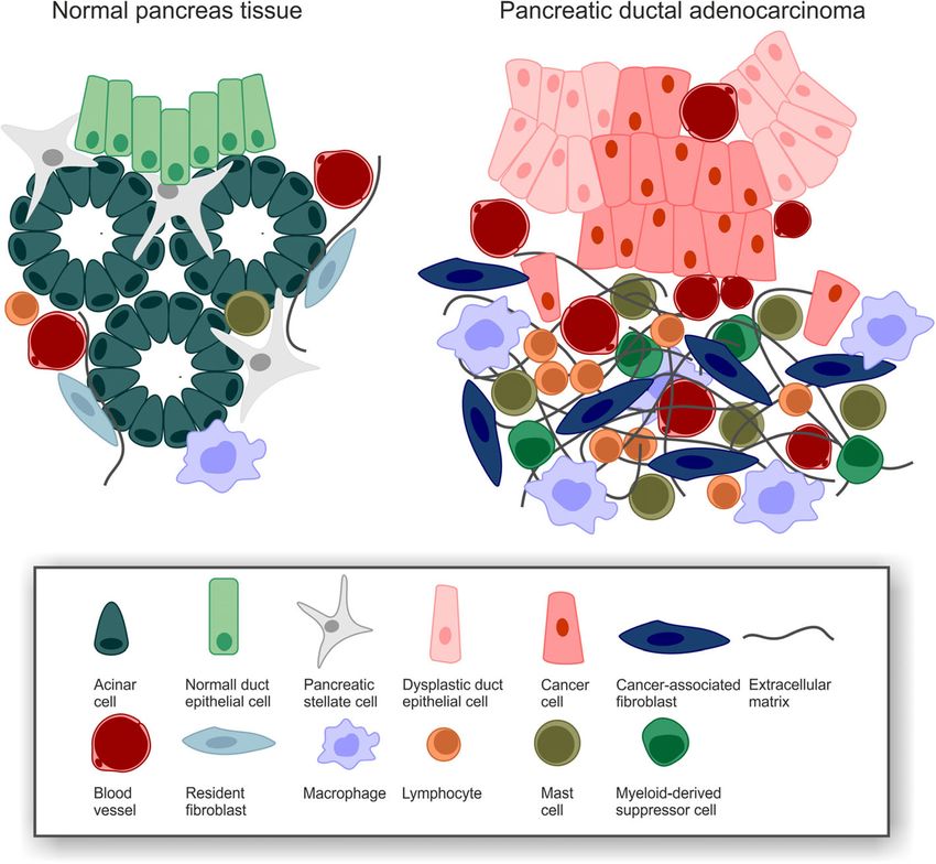
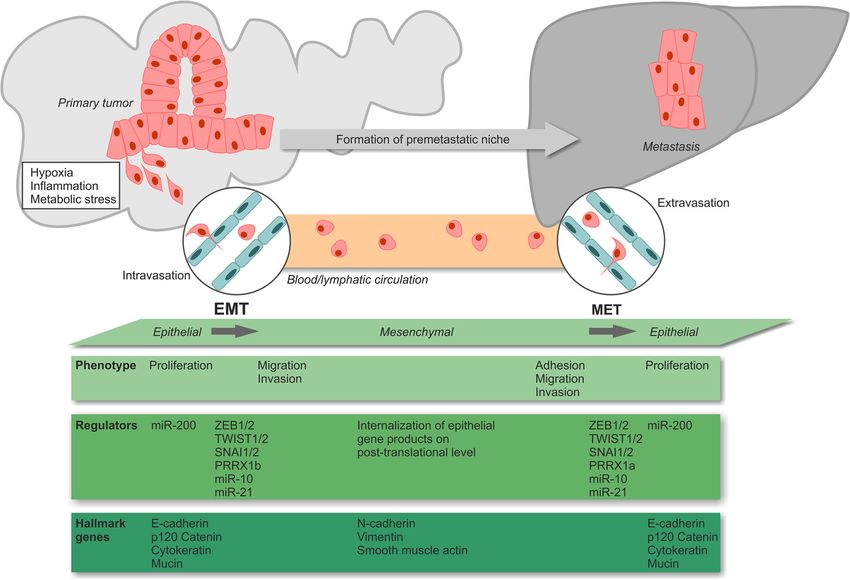
Orth et al. Radiation Oncology   (2019) 14:141                                                                    Page 2 of 20

composed of folinic acid, 5-FU, irinotecan, and oxaliplatin,     pain control paralleled by increased local tumor control
has been reported to nearly double median survival in the        [34]. Hence, SBRT can be seen as an effective and safe
metastasized stage as compared to gemcitabine alone [23],        therapeutic option, and its use in multimodality treatment
and the combination of gemcitabine and a nanoparticle            concepts and/or in palliative settings is considered more
albumin-bound paclitaxel (nab-paclitaxel) has also been          and more frequently.
shown to significantly improve overall survival [24]. How-         In several other cancer entities, e.g. in melanoma
ever, these protocols are associated with relevantly higher      and lung cancer, the implementation of immunothera-
toxicity, thus often preventing their application in elderly     peutic approaches, specifically immune checkpoint in-
patients and/or patients with poor performance status,           hibition, has proven compelling success [35–38]. Yet,
but overall quality of life was reported to increase [25].       at least so far, treatment efficacy in PDAC has been
   Radio(chemo)therapy has been rather infrequently              rather limited [35, 39], and checkpoint inhibition has
adopted for the treatment of PDAC, since the majority            only received approval for the small subset of PDAC
of patients suffer from disseminated stages in which             tumors with high microsatellite instability (1-2% of all
local treatment procedures are of secondary importance           cases) [40, 41]. This may be due to the strongly im-
[26]. Nevertheless, neoadjuvant radiotherapy has the po-         munosuppressive, desmoplastic PDAC microenviron-
tential to improve PDAC resectability in locally advanced        ment, the relatively low mutational burden (resulting
or primarily inoperable/borderline-operable patients, and        in a low number of neo-antigens), as well as other
its beneficial effects on local tumor control are well docu-     biological and/or immunological hallmarks of PDAC
mented [27, 28]. Compared to other cancer entities,              which are discussed in this review [42].
PDAC tumors exhibit a rather high degree of radioresis-
tance – a characteristic which is currently addressed by         Biological and immunological hallmarks of PDAC
combining PDAC radiotherapy with radiosensitizing                Tumor plasticity and heterogeneity
agents, including gemcitabine, capecitabine, or 5-FU, re-        The pancreas contains cells of exocrine (acinar), epithelial
spectively [28, 29]. According to the guidelines of the          (ductal), and endocrine (α, β, δ, ε) origin among which aci-
National Comprehensive Cancer Network (NCCN), the                nar cells are well known for their high degree of plasticity.
use of radio(chemo)therapy is recommended for PDAC               This plasticity is considered to drive pancreas homeostasis
patients with borderline-resectable tumors, and several          and regeneration, as – in contrast to other organs of the
regimens involving capecitabine, gemcitabine, or 5-FU            gastrointestinal tract – the pancreas seems to lack a
have been clinically implemented [29, 30]. The advances          defined stem cell compartment [43]. In a process called
of modern external beam radiation techniques, including          acinar-to-ductal metaplasia (ADM), acinar cells transdif-
image-guided radiation therapy (IGRT), stereotactic body         ferentiate to more epithelial (ductal-like) phenotypes when
radiation therapy (SBRT), and ablative radiation therapy,        experiencing certain macro- and microenvironmental
as well as the combination with novel chemotherapeutic           stimuli, e.g. tissue damage, inflammatory, or stress condi-
protocols have clearly widened the spectrum of radio-            tions [44, 45]. During ADM, acinar cells acquire ‘progeni-
therapeutic options [27, 31, 32].                                tor cell-like’ characteristics which render them more
   Expecting increased toxicities when combining more            susceptible to pro-oncogenic hits, such as activating muta-
aggressive treatment approaches, sequential application          tions in the proto-oncogene KRAS, eventually transform-
is currently being evaluated in the randomized phase III         ing them into pancreatic intra-epithelial neoplasias
CONKO-007 trial for PDAC patients with borderline-               (PanINs). This transformation is generally considered as
resectable, non-metastatic disease (NCT01827553). Pre-           the initial step in PDAC development followed by sequen-
liminary results from an interims analysis document a            tial progression involving genetic hits in several tumor
promising outcome with higher rates of resectability, con-       suppressor genes [46] (Fig. 1).
firming previous phase II findings [27, 30, 33]. As the per-        In order to examine the mutational and transcriptional
formance of systemic therapies gradually improves, local         landscape of PDAC, a number of next generation se-
tumor control moves back into the focus of interest, both        quencing approaches were initiated in the last years
with respect to symptom control as well as with respect to       [48–51]. In conjunction, these studies showed that the
quality of life. In consequence, the importance of local         gene encoding the proto-oncogenic GTPase KRAS as
radiotherapy for the treatment of PDAC patients is con-          well as several tumor suppressor genes, including tumor
stantly growing. SBRT is a highly conformal radiation            suppressor protein 53 (TP53), cyclin-dependent kinase in-
technique which is employed to deliver high doses in a           hibitor 2A (CDKN2A), and mothers against decapenta-
small number of fractions. Due to its steep dose gradients       plegic homologue 4 (SMAD4), exhibit the most frequent
around the target volume, SBRT efficiently spares adjacent       alterations and/or mutations in PDAC [49]. For instance,
organs at risk resulting in relevantly lower toxicity. In sev-   KRAS was not only found to be mutated in most PDAC
eral studies, SBRT achieved significant improvements in          tumors (> 90%), its mutant alleles were additionally
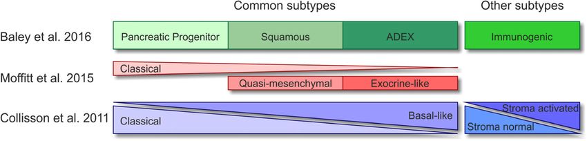
Orth et al. Radiation Oncology     (2019) 14:141                                                             Page 3 of 20

 Fig. 1 Multi-step PDAC carcinogenesis. Modified from [47].

amplified in a subgroup of samples, resulting in accel-         PDAC is a highly heterogenic disease, and various
eration of their tumor-promoting potential [52]. Fur-         attempts have been undertaken to define distinct
thermore, RAC-beta serine/threonine-protein kinase            subtypes with the aim of stratifying patients towards
(AKT2) is frequently overexpressed, and the activity          personalized treatment strategies [49, 50, 60–62]. Cur-
of its upstream regulator phosphoinositide 3-kinase           rently available transcriptome-based classifications were
(PI3K) is often elevated in PDAC leading to increased         extracted via unsupervised clustering methods and dif-
tumor cell survival [53, 54]. Apart from these key            fer in the numbers of subtypes identified. Nevertheless,
mutations, several more uncommon alterations, such            all share common subtypes, including a classical/ca-
as germline mutations in DNA damage repair genes              nonical subtype hallmarked by epithelial-like gene ex-
(e.g. breast cancer early onset genes 1/2 (BRCA1/2),          pression, and a quasi-mesenchymal/basal-like subtype
partner and localizer of BRCA2 (PALB2), and ataxia            characterized by a more mesenchymal gene expression
telangiectasia mutated protein serine/threonine kinase        pattern and poorer prognosis (Fig. 2). These subtypes
ATM), or somatic mutations in DNA mismatch repair             meanwhile can be stratified by immunohistochemistry
regulator genes leading to increased microsatellite in-       using hepatocyte nuclear factor 1A (HNF1A) and cyto-
stability have been found in certain subsets of pa-           keratin-81 (KRT81) as markers [64]. Furthermore, sub-
tients [55]. Of note, the transcriptomic landscape of         types related to exocrine pancreas function have been
PDAC is not entirely governed by genetic alterations.         described as well as subtypes with expression signatures
Integrated epigenetic regulatory circuits comprising chro-    of immune cell-related genes [50, 61, 62]. Although to
matin-based mechanisms, such as DNA methylation and           date there is still no consensus classification which
histone post-translational modification, as well as regula-   would be the prerequisite for clinical application, retro-
tion by non-coding RNAs are also largely distorted in         spective as well as prospective analyses have shown that
PDAC. In this regard, key tumor suppressor genes have         subtype-based stratification has the potential for gen-
been described to be repressed, and oncogenes upregu-         omics-driven precision medicine [64, 65]. The PDAC
lated due to epigenetic alterations [56]. Furthermore, epi-   subtypes obviously stem from inter-tumoral heterogen-
genetic (re-)programing is fundamentally linked to tumor      eity. Yet, intra-tumoral heterogeneity needs to be con-
progression and metastasis formation [57, 58], and the        sidered as well, and tumor cell plasticity might render
epigenetic landscapes of human PDAC subtypes differ           these classifications dynamic, especially upon thera-
substantially [59].                                           peutic intervention.
Orth et al. Radiation Oncology      (2019) 14:141                                                                 Page 4 of 20

 Fig. 2 Molecular classifications of PDAC. Modified from [63].

Desmoplastic, hypoxic, immunosuppressive                         extra cellular matrix, including laminins, fibronectins, col-
microenvironment                                                 lagens, and hyaluronan [69–72]. Interestingly, expression
A crucial hallmark of PDAC is the existence of extensive         of focal adhesion kinase 1 (FAK1) in PDAC cells has re-
desmoplastic stroma which can constitute up to 90% of            cently been reported to be decisive for this process as
the tumor volume and is commonly considered to origin-           pharmacological targeting of FAK1 interfered with the for-
ate from cancer-associated fibroblasts (CAFs) [42] (Fig. 3).     mation of desmoplasia, thus offering a potential target for
Distinct subtypes of CAFs with either myofibroblastic or         therapeutic intervention [73]. Hypoxia is another key fea-
inflammatory phenotypes have been identified [67, 68],           ture of the PDAC microenvironment, and it is closely
and the major source of CAFs appear to be pancreatic             interlinked with desmoplasia. It originates from desmopla-
stellate cells which upon activation, e.g. by injury or          sia-associated hypovascularization and vice versa favors
chronic inflammation, start depositing huge amounts of           desmoplastic progression by activating pancreatic stellate

 Fig. 3 PDAC desmoplasia. Modified from [66].
Orth et al. Radiation Oncology   (2019) 14:141                                                                     Page 5 of 20

cells [74–76]. PDAC hypoxia and desmoplasia, which are             of PDAC metastasis formation are still poorly under-
observed in clinical samples as well as in genetically engi-       stood, especially since the genetic composition of most
neered mouse models, seem to represent barriers to T cell          metastases is closely resembling the one of the corre-
infiltration – intriguingly both for effector as well as regula-   sponding primary tumors [96–98]. Nevertheless, metas-
tory T cells – and T cell activation [77–79]. Moreover, hyp-       tasis formation appears to be a clonal process, since
oxia and desmoplasia are accompanied by a strong                   primary PDAC tumors are composed of different sub-
accumulation of myeloid cells [80, 81]. Macrophages that           clones with individual metastatic potential, and most of
are recruited adopt an immunosuppressive, pro-angiogenic           the metastases show high levels of clonality, indicating
M2-like state, block CD4+ T cell entry into the PDAC               that they initially evolved from one or only a few dissem-
microenvironment, support PDAC progression, and thus               inated tumor cells [96, 98]. Mechanistic studies with
are a marker of negative clinical prognosis [76, 82, 83]. Sys-     genetically traceable mouse models identified a crucial in-
temic frequencies of monocytes and granulocytes are ele-           volvement of epithelial-to-mesenchymal transition (EMT)
vated in PDAC patients, and due to their pathological              explaining also why the quasi-mesenchymal PDAC sub-
activation and immunosuppressive function they are classi-         type as characterized by stronger expression of mesenchy-
fied as monocytic or polymorphonuclear myeloid-derived             mal genes may be associated with poorer prognosis due to
suppressor cells (MDSCs), respectively. Both populations           accelerated metastasis formation [61, 62, 99] (Fig. 4). EMT
are potent suppressors of T cell function and inhibit anti-        so far has been considered to be orchestrated by a com-
tumor immune responses [84, 85]. Recently, the CXCL-1/             plex network of transcription factors which repress epithe-
CXCR2-axis has been shown to be crucially involved in              lial gene expression and/or induce mesenchymal gene
intra-tumoral recruitment of MDSCs, suppressing CD8+ T             expression, including twist-related protein 1 and 2
cell infiltration and function as well as compromising re-         (TWIST1/2), snail family zinc finger protein SNAI1 and 2
sponsiveness to immunotherapy [86]. Apart from these in-           (SNAI1/2), zinc finger E-box-binding homeobox 1 and 2
nate immune cell subpopulations, immunosuppressive T               (ZEB1/2), and paired mesoderm homeobox protein 1
and B cell subpopulations, including regulatory T cells, γδ        (PRRX1a/b) [100, 101]. Especially the EMT activator ZEB1
T cells, and regulatory B cells, have been described in the        has been assigned a central role for tumor cell plasticity
PDAC microenvironment. They do not only block activa-              and metastasis formation in murine PDAC models [102].
tion but also infiltration of effector T cells resulting in low    miRNAs, particularly miR-10, miR-21 and members of the
intra-tumoral CD8+ T cell frequencies [87–89]. These ef-           miR-200 family, constitute another regulatory level of
fector T cells appear to be antigen-experienced, but tumor         EMT and are closely interlinked with the EMT transcrip-
antigen recognition and/or T cell activation seem to be dis-       tion factors via diverse feedback and feedforward circuits
turbed [90]. However, the intra-tumoral T cell repertoire          [103, 104]. Recently, a novel, partial program of EMT
shows enrichment in distinct T cell receptors, suggesting          has been described which is driven by post-transla-
that in principle PDAC tumors are sites of local T cell ex-        tional internalization of epithelial proteins resulting in
pansion [91].                                                      cluster-like rather than single-cell dissemination [105].
   On the cytokine level, the PDAC microenvironment                   Several parameters of the tumor micro- and macroen-
represents a comparable degree of complexity. Neverthe-            vironment are known to influence EMT regulation.
less, the dominating cytokines seem to be transforming             Amongst those, hypoxia, inflammation, and metabolic
growth factor beta (TGF-β), interleukin (IL-) 6, IL-8, IL-         stress appear to be of special importance [100]. Interest-
10, IL-35, granulocyte macrophage colony-stimulating               ingly, high blood glucose concentration, a crucial charac-
factor (GM-CSF), CC-chemokine ligand 2 (CCL-2), CXC-               teristic of diabetes, has also been shown to facilitate
chemokine ligand 1 (CXCL-1), and CXCL-13. In complex               EMT and metastasis formation [7], thus linking a docu-
networks they orchestrate the recruitment and education            mented risk factor to a relevant tumorbiological process.
of innate and adaptive immune cells as well as their               In order to colonize foreign tissues circulating PDAC
crosstalk with tumor cells, CAFs, and other cells in the           cells must undergo a reverse form of EMT (MET) and
PDAC microenvironment, culminating in the desmo-                   re-acquire the epithelial state [106, 107]. Morphologic-
plastic, immunosuppressive milieu that has been de-                ally and mechanistically, MET displays many features of
scribed above [92–94].                                             EMT in an inverse manner. However, the details of this
                                                                   process as well as its master regulators are still being
Metastasis formation                                               investigated.
Another feature of PDAC is its early progression to                   EMT/MET phenomena seem to be crucial elements in
metastatic disease [1]. In advanced stages, patients show          the process of metastasis formation, yet gene expression
invasion of the (retro)peritoneum, the liver, and other            profiling and epigenomic comparisons between primary
gastrointestinal organs, as well as – in some cases – the          tumor cells and metastatic cells also disclosed an in-
vascular and/or the nervous system [95]. The key drivers           volvement of other mechanisms, such as rewiring of the
Orth et al. Radiation Oncology (2019) 14:141 Page 6 of 20
Fig. 4 PDAC epithelial-mesenchymal transition and metastasis formation.
carbohydrate metabolism, e.g. in the oxidative branch of essential in order to improve the overall prognosis of
the pentose phosphate pathway, as well as shifts in en- PDAC.
ergy consumption [58, 108, 109]. Further studies re- The therapeutic success of current first-line chemother-
vealed a (re-)activation of embryonic programs and/or apy involving cytidine analogues, the poly-chemothera-
elevated expression levels of cancer stem cell markers, peutic protocol FOLFIRINOX, or gemcitabine plus nab-
including forkhead box protein A1 (FOXA1), aldehyde paclitaxel, respectively, is strongly limited by intrinsic and/
dehydrogenase 1 (ALDH1), ATP-binding cassette sub- or acquired chemoresistance, and the underlying mecha-
family G member 2 (ABCG2), and hepatocyte growth fac- nisms are only poorly understood [21, 115]. Several pre-
tor receptor (c-Met), in metastatic PDAC cells, suggesting dictive biomarkers have been identified, e.g. increased
a close relationship between retrograde developmental expression of ribonucleotide reductase catalytic subunits
transition, cancer cell stemness and biological features of M1/2 (RRM1/2), an enzyme catalyzing the reduction of ri-
metastasis formation [57, 110]. Finally, the primary bonucleotides, or human equilibrative nucleoside trans-
tumor appears to condition the future target organ of porter 1 (hENT1), a transmembrane protein which
metastasis by releasing soluble factors and/or exosomes, imports nucleosides into the cytosol [117, 118]. In preclin-
thus generating a pre-metastatic niche – even in the sta- ical studies, it was observed that elevated expression levels
tus of a premalignant lesion [111]. Key players in this re- of RRM1 indeed mediate resistance of PDAC cells to gem-
gard have been identified to be tissue inhibitor of citabine [117–119], yet no association between RRM1 ex-
metalloproteinases 1 (TIMP-1) and macrophage migra- pression and OS was detected in clinical analyses [120].
tion inhibitory factor (MIF) [112, 113]. Similar examples are given by integrin-linked kinase (ILK)
[121] and hypoxia-inducible, pro-apoptotic factor BCL2/
Therapy resistance adenovirus E1B 19 kDa protein-interacting protein 3
A signature hallmark of PDAC is its high degree of re- (BNIP3) [122]. Furthermore, cells of the microenviron-
sistance against virtually any kind of therapy [114–116]. ment limit the efficacy of gemcitabine treatment. Recent
Accordingly, overcoming treatment resistance will be data show that CAFs contribute to gemcitabine failure byOrth et al. Radiation Oncology (2019) 14:141 Page 7 of 20
metabolizing gemcitabine to the active metabolite 2′,2′- poly-chemotherapy protocols, the overall prognosis, and
difluorodeoxycytidine-5′-triphosphate (dFdCTP). How- survival rate of PDAC patients still remain poor. Hence,
ever, since dFdCTP cannot cross cell membranes, this there is a strong demand for novel, biologically motivated
process scavenges gemcitabine and reduces the effective treatment strategies with higher specificity for PDAC-
concentration of the active metabolite in the tumor cells relevant, tumor-driving targets. The genomic landscape of
[123]. In case of FOLFIRINOX treatment, increased ex- PDAC is dominated by a handful of signature genes which
pression of thymidylate synthase (TS) and the 5-FU-catab- are affected by aberrations and mutations at high frequen-
olizing enzyme dihydropyrimidine dehydrogenase (DPD) cies: KRAS, CDKN2A, TP53, and SMAD4 [49, 51]. All of
were shown to contribute to therapy resistance, both in these genes are still basically considered to be undrug-
preclinical models and in retrospective clinical analyses gable, although agents targeting mutant TP53 have been
[119, 124]. However, despite all these efforts, biomarker- developed, and attempts to pharmacologically manipulate
based, individualized chemotherapy protocols are far from RAS function are constantly increasing [137, 138]. So far,
being clinical standard. This is predominantly due to a substances targeting downstream effectors of these major
lack of prospective validation studies, let alone random- PDAC drivers or other regulators which are also fre-
ized controlled trials. quently altered, including BRAF, ERK, PI3K/AKT, and
PDAC tumors also exhibit a high degree of radioresis- mTOR, are in the focus of investigation.
tance often resulting in tumor progression even during The mitogen-activated protein kinase (MAPK) signaling
therapy [125]. As in case of chemoresistance, the cascade offers promising perspectives in this regard, be-
responsible mechanisms appear to be multifactorial. cause PDAC cells are known to depend on MAPK signal-
From a biophysical point of view, the hypoxic PDAC ing, both in terms of progression and metastasis formation
microenvironment reduces the biological effectiveness of [139, 140]. The most apical possibility to interfere with
photon irradiation by 2-3 fold as compared to well- MAPK signaling is targeting the epidermal growth factor re-
oxygenated tissues and, thus, attenuates its therapeutic ceptor (EGFR). However, a phase III trial evaluating the effi-
efficacy [126, 127]. Additionally, several studies revealed cacy of anti-EGFR treatment with cetuximab in addition to
an overexpression of key regulators of the DNA damage gemcitabine-based chemotherapy showed no significant
response, e.g. RAD51, in PDAC which contribute to ac- improvement in clinical outcome [141]. Recent data
celerated repair of radiation-induced DNA damage [128, attributed this to a compensatory activation of Integrin β1
129]. Other studies provided evidence for an implication signaling [142]. Downstream of EGFR, KRAS constitutes a
of Integrin- or SMAD signaling in PDAC radioresistance near-perfect target for PDAC treatment as revealed by pre-
[130–132]. Finally, increased recruitment of monocytes clinical RNA interference experiments [143]. However, clin-
upon irradiation stimulating tumor cell proliferation and ical RNA interference is challenging, and no reliable KRAS
neovascularization in response to therapy have been inhibitors have been described so far [144]. Nevertheless,
discussed [133]. In order to counteract PDAC radioresis- pharmacological disruption of the interaction between
tance, several approaches focused on adjusting radio- KRAS and phosphodiesterase PDEδ was shown to effi-
therapeutic protocols. As such, radiotherapy meanwhile ciently suppress PDAC progression in vitro and in vivo
is frequently combined with concomitant chemotherapy [145]. The only targeting approach for MAPK signaling that
(radiochemotherapy), using gemcitabine, 5-FU, or cape- has entered the clinical routine thus far is the combination
citabine as radiosensitizing agents [134, 135]. Addition- of gemcitabine and the EGFR-specific tyrosine kinase in-
ally, stereotactic irradiation regimens with higher single hibitor erlotinib [146]. Although EGFR is considered to be
doses, including SBRT and ablative body radiotherapy, its only target, erlotinib was reported to be similarly effect-
are increasingly being employed aiming at the delivery ive in tumors with wildtype or hyperactive mutants of
of higher biologically active doses to the tumor [26, 31, KRAS, respectively [147]. This implies that either inhibition
136]. However, therapeutic success is still rather limited, of tyrosine kinases other than EGFR or feedback regulatory
and future attempts should evaluate the clinical potential mechanisms between hyperactivated KRAS and EGFR may
of biologically and/or immunologically optimized radio- be involved, respectively [148–151]. Sunitinib, a tyrosine
chemotherapy strategies. kinase inhibitor that does not target EGFR, failed to show
similar performance when combined with gemcitabine
Novel approaches of mechanism-based, molecularly [152], and preclinical data support the notion that indeed
targeted therapies inhibition of gemcitabine-induced MAPK signaling by erlo-
Biologically targeted therapies (1,363 words) tinib accounts for the observed clinical benefits [153].
Since less than 20% of all PDAC patients exhibit surgi- Several other inhibitors of MAPK signaling, including in-
cally resectable disease at time of presentation, systemic hibitors of EGFR, MEK, ERK, and corresponding protein
chemotherapy is currently the most frequently applied phosphatases, have shown convincing performance in pre-
treatment option [21]. Albeit the development of novel clinical studies [154–156], but their potential for clinicalOrth et al. Radiation Oncology (2019) 14:141 Page 8 of 20 implementation remains to be examined, as for instance in PDAC is commonly considered a hypovascularized ACCEPT, a randomized phase II trial combining gemcita- tumor [183], but relevant expression of vascular endo- bine with the EGFR inhibitor afatinib (NCT01728818). thelial growth factor A (VEGF-A) has been observed Single-drug treatments – most likely – will not be [184]. Therefore, the VEGF-A-specific antibody bevaci- sufficient to improve the therapeutic outcome of zumab was tested in combination with gemcitabine in a PDAC [157]. Instead, dual or even multiple targeting randomized phase III trial with locally advanced PDAC strategies appear to be required in order to achieve but failed to show improved outcome [185]. A possible significant advances. One example is the concomitant explanation could be the expression of other VEGF inhibition of MAPK and PI3K/AKT signaling. Preclin- isoforms. However, complementary phase III trials ical data revealed that inhibition of MAPK signaling which evaluated the VEGF receptor tyrosine kinase in- results in potent compensatory activation of PI3K/ hibitor axitinib in combination with gemcitabine, or AKT signaling and vice versa, each being of import- the combination of bevacizumab, gemcitabine, and er- ance for PDAC progression [158, 159]. Indeed, con- lotinib, respectively, also failed [186, 187]. In sum- comitant inhibition of MAPK and PI3K/AKT signaling mary, these results render therapeutic targeting of did interfere with tumor progression to significantly angiogenesis a questionable approach for the treat- greater extent than the single-drug treatments in pre- ment of PDAC [188]. clinical PDAC models [158, 160]. However, other A subset of PDAC tumors (approximately 15% of all studies reported only modest effects of combined cases) is characterized by mutations in genes that are re- MAPK and PI3K/AKT inhibition [161–163], and lated to the DNA damage response [54]. Amongst those, clinical trialing of this combination failed [164]. One PDAC tumors carrying mutations in BRCA1/2 genes are potential explanation could be that inhibitors of dif- of highest interest as they are supposed to be defective ferent target specificities were employed. A more de- in homologous recombination DNA damage repair tailed characterization of the target spectrum of these [189]. Accordingly, patients with BRCA1/2-mutated tu- inhibitors would clarify this and could also help to mors were reported to benefit significantly more from find new targets for mechanism-based therapies. In platinum-based chemotherapy than patients with this regard, upstream and/or transcriptional regulators BRCA1/2 wildtype tumors [190, 191]. For BRCA1/2-de- of PI3K expression, such as transducin beta-like 1 ficient tumors, the inhibition of Poly-(ADP-ribose)-poly- (TBL1), may also be of interest as studies in genetic merase (PARP) may be promising, since this enzyme mouse models have identified them as crucial check- shares an axis of synthetic lethality with BRCA1/2 [192]. points in PDAC development and progression [165]. Initial trials examining the therapeutic potential of PARP Nevertheless, if this mechanism can be exploited inhibitors in patients with BRCA1/2-deficient PDAC re- therapeutically remains unclear [166]. ported promising results [193–196]. Currently, the ran- The mammalian target of rapamycin (mTOR) pathway domized phase III POLO trial is evaluating PARP is best known for its functions in cell survival, prolifera- inhibition in patients who received first-line platinum- tion, motility, and evasion of apoptosis [167]. In several based chemotherapy, and results are awaited in 2019 preclinical studies, mTOR inhibitors revealed promising (NCT02184195). Beyond BRCA1/2, mutations in other results [168–171], but it was also reported that inhibition genes of the DNA damage response, including ATM, of mTOR stimulates feedback activation mechanisms in- may select for PARP inhibitor sensitivity [197]. volving MEK/ERK or AKT signaling, respectively, further In addition to the described genetic alterations, emphasizing the need for combinatorial treatment regi- PDAC tumors display relevant changes in epigenetic mens [172–176]. Not surprisingly, multi-pathway inhib- modifications, including DNA methylation, histone ition regimens are commonly associated with higher levels post-translational modification, nucleosome remodel- of toxicity [177]. This toxicity often interferes with clinical ing, and regulation by non-coding RNAs [56]. In con- implementation. Nevertheless, clinical trials evaluating trast to genetic alterations, epigenetic modifications mTOR inhibition as monotherapy in PDAC altogether are in principle reversible, and it is plausible to as- failed [178–180], and combined modality approaches of sume that pharmacological interference with epigen- mTOR inhibition in conjunction with capecitabine re- etic mechanisms underlying PDAC pathology and vealed only limited improvements as compared to capecit- progression could open new therapeutic perspectives abine alone [181]. These findings raise the question [198]. Preclinical results of epigenetic therapies have whether mTOR inhibitors, despite their successful clinical so far been promising, PDAC cell plasticity could be implementation for the treatment of neuroendocrine pan- reduced, and resistance against standard chemother- creatic tumors, may at all represent a therapeutic alterna- apy was attenuated. However, in mono-agent settings, tive for the treatment of PDAC [182], or whether such epigenetic therapeutics did not provide any measurable approaches have been inadequately tested in the clinic. benefits, demanding for combined modality settings, e.g.
Orth et al. Radiation Oncology (2019) 14:141 Page 9 of 20
in conjunction with chemotherapy or in form of multi- (A3GALT2), a glycosylating enzyme that mainly targets
agent combinations, such as combined inhibition of bro- lipids and extracellular proteins, turned out to be the most
modomain and extra-terminal motif (BET) proteins and promising candidate for a PDAC-targeting vaccine [209].
histone deacetylases (HDACs) [199]. Currently, various However, this vaccine failed to improve treatment efficacy
phase I/II trials are ongoing which will determine the clin- when being tested in a randomized phase III trial com-
ical perspectives of such approaches. Despite all efforts, bined with the standard of care [211]. Other antigens that
individualized, mechanism-based treatment strategies for were examined include peptides derived from human tel-
PDAC are still far from being clinical standard [200]. omerase 1 (TERT1) and GVAX, a vaccine comprised of
Therapeutic targeting of hypoxia and metastasis for- autologous or allogeneic tumor cells expressing the den-
mation appears to be very attractive in the PDAC con- dritic cell-stimulating cytokine GM-CSF [212, 213]. Unfor-
text, since hypoxia is a principal determinant of therapy tunately, none of these vaccines achieved convincing
resistance and metastasis formation, and metastases are clinical results. In principle, common PDAC driver muta-
the major cause of death [20, 74]. Regardless of all pre- tions, such as KRASG12D, can harbor tumor-specific, T cell
clinical efforts [201], however, no therapeutic strategy epitopes [214]. An ongoing phase II trial first predicts
could so far be established. Sort of alternatively, efforts such neo-antigens using exome-sequencing of tumor
to (re-)activate the immune system in order to detect biopsies, followed by production of personalized den-
and combat macro- and micro-metastases have been dritic cell vaccines loaded with the respective epitopes
undertaken and will be discussed in the following. (NCT03300843) [215]. Whether this strategy turns
out to be successful needs to be awaited. Overall, sev-
Immunotherapy eral vaccination approaches could successfully elicit
Immunotherapy implementing immune checkpoint in- measurable anti-tumor T cell responses, yet so far
hibitors has revolutionized cancer treatment in the last none of these strategies resulted in clear clinical ben-
years [202]. Therapeutic antibodies targeting cytotoxic T- efits [216].
lymphocyte-associated protein 4 (CTLA-4) or the axis of Antigen-independent immunostimulatory therapies
programmed cell death protein 1 (PD-1) and its corre- aim at the activation of antigen-presenting cells. Diverse
sponding ligand PD-L1 have shown compelling results receptor-ligand-axes have been explored in this regard.
in several different cancer types, including metastasized As such, treatment with agonistic anti-CD40 antibodies
melanoma and lung cancer [36, 203]. Hence, immune is well known to activate antigen-presenting cells and to
checkpoint inhibition was also tested in PDAC [35, 39], polarize macrophages towards the pro-inflammatory
but compared to melanoma and lung cancer, consider- M1-like state [217, 218]. However, clinical evaluation of
ably smaller numbers of patients (approximately 2%) ex- this strategy in PDAC patients disclosed only short-term
hibited clinical benefits [40, 204]. Consistently, the responses, and no long-term anti-tumor immunity was
responding tumors showed high levels of microsatellite observed [219]. Nevertheless, CD40 stimulation in com-
instability, providing a mechanistic explanation as well bination with chemotherapy and immune checkpoint
as a potential future stratification marker, since micro- blockade is currently under clinical investigation in a
satellite instability is known to increase the number of phase I/II trial (NCT03214250). Complementary ap-
tumor-associated neo-antigens [205]. proaches to achieve activation of antigen-presenting cells
A major determinant of the immunotherapeutic suc- involve ligand-dependent stimulation of pattern recogni-
cess are tumor-specific T cells and their (re-)activation. tion receptors (PRRs) [220]. Indeed, agonists of toll-like
Although their numbers have been described to be ra- receptors (TLRs), RIG-I-like helicases (RLHs), and the
ther low in PDAC patients [90], recent data suggest that stimulator of interferon genes (STING) revealed encour-
the tumor-reactive T-cell repertoire is similar to the one aging results in preclinical PDAC models [221–223], but
found in melanoma where T cell-based therapies mean- their clinical potential remains to be elucidated.
while have relevant therapeutic impact [91]. Further Bypassing the in situ steps of T cell priming by anti-
studies showed that neo-antigen quality rather than gen-presenting cells, adoptive transfer of T cells carrying
quantity, and strong intra-tumoral CD8+ T cell infiltra- chimeric antigen receptors (CARs) has proven powerful
tion are associated with prolonged survival, indicating clinical performance in B-cell malignancies [224]. CAR
that the stimulation of anti-tumor T cell responses can T cells recognize specific cancer cell surface antigens
indeed be a promising strategy for the treatment of through a single-chain variable fragment (scFv) whose
PDAC [60, 206, 207]. Along these lines, different vaccin- ligation stimulates T cell activation via the intracellular
ation strategies employing various kinds of antigens have domains of the CAR construct, resulting in efficient T
already been tested [208–210]. The Algenpantucel-L vac- cell-mediated killing of the target cell [225]. PDAC ex-
cine consisting of irradiated, allogeneic pancreatic tumor hibits several tumor-specific antigens, such as carci-
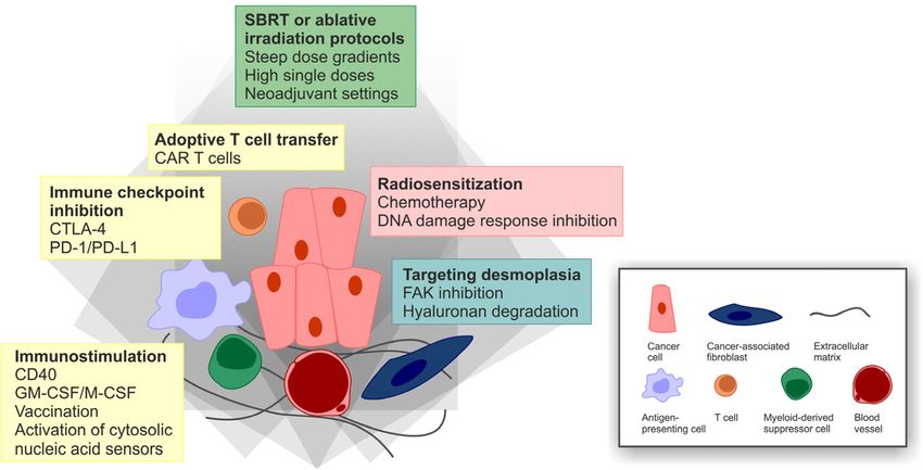
cells stably expressing alpha-1,3-galactosyltransferase 2 noembryonic antigen (CEA), mesothelin (MSLN), andOrth et al. Radiation Oncology (2019) 14:141 Page 10 of 20 mucin 1 (MUC1), which are promising determinants for but the clinical potential of this strategy remains to be ex- CAR T cell therapy [226, 227]. However, for solid cancer amined [237]. entities, intra-tumoral recruitment and trafficking of In summary, (re-)activating anti-PDAC immunity in CAR T cells as well as the commonly observed immuno- order to improve the overall clinical outcome appears suppressive tumor microenvironment appear to be clearly more challenging than extrapolated experiences major challenges. Intelligent combinations, thus, are from other cancer entities have suggested. Probably the needed in order to overcome these obstacles. most promising strategies would incorporate combina- A cardinal feature of the immunosuppressive PDAC tions of different immunotherapeutic approaches and/or microenvironment is its massive stromal content and the combinations with other (classical) treatment modalities, excessive deposition of extracellular matrix, including such as chemotherapy and/or radiotherapy [238]. hyaluronan [72]. Early phase clinical trials combining re- combinant human hyaluronidase 20 (rHuPH20) with gemcitabine and nab-paclitaxel revealed promising results, Combined modality treatment approaches encompassing particularly in those patients whose tumors were charac- radio(chemo)therapy terized by high levels of hyaluronan [228]. Reporting of In order to improve the efficacy and the outcome of the HALO-109-301 phase III trial (NCT02715804) is clinical PDAC treatment, it will be inevitable to develop awaited in order to fully assess the clinical performance of novel treatment strategies which combine different this approach [229]. Inhibition of FAK1, a tyrosine kinase therapeutic modalities aiming at achieving synergism involved in the process of CAF generation, constitutes an- [239]. The rationale for such approaches is to outcom- other approach to interfere with stromal function in pete therapy resistance, but their development remains PDAC, and pharmacological FAK1 inhibition eventually challenging as combined modality treatments are fre- rendered preclinical PDAC model systems more suscep- quently associated with higher toxicity levels [240]. We tible to T cell immunotherapy and immune checkpoint in- already discussed several combined modality attempts hibition [73]. Other studies showed that genetic ablation involving different chemotherapeutics, either with each or inhibition of FAK1 also increases PDAC responsiveness other or with novel, molecularly targeted inhibitors. At to gemcitabine and nab-paclitaxel [230, 231]. In rather this point, we want to concentrate on combinatorial ap- strong contrast, genetic deletion of stromal myofibroblasts proaches involving radiotherapy (Fig. 5). in PDAC mouse models led to disease exacerbation and Radiotherapy has rather infrequently been used for diminished animal survival due to enhanced regulatory T the treatment of PDAC. Nevertheless, there have been cell-mediated immunosuppression, clearly calling for cau- approaches to improve the efficacy of radiotherapy in tion when targeting components of PDAC stroma [78]. PDAC. One obvious strategy is to combine radiother- On a cellular level, massive infiltration by myeloid apy with radiosensitizing agents which either can be cells, such as MDSCs, and resulting exclusion of CD8+ classical chemotherapeutic drugs, such as gemcitabine T cells are major hallmarks of the immunosuppressive or 5-FU, or – as has been reported more recently – PDAC microenvironment [86, 232]. Several myeloid molecularly designed inhibitors that target specific cell-targeting approaches have been investigated in re- proteins and/or structures involved in PDAC radiore- cent years in order to overcome these mechanisms of sistance [28, 125]. The MAPK pathway is a very at- immunosuppression [82, 233, 234]. Chemokine receptor tractive target [140], and preclinical data derived from 2 (CCR2), for instance, is known to contribute to the different PDAC mouse models showed that interfer- infiltration of pancreatic tumors by monocytes and mac- ence with MAPK signaling by cetuximab treatment rophages, and this is associated with reduced patient can indeed increase the efficacy of radiochemotherapy survival and poor outcome [235]. Strikingly, the combin- [241, 242]. Encouraged by these observations, several ation of CCR2 blockade and gemcitabine/nab-paclitaxel clinical trials were initiated, yet with only modest re- chemotherapy showed promising results in phase I trials sults [243–246]. The major reason was the persist- [85, 236]. However, the follow-up phase Ib/II trial ently high rate of distant failure due to metastasis (NCT02732938) was discontinued due to strategic consid- formation, rather than poor local control [244, 246]. erations, and instead phase I/II trials with combined mo- Pharmacological intervention with the PI3K/AKT and dality approaches of CCR2 blockade in conjunction with the mTOR pathway has also been examined with regards pre-operative SBRT and immune checkpoint inhibition to its radiosensitizing potential. Several preclinical stud- were recently initiated (NCT03778879, NCT03767582). ies obtained basically positive results [247–253]. How- Another target that regulates the function of macrophages ever, due to very unfavorable pharmaceutical properties and MDSCs in PDAC is M-CSF. Preclinical data suggest of the employed substances, e.g. elevated toxicity levels that M-CSF blockade can indeed reprogram macrophages and crossover inhibition, none of these approaches have and thus, synergize with immune checkpoint inhibition, entered the clinic thus far.
Orth et al. Radiation Oncology   (2019) 14:141                                                                    Page 11 of 20

 Fig. 5 Combined modality perspectives for the treatment of PDAC.

  A very direct approach of radiosensitization is the em-           agents are required. As an example, radiotherapy plus
ployment of molecularly designed drugs which target                 GM-CSF, a potent stimulator of antigen-presenting cell
components of the DNA damage response, specifically the             maturation, produced objective abscopal responses in a
upstream kinases ATM, ATR, CHK1/2, and DNA-PK                       subset of patients with different metastatic tumors [269],
[254–256]. Several of these inhibitors displayed convin-            and a recent case report showed similar effects in a pa-
cing synergism with ionizing irradiation or DNA-dam-                tient with metastatic pancreatic cancer [270]. In preclin-
aging chemotherapy in preclinical PDAC model systems                ical model systems, PDAC tumors have been reported to
[257–263], but the transferability into the clinic remains          regress convincingly upon immunotherapeutic targeting
to be investigated – particularly in view of local control          of CCL2 or PD-L1 in combination with radiotherapy via
versus distant failure. PARP is another example for a DNA           a reduction of intra-tumoral immunosuppressive mye-
damage response regulator that can be targeted by highly            loid cells and enhanced recruitment of tumor-specific T
refined inhibitors, and preclinical data suggest that PARP          cells [133, 271], and the clinical performance of this ap-
inhibition indeed can radiosensitize PDAC cells [264].              proach will be investigated (NCT03778879, NCT03767582).
However, since PARP is known to share synthetic lethality           Similarly, radiotherapy has been described to repro-
with BRCA1/2 [192], PARP inhibition may turn out to be              gram tumor-infiltrating macrophages towards an M1-
only effective in BRCA1/2 deficient tumors [265]. This is a         like phenotype and to favor intra-tumoral recruitment
general lesson that has been learned in the era of molecu-          of adoptively transferred T cells in a mouse model of
larly targeted therapy: Molecularly designed therapy re-            neuroendocrine pancreatic cancer [272]. These obser-
quires upfront molecular diagnostics and proper patient             vations were confirmed by pilot data from patients with
stratification, since otherwise promising agents are prone          advanced PDAC stages undergoing neoadjuvant irradi-
to fail if they are trialed in the wrong subgroups of               ation prior to tumor resection revealing 3- to 5-fold in-
patients.                                                           creases in intra-epithelial CD4+ and CD8+ T cells as
  Apart from its potential to induce tumor cell death,              compared to non-irradiated control patients [272, 273].
radiotherapy is known to recondition the tumor micro-               If these findings may also be transferred to combina-
environment and to stimulate systemic anti-tumor im-                tions with PDAC-specific CAR T cells remains to be ex-
mune responses – a phenomenon summarized as                         amined. On a mechanistic level, cytosolic DNA-sensing
abscopal effects of radiotherapy [266–268]. However, in             upon irradiation-induced DNA damage and type I
the monotherapy setting, radiation is often not sufficient          interferon signaling appear to be involved in the immu-
to break the immunosuppressive milieu of established                nostimulating effects of radiotherapy [274, 275]. Ac-
tumors, and combinations with immunostimulating                     cordingly, artificial activation of cytosolic DNA sensors,
Orth et al. Radiation Oncology   (2019) 14:141                                                                                Page 12 of 20

such as STING, was shown to increase the efficacy of           as for immunotherapeutic strategies. Importantly, how-
radiotherapy by enhancing CD8+ T cell responses – at           ever, this will require in-depth optimization of timing,
least in preclinical PDAC models [276].                        dosing, and treatment sequences, as well as careful up-
   From clinical experiences with other cancer entities it     front patient stratification. Otherwise per se promising
is becoming increasingly evident that the combination of       combinations run the risk of failing prematurely.
radiotherapy and immunotherapy requires very careful
considerations regarding timing, dosing, and treatment         Abbreviations
                                                               5-FU: 5-Fluorouracil; A3GALT2: Alpha-1,3-galactosyltransferase 2; ABCG2: ATP-
sequence in order to achieve the best outcome [266].           binding cassette sub-family G member 2; ADM: Acinar-to-ductal metaplasia;
This may be of particular interest for PDAC with its           AKT: RAC-beta serine/threonine-protein kinase; ALDH1: Aldehyde
highly challenging immunosuppressive microenviron-             dehydrogenase 1; ATM: Ataxia telangiectasia mutated protein serine/
                                                               threonine kinase; ATR: ATM- and Rad3-related kinase; BET: Bromodomain and
ment. In brief, higher single doses of radiotherapy, e.g.      extra-terminal motif; BNIP3: BCL2/adenovirus E1B 19 kDa protein-interacting
SBRT or ablative protocols, applied in neoadjuvant set-        protein 3; BRAF: v-Raf murine sarcoma viral oncogene homolog B; BRCA1/
tings appear to be beneficial, and immunotherapy needs         2: Breast cancer early onset 1/2; CAF: Cancer-associated fibroblast; CAR T
                                                               cell: Chimeric antigen receptor T cell; CCL-2: CC-chemokine ligand 2;
to be started before or with the first irradiation fraction,   CD: Cluster of differentiation; CDKN2A: Cyclin-dependent kinase inhibitor 2A;
respectively [266]. However, the optimal treatment regi-       CEA: Carcinoembryonic antigen; CHK1/2: Checkpoint kinase 1/2; c-
men and the best combination of agents for PDAC remain         Met: Hepatocyte growth factor receptor; CTLA-4: Cytotoxic T lymphocyte-
                                                               associated protein 4; CXCL-1: CXC-chemokine ligand 1; CXCR2: CXC-
unclear as well as the impact of additional chemotherapy       chemokine receptor 2; dFdCTP: 2′,2′-difluorodeoxycytidine-5′-triphosphate;
and other factors, such as type II diabetes and/or obesity.    DNA-PK: DNA-dependent protein kinase; DPD: Dihydropyrimidine
A pilot study addressing some of these combinatorial is-       dehydrogenase; EGFR: Epidermal growth factor receptor; EMT: Epithelial-to-
                                                               mesenchymal transition; ERK: Extracellular signal-regulated kinase; FAK1: Focal
sues added radiotherapy to CD40-dependent immunosti-           adhesion kinase 1; FOLFIRINOX: Poly-chemotherapeutic regimen composed
mulation plus anti-CTLA-4/anti-PD-1-mediated immune            of folinic acid, 5-FU, irinotecan, and oxaliplatin; FOXA1: Forkhead box protein
checkpoint blockade in genetically engineered PDAC             A1; GM-CSF: Granulocyte macrophage stimulating factor; HDAC: Histone
                                                               deacetylases; hENT1: Human equilibrative nucleoside transporter 1;
mouse models and utilized machine learning algorithms          HNF1A: Hepatocyte nuclear factor 1A; IGRT: Image-guided radiotherapy;
to extract signature patterns for each therapeutic compo-      IL: Interleukin; ILK: Integrin-linked kinase; KRAS: Proto-oncogene from Kirsten
nent [277]. Along these lines, more in depth-analyses          rat sarcoma virus; KRT81: Cytokeratin-81; MAPK: Mitogen-activated protein
                                                               kinase; M-CSF: Macrophage colony stimulating factor; MDSC: Myeloid-derived
are needed in order to fully exploit the synergism             suppressor cell; MEK: Mitogen-activated protein kinase kinase;
between radiotherapy and immunotherapy. Neverthe-              MET: Mesenchymal-to-epithelial transition; MIF: Macrophage migration
less, several clinical phase I/II trials combining             inhibitory factor; MSLN: Mesothelin; mTOR: Mammalian target of rapamycin;
                                                               MUC1: Mucin I; NCCN: National Comprehensive Cancer Network; OS: Overall
radiotherapy with different immunotherapeutic ap-              survival; PALB2: Partner and localizer of BRCA2; PAnIN: Pancreatic intra-
proaches have been initiated for advanced PDAC,                epithelial neoplasias; PARP1/2: Poly-(ADP-ribose)-polymerase 1/2; PD-
and first results are awaited [278] (NCT02648282,              1: Programmed cell death 1; PDAC: Pancreatic ductal adenocarcinoma;
                                                               PDEδ: Photoreceptor cGMP phosphodiesterase δ subunit; PD-
NCT03161379, NCT03767582, NCT03563248).                        L1: Programmed cell death ligand 1; PI3K: Phosphoinositide 3-kinase;
                                                               PRR: Pattern recognition receptor; PRXX1a/b: Paired mesoderm homeobox
Conclusions                                                    protein 1a/b; rHuPH20: Recombinant human hyaluronidase 20; RIG-I: Retinoic
                                                               acid inducible gene I; RLH: RIG-I-like helicases; RRM1/2: Ribonucleotide
PDAC represents a cancer entity of extraordinarily high        reductase catalytic subunits M1/2; SBRT: Stereotactic body radiotherapy;
malignancy, particularly poor prognosis, and constantly        scFv: single-chain variable fragment; SMAD4: Mothers against
increasing patient numbers. Its aggressive biology and         decapentaplegic homologue 4; SNAI1/2: Snail family zinc finger protein 1/2;
                                                               STING: Stimulator of interferon genes; TBL1: Transducin beta-like 1;
the fact that most patients present in advanced or dis-        TERT1: Telomerase reverse transcriptase 1; TGF-β: Transforming growth factor
seminated stages of disease render the development of          β; TIMP-1: Tissue inhibitor of metalloproteinases 1; TLR: Toll-like receptor;
novel PDAC treatment strategies one of the super-              TP53: Tumor protein 53; TS: Thymidylate synthase; TWIST1/2: Twist-related
                                                               proteins 1/2; VEGF-A: Vascular endothelial growth factor A; ZEB1/2: Zinc finger
ordinate challenges in current oncological research. Re-       E-box-binding homeobox 1/2
sults of the last 20 years have led to the establishment
of a detailed multi-step model of PDAC development             Authors’ contributions
and progression. Although this has unquestionably re-          MO, PM, and KL: conception and writing. KL: illustration. All listed authors
                                                               were involved in drafting and revising of the manuscript. All authors have
formed our understanding of PDAC as a disease, none
                                                               read and approved the final manuscript.
of these findings could be successfully translated into a
therapeutic breakthrough so far. It is becoming increas-       Funding
ingly evident that the clinical performance of single-         This work was supported by the Deutsche Forschungsgemeinschaft
agent therapies lags behind the original expectations,         (CRC1321 projects P13, P14 and P16 to GS, KL, MS, and JM, and SCHN664/6-1
                                                               to MS), the international doctoral program ‘i-Target: Immunotargeting of
and instead intelligent combinations appear to be re-          cancer’ funded by the Elite Network of Bavaria (to MS and KL), the
quired. In this regard, radiotherapeutic protocols, and        Bundesministerium fuer Bildung und Forschung (BMBF 01EK1511A to JM),
particularly modern radiation techniques with high             and the European Union (EU-FP-7: EPC-TM to JM, Marie-Sklodowska-Curie
                                                               ‘Training Network for the Immunotherapy of Cancer (IMMUTRAIN)’ to MS).
conformality and steep dose gradients, represent at-           The funding body did not have any influence on conception and content of
tractive partners both for biologically motivated as well      this review manuscript.
You can also read