2021 IMMUNOLOGY SCIENTIFIC CONFERENCE
←
→
Page content transcription
If your browser does not render page correctly, please read the page content below
2021
IMMUNOLOGY
SCIENTIFIC
CONFERENCE
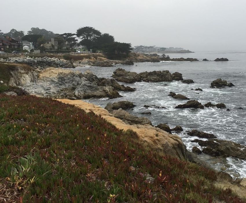
Asilomar Conference Grounds
800 Asilomar Avenue
Pacific Grove, CA
Agenda & Abstracts
Friday – Saturday
November 12-13, 2021 (updated 11/21/21)
Welcome to the 2021 Stanford Immunology Program’s
                 Conference at the Asilomar Conference Grounds!

We are pleased to present what promises to be an exciting retreat, filled with presentations
by faculty, staff, postdocs, and graduate students from across the Stanford Immunology
community. Talks on Friday and Saturday, as well as the Poster Session on Friday, will be held
in Merrill Hall. Download the Asilomar property map here. Talks are divided into sessions
with innovative research that crosses traditional boundaries at Stanford University School of
Medicine. You won’t want to miss a single talk! There are plenty of breaks to re-caffeinate,
rehydrate, sugar up, and network. Faculty talks are 20 minutes, and graduate student,
postdoc and staff talks are 10 minutes each. We will be enforcing the time limits to provide at
least 5 minutes for questions, related insights, and to catalyze discussions throughout the
weekend.

We are honored to have Keynote Speaker:

                      Atul Butte, MD, PhD
                      Professor, Pediatrics
                      Director, Institute for Computational Health Sciences at UCSF

                       Atul Butte, MD, PhD is the Priscilla Chan and Mark Zuckerberg Distinguished
                       Professor and inaugural Director of the Bakar Computational Health Sciences
Institute (bchsi.ucsf.edu) at the University of California, San Francisco (UCSF). Dr. Butte is also the
Chief Data Scientist for the entire University of California Health System, with 20 health professional
schools, 6 medical schools, 5 academic medical centers, 10 hospitals, and over 1000 care delivery
sites. Dr. Butte has been continually funded by NIH for 20 years, is an inventor on 24 patents, and has
authored over 200 publications, with research repeatedly featured in the New York Times, Wall Street
Journal, and Wired Magazine. Dr. Butte was elected into the National Academy of Medicine in 2015,
and in 2013, he was recognized by the Obama Administration as a White House Champion of Change
in Open Science for promoting science through publicly available data. Dr. Butte is also a founder of
three investor-backed data-driven companies: Personalis (IPO, 2019), providing medical genome
sequencing services, Carmenta (acquired by Progenity, 2015), discovering diagnostics for pregnancy
complications, and NuMedii, finding new uses for drugs through open molecular data. Dr. Butte
trained in Computer Science at Brown University, worked as a software engineer at Apple and
Microsoft, received his MD at Brown University, trained in Pediatrics and Pediatric Endocrinology at
Children's Hospital Boston, then received his PhD from Harvard Medical School and MIT.

 Stanford Immunology Conference 2021                                                        Page 2 of 49
Game Show Friday
On Friday night, after the keynote, we will have the traditional game show featuring our
faculty and Second Year Immunology students.
Poster Session Friday
The poster session will be on Friday night at 9:00-11:00 pm, along with a reception. A Best
Poster Prize will be given to two graduate students and two postdoctoral fellows.
• Poster Slam: Saturday, November 9 at 8:10-8:40 pm. Poster presenters may have the
podium for several minutes to exhort our conference attendees through PG-rated
means to come to their posters.
• Poster Ads will run continuously throughout the retreat. The intent of these Poster
Ads is to draw as many people as possible to the posters. Voting for Best Poster will be
done democratically – everyone votes! Faculty are encouraged to visit each poster as
they are the ‘Super Delegates’ and have decisive voting power.
Faculty-First Years only Research Blitz
On Saturday, faculty wishing to recruit rotation students to their labs may participate in a
research blitz with first year students. Each faculty member will have the opportunity to talk
about their research in several minutes and converse with a First Year student. Once the
allotted few minutes are up, the faculty member will meet the next first year student.
Birukova Midnight Swim
Take a plunge in the ocean on Friday night! In honor of Maria Birukova, a former School of
Medicine graduate student, students, postdocs, and faculty can continue the tradition of the
midnight swim on Saturday.
Walk on the Beach Saturday
Get inspired by a beach view and the smell and sound of the surf by taking a walk on the
beach. Meet in front of Crocker Dining Hall to walk with a group.
The Annual Scientific Conference is one of the highlights of the academic year for our
Stanford Immunology community – we thank you for participating in this marvelous and
enriching experience.
2021 Immunology Conference Committee
Thank you to the conference planning committee and your dedication on making the event a
success.
Conference Directors: Nima Aghaeepour and Purvesh Khatri
Social Chairs: Second Year Class, Camilo Espinosa Bernal, and Maddie Lee
Conference Committee: Lina Hansen, Olivia Martinez, and Torye Nguyen
Technical Support: Candace Liu
Page 3 of 49 Stanford Immunology Conference 2021AGENDA AT A GLANCE
Times, speakers, and topics may change without notice
General sessions are in Merrill Hall
Meals are in Crocker Hall
Friday, November 12 Saturday, November 13
9:45 AM Conference Check in 7:30 AM Breakfast
10:00 AM Welcome & Intro 9:00 AM Walk on the Beach
10:10 AM Session 1 10:00 AM CDIII Updates
11:05 AM Session 2 11:00 AM Break
12:00 PM Group Pictures 11:10 AM Session 6
12:15 PM Lunch 12:05 PM Lunch
1:10 PM Session 3 12:05 PM Faculty Meeting
2:05 PM Break 1:20 PM Research Blitz
2:20 PM Session 4 3:00 PM Session 7
3:15 PM Session 5 3:55 PM Break
4:10 PM Lodging Check in 4:05 PM Session 8
6:00 PM Dinner 5:00 PM Adjourn
7:05 PM Keynote
8:00 PM First Year/Faculty Gameshow
9:00 PM Poster Session and Reception
10:00 PM Bonfire
11:00 PM Birukova Midnight Swimming Club
12:00 AM Adjourn
Stanford Immunology Conference 2021 Page 4 of 49AGENDA
STANFORD IMMUNOLOGY SCIENTIFIC CONFERENCE
Friday, November 12, 2021 at 10:00 AM – 12:00 AM (midnight)
Talks and Poster Session are hosted in Merrill Hall
Time Speaker & Title
9:45 AM Conference Check in and pick up name badges in Merrill Hall
10:00 AM Welcome and Introductions: Conference Directors: Nima Aghaeepour, PhD, and Purvesh
Khatri, PhD
Session 1 Graduate/Postdoc Talks
10:10 AM Tobias Lanz, Robinson Lab, Clonally expanded B cells in multiple sclerosis bind EBV and
GlialCAM
Naomi Haddock, Bollyky Lab, Circulating bacteriophage in Sepsis patients reflect infection
dynamics and improve diagnosis of ‘Pseudomonas aeruginosa’ infections
Bryan Cannon, Bendall Lab, Identifying differences in cellular and disease phenotypes
across human Azheimer’s disease tissue by multiplexed ion beam imaging
Session 2 Graduate Student/Postdoc Talks
11:05 AM Guangbo Chen, Davis Lab, A cytokine surge precedes the cancer diagnosis in the elderly
Kathleen (Kassie) Dantzler, Jagannathan Lab, Diminished V2+ T cell cytokine
production and degranulation following in vitro malaria exposure
Katie Freitas, Mackall Lab, Enhanced effector activity of mediator CDK8 kinase module
deficient CAR-T Cells
12:00 PM Group Pictures for Faculty, Graduate Students, and Postdocs
12:15 PM Lunch at Crocker Dining Hall
Session 3 Graduate/Postdoc Talks
1:10 PM Joe Gonzales, Wang Lab, An enhanced IgG/CD16a axis predicts worsening disease in mild
COVID-19 patients and promotes inflammation in the lungs of Fc receptor-humanized mice
Lilit Grigoryan, Pulendran Lab, Adjuvanting a subunit SARS-CoV-2 vaccine to induce durable
protection in mice
John Hickey, Nolan Lab, T cell mediated tumor tissue restructuring: Convergence of in vivo
and in silico approaches
2:05 PM Break
Session 4 Graduate/Postdoc Talks
2:20 PM Nina Horowitz, Sunwoo Lab, Engineering highly cytotoxic tissue-resident natural killer cells
for solid tumor immunotherapy
YeEun Kim, Bendall and Greenleaf Labs, A highly multiplexed single cell proteomics screen
reveals a novel lymphoid progenitor population in human bone marrow
Hunter Martinez, Bollyky Lab, The tumor cell marker CD44v6 denotes a suppressive FoxP3+
Treg but its expression is reduced in T1D
Session 5 Graduate/Postdoc Talks
3:15 PM Tara Murty, Mackall Lab, Genomic and phenotypic correlates of GD2 chimeric antigen
receptor (CAR) T cell activity in pediatric osteosarcoma and neuroblastoma patients
Jason Nideffer, Jagannathan and Roncarolo Labs, Tolerance to malaria is marked by
homeostatic immune cell frequencies, epigenetics, and functions
Medeea Popescu, Bollyky Lab, Bacteriophage deliver small RNAs into phagocytes and
inhibit immune activation
Page 5 of 49 Stanford Immunology Conference 20214:10 PM Lodging Check in at Social Hall/Front Desk. Day guest meal tickets may be purchased at
the Front Desk.
6:00 PM Dinner at Crocker Dining Hall
7:05 PM Keynote Introduction
7:10 PM Keynote: Atul Butte, MD, PhD, Professor of Pediatrics, UCSF
8:00 PM First Year/Faculty Gameshow
9:00 PM Poster Session and Reception
10:00 PM Bonfire
11:00 PM Birukova Midnight Swimming Club
12:00 AM Adjourn
Saturday, November 13, 2021 at 7:30 AM – 5:00 PM
Talks are hosted in Merrill Hall
Time Speaker & Title
7:30 AM Breakfast at Crocker Dining Hall
9:00 AM Walk on the Beach (Meet outside Crocker Dining Hall)
10:00 AM CDIII Updates
11:00 AM Break
Session 6 Graduate/Postdoc Talks
11:10 AM Shamma Rahman, Mellins Lab, Profiling of peripheral polarized monocytes reveals a pro-
inflammatory subset in pediatric acute-onset neuropsychiatric syndrome (PANS)
Jack Silberstein, Cochran Lab, Structure-based engineering of an immune checkpoint
protein
Erin Soon, Angelo Lab, Spatio-temporal coordination at the maternal-fetal interface
promotes trophoblast invasion and vascular remodeling in the first half of human pregnancy
12:05 PM Lunch for Graduate Students and Postdocs at Crocker Dining Hall
12:05 PM Faculty Meeting: Immunology Faculty, Graduate Student, and Postdoc Representatives
remain at Merrill Hall
1:20 PM Faculty-First Year Graduate Students Research Blitz
Session 7 Graduate Student/Postdoc Talks
3:00 PM Kattria van der Ploeg, Jagannathan Lab, TNF producing CD4+ T cells dominate the SARS-
CoV-2-specific memory T cell response and are associated with antibody durability in patients
with mild to moderate COVID-19
Edward (Ted) Wilson, Andreasson Lab, Innate immune receptor TREM1 disrupts metabolic
homeostasis in aging and Alzheimer’s disease
Florian Wimmers, Pulendran Lab, The single-cell epigenomic and transcriptional landscape
of immunity to influenza vaccination in humans
3:55 PM Break
Session 8 Graduate/Postdoc Talks
4:05 PM Shady Younis, Robinson Lab, Activated ACPA pre-germinal center B cells in rheumatoid
arthritis identified by single-cell analysis
Xiang Zhao, Garcia Lab, Catch bond engineering of high-potency and low-affinity TCR
Hong Zheng, Khatri Lab, Multi-cohort analysis of host immune response identifies conserved
protective and detrimental modules associated with severity across viruses
5:00 PM Adjourn - announce winners of Talks and Posters
Stanford Immunology Conference 2021 Page 6 of 49ORAL SESSION
STANFORD IMMUNOLOGY CONFERENCE
Friday, November 12, 2021
Merrill Hall
Session 1 Graduate/Postdoc Talks
10:10 AM Tobias Lanz, Robinson Lab, Clonally expanded B cells in multiple sclerosis
bind EBV and GlialCAM
Naomi Haddock, Bollyky Lab, Circulating bacteriophage in Sepsis patients
reflect infection dynamics and improve diagnosis of ‘Pseudomonas
aeruginosa’ infections
Bryan Cannon, Bendall Lab, Identifying differences in cellular and disease
phenotypes across human Azheimer’s disease tissue by multiplexed ion beam
imaging
Tobias Lanz, MD
Basic Life Research Scientist
Advisor: Bill Robinson, MD
Department of Immunology & Rheumatology
Clonally expanded B cells in multiple sclerosis bind EBV and GlialCAM
Abstract TBA
Naomi Haddock
Immunology Graduate Student
Advisor: Paul Bollyky, MD, PhD
Department of Infectious Diseases and of Microbiology & Immunology
Circulating bacteriophage in Sepsis patients reflect infection dynamics and improve diagnosis
of ‘Pseudomonas aeruginosa’ infections.
Bacterial infections pose a major threat to global health, and call for continued improvement in
diagnostics and treatment options. Microbial cell-free DNA sequencing promises to improve non-
invasive diagnostics of bacterial infections but existing approaches often fall short. This is particularly
true for some pathogens such as Pseudomonas aeruginosa that colonize lungs and other tissues. We
have investigated whether bacteriophage, viruses produced by bacteria, can provide insight to
infection dynamics. Phages are abundant at sites of colonization and infection and are exquisitely
specific to their host bacterial strains. Because phages are able to enter circulation, they can be
assessed in cell-free DNA, thereby providing unique insights into bacterial dynamics at the strain level.
Here, we show that the circulating phageome is disrupted in individuals with Sepsis versus
asymptomatic controls, that bacterial infections are uniquely reflected in circulating phage
Page 7 of 49 Stanford Immunology Conference 2021signatures, and that identification of bacteriophage improves understanding of infection
characteristics. We propose that bacteriophage can improve the identification of pathogens in sepsis
and other infectious disease settings.
Bryan Cannon
Immunology Graduate Student
Advisor: Sean Bendall, PhD
Department of Pathology
Identifying differences in cellular and disease phenotypes across human alzheimer’s disease tissue
by multiplexed ion beam imaging
Identifying cell-based and regional phenotypes in human brain tissue is a necessary task to
understand neurodegenerative pathologies in Alzheimer’s disease. Using MIBI (multiplexed ion-beam
imaging) we have simultaneously quantified tissue-level expression of 40 different targets encoding a
deep set of neurological phenotypes with nanometer scale spatial resolution. To localize protein
expression to specific cells or tissue restricted regions, we use a combination of single cell and single
object segmentation methods. Applying this technique to multiple MIBI scans of archival human
hippocampus cross-sections, we are able to identify the spatial distribution and expression
heterogeneity of glial cell types and proteopathies in both cognitively healthy and diseased human
brain. Using off-the-shelf and in-house developed computational strategies we are able to identify
unique protein-disease relationships in neurons and glia that may lead to additional work testing the
functional significance of the descriptive work outlined here. We believe this will be a straight forward
approach to discretize and select features for multiplexed imaging technologies as applied to human
brain tissue in both degenerative and non-degenerative contexts.
Stanford Immunology Conference 2021 Page 8 of 49Session 2 Graduate Student/Postdoc Talks
11:05 AM Guangbo Chen, Davis Lab, A cytokine surge precedes the cancer diagnosis in
the elderly
Kathleen (Kassie) Dantzler, Jagannathan Lab, Diminished V2+ T cell
cytokine production and degranulation following in vitro malaria exposure
Katie Freitas, Mackall Lab, Enhanced effector activity of mediator CDK8
kinase module deficient CAR-T Cells
Guangbo Chen, PhD
Basic Life Research Scientist
Advisor: Mark Davis, PhD
Department of Microbiology & Immunology
A cytokine surge precedes the cancer diagnosis in the elderly
Immune systems function by detecting and reacting to stress, including pathogens and cancers.
However, even in healthy people not under acute stress, the baseline of immune systems varies
considerably. Previous works demonstrated that such variation arises from non-heritable factors, yet
its constitution remains largely unknown. To characterize the dynamics of immune variations during
aging and uncover its origin, we organized a longitudinal cohort of 135 participants with 557 serum
samples collected through 2007-2015 (the Stanford-Ellison Cohort).
We found outlier samples with serum cytokine concentrations far higher than the populational mean (
> 3 standard deviations). Strikingly, these outliers were clustered around the time of cancer diagnoses
in the elderly but not associated with other conditions (such as inflammatory diseases or
cardiovascular diseases). Accordingly, we found the serum concentrations of various cytokines surged
within 2 years prior to a cancer diagnosis (including non-melanoma skin cancers) in an age-dependent
manner (Pre-cancer diagnosis age-dependent surge of cytokines, pre-CAS). These highly inflamed
samples that occurred prior and post to a cancer diagnosis are the predominant source of excessive
inflammatory burden in the elderly. In cancer tissue, we also found across 10 cancer types
(particularly 4 subtypes: stomach, bladder, lung adenoma, breast), the Pre-CAS cytokine transcription
activities were significantly elevated by aging in early-stage cancers. These results support the
possibility that pre-diagnostic cancer tissue may be one major source of inflammation burden in the
“healthy” elderly.
Kathleen (Kassie) Dantzler, PhD
Postdoctoral Fellow
Advisor: Pras Jagannathan, MD
Department of Infectious Diseases and of Microbiology & Immunology
Diminished V2+ T cell cytokine production and degranulation following in vitro malaria
exposure
Kathleen Dantzler1, Sandy Klemm1, Derek Chen1, Fabian Muller2, Zicheng Hu3, John Rek4, Felistas
Nankya4, Isaac Ssewanyana4, Moses Kamya5, Bryan Greenhouse3, Grant Dorsey3, Margaret Feeney3, Will
Greenleaf1, Prasanna Jagannathan1
Page 9 of 49 Stanford Immunology Conference 20211
Stanford University, Stanford, CA
2
Max-Planck-Institut für Informatik, Saarland University, Saarbrücken, Germany
3
University of California at San Francisco, San Francisco, CA
4
Infectious Disease Research Collaboration, Kampala, Uganda
5
Makerere University College of Health Sciences, Kampala, Uganda
Natural immunity to Plasmodium falciparum (Pf) malaria provides some protection against
symptomatic disease in older children and adults, but is unable to eliminate parasite replication. A
major contributing factor to this incomplete immunity is that innate immune cells, including Vδ2+
γδ T cells. kill the malaria parasite but also cause inflammation associated with clinical symptoms.
In contrast, repeated malaria exposure leads to attenuation of the Vδ2+ T cell pro-inflammatory
response, which associates with a reduced likelihood of symptoms upon subsequent infection. We
are utilizing several innovative approaches to identify mechanisms underlying Vδ2+ T cell
dysfunction among a longitudinal cohort in Uganda, as well as to replicate this phenotype in vitro
using purified Vδ2+ T cells. Malaria-naïve Vδ2+ cells stimulated in vitro with Pf-infected red blood
cells (iRBCs) or the phosphoantigen HMBPP produce less TNFα and IFNγ and degranulate less in
response to secondary stimulation compared to unstimulated cells. In contrast, 6 -day stimulation
with iRBCs or HMBPP does not impact the ability of the cells to respond to control stimuli,
indicating that the reduced response is Pf-specific. Addition of IL2/IL15 or monocytes to Vδ2+ T
cells reduced cytokine production and degranulation compared to unstimulated cells or cells
stimulated without monocytes. Responses were further reduced when monocytes were added at
higher ratios. Together, these results support both cell-intrinsic and extrinsic mechanisms
contributing to reduced Vδ2+ T cell responsivity following malaria exposure. In parallel, we are
performing paired RNA-Seq and ATAC-Seq experiments among Vδ2+ cells from Ugandan children
at multiple timepoints in order to define transcriptional and epigenetic changes underlying altered
cell function following repeated malaria. Our in vitro system replicating this phenotype will enable
a deepened understanding of mechanisms driving inefficient acquisition of antimalarial immunity.
Ultimately, this work could inspire novel therapeutic approaches that enhance parasite clearance
and/or reduce disease severity.
Katie Freitas
Immunology Graduate Student
Advisor: Crystal Mackall, MD
Department of Pediatrics – Hematology & Oncology and of Medicine – Blood &
Marrow Transplantation
Enhanced effector activity of mediator CDK8 kinase module deficient CAR-T Cells
Adoptive T cell immune therapies mediate impressive clinical benefit in a fraction of patients, but
anti-tumor effects are often limited by inadequate T cell potency. To identify genes that limit T cell
effector function, we conducted genome-wide CRISPR knock-out screens in human primary CAR-T
cells. The top hits were MED12 and CCNC, components of the CDK8 kinase module of the Mediator
complex, an evolutionarily conserved regulator of gene transcription. MED12- or CCNC-null CAR-T
cells manifest increased expansion, cytokine production, metabolic fitness, effector function, anti-
tumor activity and reduced terminal effector differentiation. MED12-null CAR-T cells showed
widespread but selective increases in chromatin accessibility, MED1 chromatin occupancy, and H3K27
acetylation most notably involving transcription factors that play a critical role in T cell fate, including
several STAT and AP1 family members. The most pronounced enhancement was observed for STAT5
Stanford Immunology Conference 2021 Page 10 of 49which manifested as increased sensitivity to IL-2 in MED12-null T cells. These results link Mediator induced transcriptional coactivation with T cell effector programming and identify the CDK8 kinase module as a target for enhancing the potency of anti-tumor T cell responses. Page 11 of 49 Stanford Immunology Conference 2021
Session 3 Graduate/Postdoc Talks
1:10 PM Joe Gonzales, Wang Lab, An enhanced IgG/CD16a axis predicts worsening
disease in mild COVID-19 patients and promotes inflammation in the lungs of
Fc receptor-humanized mice
Lilit Grigoryan, Pulendran Lab, Adjuvanting a subunit SARS-CoV-2 vaccine to
induce durable protection in mice
John Hickey, Nolan Lab, T cell mediated tumor tissue restructuring:
Convergence of in vivo and in silico approaches
Joe Gonzales
Immunology Graduate Student
Advisor: Taia Wang, MD, PhD, MSCI
Department of Infectious Diseases and of Microbiology & Immunology
An enhanced IgG/CD16a axis predicts worsening disease in mild COVID-19 patients and promotes
inflammation in the lungs of Fc receptor-humanized mice
The identification of biological markers that predict COVID-19 disease trajectories could enable early
medical interventions. In two independent cohorts, assessed during an initial period of mild
symptoms, we found that the absence of early neutralizing antibodies, together with afucosylated
IgGs, predicted progression to more severe disease. Elevated frequencies of monocytes expressing the
receptor for afucosylated IgGs, CD16a, also predicted more severe outcomes. In mechanistic studies,
afucosylated immune complexes in the lung triggered robust immune cell activation, infiltration, and
proinflammatory cytokine production that was CD16a-dependent. This inflammatory response is
modulated by the JAK/STAT signaling pathway as treatment with baricitinib largely abrogated the
response. In contrast to natural infection, mRNA vaccination elicited neutralizing antibodies that were
enriched for Fc sialylation rather than afucosylation and were not associated with high inflammation
in vivo. Here, we show early enhancement of the IgG/CD16a signaling axis in individuals who later
progress to more severe COVID-19 and define an inflammatory lung response associated with this
signaling axis in vivo.
Lilit Grigoryan
Immunology Graduate Student
Advisor: Bali Pulendran, PhD
Department of Pathology
Adjuvanting a subunit SARS-CoV-2 vaccine to induce durable protection in mice
Adjuvants enhance the magnitude and the durability of the immune response to vaccines. However,
there is a paucity of comparative studies on the nature of the innate and adaptive immune responses
stimulated by leading adjuvant candidates. In this study, we compared five clinically relevant
adjuvants in mice – alum, AS03 (a squalene-based adjuvant supplemented with -tocopherol), AS37
(a TLR7 ligand emulsified in alum), CpG1018 (a TLR9 ligand emulsified in alum), O/W 1849101 (a
squalene-based adjuvant) – for their capacity to stimulate innate and adaptive immune responses
when combined with a subunit vaccine comprising the SARS-CoV-2 spike receptor binding domain
displayed on a protein nanoparticle (RBD-NP), which is under clinical development. We found that all
Stanford Immunology Conference 2021 Page 12 of 49four of the novel adjuvant candidates surpassed alum with respect to their capacity to induce
enhanced and durable antigen-specific antibody responses. The TLR-agonist based adjuvants
CpG1018 (TLR9) and AS37 (TLR7) induced a Th1-skewed antigen-specific CD4+ T cell response in the
lung. In contrast, alum, O/W and AS03 induced a balanced Th1/Th2 response. Consistent with this, the
adjuvants induced a distinct pattern of early innate responses, with enhanced activation of lymph
node CD8+ DC by the Th1-biased AS37 and CpG1018 adjuvants, and enhanced activation of lymph
node CD11b+ DCs by the Th2-biased O/W and SE adjuvants. Finally, vaccines adjuvanted with AS03,
AS37 and CpG1018/alum induced durable neutralizing antibody responses, and significant protection
against the variant of concern B.1.351 strain, 7 months following immunization, which was correlated
with the neutralizing antibody titers. These results in mice, together with our recent results from an
identical study in nonhuman primates (NHPs), provide a comparative benchmarking of 5 clinically
relevant vaccine adjuvants for their capacity to stimulate immunity to a subunit vaccine,
demonstrating the capacity of adjuvanted SARS-CoV-2 subunit vaccines to provide durable protection
against the B.1.351 variant. Furthermore, these results reveal differences between the widely-used
C57BL/6 mouse strain and NHP animal models, highlighting the importance of species-selection for
future vaccine and adjuvant studies.
John Hickey, PhD
Postdoctoral Fellow
Advisor: Garry Nolan, PhD
Department of Pathology
T cell mediated tumor tissue restructuring: Convergence of in vivo and in silico approaches
Immune cell therapies have shown dramatic results in achieving anti-cancer responses, yet
immune cell therapies face hurdles including complexity, cost, toxicity, and low solid-tumor
efficacy. Many studies have focused on controlling the phenotype of ex vivo expanded therapeutic
immune cells; however, little is known how this relates to changes in the tumor microenvironment
in vivo. Consequently, we metabolically altered T cells and characterized phenotypic polarization
with CyTOF. We then probed how this phenotype difference led to cancer tissue reorganization by
staining tumor samples from mice treated with differentially activated therapeutic T cells with
CODEX multiplexed imaging with 42 antibodies in the same tissue. We confirmed and further
probed critical hinge-points of the system by creating a multiscale agent-based model.
Agent-based modeling and killing assay results suggested that tumor phenotype switch to
upregulate inflammatory markers like MHCI (major histocompatibility complex) was critical for T
cell killing efficacy. When expanding to larger simulations and in vivo experiments we observed
that T cells created distinct regions of tumor phenotype change that included decrease in
proliferation of the tumor. This decrease in proliferative capacity was more important that direct
killing rates of the transferred T cells in controlling tumor growth. Furthermore, we observe
distinct cellular neighborhoods (local compositions of cell types) from our spatial multiplexed
imaging data that indicate the phenotype of the T cells creates more productive and
immunosupportive environments to facilitate continued killing of the tumor. This includes the
direct recruitment of specific immune subsets and the transformation of tumor phenotype that
were dependent on T cell phenotype.
Using and creating these tools enabled us to directly study how T cell phenotype leads to
differential tumor structure reprogramming and what cellular networks and structures are
Page 13 of 49 Stanford Immunology Conference 2021beneficial for adoptive T cell therapy. Critically this represents a shift in characterization of T cells for immunotherapy, where this adds molecular circuits involved in cancer structure reprogramming as important characteristics of the ex vivo T cell product, where the current focus is on T cell phenotype and survival markers. Particularly, this work stresses the importance of considering factors driving tumor phenotype change and recruitment of CD4+ T cells and NK cells which are necessary to maintain effective anti-cancer immune competent microenvironments. Finally, future work will investigate interesting possible mechanisms for how T cells self-regulate their environment in the tumor to maintain an effective anti-tumor phenotype. Stanford Immunology Conference 2021 Page 14 of 49
Session 4               Graduate/Postdoc Talks
 2:20 PM                 Nina Horowitz, Sunwoo Lab, Engineering highly cytotoxic tissue-resident
                         natural killer cells for solid tumor immunotherapy
                         YeEun Kim, Bendall and Greenleaf Labs, A highly multiplexed single cell
                         proteomics screen reveals a novel lymphoid progenitor population in human
                         bone marrow
                         Hunter Martinez, Bollyky Lab, The tumor cell marker CD44v6 denotes a
                         suppressive FoxP3+ Treg but its expression is reduced in T1D

                 Nina Horowitz
                 Bioengineering Graduate Student
                 Advisor: John Sunwoo, MD
                 Department of Otolaryngology – Head & Neck Surgery Divisions

Engineering highly cytotoxic tissue-resident natural killer cells for solid tumor immunotherapy

We developed methodology for differentiating peripheral blood natural killer cells (pbNKs) from
healthy human donors into tissue resident cells resembling intraepithelial group 1 innate lymphoid
cells (ieILC1s) in vitro by culturing them with irradiated squamous cell carcinoma (SCC) cell line.
We assessed the differentiation using traditional flow cytometry and further profiled the cells using
cytometry by time of flight (CyTOF) to obtain higher-dimensional data about their surface and
intracellular phenotypes. We then tested the cells for their cytotoxicity against a variety of target
cell lines using the xCELLigence platform. Next, we grew three-dimensional tumoroids from single-
cell suspensions of SCC cell lines in basement membrane extracts and added fluorescently labeled
pbNKs or ieILC1s to them. We assessed their infiltration capacity into the tumoroids using confocal
microscopy. The ieILC1-like cells generated in vitro had a comparable surface and intracellular
phenotype to ieILC1s in healthy tissue. These cells exhibited significantly increased cytotoxicity
against multiple SCC cell lines and were also capable of antibody-dependent cellular cytotoxicity,
which we tested using anti-epidermal growth factor receptor (EGFR) antibody. Importantly, the
ieILC1-like cells efficiently infiltrated the tumoroids in a manner consistent with their tissue-
resident phenotype and at higher rates than the conventional pbNKs.

                 YeEun Kim
                 Immunology Graduate Student
                 Advisors: Sean Bendall, PhD, Department of Pathology and William Greenleaf, PhD
                 Department of Genetics

A highly multiplexed single cell proteomics screen reveals a novel lymphoid progenitor population
in human bone marrow

While single-cell sequencing techniques have elucidated transcriptomic and epigenetic
heterogeneities among human hematopoietic stem and progenitor cell (HSPC) populations, the
corresponding proteomic level information, where the action of these regulatory networks
manifests, is still missing. As cell sorting relies on surface markers, the functional capabilities and
lineage specificities of HSPCs can only be evaluated after interrogation of the proteome at a single
cell resolution. To that end, based on a highly-multiplexed single-cell screening framework in our

 Page 15 of 49                                                      Stanford Immunology Conference 2021
lab (Glass and Tsai et al. Immunity, 2020) we quantified the simultaneous expression of 353 surface
molecules and 79 functional intracellular molecules (TFs, chromatin regulators, metabolic
enzymes) with mass cytometry. In doing this, we created a core panel with probes against
intracellular markers associated with lymphoid potential to better illuminate the lympho-myeloid
axis. In total, we analyzed 556,226 CD34+ bone marrow HSPCs across 3 individuals and identified
81 molecules expressed by HSPCs, with heterogeneous expression among conventionally-defined
HSPC cell types. Using unsupervised clustering, we identified 11 populations and defined their
unique proteomic composition. At the same time, we were able to infer the identity of HSPC cell
types based on prior knowledge and impute the functional proteomic data onto canonical cell
types. Most interestingly, we discovered a novel lymphoid progenitor population which was
previously misidentified as myeloid progenitors based on their surface marker expression. The
chromatin accessibility of the putative lymphoid progenitor population further supported the
lymphoid bias of this population and we further validated the lymphoid output of this population
via in vitro differentiation assay. Overall, we supply a quantified summary of the proteomes of
human HSPCs and create a framework to identify and characterize progenitor populations with
unique functional states along the lympho-myeloid developmental process.
Hunter Martinez
Immunology Graduate Student
Advisor: Paul Bollyky, MD, PhD
Department of Infectious Diseases and of Microbiology & Immunology
The tumor cell marker CD44v6 denotes a suppressive FoxP3+ Treg but its expression is reduced in
T1D
In this study we have characterized a CD44 variant isoform in FoxP3+ T-cells, which function to
prevent autoimmunity. Flow cytometry data indicate an increase in expression of CD44 variant 6
(CD44v6) upon CD3/CD28 activation in human CD4+ T-cells. Additionally, activated cells gated for
CD4+CD25+FoxP3+, characteristic of regulatory T-cells, display a significant increase in CD44v6
expression. Conversely, CD4+CD25+FoxP3+ T-cells obtained from patients with autoimmune type 1
diabetes, express significantly less CD44v6. Further flow cytometry data correlate CD44v6 expression
with activation markers, while unstimulated cells express little CD44v6. Treg suppression assays show
the population of CD4+CD25+ Tregs containing CD44v6 robustly suppresses T-cell proliferation. These
findings indicate a potential role for CD44v6 in regulatory T-cell characterization and function.
Stanford Immunology Conference 2021 Page 16 of 49Session 5 Graduate/Postdoc Talks
3:15 PM Tara Murty, Mackall Lab, Genomic and phenotypic correlates of GD2 chimeric antigen
receptor (CAR) T cell activity in pediatric osteosarcoma and neuroblastoma patients
Jason Nideffer, Jagannathan and Roncarolo Labs, Tolerance to malaria is marked by
homeostatic immune cell frequencies, epigenetics, and functions
Medeea Popescu, Bollyky Lab, Bacteriophage deliver small RNAs into phagocytes and
inhibit immune activation
Tara Murty
Biophysics MSTP Graduate Student
Advisor: Crystal Mackall, MD
Department of Pediatrics (Hematology & Oncology) and of Medicine (Blood & Marrow
Transplantation)
Genomic and phenotypic correlates of GD2 chimeric antigen receptor (CAR) T cell activity in
pediatric osteosarcoma and neuroblastoma patients
Chimeric antigen receptor (CAR) T cell therapy has revolutionized oncology through engineered
targeting of antigens on previously untreatable cancers. However, less than half of patients on CAR T
cell therapy experience long-term disease control, and CAR T cells have not mediated sustained
responses in solid tumors, which account for one third of deaths due to pediatric cancer.
Neuroectoderm-derived pediatric solid tumors have been found to express high levels of tumor-
associated antigen GD2, motivating design of a clinical trial (NCT02107963) to determine the safety of
administering escalating doses of autologous anti-GD2 CAR (anti-GD2.28.z.OX40.ICD9) T cells in
children and young adults with GD2+ solid tumors, including osteosarcoma and neuroblastoma. GD2
CAR T cells were successfully manufactured for all patients and were administered to 13 of 15
patients, following cyclophosphamide-based lymphodepleting chemotherapy. Half of these patients
exhibited adequate CAR T cell expansion, but all patients exhibited limited CAR persistence. Since a
major barrier to CAR T cell efficacy in children and young adults with advanced solid tumors is
inadequate CAR expansion, we comprehensively evaluated the phenotypic, transcriptomic, and
epigenetic immune profile of patient apheresis, infused CAR T cell product, and post-treatment
peripheral blood samples to identify determinants of CAR expansion. Profiles generated from
ATACseq, RNAseq, and CyTOF analyses indicate relationships between exhaustion and T cell memory
programs and CAR expansion. These findings suggest key mechanisms underlying CAR T cell biology
and highlight the potential of defining and applying molecular signatures in apheresis and CAR T
product as prognostic indicators of CAR expansion, with promise for advancing immunotherapies for
solid tumor patients in the future.
Jason Nideffer
Immunology Graduate Student
Advisors: Pras Jagannathan, MD, Department of Infectious Diseases and of
Microbiology & Immunology and Maria Grazia Roncarolo, MD, Department Pediatrics
(Stem Cell Transplantation) and of Medicine (Blood & Marrow Transplantation)
Tolerance to malaria is marked by homeostatic immune cell frequencies, epigenetics, and functions
Page 17 of 49 Stanford Immunology Conference 2021Malaria remains one of the greatest threats to public health in developing countries and primarily
affects young children living in endemic settings. The high incidence of Plasmodium infections and
the disproportionate disease burden experienced by children can be explained by the acquisition of
parasite tolerance rather than anti-parasite immunity following repeated malaria exposure. Although
several studies have speculated the mechanistic underpinnings of this phenomenon of acquired
tolerance, no study has assessed and compared the relevance of the many thousands of potential
cellular and molecular factors to date. Here, we took advantage of longitudinally collected blood
samples from children living in malaria-endemic Tororo, Uganda and utilized several next-generation
technologies to study how the immune system changes as an individual transitions between a state of
being uninfected and a state of symptomatic malaria or asymptomatic parasitemia. This systems
immunology approach has allowed us to test a plethora of hypotheses regarding acquired tolerance
while validating conclusions using independent and heterogeneous datasets. Ultimately, our analysis
revealed differences in cell proportions, systemic transcriptional programs, epigenetic landscapes,
and cellular functions capable of distinguishing symptomatic malaria from both asymptomatic
parasitemia and the uninfected state. There were not, however, any strong immunological signatures
capable of distinguishing asymptomatic parasitemia from the uninfected state, suggesting that
tolerance to malaria is mediated by the host’s ability to maintain a “homeostatic” immune response
in the face of extraordinary parasitemia.
Medeea Popescu
Immunology Graduate Student
Advisor: Paul Bollyky, MD, PhD
Department of Infectious Diseases and of Microbiology & Immunology
Bacteriophage deliver small RNAs into phagocytes and inhibit immune activation
Small regulatory RNAs play critical roles in directing the course of an inflammatory response, but
cross-kingdom interactions between bacterial RNAs and mammalian targets are just beginning to be
investigated. We report here that Pf phage, a temperate filamentous bacteriophage produced by
Pseudomonas aeruginosa, delivers small dsRNAs into phagocytes via bacterial outer membrane
vesicles (OMVs). Both Pf phage and OMV are taken up robustly by macrophages and co-localize within
the cell. We show that OMV-carrying phage inhibits macrophage activation and downregulates
neutrophil chemoattractants such as CXCL5. We demonstrate that phage-carried RNA activates the
dsRNA sensor TLR3 and induces type I IFN production. Furthermore, blocking IFNAR or TLR3 signaling
abrogates the effect of RNA-carrying phage on macrophage activation. These changes in macrophage
phenotype are functionally relevant: conditioned media from cells exposed to RNA-carrying phage are
significantly less effective at inducing neutrophil migration. These data suggest that OMV-Pf phage
complexes interfere with the immune response to P. aeruginosa and when present at the site of
infection may lead to impaired bacterial control.
Stanford Immunology Conference 2021 Page 18 of 49Session 6 Graduate/Postdoc Talks
11:10 AM Shamma Rahman, Mellins Lab, Profiling of peripheral polarized monocytes reveals a pro-
inflammatory subset in pediatric acute-onset neuropsychiatric syndrome (PANS)
Jack Silberstein, Cochran Lab, Structure-based engineering of an immune checkpoint
protein
Erin Soon, Angelo Lab, Spatio-temporal coordination at the maternal-fetal interface
promotes trophoblast invasion and vascular remodeling in the first half of human pregnancy
Shamma Rahman, PhD
Postdoctoral Fellow
Advisor: Elizabeth Mellins, MD
Department of Pediatrics (Human Gene Therapy)
Profiling of peripheral polarized monocytes reveals a pro-inflammatory subset in pediatric acute-
onset neuropsychiatric syndrome (PANS)
Background and relevance:
Pediatric Acute-onset Neuropsychiatric Syndrome (PANS) is characterized by an abrupt onset of
obsessive-compulsive disorder and a constellation of other psychiatric and somatic symptoms. 65%
PANS patients undergo repeated episodes of flare and remission. In 2015, Frankovich reported
monocytosis during PANS. Monocytes are known to differentiate to scavenging macrophages as they
cross the endothelium during inflammation. Subsets of macrophages are present in humans.
Classically activated macrophages (or M1 macrophages) are described as pro-inflammatory in nature,
whereas the alternatively activated macrophages (or M2 macrophages) are predominantly anti-
inflammatory in nature. We compared the immunophenotypes of circulating polarized monocytes in
PANS patients and controls to understand their pro- vs anti-inflammatory role in the pathogenesis of
PANS.
Design and method:
To evaluate the inflammatory state of monocytes during PANS, we analyzed the circulatory polarized
monocyte population in unstimulated peripheral blood of relapsing/remitting male and female PANS
patients (n = 25 pairs) with age- and sex-matched healthy controls (HC) (n = 26). CD64 and CD86 are
well-established markers to identify classically activated macrophages (or M1 macrophages), whereas
CD163 and CD206 are used to identify alternatively activated macrophages (or M2 macrophages).
Results:
We observed an increased frequency of CD14+CD64+, CD14+CD86+, and CD14+CD64+CD86+ cells in
PANS patients in flare compared to paired improved patients and healthy controls. Additionally, we
found an increase in the frequency of CD163+ cells within the CD14+ monocyte population and
elevated levels of CD163 expression in patients with reduced or remitted disease. Interestingly, we
also discovered a novel M1/M2-mixed subset of polarized monocytes that expresses both CD64 and
CD163 and found this subset to be elevated in improved PANS patients. These observations suggest
that the polarization of circulating monocytes and macrophage-like cells in PANS patients differs
during flare compared to improved state.
Conclusion
Our current hypothesis that monocytes adopt an anti-inflammatory state during remission is
Page 19 of 49 Stanford Immunology Conference 2021supported by our phenotyping study of macrophage-like cells. Although the abundance of
macrophage-like cells is very low in the circulation, we were nevertheless able to identify subsets of
CD14+ cells that express macrophage markers. Our data show elevated levels of cells with an M1-type
macrophage phenotype in the circulation of PANS patients in flare, suggesting the presence of a pro-
inflammatory environment in these patients during active disease.
Jack Silberstein
Immunology Graduate Student
Advisor: Jennifer Cochran, PhD
Department of Bioengineering
Structure-based engineering of an immune checkpoint protein
Due to its similarity to CD4 in sequence and chromosomal location, LAG-3 was thought to bind MHCII
and be a negative regulator of CD4 signaling. However, LAG-3 expression on CD8+ T cells and NK cells,
which do not recognize MHCII, led to a search for a functional ligand independent of MHCII. A genome-
scale receptor array identified fibrinogen-like protein 1 (FGL1) as the functional ligand for LAG-3
inhibitory signaling. Our group has engineered a soluble version of FGL1 to potently agonize LAG-3,
thereby creating a novel autoimmune disease treatment.
Erin Soon
Immunology Graduate Student
Advisor: Mike Angelo, MD, PhD
Department of Pathology
Spatio-temporal coordination at the maternal-fetal interface promotes trophoblast invasion and
vascular remodeling in the first half of human pregnancy
Beginning in the first trimester, fetally derived extravillous trophoblasts (EVTs) invade the uterus and
remodel its spiral arteries, transforming them into large, dilated blood vessels that lack smooth
muscle and are partially lined with EVTs instead of vascular endothelium. Several mechanisms have
been proposed to explain how EVTs coordinate with decidual cells to promote a tissue
microenvironment conducive to spiral artery remodeling (SAR). However, it remains a matter of
debate which immune and stromal cell types participate in these interactions, how this process
evolves with respect to gestational age, and which anatomic routes are the predominate path of EVT
invasion in humans. To elucidate this complex interplay, we used multiplexed ion beam imaging by
time of flight with a 37-plex antibody panel to build the first spatio-temporal atlas of the human
maternal-fetal interface in the first half of pregnancy at single-cell resolution. We analyzed ~500,000
cells and 588 spiral arteries within intact decidua from 66 patients between 6-20 weeks of gestation.
Using custom machine learning algorithms for cell segmentation and classification, we evaluated the
spatial distributions and phenotype of 20 maternal and five EVT populations with respect to
gestational age and SAR. Gestational age substantially influenced the frequency of most maternal
immune and stromal cells, with tolerogenic subsets expressing CD206, CD163, TIM-3, Galectin-9, and
IDO-1 preferentially enriched at later time points. In contrast, SAR progression, and not gestational
age, preferentially correlated with local invasion of EVTs. Lastly, by comparing spatial co-occurrence
and phenotype of decidual interstitial, perivascular and intravascular EVTs with respect to SAR
Stanford Immunology Conference 2021 Page 20 of 49progression, we developed a statistical model suggesting an intravasation mechanism as the predominant route of EVT invasion in superficial decidua. Taken together, these results support a coordinated model of decidualization in which increasing gestational age drives a transition in maternal decidua towards a tolerogenic niche conducive to locally regulated, EVT-dependent SAR. Page 21 of 49 Stanford Immunology Conference 2021
Session 7 Graduate Student/Postdoc Talks
3:00 PM Kattria van der Ploeg, Jagannathan Lab, TNF producing CD4+ T cells dominate the SARS-
CoV-2-specific memory T cell response and are associated with antibody durability in patients
with mild to moderate COVID-19
Edward Wilson, Andreasson Lab, Innate immune receptor TREM1 disrupts metabolic
homeostasis in aging and Alzheimer’s disease
Florian Wimmers, Pulendran Lab, The single-cell epigenomic and transcriptional landscape
of immunity to influenza vaccination in humans
Kattria van der Ploeg, PhD
Postdoctoral Fellow
Advisor: Pras Jagannathan, MD
Department of Infectious Diseases and of Microbiology & Immunology
TNF producing CD4+ T cells dominate the SARS-CoV-2-specific memory T cell response and are
associated with antibody durability in patients with mild to moderate COVID-19
Although evidence suggests that CD4+ T cells are critical for immunity to SARS-CoV-2, our
understanding of longitudinal dynamics of SARS-CoV-2 specific T cell magnitude and quality remain
limited. Furthermore, it remains unclear whether specific features of the SARS-CoV-2 specific CD4+ T
helper response are associated with the generation and/or maintenance of neutralizing antibodies
against SARS-CoV-2. We used intracellular cytokine staining and an activation-induced marker (AIM)
assay to perform a detailed functional characterization of SARS-CoV-2-specific memory CD4+ T cells in
108 adults with mild to moderate COVID-19 enrolled in a Phase 2, randomized, placebo-controlled
trial of Peginterferon Lambda. T cell responses to SARS-CoV-2 membrane, nucleocapsid, and spike
proteins were assessed at day 0, 5, 14, 28, and month 4 post-enrollment, and neutralizing antibody
titers against SARS-CoV-2 measured at day 28 and month 7. We observed a shift in the quality of the
SARS-CoV-2-specific CD4 response from a IFNγ-producing to a TNFα-producing response between day
5 (early) and day 28 post-enrollment, with TNFα+/IFNγ-/IL21- (TNFsp) cells the predominant CD4+ T
cell response detected at day 28 and month 4. These TNFsp CD4 T cell populations highly correlated
with AIM+ CD4+ T cell populations, including ICOS+ T follicular helper cells (Tfh). Although we did not
observe significant associations between CD4+ T cell features measured at day 5 and neutralizing
antibody magnitude at day 28, we found a significant positive correlation between SARS-CoV-2-
specific TNFsp CD4+ T cells and neutralizing antibodies at day 28 (Rho=0.65, PHuman genetics implicate defective myeloid responses in age-associated development of Alzheimer’s
disease (AD). Aging is characterized by a significant decline in myeloid metabolism that triggers
maladaptive, neurotoxic immune responses. TREM1 is an amplifier of pro-inflammatory myeloid
responses, and we find that Trem1 deficiency prevents age-dependent changes in macrophage
metabolism, inflammation, and hippocampal memory function. TREM1 suppresses generation of
ribose-5P, a pentose phosphate pathway intermediate required for glycolysis and purine/pyrimidine
biosynthesis. In aging APPSwe mice, Trem1 deficiency restores synaptic mitochondrial function and
hippocampal glucose uptake and prevents hippocampal memory decline. In human brain, microglial
TREM1 expression increases with clinical and neuropathological severity. Thus, TREM1-mediated
disruption of myeloid metabolism promotes neurotoxic inflammation and cognitive decline in aging
and amyloid accumulation, two major risk factors for AD development.
Florian Wimmers, PhD
Postdoctoral Fellow
Advisor: Bali Pulendran, PhD
Department of Pathology
The single-cell epigenomic and transcriptional landscape of immunity to influenza vaccination in
humans
Wimmers F, Donato M, Kuo A, Ashuach T, Gupta S, et al. The single-cell epigenomic and transcriptional
landscape of immunity to influenza vaccination. Cell. 2021 Jul 22;184(15):3915-3935.e21. doi:
10.1016/j.cell.2021.05.039. Epub 2021 Jun 25. PMID: 34174187; PMCID: PMC8316438.
Background: Emerging evidence highlights a fundamental role for the epigenome in immunity. In this
context, molecular and epidemiological analyses indicate that certain vaccines mediate non-specific
protection against a broad range of pathogens by modulating the epigenomic immune cell landscape.
This mechanism, often referred to as “Trained Immunity”, could be of benefit in situations where
specific vaccines are not available, such as the early phase of a global pandemic, or less effective, such
as in many Elderly people. However, it is currently unclear how different types of vaccines modulate
the epigenome, and which immune cell compartments are precisely affected by vaccine-induced
epigenomic reprogramming.
Aim and approach: To answer these questions, here, we mapped the epigenomic and transcriptional
landscape of immunity to influenza vaccination in humans at the single-cell level.
Results: Vaccination against seasonal influenza induced persistently diminished H3K27ac in
monocytes and myeloid dendritic cells (mDCs), which was associated with impaired cytokine
responses to TLR stimulation. Single-cell ATAC-seq analysis revealed an epigenomically distinct
subcluster of monocytes with reduced chromatin accessibility at AP-1-targeted loci after vaccination.
Similar effects were observed in response to vaccination with the AS03-adjuvanted H5N1 pandemic
influenza vaccine. However, this vaccine also stimulated persistently increased chromatin
accessibility at interferon response factor (IRF) loci in monocytes and mDCs. This was associated with
elevated expression of antiviral genes and heightened resistance to the unrelated Zika and Dengue
viruses.
Conclusion: These results demonstrate that influenza vaccination stimulates persistent epigenomic
remodeling of the innate immune system and reveal AS03’s potential as an epigenetic adjuvant.
Page 23 of 49 Stanford Immunology Conference 2021Outlook: Currently, we are analyzing data from additional vaccines against a range of viral infections, including SARS-COV-2, Yellow Fever Virus, and Herpes Zoster. The goal of this follow-up project is to extend our findings beyond influenza in order to determine the overarching paradigms of vaccine- induced epigenomic changes and associated non-specific protection. Stanford Immunology Conference 2021 Page 24 of 49
Session 8        Graduate/Postdoc Talks
4:05 PM          Shady Younis, Robinson Lab, Activated ACPA pre-germinal center B cells in rheumatoid
                 arthritis identified by single-cell analysis
                 Xiang Zhao, Garcia Lab, Catch bond engineering of high-potency and low-affinity TCR
                 Hong Zheng, Khatri Lab, Multi-cohort analysis of host immune response identifies conserved
                 protective and detrimental modules associated with severity across viruses

                 Shady Younis, PhD
                 Postdoctoral Fellow
                 Advisor: Bill Robinson, MD
                 Department of Immunology & Rheumatology

Activated ACPA pre-germinal center B cells in rheumatoid arthritis identified by single-cell analysis

Rheumatoid arthritis (RA) is a systemic autoimmune disease that involves synovial joints. A distinct
feature of autoimmune diseases is the localization of immune infiltrates in target tissues and the
specificity of the recognition of self-antigens found in each disease. This specificity is the result of
expansion and altered function of autoreactive lymphocytes. Although these autoreactive
lymphocytes are numerically rare in vivo, there is strong evidence that collaboration between self-
reactive T and B cells plays a major role in the etiology of autoimmune disease. Here we have used
combined single cell transcriptomics and VDJ repertoire analyses of B cells to analyze the
transcriptional profile and the of antigen-specific B cells in blood from individuals with ACPA+ RA and
to investigate the dynamic changes in B cell responses. Unsupervised clustering of single B cells
identified distinct cell populations including naïve, memory and plasmablast clusters based on the
differential expression of B cell markers. Pathway and gene ontogeny analyses were performed at
cluster-specific levels and revealed a population of activated, naïve antigen-specific B cells that
expressed high levels of IL-4R and CD83. These activated naïve B cells exhibit increased expression of
LYN and BTK genes and lower levels of expression of the anti-proliferation gene BTG1, compared to
healthy donors, suggesting a higher proliferative capacity. Furthermore, the activated naïve-like B
cells in RA exhibited an accrual of somatic hypermutation (SHM) that correlates with loss of naïve B
cell status. Collectively, scRNA-seq analysis of B cells suggests a continual activation of B cells in
ACPA+ RA. These findings are in agreement with the hypothesized role of mucosal breaks in the
initiation and progression of rheumatoid arthritis where the bacteria at mucosal surfaces can trigger a
chain of events that mediate both the development and progression of RA.

                  Xiang Zhao, PhD
                  Postdoctoral Fellow
                  Advisor: Chris Garcia, PhD
                  Department of Molecular & Cellular Physiology and of Structural Biology

Catch bond engineering of high-potency and low-affinity TCR

Adoptive cell therapy using engineered T cell receptors (TCRs) is a promising approach for targeting
cancer antigens, but tumor-reactive TCRs are often only weak responsive to their target ligands

 Page 25 of 49                                                      Stanford Immunology Conference 2021
You can also read