Cancer as a metabolic disease - REVIEW
←
→
Page content transcription
If your browser does not render page correctly, please read the page content below
Seyfried and Shelton Nutrition & Metabolism 2010, 7:7
http://www.nutritionandmetabolism.com/content/7/1/7

 REVIEW                                                                                                                                      Open Access

Cancer as a metabolic disease
Thomas N Seyfried*, Laura M Shelton

  Abstract
  Emerging evidence indicates that impaired cellular energy metabolism is the defining characteristic of nearly all
  cancers regardless of cellular or tissue origin. In contrast to normal cells, which derive most of their usable energy
  from oxidative phosphorylation, most cancer cells become heavily dependent on substrate level phosphorylation
  to meet energy demands. Evidence is reviewed supporting a general hypothesis that genomic instability and
  essentially all hallmarks of cancer, including aerobic glycolysis (Warburg effect), can be linked to impaired mito-
  chondrial function and energy metabolism. A view of cancer as primarily a metabolic disease will impact
  approaches to cancer management and prevention.

Introduction                                                                          In a landmark review, Hanahan and Weinberg sug-
Cancer is a complex disease involving numerous tempo-                               gested that six essential alterations in cell physiology
spatial changes in cell physiology, which ultimately lead                           could underlie malignant cell growth [6]. These six
to malignant tumors. Abnormal cell growth (neoplasia)                               alterations were described as the hallmarks of nearly all
is the biological endpoint of the disease. Tumor cell                               cancers and included, 1) self-sufficiency in growth sig-
invasion of surrounding tissues and distant organs is the                           nals, 2) insensitivity to growth inhibitory (antigrowth)
primary cause of morbidity and mortality for most can-                              signals, 3) evasion of programmed cell death (apoptosis),
cer patients. The biological process by which normal                                4) limitless replicative potential, 5) sustained vascularity
cells are transformed into malignant cancer cells has                               (angiogenesis), and 6) tissue invasion and metastasis.
been the subject of a large research effort in the biome-                           Genome instability, leading to increased mutability, was
dical sciences for many decades. Despite this research                              considered the essential enabling characteristic for man-
effort, cures or long-term management strategies for                                ifesting the six hallmarks [6]. However, the mutation
metastatic cancer are as challenging today as they were                             rate for most genes is low making it unlikely that the
40 years ago when President Richard Nixon declared a                                numerous pathogenic mutations found in cancer cells
war on cancer [1,2].                                                                would occur sporadically within a normal human life-
  Confusion surrounds the origin of cancer. Contradic-                              span [7]. This then created another paradox. If muta-
tions and paradoxes have plagued the field [3-6]. With-                             tions are such rare events, then how is it possible that
out a clear idea on cancer origins, it becomes difficult to                         cancer cells express so many different types and kinds
formulate a clear strategy for effective management.                                of mutations?
Although very specific processes underlie malignant                                   The loss of genomic “caretakers” or “guardians”,
transformation, a large number of unspecific influences                             involved in sensing and repairing DNA damage, was
can initiate the disease including radiation, chemicals,                            proposed to explain the increased mutability of tumor
viruses, inflammation, etc. Indeed, it appears that pro-                            cells [7,9]. The loss of these caretaker systems would
longed exposure to almost any provocative agent in the                              allow genomic instability thus enabling pre-malignant
environment can potentially cause cancer [7,8]. That a                              cells to reach the six essential hallmarks of cancer [6]. It
very specific process could be initiated in very unspecific                         has been difficult, however, to define with certainty the
ways was considered “the oncogenic paradox” by Szent-                               origin of pre-malignancy and the mechanisms by which
Gyorgyi [8]. This paradox has remained largely unre-                                the caretaker/guardian systems themselves are lost dur-
solved [7].                                                                         ing the emergent malignant state [5,7].
                                                                                      In addition to the six recognized hallmarks of cancer,
                                                                                    aerobic glycolysis or the Warburg effect is also a robust
* Correspondence: thomas.seyfried@bc.edu                                            metabolic hallmark of most tumors [10-14]. Although
Biology Department, Boston College, Chestnut Hill, MA 02467, USA

                                      © 2010 Seyfried and Shelton; licensee BioMed Central Ltd. This is an Open Access article distributed under the terms of the Creative
                                      Commons Attribution License (http://creativecommons.org/licenses/by/2.0), which permits unrestricted use, distribution, and
                                      reproduction in any medium, provided the original work is properly cited.
Seyfried and Shelton Nutrition & Metabolism 2010, 7:7 Page 2 of 22 http://www.nutritionandmetabolism.com/content/7/1/7 no specific gene mutation or chromosomal abnormality of gene expression. The view of cancer as a metabolic is common to all cancers [7,15-17], nearly all cancers disease was gradually displaced with the view of cancer express aerobic glycolysis, regardless of their tissue or as a genetic disease. While there is renewed interest in cellular origin. Aerobic glycolysis in cancer cells involves the energy metabolism of cancer cells, it is widely elevated glucose uptake with lactic acid production in thought that the Warburg effect and the metabolic the presence of oxygen. This metabolic phenotype is the defects expressed in cancer cells arise primarily from basis for tumor imaging using labeled glucose analogues genomic mutability selected during tumor progression and has become an important diagnostic tool for cancer [36-39]. Emerging evidence, however, questions the detection and management [18-20]. Genes for glycolysis genetic origin of cancer and suggests that cancer is pri- are overexpressed in the majority of cancers examined marily a metabolic disease. [21,22]. Our goal is to revisit the argument of tumor cell ori- The origin of the Warburg effect in tumor cells has gin and to provide a general hypothesis that genomic been controversial. The discoverer of this phenomenon, mutability and essentially all hallmarks of cancer, Otto Warburg, initially proposed that aerobic glycolysis including the Warburg effect, can be linked to impaired was an epiphenomenon of a more fundamental problem respiration and energy metabolism. In brief, damage to in cancer cell physiology, i.e., impaired or damaged cellular respiration precedes and underlies the genome respiration [23,24]. An increased glycolytic flux was instability that accompanies tumor development. Once viewed as an essential compensatory mechanism of established, genome instability contributes to further energy production in order to maintain the viability of respiratory impairment, genome mutability, and tumor tumor cells. Although aerobic glycolysis and anaerobic progression. In other words, effects become causes. This glycolysis are similar in that lactic acid is produced hypothesis is based on evidence that nuclear genome under both situations, aerobic glycolysis can arise in integrity is largely dependent on mitochondrial energy tumor cells from damaged respiration whereas anaerobic homeostasis and that all cells require a constant level of glycolysis arises from the absence of oxygen. As oxygen useable energy to maintain viability. While Warburg will reduce anaerobic glycolysis and lactic acid produc- recognized the centrality of impaired respiration in the tion in most normal cells (Pasteur effect), the continued origin of cancer, he did not link this phenomenon to production of lactic acid in the presence of oxygen can what are now recognize as the hallmarks of cancer. We represent an abnormal Pasteur effect. This is the situa- review evidence that make these linkages and expand tion in most tumor cells. Only those body cells able to Warburg’s ideas on how impaired energy metabolism increase glycolysis during intermittent respiratory can be exploited for tumor management and prevention. damage were considered capable of forming cancers [24]. Cells unable to elevate glycolysis in response to Energetics of the living cell respiratory insults, on the other hand, would perish due In order for cells to remain viable and to perform their to energy failure. Cancer cells would therefore arise genetically programmed functions they must produce from normal body cells through a gradual and irreversi- usable energy. This energy is commonly stored in ATP ble damage to their respiratory capacity. Aerobic glyco- and is released during the hydrolysis of the terminal lysis, arising from damaged respiration, is the single phosphate bond. This is generally referred to as the free most common phenotype found in cancer. energy of ATP hydrolysis [40-42]. The standard energy Based on metabolic data collected from numerous ani- of ATP hydrolysis under physiological conditions is mal and human tumor samples, Warburg proposed with known as ΔG’ ATP and is tightly regulated in all cells considerable certainty and insight that irreversible between -53 to -60 kJ/mol [43]. Most of this energy is damage to respiration was the prime cause of cancer used to power ionic membrane pumps [10,40]. In cells [23-25]. Warburg’s theory, however, was attacked as with functional mitochondria, this energy is derived being too simplistic and not consistent with evidence of mostly from oxidative phosphorylation where approxi- apparent normal respiratory function in some tumor mately 88% of total cellular energy is produced (about cells [26-34]. The theory did not address the role of 28/32 total ATP molecules). The other approximate tumor-associated mutations, the phenomenon of metas- 12% of energy is produced about equally from substrate tasis, nor did it link the molecular mechanisms of level phosphorylation through glycolysis in the cyto- uncontrolled cell growth directly to impaired respiration. plasm and through the TCA cycle in the mitochondrial Indeed, Warburg’s biographer, Hans Krebs, mentioned matrix (2 ATP molecules each). Veech and co-workers that Warburg’s idea on the primary cause of cancer, i.e., showed that the ΔG’ATP of cells was empirically forma- the replacement of respiration by fermentation (glycoly- lized and measurable through the energies of ion distri- sis), was only a symptom of cancer and not the cause butions via the sodium pump and its linked transporters [35]. The primary cause was assumed to be at the level [42]. The energies of ion distributions were explained in
Seyfried and Shelton Nutrition & Metabolism 2010, 7:7                                                         Page 3 of 22
http://www.nutritionandmetabolism.com/content/7/1/7

terms of the Gibbs-Donnan equilibrium, which was              energy produced since approximately -56 kJ/mol is the
essential for producing electrical, concentration, and        amount of energy required for cell survival regardless of
pressure work.                                                whether cells are quiescent or proliferating or are mostly
  A remarkable finding was the similarity of the ΔG’ATP       glycolytic or respiratory. It is important to recognize,
among cells with widely differing resting membrane            however, that a prolonged reliance on substrate level
potentials and mechanisms of energy production. For           phosphorylation for energy production produces gen-
example, the ΔG’ ATP in heart, liver, and erythrocytes        ome instability, cellular disorder, and increased entropy,
was approximately - 56 kJ/mol despite having very dif-        i.e., characteristics of cancer [8,24].
ferent electrical potentials of - 86, - 56, and - 6 mV,
respectively [42]. Moreover, energy production in heart       Mitochondrial function in cancer cells
and liver, which contain many mitochondria, is largely        Considerable controversy has surrounded the issue of
through respiration, whereas energy production in the         mitochondrial        function       in     cancer      cells
erythrocyte, which contains no nucleus or mitochondria,       [18,29,30,33,34,51-57]. Sidney Weinhouse and Britton
is entirely through glycolysis. Warburg also showed that      Chance initiated much of this controversy through their
the total energy production in quiescent kidney and           critical evaluation of the Warburg theory and the role of
liver cells was remarkably similar to that produced in        mitochondrial function [33,34]. Basically, Weinhouse felt
proliferating cancer cells [24]. Despite the profound dif-    that quantitatively and qualitatively normal carbon and
ferences in resting potentials and in mechanisms of           electron transport could occur in cancer cells despite
energy production among these disparate cell types, they      the presence of elevated glycolysis [33,34]. Weinhouse
all require a similar amount of total energy to remain        assumed that oxygen consumption and CO2 production
viable.                                                       were indicative of coupled respiration. However, exces-
  The constancy of the ΔG’ATP of approximately -56 kJ/        sive amounts of Donnan active material (ATP) would be
mol is fundamental to cellular homeostasis and its rela-      produced if elevated glycolysis were expressed together
tionship to cancer cell energy is pivotal. The mainte-        with coupled respiration [42]. Accumulation of Donnan
nance of the ΔG’ATP is the “end point” of both genetic        active material will induce cell swelling and produce a
and metabolic processes and any disturbance in this           physiological state beyond the Gibbs-Donnan equili-
energy level will compromise cell function and viability      brium. The occurrence of up-regulated glycolysis
[40]. Cells can die from either too little or too much        together with normal coupled respiration is incompati-
energy. Too little energy will lead to cell death by either   ble with metabolic homeostasis and cell viability. Chance
necrotic or apoptotic mechanisms, whereas over produc-        and Hess also argued against impaired respiration in
tion of ATP, a polyanionic Donnan active material, will       cancer based on their spectrophotometric studies show-
disrupt the Gibbs-Donnan equilibrium, alter the func-         ing mostly normal electron transfer in ascites tumor
tion of membrane pumps, and inhibit respiration and           cells [58]. These studies, however, failed to assess the
viability [42]. Glycolysis or glutaminolysis must increase    level of ATP production as a consequence of normal
in cells suffering mitochondrial impairment in order to       electron transfer and did not exclude the possibility of
maintain an adequate ΔG’ATP for viability. This fact was      elevated ATP production through TCA cycle substrate
clearly illustrated in showing that total cellular energy     level phosphorylation. As discussed below, mitochon-
production was essentially the same in respiration-nor-       drial uncoupling can give the false impression of func-
mal and respiration-deficient fibroblasts [44].               tional respiratory capacity.
  In addition to its role in replenishing TCA cycle inter-      Oxygen uptake and CO 2 production can occur in
mediates (anaplerosis), glutamine can also provide            mitochondria that are uncoupled and/or dysfunctional
energy through stimulation of glycolysis in the cyto-         [24,59]. While reduced oxygen uptake can be indicative
plasm and through substrate level phosphorylation in          of reduced oxidative phosphorylation, increased oxygen
the TCA cycle (glutaminolysis) [45-49]. Energy obtained       uptake may or may not be indicative of increased oxida-
through substrate level phosphorylation in the TCA            tive phosphorylation and ATP production [59-62].
cycle can compensate for deficiencies in either glycolysis    Ramanathan and co-workers showed that oxygen con-
or oxidative phosphorylation [46,48,50], and can repre-       sumption was greater, but oxygen dependent (aerobic)
sent a major source of energy for the glutamine-depen-        ATP synthesis was less in cells with greater tumorigenic
dent cancers. More energy is produced through                 potential than in cells with lower tumorigenic potential
substrate level phosphorylation in cancer cells than in       [61]. These findings are consistent with mitochondrial
normal cells, which produce most of their energy              uncoupling in tumor cells. It was for these types of
through oxidative phosphorylation. A major difference         observations in other systems that Warburg considered
between normal cells and cancer cells is in the origin of     the phenomenon of aerobic glycolysis as too capricious
the energy produced rather than in the amount of              to serve as a reliable indicator of respiratory status [24].
Seyfried and Shelton Nutrition & Metabolism 2010, 7:7                                                           Page 4 of 22
http://www.nutritionandmetabolism.com/content/7/1/7

Heat production is also greater in poorly differentiated        with concomitant reduction in respiratory energy pro-
high glycolytic tumor cells than in differentiated low gly-     duction [41,73,79-82]. Cancer cells contain abnormalities
colytic cells [63]. Heat production is consistent with          in cardiolipin content or composition, which are asso-
mitochondrial uncoupling in these highly tumorigenic            ciated with electron transport abnormalities [73]. Cardi-
cells. Although Burk, Schade, Colowick and others con-          olipin is the only lipid synthesized almost exclusively in
vincingly dispelled the main criticisms of the Warburg          the mitochondria. Proteins of the electron transport
theory [55,57,64], citations to the older arguments for         chain evolved to function in close association with car-
normal respiration in cancer cells persist in current dis-      diolipin. Besides altering the function of most electron
cussions of the subject.                                        transport chain complexes including the F1-ATPase,
  Besides glucose, glutamine can also serve as a major          abnormalities in cardiolipin content and composition
energy metabolite for some cancers [65-67]. Glutamine           can also inhibit uptake of ADP through the adenine
is often present in high concentrations in culture media        nucleotide transporter thus altering the efficiency of oxi-
and serum. Cell viability and growth can be maintained          dative phosphorylation [41,79-81,83]. Abnormalities in
from energy generated through substrate level phos-             the content and composition of cardiolipin will also pre-
phorylation in the TCA cycle using glutamine as a sub-          vent oxidation of the coenzyme Q couple thus produ-
strate [47,48]. Energy obtained through this pathway            cing reactive oxygen species during tumor progression
could give the false impression of normal oxidative             [73,84]. Increased ROS production can impair genome
phosphorylation, as oxygen consumption and CO2 pro-             stability, tumor suppressor gene function, and control
duction can arise from glutaminolysis and uncoupled             over cell proliferation [7,85]. Hence, abnormalities in CL
oxidative phosphorylation. Hence, evidence suggesting           can alter cancer cell respiration in numerous ways.
that mitochondrial function is normal in cancer cells             Cardiolipin abnormalities in cancer cells can arise
should be considered with caution unless data are pro-          from any number of unspecific influences to include
vided, which exclude substrate level phosphorylation            damage from mutagens and carcinogens, radiation, low
through glutaminolysis or glycolysis as alternative             level hypoxia, inflammation, ROS, or from inherited
sources of energy.                                              mutations that alter mitochondrial energy homeostasis
                                                                [73]. Considering the dynamic behavior of mitochondria
Mitochondrial dysfunction in cancer cells                       involving regular fusions and fissions [86], abnormalities
Numerous studies show that tumor mitochondria are               in mitochondrial lipid composition and especially of car-
structurally and functionally abnormal and incapable of         diolipin could be rapidly disseminated throughout the
generating normal levels of energy [10,60,61,68-74].            cellular mitochondrial network and could even be
Recent evidence also shows that the in vitro growth             passed along to daughter cells somatically, through cyto-
environment alters the lipid composition of mitochon-           plasmic inheritance.
drial membranes and electron transport chain function             Besides lipidomic evidence supporting the Warburg
[75]. Moreover, the mitochondrial lipid abnormalities           cancer theory [73], recent studies from Cuezva and col-
induced from the in vitro growth environment are dif-           leagues also provide compelling proteomic evidence
ferent from the lipid abnormalities found between nor-          supporting the theory [21]. Their results showed a drop
mal tissue and tumors that are grown in vivo. It appears        in the b-F1-ATPase/Hsp60 ratio concurrent with an
that the in vitro growth environment reduces Complex I          upregulation of the glyceraldehyde-3-phosphate dehy-
activity and obscures the boundaries of the Crabtree            drogenase potential in most common human tumors
and the Warburg effects. The Crabtree effect involves           [72]. These and other observations indicate that the
the inhibition of respiration by high levels of glucose         bioenergetic capacity of tumor cells is largely defective
[76,77], whereas the Warburg effect involves inhibition         [87-89]. Viewed collectively, the bulk of the experimen-
of respiration from impaired oxidative phosphorylation.         tal evidence indicates that mitochondria structure and
While the Crabtree effect is reversible, the Warburg            function is abnormal in cancer cells. Hence, mitochon-
effect is largely irreversible. Similarities in mitochondrial   drial dysfunction will cause cancer cells to rely more
lipids found between lung epidermoid carcinoma and              heavily than non-cancer cells on substrate level phos-
fetal lung cells are also consistent with respiratory           phorylation for energy production in order to maintain
defects in tumor cells [78]. The bioenergetic capacity of       membrane pump function and cell viability.
mitochondria is dependent to a large extent on the con-
tent and composition of mitochondrial lipids.                   Linking genome instability to mitochondrial dysfunction
  Alterations in mitochondrial membrane lipids and              Is it genomic instability or is it impaired energy metabo-
especially the inner membrane enriched lipid, cardioli-         lism that is primarily responsible for the origin of can-
pin, disrupt the mitochondrial proton motive gradient           cer? This is more than an academic question, as the
(ΔΨ m) thus inducing protein-independent uncoupling             answer will impact approaches to cancer management
Seyfried and Shelton Nutrition & Metabolism 2010, 7:7 Page 5 of 22 http://www.nutritionandmetabolism.com/content/7/1/7 and prevention. Metabolic studies in a variety of human involvement of this and other genome guardians in car- cancers previously showed that that loss of mitochon- cinogenesis [7,101-104]. While numerous genetic drial function preceded the appearance of malignancy abnormalities have been described in most human can- and aerobic glycolysis [90]. However, the general view cers, no specific mutation is reliably diagnostic for any over the last 50 years has been that gene mutations and specific type of tumor [7,17,105]. On the other hand, chromosomal abnormalities underlie most aspects of few if any tumors are known, which express normal tumor initiation and progression including the Warburg respiration. effect and impaired respiratory function. The gene the- ory of cancer would argue that mitochondrial dysfunc- Retrograde response and genomic instability tion is an effect rather than a cause of cancer, whereas As an alternative to the genome guardian hypothesis for the metabolic impairment theory would argue the the origin of somatic mutations, a persistent retrograde reverse. If gene mutations are the primary cause of can- response can underlie the genomic instability and mut- cer then the disease can be considered etiologically ability of tumor cells. The retrograde (RTG) response is complicated requiring multiple solutions for manage- the general term for mitochondrial signaling and ment and prevention. This comes from findings that the involves cellular responses to changes in the functional numbers and types of mutations differ markedly among state of mitochondria [106-110]. Although the RTG and within different types of tumors. If, on the other response has been most studied in yeast, mitochondrial hand, impaired energy metabolism is primarily responsi- stress signaling is an analogous response in mammalian ble for cancer, then most cancers can be considered a cells [110,111]. Expression of multiple nuclear genes type of metabolic disease requiring fewer and less com- controlling energy metabolism is profoundly altered fol- plicated solutions. lowing impairment in mitochondrial energy homeostasis Although mitochondrial function and oxidative phos- [112,113]. Mitochondrial impairment can arise from phorylation is impaired in tumor cells, it remains abnormalities in mtDNA, the TCA cycle, the electron unclear how these impairments relate to carcinogenesis transport chain, or in the proton motive gradient (ΔΨm) and to the large number of somatic mutations and chro- of the inner membrane. Any impairment in mitochon- mosomal abnormalities found in tumors [7,15,91-93]. drial energy production can trigger an RTG response. Most inherited “inborn errors of metabolism” do not The RTG response evolved in yeast to maintain cell via- specifically compromise mitochondrial function or cause bility following periodic disruption of mitochondrial cancer in mammals. There are some exceptions, how- ATP production [110,114]. This mostly involves an ever, as germ-line mutations in genes encoding proteins energy transition from oxidative phosphorylation to sub- of the TCA cycle can increase risk to certain human strate level phosphorylation. Similar systems are also cancers [94]. For example, risk for paraganglioma expressed in mammalian cells [110-113]. Prolonged or involves mutations in the succinate dehydrogenase gene, continued activation of the retrograde response, how- whereas risk for leiomyomatosis and renal cell carci- ever, can have dire consequences on nuclear genome noma involves mutations in the fumarate hydratase stability and function. (fumarase) gene [94-97]. These and similar mutations Three main regulatory elements define the RTG directly impair mitochondrial energy production leading response in yeast to include the Rtg2 signaling protein, to increased glycolysis and the Warburg effect [98]. and the Rtg1/Rtg-3 transcriptional factor complex (both Although rare inherited mutations in the p53 tumor are basic helix-loop-helix-leucine zippers) [110]. Rtg2 suppressor gene can increase risk for some familial can- contains an N-terminal ATP binding motif that senses cers of the Li Fraumeni syndrome [99], most p53 defects changes in mitochondrial ATP production. Rtg2 also found in cancers are not inherited and appear to arise regulates the function and cellular localization of the sporadically, as do the vast majority of cancer-associated heterodimeric Rtg1/Rtg-3 complex (Figure 1). The RTG mutations [6,7,100]. In general, cancer-causing germline response is “off” in healthy cells with normal mitochon- mutations are rare and contribute to only about 5-7% of drial function. In the off state, the Rtg1/Rtg3 complex is all cancers [5,7]. While germline mutations can cause a sequestered in the cytoplasm with Rtg1 attached (dimer- few cancers, most cancer mutations are somatic and will ized) to a highly phosphorylated form of Rtg3 [110]. contribute more to the progression than to the origin of Besides its role in the cytoplasm as an energy sensor, most cancers. Rtg2 also functions in the nucleus as a regulator of The cancer mutator phenotype was invoked to explain chromosomal integrity [115]. the large number of somatic mutations found in cancer, The RTG response is turned “on” following impair- but mutations in the p53 caretaker gene are not ment in mitochondrial energy production. In the on expressed in all cancers nor does p53 deletion produce state, cytoplasmic Rtg2 disengages the Rtg1/Rtg-3 com- cancer in mice suggesting a more complicated plex through a dephosphorylation of Rtg3 [110]. The
Seyfried and Shelton Nutrition & Metabolism 2010, 7:7                                                                         Page 6 of 22
http://www.nutritionandmetabolism.com/content/7/1/7

             Inactive                                                                        Active

                Rtg1
                                      Rtg2
                             P
                                 P                                                Rtg1
               Rtg3                                                                                                     P
                                                                                                        Rtg3
                                 P
                         P
                                                        P
                                                            P
                                                P

             Ca+2        ATP
                                                                          Rtg1
  ROS                                ΔΨm
                                                                                                                nucleus
                                                                                         P
                                                                        Rtg3

             mitochondria                                                 R Box

                                                                                              Metabolic adaptation
                                                                                              (SLP)
                                                                                              Cell proliferation and
                                                                                              survival
                                                                                              Genome instability

              damage                    Cytosol
 Figure 1 Activation of the retrograde response (RTG) response in yeast cells. The circled Ps are phosphate groups. SLP, (substrate level
 phosphorylation). See text for description of the RTG response.

Rtg1 and Rtg3 proteins then individually enter the                     the multi-drug resistance phenotype, production of reac-
nucleus where Rtg3 binds to R box sites, Rtg1 reengages                tive oxygen species, and abnormalities in iron-sulfur
Rtg3, and transcription and signaling commences for                    complexes, which together would further accelerate
multiple energy and anti-apoptotic related genes and                   aberrant RTG signaling and genome mutability
proteins to include MYC, TOR, p53, Ras, CREB, NFkB,                    [85,106,107,110,111,120-122]. Chronic tissue inflamma-
and CHOP [110,112,113,116-118]. The RTG response                       tion could further damage mitochondria, which would
also involves the participation of multiple negative and               accelerate these processes [123,124]. Considered collec-
positive regulators, which facilitate the bioenergetic                 tively, these findings indicate that the integrity of the
transition from respiration to substrate level phosphory-              nuclear genome is dependent to a large extent on the
lation [110].                                                          functionality and energy production of the
  The primary role of the RTG response is to coordi-                   mitochondria.
nate the synthesis of ATP through glycolysis alone or
through a combination of glycolysis and glutaminolysis                 Similarities between yeast cells and mammalian cells to
when respiratory function is impaired [110,111]. The                   impaired respiration
RTG response would be essential for maintaining a                      Interesting analogies exist between yeast and mamma-
stable ΔG’ ATP for cell viability during periods when                  lian cells for the physiological response to impaired
respiration is impaired. A prolonged RTG response,                     respiration [76,112,117,125,126]. Mammalian cells
however, would leave the nuclear genome vulnerable to                  increase expression of hypoxia-inducible factor-1a (HIF-
instability and mutability [112,117,119]. Mitochondrial                1a) in response to transient hypoxia [127]. HIF-1a is
dysfunction also increases levels of cytoplasmic calcium,              rapidly degraded under normoxic conditions, but
Seyfried and Shelton Nutrition & Metabolism 2010, 7:7 Page 7 of 22
http://www.nutritionandmetabolism.com/content/7/1/7
becomes stabilized under hypoxia. This is a conserved gene expression is different in different tissues, it is
physiological response that evolved to protect mamma- expected that disturbed energy metabolism would pro-
lian mitochondria from hypoxic damage and to provide duce different kinds of mutations in different types of
an alternative source of energy to respiration, as HIF-1a cancers. Even different tumors within the same cancer
induces expression of pyruvate dehydrogenase kinase 1 type could appear to represent different diseases when
and most major genes involved with glucose uptake, gly- evaluated at the genomic level. When evaluated at the
colysis, and lactic acid production [127]. HIF-1a expres- metabolic level, however, most cancers and tumors are
sion is also elevated in most tumor cells whether or not alike in expressing mitochondrial dysfunction and ele-
hypoxia is present and could mediate in part aerobic vated substrate level phosphorylation. Emerging evi-
glycolysis [20,28,98,128,129]. Although the mechanisms dence suggests that mitochondrial dysfunction underlies
of HIF-1a stabilization under hypoxic conditions are the mutator phenotype of tumor cells.
well defined, the mechanisms by which HIF-1a is stabi- Impaired mitochondrial function can induce abnorm-
lized under aerobic or normoxic conditions are less alities in tumor suppressor genes and oncogenes. For
clear [129,130]. example, impaired mitochondrial function can induce
HIF-1a is generally unstable in cells under normal abnormalities in p53 activation, while abnormalities in
aerobic conditions through its interaction with the von p53 expression and regulation can further impair mito-
Hippel-Lindau (VHL) tumor suppressor protein, which chondrial function [85,103,113,116,140-143]. The func-
facilitates HIF-1a hydroxylation, ubiquitination, and tion of the pRB tumor suppressor protein, which
proteasomal degradation [28]. HIF-1a stabilization controls the cell cycle, is also sensitive to ROS produc-
under aerobic conditions can be linked to mitochondrial tion through the redox state of the cell [144]. Elevated
dysfunction through abnormalities in calcium homeosta- expression of the MYC and Ras oncogenes can be linked
sis, ROS generation, NFkB signaling, accumulation of to the requirements of substrate level phosphorylation
TCA cycle metabolites (succinate and fumarate), and to maintain tumor cell viability. Hence, the numerous
oncogenic viral infections [131-135]. It is not yet clear if gene defects found in various cancers can arise as sec-
genomic instability can arise through prolonged HIF-1a ondary consequences of mitochondrial dysfunction.
stabilization under aerobic conditions as would occur Calcium homeostasis is also dependent on mitochon-
during tumor initiation and progression. drial function [110]. It appears that calcium homeostasis
Besides HIF-1a function, the human MYC transcrip- is essential for the fidelity of mitosis to include spindle
tion factor also shows homology to the yeast Rtg3 tran- assembly, sister chromosome separation, and cytokinesis
scription factor [112]. MYC is also a member of the [145-150]. Disturbances in cytoplasmic calcium homeos-
basic, helix-loop-helix leucine zipper family of transcrip- tasis, arising as a consequence of mitochondrial dysfunc-
tion factors as are RTG1 and RTG3. HIF-1a and MYC tion [111], could contribute to abnormalities in
also up-regulate many of the same genes for glycolysis chromosomal segregation during mitosis. These findings
[136]. Hence, both HIF-1a and MYC share similarities suggest that the numerous chromosomal abnormalities
with components of the yeast RTG system. found in cancer cells can arise as a consequence of
mitochondrial damage.
Mitochondrial dysfunction and the mutator phenotype Recent studies in yeast indicate that damage to the
Most human cancer cells display genome instability inner mitochondrial membrane potential (ΔΨm) follow-
involving elevated mutation rates, gross chromosomal ing loss of mtDNA alters the function of several nuclear
rearrangements, and alterations in chromosome number iron-sulfur-dependent DNA repair enzymes involving
[15,17,100,137]. The recent studies of the Singh and the the Rad3 helicase, the Pri2 primase, and the Ntg2 gly-
Jazwinski groups provide compelling evidence that mito- case [107]. Abnormalities in these DNA repair enzymes
chondrial dysfunction, operating largely through the contribute to the loss of heterozygosity (LOH) pheno-
RTG response (mitochondrial stress signaling), can type in specific genes of the affected yeast cells. These
underlie the mutator phenotype of tumor cells findings indicate that LOH, which is commonly
[71,113,115,117,138]. Chromosomal instability, expres- observed in many genes of cancer cells [100], can also
sion of gene mutations, and the tumorigenic phenotype be linked to mitochondrial dysfunction. Considered col-
were significantly greater in human cells with mtDNA lectively, these observations suggest that the bulk of the
depletion than in cells with normal mtDNA. Mitochon- genetic abnormalities found in cancer cells, ranging
drial dysfunction can also down-regulate expression of from point mutations to gross chromosomal rearrange-
the apurinic/apyrimidinic endonuclease APE1. This is a ments, can arise following damage to the structure and
redox-sensitive multifunctional endonuclease that regu- function of mitochondria.
lates DNA transcription and repair [113,139]. APE1 Impairment of mitochondrial function can occur fol-
down regulation will increase genomic mutability. Since lowing prolonged injury or irritation to tissues includingSeyfried and Shelton Nutrition & Metabolism 2010, 7:7 Page 8 of 22
http://www.nutritionandmetabolism.com/content/7/1/7
disruption of morphogenetic fields [123,151]. This follow this energy transformation according to the gen-
tumorigenic process could be initiated in the cells of eral hypothesis presented here.
any tissue capable of producing mitochondrial stress sig-
naling following repetitive sub-lethal respiratory damage Mitochondrial suppression of tumorigenicity
over prolonged periods. The accumulation of mitochon- While the mutator phenotype of cancer can be linked to
drial damage over time is what ultimately leads to impaired mitochondrial function, normal mitochondrial
malignant tumor formation. Acquired abnormalities in function can also suppress tumorigenesis. It is well
mitochondrial function would produce a type of vicious documented that tumorigenicity can be suppressed
cycle where impaired mitochondrial energy production when cytoplasm from enucleated normal cells is fused
initiates genome instability and mutability, which then with tumor cells to form cybrids, suggesting that normal
accelerates mitochondrial dysfunction and energy mitochondria can suppress the tumorigenic phenotype
impairment and so on in a cumulative way. An [156-158]. Singh and co-workers provided additional
increased dependency on substrate level phosphorylation evidence for the role of mitochondria in the suppression
for survival would follow each round of metabolic and of tumorigenicity by showing that exogenous transfer of
genetic damage thus initiating uncontrolled cell growth wild type mitochondria to cells with depleted mitochon-
and eventual formation of a malignant neoplasm. In dria (rho0 cells) could reverse the altered expression of
other words, the well-documented tumor-associated the APE1 multifunctional protein and the tumorigenic
abnormalities in oncogenes, tumor suppressor genes, phenotype [113]. On the other hand, introduction of
and chromosomal imbalances can arise as a conse- mitochondrial mutations can reverse the anti-tumori-
quence of the progressive impairment of mitochondrial genic effect of normal mitochondria in cybrids [159]. It
function. is also well documented that nuclei from cancer cells
can be reprogrammed to form normal tissues when
Mitochondrial dysfunction following viral infection transplanted into normal cytoplasm despite the contin-
Viruses have long been recognized as the cause of some ued presence of the tumor-associated genomic defects
cancers [152]. It is interesting that several cancer-asso- in the cells of the derived tissues [160-162]. These find-
ciated viruses localize to, or accumulate in, the mito- ings indicate that nuclear gene mutations alone cannot
chondria. Viral alteration of mitochondrial function account for the origin of cancer and further highlight
could potentially disrupt energy metabolism thus alter- the dynamic role of mitochondria in the epigenetic reg-
ing expression of tumor suppressor genes and onco- ulation of carcinogenesis.
genes over time. Viruses that can affect mitochondrial It is expected that the presence of normal mitochon-
function include the Rous sarcoma virus, Epstein-Barr dria in tumor cells would restore the cellular redox sta-
virus (EBV), Kaposi’s sarcoma-associated herpes virus tus, turn off the RTG response, and reduce or eliminate
(KSHV), human papilloma virus (HPV), hepatitis B virus the need for glycolysis (Warburg effect) and glutamino-
(HBV), hepatitis C virus (HCV), and human T-cell leu- lysis to maintain viability. In other words, normal mito-
kemia virus type 1 (HTLV-1) [64,153-155]. Although chondrial function would facilitate expression of the
viral disruption of mitochondrial function will kill most differentiated state thereby suppressing the tumorigenic
cells through apoptosis following an acute infection, or undifferentiated state. This concept can link mito-
those infected cells that can up-regulate substrate level chondrial function to the long-standing controversy on
phosphorylation will survive and potentially produce a cellular differentiation and tumorigenicity [5,163].
neoplasm following chronic infection. Indeed, the hepa- Respiration is required for the emergence and mainte-
titis B × protein (HBx) blocks HIF-1a ubiquitination nance of differentiation, while loss of respiration leads
thus increasing HIF-1a stability and activity in a to glycolysis, dedifferentiation, and unbridled prolifera-
hypoxia-independent manner [135]. Alterations in cal- tion [8,25]. These observations are consistent with the
cium homeostasis, ROS production, and expression of general hypothesis presented here, that prolonged
NF-kB and HIF-1a are also expected to alter the meta- impairment of mitochondrial energy metabolism under-
bolic state as was previously found for some viral infec- lies carcinogenesis. New studies are necessary to assess
tions [153,154]. It is interesting in this regard that the degree to which cellular energy balance is restored
carcinogenesis, whether arising from viral infection or in cybrids and in reprogrammed tumor cells.
from chemical agent, produces similar impairment in
respiratory enzyme activity and mitochondrial function Linking the acquired capabilities of cancer to impaired
[90]. Thus, viruses can potentially cause cancer through energy metabolism
displacement of respiration with substrate level phos- Although the mutator phenotype was considered the
phorylation in the infected cells. Alterations in expres- essential enabling characteristic for manifesting the six
sion of tumor suppressor genes and oncogenes will hallmarks of cancer, the pathways by which the acquiredSeyfried and Shelton Nutrition & Metabolism 2010, 7:7 Page 9 of 22
http://www.nutritionandmetabolism.com/content/7/1/7
capabilities of cancer are linked specifically to impaired the loss of mitochondrial function, the greater is the
energy metabolism remain poorly defined. Kromer and induction of the RTG response, and the greater the
Pouyssegur recently provided an overview on how the longevity (bud production) [108]. As mitochondrial
hallmarks of cancer could be linked to signaling cas- function declines with age, substrate level phosphoryla-
cades and to the metabolic reprogramming of cancer tion becomes necessary to compensate for the lost
cells [164]. As the acquired capabilities of self-sufficiency energy from respiration if a cell is to remain alive. A
in growth signals, insensitivity to growth inhibitory greater reliance on substrate level phosphorylation will
(antigrowth) signals, and limitless replicative potential induce oncogene expression and unbridled proliferation,
are similar, these capabilities can be grouped and dis- which could underlie in part the enhanced longevity in
cussed together. The acquired capabilities of evasion of yeast [110,112,119]. When this process occurs in mam-
programmed cell death, angiogenesis, and metastasis malian cells, however, the phenomenon is referred to as
can be discussed separately. neoplasia or “new growth”. We propose that replicative
life span extension in yeast and limitless replicative
Growth signaling abnormalities and limitless replicative potential in tumor cells can be linked through common
potential bioenergetic mechanisms involving impaired mitochon-
A central concept in linking abnormalities of growth drial function.
signaling and replicative potential to impaired energy
metabolism is in recognizing that proliferation rather Linking telomerase to mitochondrial function
than quiescence is the default state of both microorgan- Emerging evidence indicates that telomerase, a ribonu-
isms and metazoans [5,8,165,166]. The cellular default cleoprotein complex, plays a role in tumor progression
state is the condition under which cells are found when [171]. Although still somewhat sparse, data suggest that
they are freed from any active control. Respiring cells in mitochondrial dysfunction could underlie the relocation
mature organ systems are quiescent largely because of telomerase from the mitochondria, where it seems to
their replicative potential is under negative control have a protective role, to the nucleus where it maintains
through the action of tumor suppressor genes like p53 telomere integrity necessary for limitless replicative
and the retinoblastoma protein, pRB [144,165]. As p53 potential [172-174]. Interestingly, telomerase activity is
function is linked to cellular respiration, prolonged high during early embryonic development when anaero-
damage to respiration will gradually reduce p53 function bic glycolysis and cell proliferation is high, but telomer-
thus inactivating the negative control of p53 and of ase expression is suppressed in adult tissues, where
other tumor suppressor genes on cell proliferation. cellular energy is derived largely from respiration.
A persistent impairment in respiratory function will Further studies will be necessary to determine how
trigger the RTG response, which is necessary for up-reg- changes in telomerase expression and subcellular locali-
ulating the pathways of glycolysis and glutaminolysis in zation could be related to mitochondrial dysfunction,
order to maintain the ΔG’ ATP for viability. The RTG elevated substrate level phosphorylation, and to the lim-
response will activate MYC, Ras, HIF-1a, Akt, and m- itless replication of tumor cells.
Tor etc, which are required to facilitate and to sustain
up-regulation of substrate level phosphorylation Evasion of programmed cell death (apoptosis)
[61,110,113,167,168]. In addition to facilitating the Apoptosis is a coordinated process that initiates cell
uptake and metabolism of alternative energy substrates death following a variety of cellular insults. Damage to
through substrate level phosphorylation, MYC and Ras mitochondrial energy production is one type of insult
further stimulate cell proliferation [136,169,170]. Part of that can trigger the apoptotic cascade, which ultimately
this mechanism also includes inactivation of pRB, the involves release of mitochondrial cytochrome c, activa-
function of which is dependent on mitochondrial activ- tion of intracellular caspases, and death [6]. In contrast
ities and the cellular redox state [144]. Disruption of the to normal cells, acquired resistance to apoptosis is a
pRB signaling pathway will contribute to cell prolifera- hallmark of most types of cancer cells [6]. The evasion
tion and neoplasia [6]. Hence, the growth signaling of apoptosis is a predictable physiological response of
abnormalities and limitless replicative potential of tumor tumor cells that up-regulate substrate level phosphoryla-
cells can be linked directly to the requirements of glyco- tion for energy production following respiratory damage
lysis and glutaminolysis and ultimately to impaired during the protracted process of carcinogenesis. Only
respiration. those cells capable of making the gradual energy transi-
It is interesting that RTG signaling also underlies tion from respiration to substrate level phosphorylation
replicative life span extension in budding yeast. Yeast in response to respiratory damage will be able to evade
longevity is manifested by the number of buds that a apoptosis. Cells unable to make this energy transition
mother cell produces before it dies [110]. The greater will die and thus never become tumor cells.Seyfried and Shelton Nutrition & Metabolism 2010, 7:7 Page 10 of 22
http://www.nutritionandmetabolism.com/content/7/1/7
Numerous findings indicate that the genes and signal- cascade, cancer cells must detach from the primary
ing pathways needed to up-regulate and sustain sub- tumor, intravasate into the circulation and lymphatic
strate level phosphorylation are themselves anti- system, evade immune attack, extravasate at a distant
apoptotic. For example, sustained glycolysis requires capillary bed, and invade and proliferate in distant
participation of mTOR, MYC, Ras, HIF-1a, and the organs [185-189]. Metastatic cells also establish a micro-
IGF-1/PI3K/Akt signaling pathways environment that facilitates angiogenesis and prolifera-
[28,110,112,113,128,168]. The up-regulation of these tion, resulting in macroscopic, malignant secondary
genes and pathways together with inactivation of tumor tumors. A difficulty in better characterizing the molecu-
suppressor genes like p53, which is required to initiate lar mechanisms of metastasis comes in large part from
apoptosis, will disengage the apoptotic-signaling cascade the lack of animal models that manifest all steps of the
thus preventing programmed cell death [142]. cascade. Tumor cells that are naturally metastatic
Abnormalities in the mitochondrial membrane poten- should not require intravenous injection in animal mod-
tial (ΔΨm ) can also induce expression of known anti- els to initiate the metastatic phenotype [190,191]. In
apoptotic genes (Bcl2 and Ccl-XL ) [111]. Tumor cells vitro models, on the other hand, do not replicate all the
will continue to evade apoptosis as long as they have steps required for systemic metastasis in vivo. Although
access to glucose and glutamine, which are required to the major steps of metastasis are well documented, the
maintain substrate level phosphorylation. Glycolytic process by which metastatic cells arise from within
tumor cells, however, can readily express a robust apop- populations of non-metastatic cells of the primary
totic phenotype if their glucose supply is targeted. This tumor is largely unknown [185,192,193].
was clearly illustrated in experimental brain tumors Several mechanisms have been advanced to account
using calorie restriction [168,175,176]. Hence, the eva- for the origin of metastasis. The epithelial-mesenchymal
sion of apoptosis in tumor cells can be linked directly to transition (EMT) posits that metastatic cells arise from
a dependency on substrate level phosphorylation, which epithelial cells through a step-wise accumulation of gene
itself is a consequence of impaired respiratory function. mutations that eventually transform an epithelial cell
into a tumor cell with mesenchymal features
Sustained vascularity (angiogenesis) [6,100,194-196]. The idea comes from findings that
Angiogenesis involves neovascularization or the forma- many cancers generally arise in epithelial tissues where
tion of new capillaries from existing blood vessels and is abnormalities in cell-cell and cell-matrix interactions
associated with the processes of tissue inflammation, occur during tumor progression. Eventually neoplastic
wound healing, and tumorigenesis [123,124,177,178]. cells emerge that appear as mesenchymal cells, which
Angiogenesis is required for most tumors to grow lack cell-cell adhesion and are dysmorphic in shape
beyond an approximate size of 0.2-2.0 mm [179]. Vascu- [185]. These transformed epithelial cells eventually
larity is necessary in order to provide the tumor with acquire the multiple effector mechanisms of metastasis
essential energy nutrients to include glucose and gluta- [185]. Recent studies suggest that ectopic co-expression
mine, and to remove toxic tumor waste products such of only two genes might be all that is necessary to facili-
as lactic acid and ammonia [49]. In addition to its role tate EMT in some gliomas [197]. Considerable contro-
in up-regulating glycolysis in response to hypoxia, HIF- versy surrounds the EMT hypothesis of metastasis,
1a is also the main transcription factor for vascular however, as EMT is not often detected in tumor patho-
endothelial growth factor (VEGF), which stimulates logical preparations [198,199].
angiogenesis [168,180-182]. HIF-1a is part of the IGF-1/ The macrophage hypothesis of metastasis suggests
PI3K/Akt signaling pathway that also indirectly influ- that metastatic cells arise following fusions of macro-
ences expression of b FGF, another key angiogenesis phages or bone marrow derived hematopoietic cells with
growth factor [168,183]. Hence the sustained vascularity committed tumor cells [193,200,201]. It is well docu-
of tumors can be linked mechanistically to the metabolic mented that metastatic cancer cells, arising from a vari-
requirements of substrate level phosphorylation neces- ety of tissues, possess numerous properties of
sary for tumor cell survival. macrophages or cells of myeloid lineage including pha-
gocytosis and fusogenicity [190,202-208]. Macrophages
Invasion and metastasis and other types of myeloid cells are already genetically
Metastasis is the general term used to describe the programmed to enter and exit tissues. Many of the nor-
spread of cancer cells from the primary tumor to sur- mal behaviors of macrophages elaborate each step of the
rounding tissues and to distant organs and is a primary metastatic cascade [204]. Fusion of a myeloid cell
cause of cancer morbidity and mortality [6,184,185]. (macrophage) with a tumor cell could produce a hybrid
Metastasis involves a complex series of sequential and cell possessing the replicative capacity of the tumor cell
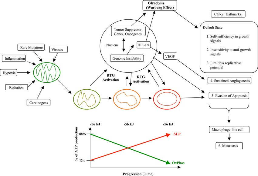
interrelated steps. In order to complete the metastatic and the properties of macrophages including theSeyfried and Shelton Nutrition & Metabolism 2010, 7:7 Page 11 of 22 http://www.nutritionandmetabolism.com/content/7/1/7 invasive and inflammatory properties [193,205,209]. As the non-neoplastic TAM, which are also present in myeloid cells are also part of the immune system, eva- tumors and can facilitate tumor progression sion of immune surveillance would be another expected [190,213,215,216,245]. Poor prognosis is generally asso- characteristic of metastatic cells derived from macro- ciated with those cancers that display characteristics of phage-like cells [205]. Indeed, metastatic melanoma cells macrophages [210,221]. Hence, damage to the respira- can phagocytose live T-cells, which are supposed to kill tory capacity of myeloid or macrophage-like cells would the tumor cells [210]. produce “rogue macrophages” leading to cancers with Fusions among metastatic myeloid cells at the primary the highest metastatic behavior. tumor site could, through reprogramming strategies, The plethora of the cell surface molecules thought to also produce functional epithelial cells at secondary sites participate in metastatic tumor cell behavior are also potentially simulating the histological characteristics of expressed on myeloid cells especially macrophages the original tissue of origin [200,211,212]. The macro- [185,213]. A robust Warburg effect in human metastatic phage fusion hypothesis would also fit with the roles of lesions, detected with combined 18F-fluorodeoxyglucose- hematopoietic stem cells in the metastatic niche positron emission tomography imaging, indicates that [208,213]. While the fusion hypothesis is attractive, it metastatic cells have impaired energy metabolism like would be an exception to the observations showing sup- that of most cancer cells [18,20,246]. Hence, invasion pressed tumorigenicity following hybridization between and metastasis can be linked to impaired energy meta- normal cells and tumor cells [163], though some excep- bolism if this impairment occurs in cells of hematopoie- tions have been reported [205,206]. However, neither tic or myeloid origin. the EMT hypothesis nor the macrophage fusion hypoth- esis link the origin of metastasis to the Warburg effect Connecting the links or to impaired energy metabolism. The path from normal cell physiology to malignant Recent findings of cardiolipin abnormalities in sys- behavior, where all major cancer hallmarks are temic metastatic mouse tumor cells with macrophage expressed, is depicted in Figure 2 and is based on the properties can link metastasis to impaired respiratory evidence reviewed above. Any unspecific condition that function in these cells [73,190,204]. Most tissues contain damages a cell’s oxidative phosphorylation, but is not resident phagocytes as part of their histoarchitecture or severe enough to induce apoptosis, can potentially initi- stroma [214]. Tumor associated macrophages (TAM) ate the path to a malignant cancer. Some of the many also become a major cell type in many cancers [215]. unspecific conditions contributing to carcinogenesis can While TAM can facilitate the invasive and metastatic include inflammation, carcinogens, radiation (ionizing or properties of tumor cells [213,216], metastatic tumor ultraviolet), intermittent hypoxia, rare germline muta- cells can also express several properties of TAM tions, viral infections, and disruption of tissue morpho- [190,204]. genetic fields. Any of these conditions can damage the Damage to the respiratory capacity of resident tissue structure and function of mitochondria thus activating a phagocytes, TAM, or macrophage hybrids would trigger specific RTG response in the damaged cell. If the mito- a RTG response, force a reliance on substrate level chondrial damage persists, the RTG response will per- phosphorylation for energy, and eventually, over time, sist. Uncorrected mitochondrial damage will require a lead to dysregulated growth and genomic instability as continuous compensatory energy response involving described in the general hypothesis. Metastatic behavior substrate level phosphorylation in order to maintain the would be an expected outcome following impaired ΔG’ATP of approximately -56 kJ/mol for cell viability. respiratory function in hematopoietic or myeloid-type Tumor progression is linked to a greater dependence on cells, as macrophages are already mesenchymal cells substrate level phosphorylation, which eventually that embody the capacity to degrade the extracellular becomes irreversible. As the integrity of the nuclear gen- matrix, to enter and to exit tissues from the blood ome is dependent on the efficiency of mitochondrial stream, to migrate through tissues, and to survive in energy production, the continued impairment of mito- hypoxic environments. A sampling of human metastatic chondrial energy production will gradually undermine cancers with properties of macrophage-like cells include nuclear genome integrity leading to a mutator pheno- brain [204,217-220], breast [221-225], lung type and a plethora of somatic mutations. Activation of [202,225-229], skin [203,205,209,210,230-233], gastric oncogenes, inactivation of tumor suppressor genes, and [234], colon [235,236], pancreas [237,238], bladder [239], aneuploidy will be the consequence of protracted mito- kidney [240], ovarian [241,242], and muscle [243,244]. It chondrial dysfunction. These gene abnormalities will is important to mention that these macrophage proper- contribute further to mitochondrial dysfunction while ties are expressed in the tumor cells themselves and are also enhancing those energy pathways needed to up-reg- not to be confused with similar properties expressed in ulate and sustain substrate level phosphorylation. The
You can also read