HHS Public Access Author manuscript Cell Metab. Author manuscript; available in PMC 2018 February 07.
←
→
Page content transcription
If your browser does not render page correctly, please read the page content below
HHS Public Access
                                 Author manuscript
                                 Cell Metab. Author manuscript; available in PMC 2018 February 07.
Author Manuscript

                    Published in final edited form as:
                     Cell Metab. 2017 February 07; 25(2): 262–284. doi:10.1016/j.cmet.2016.12.022.

                    Multi-dimensional roles of ketone bodies in fuel metabolism,
                    signaling, and therapeutics
                    Patrycja Puchalska and Peter A. Crawford1
                    Center for Metabolic Origins of Disease, Sanford Burnham Prebys Medical Discovery Institute,
                    Orlando, Florida, USA
Author Manuscript

                    Abstract
                         Ketone body metabolism is a central node in physiological homeostasis. In this review, we discuss
                         how ketones serve discrete fine-tuning metabolic roles that optimize organ and organism
                         performance in varying nutrient states, and protect from inflammation and injury in multiple organ
                         systems. Traditionally viewed as metabolic substrates enlisted only in carbohydrate restriction,
                         recent observations underscore the importance of ketone bodies as vital metabolic and signaling
                         mediators when carbohydrates are abundant. Complementing a repertoire of known therapeutic
                         options for diseases of the nervous system, prospective roles for ketone bodies in cancer have
                         arisen, as have intriguing protective roles in heart and liver, opening therapeutic options in obesity-
                         related and cardiovascular disease. Controversies in ketone metabolism and signaling are
                         discussed to reconcile classical dogma with contemporary observations.
Author Manuscript

                    Introduction
                                       Ketone bodies are a vital alternative metabolic fuel source for all the domains of life,
                                       eukarya, bacteria, and archaea (Aneja et al., 2002; Cahill GF Jr, 2006; Krishnakumar et al.,
                                       2008). Ketone body metabolism in humans has been leveraged to fuel the brain during
                                       episodic periods of nutrient deprivation. Ketone bodies are interwoven with crucial
                                       mammalian metabolic pathways such as β-oxidation (FAO), the tricarboxylic acid cycle
                                       (TCA), gluconeogenesis, de novo lipogenesis (DNL), and biosynthesis of sterols. In
                                       mammals, ketone bodies are produced predominantly in the liver from FAO-derived acetyl-
                                       CoA, and they are transported to extrahepatic tissues for terminal oxidation. This physiology
                                       provides an alternative fuel that is augmented by relatively brief periods of fasting, which
                                       increases fatty acid availability and diminishes carbohydrate availability (Cahill GF Jr, 2006;
Author Manuscript

                                       McGarry and Foster, 1980; Robinson and Williamson, 1980). Ketone body oxidation

                    1
                     Lead contact: Peter A. Crawford MD, PhD, Director, Cardiovascular Metabolism Program, Associate Professor, Center for Metabolic
                    Origins of Disease, Sanford Burnham Prebys Medical Discovery Institute, 6400 Sanger Rd., Orlando, FL 32827, USA, Tel: +1
                    407-745-2079, pcrawford@sbpdiscovery.org.
                    Publisher's Disclaimer: This is a PDF file of an unedited manuscript that has been accepted for publication. As a service to our
                    customers we are providing this early version of the manuscript. The manuscript will undergo copyediting, typesetting, and review of
                    the resulting proof before it is published in its final citable form. Please note that during the production process errors may be
                    discovered which could affect the content, and all legal disclaimers that apply to the journal pertain.
                    Author Contribution
                    Conceptualization, P.P. and P.A.C.; Methodology, P.P.; Investigation, P.P.; Resources, P.A.C.; Writing – Original Draft, P.P. and P.A.C.;
                    Writing – Review & Editing, P.P. and P.A.C.; Visualization, P.P. and P.A.C.; Supervision, P.A.C.; Funding Acquisition, P.A.C.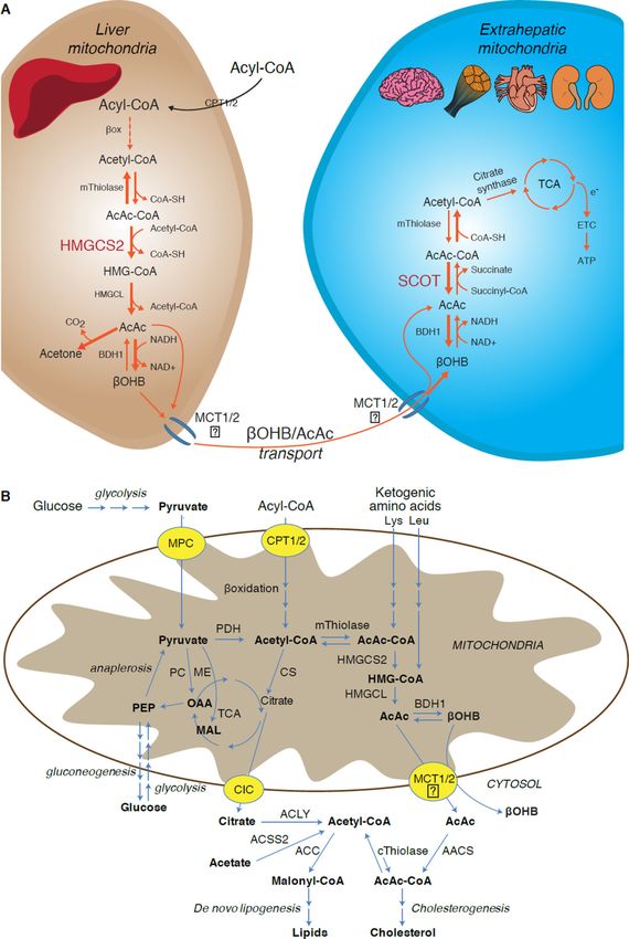
Puchalska and Crawford                                                                                          Page 2

                                        becomes a significant contributor to overall energy mammalian metabolism within
Author Manuscript

                                        extrahepatic tissues in a myriad of physiological states, including fasting, starvation, the
                                        neonatal period, post-exercise, pregnancy, and adherence to low carbohydrate diets.
                                        Circulating total ketone body concentrations in healthy adult humans normally exhibit
                                        circadian oscillations between approximately 100–250 µM, rise to ~1 mM after prolonged
                                        exercise or 24h of fasting, and can accumulate to as high as 20 mM in pathological states
                                        like diabetic ketoacidosis (Cahill GF Jr, 2006; Johnson et al., 1969b; Koeslag et al., 1980;
                                        Robinson and Williamson, 1980; Wildenhoff et al., 1974). The human liver produces up to
                                        300 g of ketone bodies per day (Balasse and Fery, 1989), which contribute between 5–20%
                                        of total energy expenditure in fed, fasted, and starved states (Balasse et al., 1978; Cox et al.,
                                        2016).

                                        Recent studies now highlight imperative roles for ketone bodies in mammalian cell
                                        metabolism, homeostasis, and signaling under a wide variety of physiological and
Author Manuscript

                                        pathological states. Apart from serving as energy fuels for extrahepatic tissues like brain,
                                        heart, or skeletal muscle, ketone bodies play pivotal roles as signaling mediators, drivers of
                                        protein post-translational modification (PTM), and modulators of inflammation and
                                        oxidative stress. In this review, we provide both classical and modern views of the
                                        pleiotropic roles of ketone bodies and their metabolism.

                       Overview of ketone body metabolism
                                        The rate of hepatic ketogenesis is governed by an orchestrated series of physiological and
                                        biochemical transformations of fat. Primary regulators include lipolysis of fatty acids from
                                        triacylglycerols, transport to and across the hepatocyte plasma membrane, transport into
                                        mitochondria via carnitine palmitoyltransferase 1 (CPT1), the β-oxidation spiral, TCA cycle
Author Manuscript

                                        activity and intermediate concentrations, redox potential, and the hormonal regulators of
                                        these processes, predominantly glucagon and insulin [reviewed in (Arias et al., 1995; Ayte et
                                        al., 1993; Ehara et al., 2015; Ferre et al., 1983; Kahn et al., 2005; McGarry and Foster, 1980;
                                        Williamson et al., 1969)]. Classically ketogenesis is viewed as a spillover pathway, in which
                                        β-oxidation-derived acetyl-CoA exceeds citrate synthase activity and/or oxaloacetate
                                        availability for condensation to form citrate. Three-carbon intermediates exhibit anti-
                                        ketogenic activity, presumably due to their ability to expand the oxaloacetate pool for acetyl-
                                        CoA consumption, but hepatic acetyl-CoA concentration alone does not determine ketogenic
                                        rate (Foster, 1967; Rawat and Menahan, 1975; Williamson et al., 1969). The regulation of
                                        ketogenesis by hormonal, transcriptional, and post-translational events together support the
                                        notion that the molecular mechanisms that fine tune ketogenic rate remain incompletely
                                        understood (see Regulation of HMGCS2 and SCOT/OXCT1).
Author Manuscript

                                        Ketogenesis occurs primarily in hepatic mitochondrial matrix at rates proportional to total
                                        fat oxidation. After transport of acyl chains across the mitochondrial membranes and β-
                                        oxidation, the mitochondrial isoform of 3-hydroxymethylglutaryl-CoA synthase (HMGCS2)
                                        catalyzes the fate committing condensation of acetoacetyl-CoA (AcAc-CoA) and acetyl-
                                        CoA to generate HMG-CoA (Fig. 1A). HMG-CoA lyase (HMGCL) cleaves HMG-CoA to
                                        liberate acetyl-CoA and acetoacetate (AcAc), and the latter is reduced to D-β-
                                        hydroxybutyrate (D-βOHB) by phosphatidylcholine-dependent mitochondrial D-βOHB

                                             Cell Metab. Author manuscript; available in PMC 2018 February 07.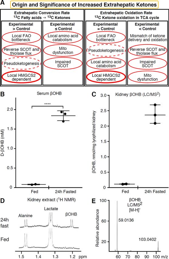
Puchalska and Crawford                                                                                        Page 3

                                        dehydrogenase (BDH1) in a NAD+/NADH-coupled near-equilibrium reaction (Bock and
Author Manuscript

                                        Fleischer, 1975; LEHNINGER et al., 1960). The BDH1 equilibrium constant favors D-
                                        βOHB production, but the ratio of AcAc/D-βOHB ketone bodies is directly proportional to
                                        mitochondrial NAD+/NADH ratio, and thus BDH1 oxidoreductase activity modulates
                                        mitochondrial redox potential (Krebs et al., 1969; Williamson et al., 1967). AcAc can also
                                        spontaneously decarboxylate to acetone (Pedersen, 1929), the source of sweet odor in
                                        humans suffering ketoacidosis (i.e., total serum ketone bodies > ~7 mM; AcAc pKa 3.6,
                                        βOHB pKa 4.7). The mechanisms through which ketone bodies are transported across the
                                        mitochondrial inner membrane are not known, but AcAc/D-βOHB are released from cells
                                        via monocarboxylate transporters (in mammals, MCT 1 and 2, also known as solute carrier
                                        16A family members 1 and 7) and transported in the circulation to extrahepatic tissues for
                                        terminal oxidation (Cotter et al., 2011; Halestrap and Wilson, 2012; Halestrap, 2012; Hugo
                                        et al., 2012). Concentrations of circulating ketone bodies are higher than those in the
Author Manuscript

                                        extrahepatic tissues (Harrison and Long, 1940) indicating ketone bodies are transported
                                        down a concentration gradient. Loss-of-function mutations in MCT1 are associated with
                                        spontaneous bouts of ketoacidosis, suggesting a critical role in ketone body import.

                                        With the exception of potential diversion of ketone bodies into non-oxidative fates (see Non-
                                        oxidative metabolic fates of ketone bodies), hepatocytes lack the ability to metabolize the
                                        ketone bodies they produce. Ketone bodies synthesized de novo by liver are (i) catabolized
                                        in mitochondria of extrahepatic tissues to acetyl-CoA, which is available to the TCA cycle
                                        for terminal oxidation (Fig. 1A), (ii) diverted to the lipogenesis or sterol synthesis pathways
                                        (Fig. 1B), or (iii) excreted in the urine. As an alternative energetic fuel, ketone bodies are
                                        avidly oxidized in heart, skeletal muscle, and brain (Balasse and Fery, 1989; Bentourkia et
                                        al., 2009; Owen et al., 1967; Reichard et al., 1974; Sultan, 1988). Extrahepatic
                                        mitochondrial BDH1 catalyzes the first reaction of βOHB oxidation, converting it to back
Author Manuscript

                                        AcAc (LEHNINGER et al., 1960; Sandermann et al., 1986). A cytoplasmic D-βOHB-
                                        dehydrogenase (BDH2) with only 20% sequence identity to BDH1 has a high Km for ketone
                                        bodies, and also plays a role in iron homeostasis (Davuluri et al., 2016; Guo et al., 2006). In
                                        extrahepatic mitochondrial matrix, AcAc is activated to AcAc-CoA through exchange of a
                                        CoA-moiety from succinyl-CoA in a reaction catalyzed by a unique mammalian CoA
                                        transferase, succinyl-CoA:3-oxoacid-CoA transferase (SCOT, CoA transferase; encoded by
                                        OXCT1), through a near equilibrium reaction. The free energy released by hydrolysis of
                                        AcAc-CoA is greater than that of succinyl-CoA, favoring AcAc formation. Thus ketone
                                        body oxidative flux occurs due to mass action: an abundant supply of AcAc and rapid
                                        consumption of acetyl-CoA through citrate synthase favors AcAc-CoA (+ succinate)
                                        formation by SCOT. Notably, in contrast to glucose (hexokinase) and fatty acids (acyl-CoA
                                        synthetases), the activation of ketone bodies (SCOT) into an oxidizable form does not
Author Manuscript

                                        require the investment of ATP. A reversible AcAc-CoA thiolase reaction [catalyzed by any
                                        of the four mitochondrial thiolases encoded by either ACAA2 (encoding an enzyme known
                                        as T1 or CT), ACAT1 (encoding T2), HADHA, or HADHB] yields two molecules of acetyl-
                                        CoA, which enter the TCA cycle (Hersh and Jencks, 1967; Stern et al., 1956; Williamson et
                                        al., 1971). During ketotic states (i.e., total serum ketones > 500 µM), ketone bodies become
                                        significant contributors to energy expenditure, and are utilized in tissues rapidly until uptake
                                        or saturation of oxidation occurs (Balasse et al., 1978; Balasse and Fery, 1989; Edmond et

                                             Cell Metab. Author manuscript; available in PMC 2018 February 07.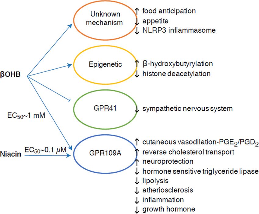
Puchalska and Crawford                                                                                       Page 4

                                        al., 1987). A very small fraction of liver-derived ketone bodies can be readily measured in
Author Manuscript

                                        the urine, and utilization and reabsorption rates by the kidney are proportionate to
                                        circulating concentration (Goldstein, 1987; Robinson and Williamson, 1980). During highly
                                        ketotic states (> 1 mM in plasma), ketonuria serves as a semi-quantitative reporter of ketosis,
                                        although most clinical assays of urine ketone bodies detect AcAc but not βOHB (Klocker et
                                        al., 2013).

                       Ketogenic substrates and their impact on hepatocyte metabolism
                                        Ketogenic substrates include fatty acids and amino acids (Fig. 1B). The catabolism of amino
                                        acids, especially leucine, generates about 4% of ketone bodies in post-absorptive state
                                        (Thomas et al., 1982). Thus the acetyl-CoA substrate pool to generate ketone bodies mainly
                                        derives from fatty acids, because during states of diminished carbohydrate supply, pyruvate
                                        enters the hepatic TCA cycle primarily via anaplerosis, i.e., ATP-dependent carboxylation to
Author Manuscript

                                        oxaloacetate (OAA), or to malate (MAL), and not oxidative decarboxylation to acetyl-CoA
                                        (Jeoung et al., 2012; Magnusson et al., 1991; Merritt et al., 2011). In liver, glucose and
                                        pyruvate contribute negligibly to ketogenesis, even when pyruvate decarboxylation to acetyl-
                                        CoA is maximal (Jeoung et al., 2012).

                                        Acetyl-CoA subsumes several roles integral to hepatic intermediary metabolism beyond
                                        ATP generation via terminal oxidation (also see The integration of ketone body
                                        metabolism, post-translational modification, and cell physiology). Acetyl-CoA
                                        allosterically activates (i) pyruvate carboxylase (PC), thereby activating a metabolic control
                                        mechanism that augments anaplerotic entry of metabolites into the TCA cycle (Owen et al.,
                                        2002; Scrutton and Utter, 1967) and (ii) pyruvate dehydrogenase kinase, which
                                        phosphorylates and inhibits pyruvate dehydrogenase (PDH) (Cooper et al., 1975), thereby
Author Manuscript

                                        further enhancing flow of pyruvate into the TCA cycle via anaplerosis. Furthermore,
                                        cytoplasmic acetyl-CoA, whose pool is augmented by mechanisms that convert
                                        mitochondrial acetyl-CoA to transportable metabolites, inhibits fatty acid oxidation: acetyl-
                                        CoA carboxylase (ACC) catalyzes the conversion of acetyl-CoA to malonyl-CoA, the
                                        lipogenic substrate and allosteric inhibitor of mitochondrial CPT1 [reviewed in (Kahn et al.,
                                        2005; McGarry and Foster, 1980)]. Thus, the mitochondrial acetyl-CoA pool both regulates
                                        and is regulated by the spillover pathway of ketogenesis, which orchestrates key aspects of
                                        hepatic intermediary metabolism.

                       Non-oxidative metabolic fates of ketone bodies
                                        The predominant fate of liver derived ketones is SCOT-dependent extrahepatic oxidation.
                                        However, AcAc can be exported from mitochondria and utilized in anabolic pathways via
Author Manuscript

                                        conversion to AcAc-CoA by an ATP-dependent reaction catalyzed by cytoplasmic
                                        acetoacetyl-CoA synthetase (AACS, Fig. 1B). This pathway is active during brain
                                        development and in lactating mammary gland (Morris, 2005; Robinson and Williamson,
                                        1978; Ohgami et al., 2003). AACS is also highly expressed in adipose tissue, and activated
                                        osteoclasts (Aguilo et al., 2010; Yamasaki et al., 2016). Cytoplasmic AcAc-CoA can be
                                        either directed by cytosolic HMGCS1 toward sterol biosynthesis, or cleaved by either of two
                                        cytoplasmic thiolases to acetyl-CoA (ACAA1 and ACAT2), carboxylated to malonyl-CoA,

                                             Cell Metab. Author manuscript; available in PMC 2018 February 07.
Puchalska and Crawford                                                                                      Page 5

                                        and contribute to the synthesis of fatty acids (Bergstrom et al., 1984; Edmond, 1974;
Author Manuscript

                                        Endemann et al., 1982; Geelen et al., 1983; Webber and Edmond, 1977).

                                        While the physiological significance is yet to be established, ketones can serve as anabolic
                                        substrates even in the liver. In artificial experimental contexts, AcAc can contribute to as
                                        much as half of newly synthesized lipid, and up to 75% of new synthesized cholesterol
                                        (Endemann et al., 1982; Geelen et al., 1983; Freed et al., 1988). Because AcAc is derived
                                        from incomplete hepatic fat oxidation, the ability of AcAc to contribute to lipogenesis in
                                        vivo would imply hepatic futile cycling, where fat-derived ketones can be utilized for lipid
                                        production, a notion whose physiological significance requires experimental validation, but
                                        could serve adaptive or maladaptive roles (Solinas et al., 2015). AcAc avidly supplies
                                        cholesterogenesis, with a low AACS Km-AcAc (~50 µM) favoring AcAc activation even in
                                        the fed state (Bergstrom et al., 1984). The dynamic role of cytoplasmic ketone metabolism
                                        has been suggested in primary mouse embryonic neurons and in 3T3-L1 derived-adipocytes,
Author Manuscript

                                        as AACS knockdown impaired differentiation of each cell type (Hasegawa et al., 2012a;
                                        Hasegawa et al., 2012b). Knockdown of AACS in mice in vivo decreased serum cholesterol
                                        (Hasegawa et al., 2012c). SREBP-2, a master transcriptional regulator of cholesterol
                                        biosynthesis, and peroxisome proliferator activated receptor (PPAR)-γ are AACS
                                        transcriptional activators, and regulate its transcription during neurite development and in
                                        the liver (Aguilo et al., 2010; Hasegawa et al., 2012c). Taken together, cytoplasmic ketone
                                        body metabolism may be important in select conditions or disease natural histories, but are
                                        inadequate to dispose of liver derived ketone bodies, as massive hyperketonemia occurs in
                                        the setting of selective impairment of the primary oxidative fate via loss of function
                                        mutations to SCOT (Berry et al., 2001; Cotter et al., 2011).

                       Regulation of HMGCS2 and SCOT/OXCT1
Author Manuscript

                                        The divergence of a mitochondrial from the gene encoding cytosolic HMGCS occurred early
                                        in vertebrate evolution due to the need to support hepatic ketogenesis in species with higher
                                        brain to body weight ratios (Boukaftane et al., 1994; Cunnane and Crawford, 2003).
                                        Naturally occurring loss-of-function HMGCS2 mutations in humans cause bouts of
                                        hypoketotic hypoglycemia (Pitt et al., 2015; Thompson et al., 1997). Robust HMGCS2
                                        expression is restricted to hepatocytes and colonic epithelium, and its expression and
                                        enzymatic activity are coordinated through diverse mechanisms (Mascaro et al., 1995;
                                        McGarry and Foster, 1980; Robinson and Williamson, 1980). While the full scope of
                                        physiological states that influence HMGCS2 requires further elucidation, its expression
                                        and/or activity is regulated during the early postnatal period, aging, diabetes, starvation or
                                        ingestion of ketogenic diet (Balasse and Fery, 1989; Cahill GF Jr, 2006; Girard et al., 1992;
Author Manuscript

                                        Hegardt, 1999; Satapati et al., 2012; Sengupta et al., 2010). In the fetus, methylation of 5’
                                        flanking region of Hmgcs2 gene inversely correlates with its transcription, and is partially
                                        reversed after birth (Arias et al., 1995; Ayte et al., 1993; Ehara et al., 2015; Ferre et al.,
                                        1983). Similarly, hepatic Bdh1 exhibits a developmental expression pattern, increasing from
                                        birth to weaning, and is also induced by ketogenic diet in a fibroblast growth factor
                                        (FGF)-21-dependent manner (Badman et al., 2007; Zhang et al., 1989). Ketogenesis in
                                        mammals is highly responsive to both insulin and glucagon, being suppressed and
                                        stimulated, respectively (McGarry and Foster, 1977). Insulin suppresses adipose tissue

                                             Cell Metab. Author manuscript; available in PMC 2018 February 07.
Puchalska and Crawford Page 6
lipolysis, thus depriving ketogenesis of its substrate, while glucagon increases ketogenic flux
Author Manuscript
though a direct effect on the liver (Hegardt, 1999). Hmgcs2 transcription is stimulated by
forkhead transcriptional factor FOXA2, which is inhibited via insulin-
phosphatidylinositol-3-kinase/Akt, and is induced by glucagon-cAMP-p300 signaling (Arias
et al., 1995; Hegardt, 1999; Quant et al., 1990; Thumelin et al., 1993; von Meyenn et al.,
2013; Wolfrum et al., 2004; Wolfrum et al., 2003). PPARα (Rodriguez et al., 1994) together
with its target, FGF21 (Badman et al., 2007) also induce Hmgcs2 transcription in the liver
during starvation or administration of ketogenic diet (Badman et al., 2007; Inagaki et al.,
2007). Induction of PPARα may occur before the transition from fetal to neonatal
physiology, while FGF21 activation may be favored in the early neonatal period via βOHB-
mediated inhibition of histone deacetylase (HDAC)-3 (Rando et al., 2016). mTORC1
(mammalian target of rapamycin complex 1) dependent inhibition of PPARα transcriptional
activity is also a key regulator of Hmgcs2 gene expression (Sengupta et al., 2010), and liver
Author Manuscript
PER2, a master circadian oscillator, indirectly regulates Hmgcs2 expression (Chavan et al.,
2016). Recent observations indicate that extrahepatic tumor-induced interleukin-6 impairs
ketogenesis via PPARα suppression (Flint et al., 2016). Despite these observations, it is
important to note that physiological shifts in Hmgcs2 gene expression have not been
mechanistically linked to HMGCS2 protein abundance or to variations of ketogenic rate.
HMGCS2 enzyme activity is regulated through multiple PTMs. HMGCS2 serine
phosphorylation enhanced its activity in vitro (Grimsrud et al., 2012). HMGCS2 activity is
allosterically inhibited by succinyl-CoA and lysine residue succinylation (Arias et al., 1995;
Hegardt, 1999; Lowe and Tubbs, 1985; Quant et al., 1990; Rardin et al., 2013; Reed et al.,
1975; Thumelin et al., 1993). Succinylation of HMGCS2, HMGCL, and BDH1 lysine
residues in hepatic mitochondria are targets of the NAD+ dependent deacylase sirtuin 5
(SIRT5) (Rardin et al., 2013). HMGCS2 activity is also enhanced by SIRT3 lysine
Author Manuscript
deacetylation, and it is possible that crosstalk between acetylation and succinylation
regulates HMGCS2 activity (Rardin et al., 2013; Shimazu et al., 2013). Despite the ability of
these PTMs to regulate HMGCS2 Km and Vmax, fluctuations of these PTMs have not yet
been carefully mapped and have not been confirmed as mechanistic drivers of ketogenesis in
vivo.
SCOT is expressed in all mammalian cells that harbor mitochondria, except those of
hepatocytes. The importance of SCOT activity and ketolysis was demonstrated in SCOT-KO
mice, which exhibited uniform lethality due to hyperketonemic hypoglycemia within 48h
after birth (Cotter et al., 2011). Tissue-specific loss of SCOT in neurons or skeletal myocytes
induces metabolic abnormalities during starvation but is not lethal (Cotter et al., 2013b). In
humans, SCOT deficiency presents early in life with severe ketoacidosis, causing lethargy,
Author Manuscript
vomiting, and coma (Berry et al., 2001; Fukao et al., 2000; Kassovska-Bratinova et al.,
1996; Niezen-Koning et al., 1997; Saudubray et al., 1987; Snyderman et al., 1998; Tildon
and Cornblath, 1972). Relatively little is known at the cellular level about SCOT gene and
protein expression regulators. Oxct1 mRNA expression and SCOT protein and activity are
diminished in ketotic states, possibly through PPAR-dependent mechanisms (Fenselau and
Wallis, 1974; Fenselau and Wallis, 1976; Grinblat et al., 1986; Okuda et al., 1991; Turko et
al., 2001; Wentz et al., 2010). In diabetic ketoacidosis, the mismatch between hepatic
ketogenesis and extrahepatic oxidation becomes exacerbated by impairment of SCOT
Cell Metab. Author manuscript; available in PMC 2018 February 07.Puchalska and Crawford Page 7
activity. Overexpression of insulin independent glucose transporter (GLUT1/SLC2A1) in
Author Manuscript
cardiomyocytes also inhibits Oxct1 gene expression and downregulates ketones terminal
oxidation in a non-ketotic state (Yan et al., 2009). In liver, Oxct1 mRNA abundance is
suppressed by microRNA-122 and histone methylation H3K27me3 that are evident during
the transition from fetal to the neonatal period (Thorrez et al., 2011). However, suppression
of hepatic Oxct1 expression in the postnatal period is primarily attributable to the evacuation
of Oxct1-expressing hematopoietic progenitors from the liver, rather than a loss of
previously existing Oxct1 expression in terminally differentiated hepatocytes. In fact,
expression of Oxct1 mRNA and SCOT protein in differentiated hepatocytes are extremely
low (Orii et al., 2008).
SCOT is also regulated by PTMs. The enzyme is hyper-acetylated in brains of SIRT3 KO
mice, which also exhibit diminished AcAc dependent acetyl-CoA production (Dittenhafer-
Reed et al., 2015). Non-enzymatic nitration of tyrosine residues of SCOT also attenuates its
Author Manuscript
activity, which has been reported in hearts of various diabetic mice models (Marcondes et
al., 2001; Turko et al., 2001; Wang et al., 2010a). In contrast, tryptophan residue nitration
augments SCOT activity (Brégère et al., 2010; Rebrin et al., 2007). Molecular mechanisms
of residue-specific nitration or de-nitration designed to modulate SCOT activity may exist
and require elucidation.
Controversies in extrahepatic ketogenesis
In mammals the primary ketogenic organ is liver, and only hepatocytes and gut epithelial
cells abundantly express the mitochondrial isoform of HMGCS2 (Cotter et al., 2013a; Cotter
et al., 2014; McGarry and Foster, 1980; Robinson and Williamson, 1980). Anaerobic
bacterial fermentation of complex polysaccharides yields butyrate, which is absorbed by
Author Manuscript
colonocytes in mammalians for terminal oxidation or ketogenesis (Cherbuy et al., 1995),
which may play a role in colonocyte differentiation (Wang et al., 2016). Excluding gut
epithelial cells and hepatocytes, HMGCS2 is nearly absent in almost all other mammalian
cells, but the prospect of extrahepatic ketogenesis has been raised in tumor cells, astrocytes
of the central nervous system, the kidney, pancreatic β cells, retinal pigment epithelium
(RPE), and even in skeletal muscle (Adijanto et al., 2014; Avogaro et al., 1992; El Azzouny
et al., 2016; Grabacka et al., 2016; Kang et al., 2015; Le Foll et al., 2014; Nonaka et al.,
2016; Takagi et al., 2016a; Thevenet et al., 2016; Zhang et al., 2011). Ectopic HMGCS2 has
been observed in tissues that lack net ketogenic capacity (Cook et al., 2016; Wentz et al.,
2010), and HMGCS2 exhibits prospective ketogenesis-independent ‘moonlighting’
activities, including within the cell nucleus (Chen et al., 2016; Kostiuk et al., 2010; Meertens
et al., 1998).
Author Manuscript
Any extrahepatic tissue that oxidizes ketone bodies also has the potential to accumulate
ketone bodies via HMGCS2 independent mechanisms (Fig. 2A). However, there is no
extrahepatic tissue in which a steady state ketone body concentration exceeds that in the
circulation (Cotter et al., 2011; Cotter et al., 2013b; Harrison and Long, 1940), underscoring
that ketone bodies are transported down a concentration gradient via MCT1/2-dependent
mechanisms. One mechanism of apparent extrahepatic ketogenesis may actually reflect
relative impairment of ketone oxidation. Additional potential explanations fall within the
Cell Metab. Author manuscript; available in PMC 2018 February 07.Puchalska and Crawford Page 8
realm of ketone body formation. First, de novo ketogenesis may occur via reversible
Author Manuscript
enzymatic activity of thiolase and SCOT (Weidemann and Krebs, 1969). When the
concentration of acetyl-CoA is relatively high, reactions normally responsible for AcAc
oxidation operate in the reverse direction (GOLDMAN, 1954). A second mechanism occurs
when β-oxidation-derived intermediates accumulate due to a TCA cycle bottleneck, AcAc-
CoA is converted to L-βOHB-CoA through a reaction catalyzed by mitochondrial 3-
hydroxyacyl-CoA dehydrogenase, and further by 3-hydroxybutyryl CoA deacylase to L-
βOHB, which is indistinguishable by mass spectrometry or resonance spectroscopy from the
physiological enantiomer D-βOHB (Reed and Ozand, 1980). L-βOHB can be
chromatographically or enzymatically distinguished from D-βOHB, and is present in
extrahepatic tissues, but not in liver or blood (Hsu et al., 2011). Hepatic ketogenesis
produces only D-βOHB, the only enantiomer that is a BDH substrate (Ito et al., 1984;
Lincoln et al., 1987; Reed and Ozand, 1980; Scofield et al., 1982; Scofield et al., 1982). A
Author Manuscript
third HMGCS2-independent mechanism generates D-βOHB through amino acid catabolism,
particularly that of leucine and lysine. A fourth mechanism is only apparent because it is due
to a labeling artifact and is thus termed pseudoketogenesis. This phenomenon is attributable
to the reversibility of the SCOT and thiolase reactions, and can cause overestimation of
ketone body turnover due to the isotopic dilution of ketone body tracer in extrahepatic tissue
(Des Rosiers et al., 1990; Fink et al., 1988). Nonetheless, pseudoketogenesis may be
negligible in most contexts (Bailey et al., 1990; Keller et al., 1978). A schematic (Fig. 2A)
indicates a useful approach to apply while considering elevated tissue steady state
concentration of ketones.
Kidney has recently received attention as a potentially ketogenic organ. In the vast majority
of states, the kidney is a net consumer of liver-derived ketone bodies, excreting or
reabsorbing ketone bodies from the bloodstream, and kidney is generally not a net ketone
Author Manuscript
body generator or concentrator (Robinson and Williamson, 1980). The authors of a classical
study concluded that minimal renal ketogenesis quantified in an artificial experimental
system was not physiologically relevant (Weidemann and Krebs, 1969). Recently, renal
ketogenesis has been inferred in diabetic and autophagy deficient mouse models, but it is
more likely that multi-organ shifts in metabolic homeostasis alter integrative ketone
metabolism through inputs on multiple organs (Takagi et al., 2016a; Takagi et al., 2016b;
Zhang et al., 2011). One recent publication suggested renal ketogenesis as a protective
mechanism against ischemia-reperfusion injury in the kidney (Tran et al., 2016). Absolute
steady state concentrations of βOHB from extracts of mice renal tissue were reported at ~4–
12 mM. To test whether this was tenable, we quantified βOHB concentrations in renal
extracts from fed and 24h fasted mice. Serum βOHB concentrations increased from ~100
µM to 2 mM with 24h fasting (Fig. 2B), while renal steady state βOHB concentrations
Author Manuscript
approximate 100 µM in the fed state, and only 1 mM in the 24h fasted state (Fig. 2C–E),
observations that are consistent with concentrations quantified over 45 years ago (Hems and
Brosnan, 1970). It remains possible that in ketotic states, liver-derived ketone bodies could
be renoprotective, but evidence for renal ketogenesis requires further substantiation.
Compelling evidence that supports true extrahepatic ketogenesis was presented in RPE
(Adijanto et al., 2014). This intriguing metabolic transformation was suggested to
Cell Metab. Author manuscript; available in PMC 2018 February 07.Puchalska and Crawford Page 9
potentially allow RPE-derived ketones to flow to photoreceptor or Müller glia cells, which
Author Manuscript
could aid in the regeneration of photoreceptor outer segment.
βOHB as a signaling mediator
Although they are energetically rich, ketone bodies exert provocative ‘non-canonical’
signaling roles in cellular homeostasis (Fig. 3) (Newman and Verdin, 2014; Rojas-Morales et
al., 2016). For example, βOHB inhibits Class I HDACs, which increases histone acetylation
and thereby induces the expression of genes that curtail oxidative stress (Shimazu et al.,
2013). βOHB itself is a histone covalent modifier at lysine residues in livers of fasted or
streptozotocin induced diabetic mice (Xie et al., 2016) (also see below, The integration of
ketone body metabolism, post-translational modification, and cell physiology, and
Ketone bodies, oxidative stress, and neuroprotection).
Author Manuscript
βOHB is also an effector via G-protein coupled receptors. Through unclear molecular
mechanisms, it suppresses sympathetic nervous system activity and reduces total energy
expenditure and heart rate by inhibiting short chain fatty acid signaling through G protein
coupled receptor 41 (GPR41) (Kimura et al., 2011). One of the most studied signaling
effects of βOHB proceeds through GPR109A (also known as HCAR2), a member of the
hydrocarboxylic acid GPCR sub-family expressed in adipose tissues (white and brown)
(Tunaru et al., 2003), and in immune cells (Ahmed et al., 2009). βOHB is the only known
endogenous ligand of GPR109A receptor (EC50 ~770 µM) activated by D-βOHB, L-βOHB,
and butyrate, but not AcAc (Taggart et al., 2005). The high concentration threshold for
GPR109A activation is achieved through adherence to a ketogenic diet, starvation, or during
ketoacidosis, leading to inhibition of adipose tissue lipolysis. The anti-lipolytic effect of
GPR109A proceeds though inhibition of adenylyl cyclase and decreased cAMP, inhibiting
Author Manuscript
hormone sensitive triglyceride lipase (Ahmed et al., 2009; Tunaru et al., 2003). This creates
a negative feedback loop in which ketosis places a modulatory brake on ketogenesis by
diminishing the release of non-esterified fatty acids from adipocytes (Ahmed et al., 2009;
Taggart et al., 2005), an effect that can be counterbalanced by the sympathetic drive that
stimulates lipolysis. Niacin (vitamin B3, nicotinic acid) is a potent (EC50 ~ 0.1 µM) ligand
for GRP109A, effectively employed for decades for dyslipidemias (Benyo et al., 2005;
Benyo et al., 2006; Fabbrini et al., 2010a; Lukasova et al., 2011; Tunaru et al., 2003). While
niacin enhances reverse cholesterol transport in macrophages and reduces atherosclerotic
lesions (Lukasova et al., 2011), the effects of βOHB on atherosclerotic lesions remain
unknown. Although GPR109A receptor exerts protective roles, and intriguing connections
exist between ketogenic diet use in stroke and neurodegenerative diseases (Fu et al., 2015;
Rahman et al., 2014), a protective role of βOHB via GPR109A has not been demonstrated in
Author Manuscript
vivo.
Finally, βOHB may influence appetite and satiety. A meta-analysis of studies that measured
the effects of ketogenic and very low energy diets concluded that participants consuming
these diets exhibit higher satiety, compared to control diets (Gibson et al., 2015). However, a
plausible explanation for this effect is the additional metabolic or hormonal elements that
might modulate appetite. For example, mice maintained on a rodent ketogenic diet exhibited
increased energy expenditure compared to chow control-fed mice, despite similar caloric
Cell Metab. Author manuscript; available in PMC 2018 February 07.Puchalska and Crawford Page 10
intake, and circulating leptin or genes of peptides regulating feeding behavior were not
Author Manuscript
changed (Kennedy et al., 2007). Among proposed mechanisms that suggest appetite
suppression by βOHB includes both signaling and oxidation (Laeger et al., 2010).
Hepatocyte specific deletion of circadian rhythm gene (Per2), and chromatin
immunoprecipitation studies revealed that PER2 directly activates the Cpt1a gene, and
indirectly regulates Hmgcs2, together leading to impaired ketosis in Per2 knockout mice
(Chavan et al., 2016). These mice exhibited impaired food anticipation, which was partially
restored by systemic βOHB administration. Future studies will be needed to confirm the
central nervous system as a direct βOHB target, and whether ketone oxidation is required for
the observed effects, or whether another signaling mechanism is involved. Other
investigators have invoked the possibility of local astrocyte-derived ketogenesis within the
ventromedial hypothalamus as a regulator of food intake, but these preliminary observations
also will benefit from genetic and flux-based assessments (Le Foll et al., 2014). The
Author Manuscript
relationship between ketosis and nutrient deprivation remains of interest because hunger and
satiety are important elements in failed weight loss attempts.
Integration of ketone body metabolism, post-translational modification, and
cell physiology
Ketone bodies contribute to compartmentalized pools of acetyl-CoA, a key intermediate that
exhibits prominent roles in cellular metabolism (Pietrocola et al., 2015). One role of acetyl-
CoA is to serve as a substrate for acetylation, an enzymatically-catalyzed histone covalent
modification (Choudhary et al., 2014; Dutta et al., 2016; Fan et al., 2015; Menzies et al.,
2016). A large number of dynamically acetylated mitochondrial proteins, many of which
may occur through non-enzymatic mechanisms, have also emerged from computational
proteomics studies (Dittenhafer-Reed et al., 2015; Hebert et al., 2013; Rardin et al., 2013;
Author Manuscript
Shimazu et al., 2010). Lysine deacetylases use a zinc cofactor (e.g., nucleocytosolic HDACs)
or NAD+ as co-substrate (sirtuins, SIRTs) (Choudhary et al., 2014; Menzies et al., 2016).
The acetylproteome serves as both sensor and effector of the total cellular acetyl-CoA pool,
as physiological and genetic manipulations each result in non-enzymatic global variations of
acetylation (Weinert et al., 2014). As intracellular metabolites serve as modulators of lysine
residue acetylation, it is important to consider the role of ketone bodies, whose abundance is
highly dynamic.
βOHB is an epigenetic modifier through at least two mechanisms. Increased βOHB levels
induced by fasting, caloric restriction, direct administration or prolonged exercise provoke
HDAC inhibition or histone acetyltransferase activation (Marosi et al., 2016; Sleiman et al.,
2016) or to oxidative stress (Shimazu et al., 2013). βOHB inhibition of HDAC3 could
Author Manuscript
regulate newborn metabolic physiology (Rando et al., 2016). Independently, βOHB itself
directly modifies histone lysine residues (Xie et al., 2016). Prolonged fasting, or
steptozotocin-induced diabetic ketoacidosis increased histone β-hydroxybutyrylation.
Although the number of lysine β-hydroxybutyrylation and acetylation sites was comparable,
stoichiometrically greater histone β-hydroxybutyrylation than acetylation was observed.
Distinct genes were impacted by histone lysine β-hydroxybutyrylation, versus acetylation or
methylation, suggesting distinct cellular functions. Whether β-hydroxybutyrylation is
Cell Metab. Author manuscript; available in PMC 2018 February 07.Puchalska and Crawford Page 11
spontaneous or enzymatic is not known, but expands the range of mechanisms through
Author Manuscript
ketone bodies dynamically influence transcription.
Essential cell reprogramming events during caloric restriction and nutrient deprivation may
be mediated in SIRT3- and SIRT5-dependent mitochondrial deacetylation and
desuccinylation, respectively, regulating ketogenic and ketolytic proteins at post-
translational level in liver and extrahepatic tissues (Dittenhafer-Reed et al., 2015; Hebert et
al., 2013; Rardin et al., 2013; Shimazu et al., 2010). Even though stoichiometric comparison
of occupied sites does not necessarily link directly to shifts in metabolic flux, mitochondrial
acetylation is dynamic and may be driven by acetyl-CoA concentration or mitochondrial pH,
rather than enzymatic acetyltransferases (Wagner and Payne, 2013). That SIRT3 and SIRT5
modulate activities of ketone body metabolizing enzymes provokes the question of the
reciprocal role of ketones in sculpting the acetylproteome, succinylproteome, and other
dynamic cellular targets. Indeed, as variations of ketogenesis reflect NAD+ concentrations,
Author Manuscript
ketone production and abundance could regulate sirtuin activity, thereby influencing total
acetyl-CoA/succinyl-CoA pools, the acylproteome, and thus mitochondrial and cell
physiology. β-hydroxybutyrylation of enzyme lysine residues could add another layer to
cellular reprogramming. In extrahepatic tissues, ketone body oxidation may stimulate
analogous changes in cell homeostasis. While compartmentation of acetyl-CoA pools is
highly regulated and coordinates a broad spectrum of cellular changes, the ability of ketone
bodies to directly shape both mitochondrial and cytoplasmic acetyl-CoA concentrations
requires elucidation (Chen et al., 2012; Corbet et al., 2016; Pougovkina et al., 2014; Schwer
et al., 2009; Wellen and Thompson, 2012). Because acetyl-CoA concentrations are tightly
regulated, and acetyl-CoA is membrane impermeant, it is crucial to consider the driver
mechanisms coordinating acetyl-CoA homeostasis, including the rates of production and
terminal oxidation in the TCA cycle, conversion into ketone bodies, mitochondrial efflux via
Author Manuscript
carnitine acetyltransferase (CrAT), or acetyl-CoA export to cytosol after conversion to citrate
and release by ATP citrate lyase (ACLY). The key roles of these latter mechanisms in cell
acetylproteome and homeostasis require matched understanding of the roles of ketogenesis
and ketone oxidation (Das et al., 2015; McDonnell et al., 2016; Moussaieff et al., 2015;
Overmyer et al., 2015; Seiler et al., 2014; Seiler et al., 2015; Wellen et al., 2009; Wellen and
Thompson, 2012). Convergent technologies in metabolomics and acylproteomics in the
setting of genetically manipulated models will be required to specify targets and outcomes.
Anti- and pro-inflammatory responses to ketone bodies
Ketosis and ketone bodies modulate inflammation and immune cell function, but varied and
even discrepant mechanisms have been posed. Prolonged nutrient deprivation reduces
Author Manuscript
inflammation (Youm et al., 2015), but the chronic ketosis of type 1 diabetes is a pro-
inflammatory state (Jain et al., 2002; Kanikarla-Marie and Jain, 2015; Kurepa et al., 2012).
Mechanism-based signaling roles for βOHB in inflammation emerge because many immune
system cells, including macrophages or monocytes, abundantly express GPR109A. While
βOHB exerts a predominantly anti-inflammatory response (Fu et al., 2014; Gambhir et al.,
2012; Rahman et al., 2014; Youm et al., 2015), high concentrations of ketone bodies,
particularly AcAc, may trigger a pro-inflammatory response (Jain et al., 2002; Kanikarla-
Marie and Jain, 2015; Kurepa et al., 2012).
Cell Metab. Author manuscript; available in PMC 2018 February 07.Puchalska and Crawford Page 12
Anti-inflammatory roles of GPR109A ligands in atherosclerosis, obesity, inflammatory
Author Manuscript
bowel disease, neurological disease, and cancer have been reviewed (Graff et al., 2016).
GPR109A expression is augmented in RPE cells of diabetic models, human diabetic patients
(Gambhir et al., 2012), and in microglia during neurodegeneration (Fu et al., 2014). Anti-
inflammatory effects of βOHB are enhanced by GPR109A overexpression in RPE cells, and
abrogated by pharmacological inhibition or genetic knockout of GPR109A (Gambhir et al.,
2012). βOHB and exogenous nicotinic acid (Taggart et al., 2005), both confer anti-
inflammatory effects in TNFα or LPS-induced inflammation by decreasing the levels of pro-
inflammatory proteins (iNOS, COX-2), or secreted cytokines (TNFα, IL-1β, IL-6, CCL2/
MCP-1), in part through inhibiting NF-κB translocation (Fu et al., 2014; Gambhir et al.,
2012). βOHB decreases ER stress and the NLRP3 inflammasome, activating the
antioxidative stress response (Bae et al., 2016; Youm et al., 2015). However, in
neurodegenerative inflammation, GPR109A-dependent βOHB-mediated protection does not
Author Manuscript
involve inflammatory mediators like MAPK pathway signaling (e.g., ERK, JNK, p38) (Fu et
al., 2014), but may require COX-1-dependent PGD2 production (Rahman et al., 2014). It is
intriguing that macrophage GPR109A is required to exert a neuroprotective effect in an
ischemic stroke model (Rahman et al., 2014), but the ability of βOHB to inhibit the NLRP3
inflammasome in bone marrow derived macrophages is GPR109A independent (Youm et al.,
2015). Although most studies link βOHB to anti-inflammatory effects, βOHB may be pro-
inflammatory and increase markers of lipid peroxidation in calf hepatocytes (Shi et al.,
2014). Anti- versus pro- inflammatory effects of βOHB may thus depend on cell type,
βOHB concentration, exposure duration, and the presence or absence of co-modulators.
Unlike βOHB, AcAc may activate pro-inflammatory signaling. Elevated AcAc, especially
with a high glucose concentration, intensifies endothelial cell injury through an NADPH
oxidase/oxidative stress dependent mechanism (Kanikarla-Marie and Jain, 2015). High
Author Manuscript
AcAc concentrations in umbilical cord of diabetic mothers were correlated with higher
protein oxidation rate and MCP-1 concentration (Kurepa et al., 2012). High AcAc in
diabetic patients was correlated with TNFα expression (Jain et al., 2002), and AcAc, but not
βOHB, induced TNFα, MCP-1 expression, ROS accumulation, and diminished cAMP level
in U937 human monocyte cells (Jain et al., 2002; Kurepa et al., 2012).
Ketone body dependent signaling phenomena are frequently triggered only with high ketone
body concentrations (> 5 mM), and in the case of many studies linking ketones to pro- or
anti-inflammatory effects, through unclear mechanisms. In addition, due to the contradictory
effects of βOHB versus AcAc on inflammation, and the ability of AcAc/βOHB ratio to
influence mitochondrial redox potential, the best experiments assessing the roles of ketone
bodies on cellular phenotypes compare the effects of AcAc and βOHB in varying ratios, and
Author Manuscript
at varying cumulative concentrations [e.g., (Saito et al., 2016)]. Finally, AcAc can be
purchased commercially only as a lithium salt or as an ethyl ester that requires base
hydrolysis before use. Lithium cation independently induces signal transduction cascades
(Manji et al., 1995), and AcAc anion is labile. Finally, studies using racemic D/L-βOHB can
be confounded, as only the D-βOHB stereoisomer can be oxidized to AcAc, but D-βOHB
and L-βOHB can each signal through GPR109A, inhibit the NLRP3 inflammasome, and
serve as lipogenic substrates.
Cell Metab. Author manuscript; available in PMC 2018 February 07.Puchalska and Crawford Page 13
Ketone bodies, oxidative stress, and neuroprotection
Author Manuscript
Oxidative stress is typically defined as a state in which ROS are presented in excess, due to
excessive production and/or impaired elimination. Antioxidant and oxidative stress
mitigating roles of ketone bodies have been widely described both in vitro and in vivo,
particularly in the context of neuroprotection. As most neurons do not effectively generate
high-energy phosphates from fatty acids, but do oxidize ketone bodies when carbohydrates
are in short supply, neuroprotective effects of ketone bodies are especially important (Cahill
GF Jr, 2006; Edmond et al., 1987; Yang et al., 1987). In oxidative stress models, BDH1
induction and SCOT suppression suggest that ketone body metabolism can be
reprogrammed to sustain diverse cell signaling, redox potential, or metabolic requirements
(Nagao et al., 2016; Tieu et al., 2003).
Ketone bodies decrease the grades of cellular damage, injury, death and lower apoptosis in
Author Manuscript
neurons and cardiomyocytes (Haces et al., 2008; Maalouf et al., 2007; Nagao et al., 2016;
Tieu et al., 2003). Invoked mechanisms are varied and not always linearly related to
concentration. Low millimolar concentrations of (D or L)-βOHB scavenge ROS (hydroxyl
anion), while AcAc scavenges numerous ROS species, but only at concentrations that
exceed the physiological range (IC50 20–67 mM) (Haces et al., 2008). Conversely, a
beneficial influence over the electron transport chain’s redox potential is a mechanism
commonly linked to D-βOHB. While all three ketone bodies (D/L-βOHB and AcAc) reduced
neuronal cell death and ROS accumulation triggered by chemical inhibition of glycolysis,
only D-βOHB and AcAc prevented neuronal ATP decline. Conversely, in a hypoglycemic in
vivo model, (D or L)-βOHB, but not AcAc prevented hippocampal lipid peroxidation (Haces
et al., 2008; Maalouf et al., 2007; Marosi et al., 2016; Murphy, 2009; Tieu et al., 2003). In
vivo studies of mice fed a ketogenic diet (87% kcal fat and 13% protein) exhibited
Author Manuscript
neuroanatomical variation of antioxidant capacity (Ziegler et al., 2003), where the most
profound changes were observed in hippocampus, with increase glutathione peroxidase and
total antioxidant capacities.
Ketogenic diet, ketone esters (also see Therapeutic use of ketogenic diet and exogenous
ketone bodies), or βOHB administration exert neuroprotection in models of ischemic stroke
(Rahman et al., 2014); Parkinson’s disease (Tieu et al., 2003); central nervous system
oxygen toxicity seizure (D'Agostino et al., 2013); epileptic spasms (Yum et al., 2015);
mitochondrial encephalomyopathy, lactic acidosis and stroke-like (MELAS) episodes
syndrome (Frey et al., 2016) and Alzheimer’s disease (Cunnane and Crawford, 2003; Yin et
al., 2016). Conversely, a recent report demonstrated histopathological evidence of
neurodegenerative progression by a ketogenic diet in a transgenic mouse model of abnormal
Author Manuscript
mitochondrial DNA repair, despite increases in mitochondrial biogenesis and antioxidant
signatures (Lauritzen et al., 2016). Other conflicting reports suggest that exposure to high
ketone body concentrations elicits oxidative stress. High βOHB or AcAc doses induced
nitric oxide secretion, lipid peroxidation, reduced expression of SOD, glutathione peroxidase
and catalase in calf hepatocytes, while in rat hepatocytes the MAPK pathway induction was
attributed to AcAc but not βOHB (Abdelmegeed et al., 2004; Shi et al., 2014; Shi et al.,
2016).
Cell Metab. Author manuscript; available in PMC 2018 February 07.Puchalska and Crawford Page 14
Taken together, most reports link βOHB to attenuation of oxidative stress, as its
Author Manuscript
administration inhibits ROS/superoxide production, prevents lipid peroxidation and protein
oxidation, increases antioxidant protein levels, and improves mitochondrial respiration and
ATP production (Abdelmegeed et al., 2004; Haces et al., 2008; Jain et al., 1998; Jain et al.,
2002; Kanikarla-Marie and Jain, 2015; Maalouf et al., 2007; Maalouf and Rho, 2008;
Marosi et al., 2016; Tieu et al., 2003; Yin et al., 2016; Ziegler et al., 2003). While AcAc has
been more directly correlated than βOHB with the induction of oxidative stress, these effects
are not always easily dissected from prospective pro-inflammatory responses (Jain et al.,
2002; Kanikarla-Marie and Jain, 2015; Kanikarla-Marie and Jain, 2016). Moreover, it is
critical to consider that the apparent antioxidative benefit conferred by pleiotropic ketogenic
diets may not be transduced by ketone bodies themselves, and neuroprotection conferred by
ketone bodies may not entirely be attributable to oxidative stress. For example during
glucose deprivation, in a model of glucose deprivation in cortical neurons, βOHB stimulated
Author Manuscript
autophagic flux and prevented autophagosome accumulation, which was associated with
decreased neuronal death (Camberos-Luna et al., 2016). D-βOHB induces also the canonical
antioxidant proteins FOXO3a, SOD, MnSOD, and catalase, prospectively through HDAC
inhibition (Nagao et al., 2016; Shimazu et al., 2013).
Non-alcoholic fatty liver disease (NAFLD) and ketone body metabolism
Obesity-associated NAFLD and nonalcoholic steatohepatitis (NASH) are the most common
causes of liver disease in Western countries (Rinella and Sanyal, 2016), and NASH-induced
liver failure is one of the most common reasons for liver transplantation. While excess
storage of triacylglycerols in hepatocytes >5% of liver weight (NAFL) alone does not cause
degenerative liver function, the progression to NAFLD in humans correlates with systemic
insulin resistance and increased risk of type 2 diabetes, and may contribute to the
Author Manuscript
pathogenesis of cardiovascular disease and chronic kidney disease (Fabbrini et al., 2009;
Targher et al., 2010; Targher and Byrne, 2013). The pathogenic mechanisms of NAFLD and
NASH are incompletely understood but include abnormalities of hepatocyte metabolism,
hepatocyte autophagy and endoplasmic reticulum stress, hepatic immune cell function,
adipose tissue inflammation, and systemic inflammatory mediators (Fabbrini et al., 2009;
Masuoka and Chalasani, 2013; Targher et al., 2010; Yang et al., 2010). Perturbations of
carbohydrate, lipid, and amino acid metabolism occur in and contribute to obesity, diabetes,
and NAFLD in humans and in model organisms [reviewed in (Farese et al., 2012; Lin and
Accili, 2011; Newgard, 2012; Samuel and Shulman, 2012; Sun and Lazar, 2013)]. While
hepatocyte abnormalities in cytoplasmic lipid metabolism are commonly observed in
NAFLD (Fabbrini et al., 2010b), the role of mitochondrial metabolism, which governs
oxidative disposal of fats is less clear in NAFLD pathogenesis. Abnormalities of
Author Manuscript
mitochondrial metabolism occur in and contribute to NAFLD/NASH pathogenesis
(Hyotylainen et al., 2016; Serviddio et al., 2011; Serviddio et al., 2008; Wei et al., 2008).
There is general (Felig et al., 1974; Iozzo et al., 2010; Koliaki et al., 2015; Satapati et al.,
2015; Satapati et al., 2012; Sunny et al., 2011) but not uniform (Koliaki and Roden, 2013;
Perry et al., 2016; Rector et al., 2010) consensus that, prior to the development of bona fide
NASH, hepatic mitochondrial oxidation, and in particular fat oxidation, is augmented in
obesity, systemic insulin resistance, and NAFLD. It is likely that as NAFLD progresses,
Cell Metab. Author manuscript; available in PMC 2018 February 07.Puchalska and Crawford Page 15
oxidative capacity heterogenity, even among individual mitochondria, emerges, and
Author Manuscript
ultimately oxidative function becomes impaired (Koliaki et al., 2015; Rector et al., 2010;
Satapati et al., 2008; Satapati et al., 2012).
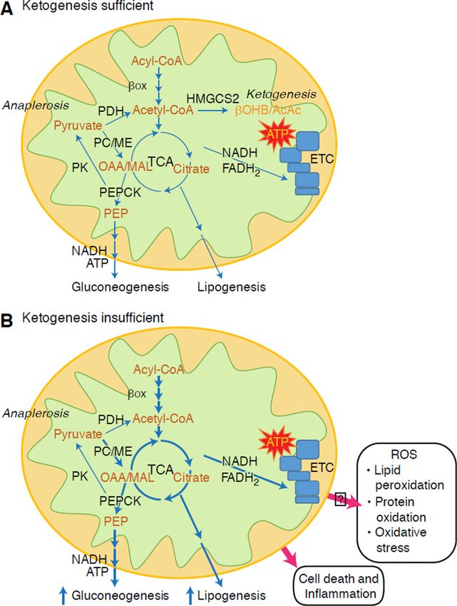
Ketogenesis is often used as a proxy for hepatic fat oxidation. Impairments of ketogenesis
emerge as NAFLD progresses in animal models, and likely in humans. Through
incompletely defined mechanisms, hyperinsulinemia suppresses ketogenesis, possibly
contributing to hypoketonemia compared to lean controls (Bergman et al., 2007; Bickerton
et al., 2008; Satapati et al., 2012; Soeters et al., 2009; Sunny et al., 2011; Vice et al., 2005).
Nonetheless, the ability of circulating ketone body concentrations to predict NAFLD is
controversial (Männistö et al., 2015; Sanyal et al., 2001). Robust quantitative magnetic
resonance spectroscopic methods in animal models revealed increased ketone turnover rate
with moderate insulin resistance, but decreased rates were evident with more severe insulin
resistance (Satapati et al., 2012; Sunny et al., 2010). In obese humans with fatty liver,
Author Manuscript
ketogenic rate is normal (Bickerton et al., 2008; Sunny et al., 2011), and hence, the rates of
ketogenesis are diminished relative to the increased fatty acid load within hepatocytes.
Consequently, β-oxidation-derived acetyl-CoA may be directed to terminal oxidation in the
TCA cycle, increasing terminal oxidation, phosphoenolpyruvate-driven gluconeogenesis via
anaplerosis/cataplerosis, and oxidative stress. Acetyl-CoA also possibly undergoes export
from mitochondria as citrate, a precursor substrate for lipogenesis (Fig. 4) (Satapati et al.,
2015; Satapati et al., 2012; Solinas et al., 2015). While ketogenesis becomes less responsive
to insulin or fasting with prolonged obesity (Satapati et al., 2012), the underlying
mechanisms and downstream consequences of this remain incompletely understood. Recent
evidence indicates that mTORC1 suppresses ketogenesis in a manner that may be
downstream of insulin signaling (Kucejova et al., 2016), which is concordant with the
observations that mTORC1 inhibits PPARα-mediated Hmgcs2 induction (Sengupta et al.,
Author Manuscript
2010) (also see Regulation of HMGCS2 and SCOT/OXCT1).
Preliminary observations from our group suggest adverse hepatic consequences of ketogenic
insufficiency (Cotter et al., 2014). To test the hypothesis that impaired ketogenesis, even in
carbohydrate-replete and thus ‘non-ketogenic’ states, contributes to abnormal glucose
metabolism and provokes steatohepatitis, we generated a mouse model of marked ketogenic
insufficiency by administration of antisense oligonucleotides (ASO) targeted to Hmgcs2.
Loss of HMGCS2 in standard low fat chow-fed adult mice caused mild hyperglycemia and
markedly increased production of hundreds of hepatic metabolites, a suite of which strongly
suggested lipogenesis activation. High fat diet feeding of mice with insufficient ketogenesis
resulted in extensive hepatocyte injury and inflammation. These findings support the central
hypotheses that (i) ketogenesis is not a passive overflow pathway but rather a dynamic node
Author Manuscript
in hepatic and integrated physiological homeostasis, and (ii) prudent ketogenic augmentation
to mitigate NAFLD/NASH and disordered hepatic glucose metabolism is worthy of
exploration.
How might impaired ketogenesis contribute to hepatic injury and altered glucose
homeostasis? The first consideration is whether the culprit is deficiency of ketogenic flux, or
ketones themselves. A recent report suggests that ketone bodies may mitigate oxidative
stress-induced hepatic injury in response to n-3 polyunsaturated fatty acids (Pawlak et al.,
Cell Metab. Author manuscript; available in PMC 2018 February 07.You can also read