HHS Public Access Author manuscript J Diagn Med Sonogr. Author manuscript; available in PMC 2021 January 01 - CDC stacks
←
→
Page content transcription
If your browser does not render page correctly, please read the page content below
HHS Public Access
                               Author manuscript
                               J Diagn Med Sonogr. Author manuscript; available in PMC 2021 January 01.
Author Manuscript

                    Published in final edited form as:
                     J Diagn Med Sonogr. 2020 ; 2020: . doi:10.1177/8756479319898471.

                    Comparing Shape Categorization to Circularity Measurement in
                    the Evaluation of Median Nerve Compression Using Sonography
                    Buwen Yao, MBBS, Kayla Gan, Annie Lee, Shawn C. Roll, PhD, OTR/L, RMSKS, FAOTA,
                    FAIUM
                    Chan Division of Occupational Science and Occupational Therapy, University of Southern
                    California, Los Angeles, CA
Author Manuscript

                    Abstract
                         Purpose—This study aimed to develop a subjective categorization of nerve shape and to examine
                         the relationship of shape categorizations to measurement of nerve circularity.

                         Methods—Wrists were evaluated with sonography in healthy participants. Images of the median
                         nerve were obtained in the transverse plane at the level of pisiform with the fingers resting,
                         gripping, and pinching. Nerves were categorized as ovoid, angular, or irregular, and the cross-
                         sectional area and perimeter were measured to calculate nerve circularity.

                         Results—Across 167 participants, the median nerve shifted from being primarily ovoid at rest to
                         angular shaped when the fingers were in a full fist or pinching. Approximately three-quarters of
                         subjects exhibited a shape change during dynamic movement. Irregular nerves had the lowest
Author Manuscript

                         circularity values; however, the majority of nerves had similar circularity measures despite having
                         different shapes.

                         Conclusions—Subjective categorization of shape has the potential to be a valid technique for
                         evaluation of the median nerve using sonography, and this evaluation may provide additional
                         information regarding nerve compression that is not fully captured by a circularity measure.
                         Further investigation is needed to determine how these two techniques may be best used
                         individually or together to advance clinical diagnosis, prevention, and rehabilitative interventions.

                    Keywords
                         Sonography; carpal tunnel syndrome; median nerve; circularity

                                     Carpal tunnel syndrome (CTS) is the most common nerve entrapment of the upper extremity
Author Manuscript

                                     caused by compression of the median nerve in the carpal tunnel.1,2 Approximately 3.8% of
                                     the general population has CTS,3 and the economic costs of CTS are a heavy burden to
                                     society.4 Most cases of CTS are considered to be idiopathic;5 however, several
                                     environmental risk factors are related to the condition, such as repetitive hand motions6–8
                                     and prolonged postures in extremes of wrist flexion or extension.4 One underlying
                                     pathophysiologic theory behind these risk factors is that during hand motion the median
                                     nerve is deformed due to a combination of pressure and available space within the carpal

                    Corresponding Author: Shawn C. Roll, 1540 Alcazar Street, CHP 133, Los Angeles, CA 90089-9003, sroll@usc.edu.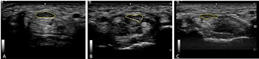
Yao et al.                                                                                                Page 2

                                 tunnel.9,10 For some hand motions or positions, the median nerve is more distorted and more
Author Manuscript

                                 vulnerable to pressures in the carpal tunnel.

                                 Recently, sonography has been used to evaluate the median nerve in the carpal tunnel due to
                                 its ability to capture nerve anatomy and other advantages such as accessibility, low-cost, and
                                 noninvasiveness.7,11 Most studies have used sonography to identify morphologic features
                                 and changes of the median nerve with quantitative measurements. Common nerve measures
                                 include cross-sectional area (CSA) and flattening ratio, that is, medial-lateral diameter
                                 divided by anterior-posterior diameter.12–14 A less-common measure more recently
                                 appearing in literature is median nerve circularity.15,16 Circularity is a measurement
                                 calculated using the CSA and perimeter of the median nerve (4π*CSA/perimeter2), which
                                 indicates how close the median nerve’s shape is to a circle.15–17 Using circularity
                                 measurement, studies have shown that the median nerve is deformed during hand motions
                                 and during forced gripping, that is, when grip force increases, the median nerve becomes
Author Manuscript

                                 more flattened.10,15,18–20 Identifying nerves that are more flat than round may serve as an
                                 indicator of increased compression, which is a risk factor for development of nerve
                                 pathology.

                                 However, the quantitative measurement of circularity has several limitations. Calculating
                                 circularity is a challenging procedure that requires reliable and valid measurements of the
                                 CSA and perimeter of the median nerve. This process takes practice and time to develop the
                                 necessary skills and expertise. Calculating circularity also raises the potential for errors,
                                 because it is not an automatic value. Rather, the calculation requires additional steps beyond
                                 the measures directly reported on the machine or other imaging software. Additionally,
                                 current studies using circularity have conflicting results,10,15,18–20 such that median nerves
                                 with a similar circularity can exhibit different shapes, which may correlate with different
Author Manuscript

                                 amounts of compression. The median nerve could be compressed by surrounding tendons
                                 and structures becoming flattened or irregular in shape during hand movement, or have a
                                 different circular or ovoid shape than when the hand is at rest due to varied compression by
                                 different surrounding tissues (e.g., slightly taller than longer vs. slightly longer than taller).

                                 As such, it may be useful to further understand circularity by evaluating the amount of
                                 compression or variations in shape, thereby improving upon the use of circularity as a
                                 singular method for evaluating nerve pathology. Given the current limitations of circularity
                                 measurement and need for further refinement of our understanding of the applicability of the
                                 circularity measurement, we aimed to develop a subjective categorization of nerve shape and
                                 explore the usefulness of adding the proposed subjective categorization of nerve shape to
                                 existing protocols for measuring nerve circularity. This process can help to further a more
Author Manuscript

                                 nuanced understanding of nerve compression as it contributes to the identification of
                                 morphologic changes or individual morphologic patterns that are predictive of the onset of
                                 CTS in future research.

                                  J Diagn Med Sonogr. Author manuscript; available in PMC 2021 January 01.
Yao et al. Page 3
Methods
Author Manuscript
Participants
A total of 172 asymptomatic students were recruited from two universities between June
2015 to September 2018. Participants were excluded if they reported history of a carpal
tunnel release surgery, median nerve pathology, or other polyneuropathy that involved the
median nerve. Participants with bilateral bifid median nerves in the carpal tunnel were
excluded. For participants with a unilateral bifid median nerve, the wrist with the bifid
median nerve was excluded and the wrist with non-bifid median nerve was included in the
final analysis. Demographic data including age, gender, ethnicity/race, and hand dominance
were obtained via self-report. The study was approved by the institutional review board and
written informed consent was signed by participants prior to participation in the study.
Ultrasound Technique
Author Manuscript
Participants were seated facing the sonographer with the shoulder adducted, elbow
comfortably extended, the forearm fully supinated and resting on the table, and the hand in a
relaxed position. Imaging was conducted using a Logiq-e portable sonography machine (GE
Healthcare, Milwaukee, WI) with a 12-MHz linear array transducer, by two experienced
musculoskeletal sonographers. The transducer was placed on top of the wrist with no
additional force than the probe weight to the skin. Sonographic images were taken on both
wrists by placing the transducer over the carpal tunnel at the level of pisiform in the
transverse plane to obtain short-axis views of the median nerve.
Static images of carpal tunnel and median nerve were taken at three positions: resting,
gripping, and pinching. For resting, the fingers were held in a naturally relaxed position,
which resulted in slight composite flexion throughout all digits. During gripping, the
Author Manuscript
participant started with the fingers fully extended, slowly flexed the fingers into a closed fist
without any forceful gripping or squeezing with the fingers. After holding the closed fist for
a brief moment, the participant opened the hand to a fully extended position. To capture the
median nerve during pinching, participants held a pen in their hand using a tripod or
modified tripod pinch, as if they were to write. While the pinch position was not
standardized across participants, the technique was intended to capture the functional
position most often used by each individual participant. Static images were obtained in both
the resting and pinching positions, with image optimization focused on the median nerve.
Dynamic cine clips were recorded while the participant completed the gripping motion, each
lasting approximately 6 seconds. The sonographer ensured that the transducer was
maintained in a stable position over the wrist throughout the motion, using pisiform as bony
landmarks. Each image and cine clip were obtained at least twice to ensure that a high-
Author Manuscript
quality, valid image was obtained for evaluation of the median nerve.
Image Analysis
Image Processing—Images and cine clips were archived and analyzed using ViewPoint 6
(GE Healthcare, Milwaukee, WI). Prior to analysis, all images within a participant file were
screened to determine if a bifid nerve was present within the carpal tunnel. During this
screening process, the resting and pinching images that most clearly depicted the median
J Diagn Med Sonogr. Author manuscript; available in PMC 2021 January 01.Yao et al. Page 4
nerve were identified for analysis. Similarly, each of the cine clips were evaluated to select
Author Manuscript
the clip that most clearly represented the median nerve throughout the movement of the
fingers and was most stable as identified by no shifting of bony landmarks of pisiform
within the frame. The rater then identified a frame at the start of the clip prior to initiation of
movement and a frame in the middle of the cine clip where motion had paused, thus
indicating the hand was in a full grip position. These frames were used along with the still
images in the analysis of median nerve shape.
Nerve Shape—Through an iterative process of image review and discussion, the research
team developed consensus on three categories of median nerve shape, which was followed
by further iterations of image evaluation, review, and discussion to refine the evaluation
criteria for categorizing nerve images. The research team consisted of a registered
musculoskeletal sonographer and three students with limited previous sonography
experience. As part of the criteria development process, three student raters analyzed nerve
Author Manuscript
shapes in images obtained from the first 25 participants in the study across multiple rounds
of 5–10 cases per round. In each round, we noted that a variety of shapes were represented
within the images. Pairwise percent agreement between the three raters was calculated each
time the raters completed analysis on a series of images, and the evaluation criteria were
refined through discussions with the sonographer until the research team agreed upon
definitions which resulted in acceptable inter-rater reliability (i.e., >80%). The final
evaluation criteria used to categorize nerves into ovoid, angular, or irregular shapes are
detailed in Table 1, and examples of nerve with each shape are shown in Figure 1. Using
these criteria and the images or still frames selected in the screening process, the shape of
the median nerve at the pisiform was subjective categorized for each wrist of every
participant in the resting, gripping, and pinching position. All three trained raters evaluated
images; only one rater categorized each image.
Author Manuscript
Shape Change—In addition to identifying the shape of the median nerve in specific static
positions, the cine clips were subjectively evaluated to determine if the shape of the median
nerve changed at any point during the gripping motion. Any indication that the nerve was
compressed or impacted by the surrounding tendons or other structures during the gripping
motion was recorded as a ‘change.’ This dynamic evaluation allowed for identification of
nerve compression during functional movement, which may not be apparent if the shape of
the median nerve were categorized as being same using static images at the beginning and
end of the gripping movement. If the nerve shape remained the same and was not visually
impacted by the surrounding structures during the entire gripping movement, shape change
was recorded as ‘no change.’
Author Manuscript
Nerve Circularity—Nerve circularity was calculated using the CSA and perimeter of the
median nerve measured on the static images of resting, gripping, and pinching. A direct
trace along the inner hyper-echoic border of the nerve was used to obtain CSA and
perimeter.21 CSA and perimeter of the nerve were measured three times and the average of
the three measurements was used to calculate circularity as follows:17
Circularity = 4π*CSA/perimeter2
J Diagn Med Sonogr. Author manuscript; available in PMC 2021 January 01.Yao et al. Page 5
Using this formula, a circularity of 1.0 indicates that the median nerve shape is a perfect
Author Manuscript
circle; lower values are interpreted as the nerve being further away from a circle; however,
the values do not indicate any distinct shape or type of compression on the nerve, merely
that it is not circular.
Statistical Analysis
Descriptive statistics were calculated for all demographic data. A chi-squared test was
performed to examine if median nerve shape and shape change was associated with hand
dominance. A Shapiro-Wilk test for normality was performed for median nerve CSA,
perimeter, and circularity data, confirming that these data were normally distributed; as such,
these data were expressed and evaluated as parametric data using mean ± standard deviation
(SD). A two-sample independent t-test was used to analyze the differences in the means of
CSA, perimeter, and circularity between dominant and non-dominant sides. An analysis of
Author Manuscript
variance was performed to examine differences in mean values of CSA, perimeter, and
circularity between rest, grip, and pinch positions, and between three categorizations of
nerve shapes. A post-hoc Bonferroni test was performed to identify the significant group
differences. SPSS (IBM, version 24) was used for statistical analysis, and p-values of < 0.05
were considered statistically significant.
Results
A total of 172 asymptomatic participants were enrolled in the study. Five participants had
bilateral bifid median nerves, resulting in data from 167 participants for the final analysis.
These participants were predominately right-handed (92.2%) females (85.0%) who had a
mean age of 24.6 ± 3.3 years (Table 2). Across the sample, 11 participants had a unilateral
bifid median nerve in the carpal tunnel on their dominant hand, and 10 participants had a
Author Manuscript
unilateral bifid median nerve in the carpal tunnel on their non-dominant hand. The analyses
included data from 156 dominant wrists and 157 non-dominant wrists.
The frequency of the median nerve shapes across various finger positions in the static
images and shape changes during dynamic gripping are reported in Table 3. When the hand
is in the rest position, the largest number of the subjects exhibited an ovoid median nerve
(48.7% and 38.2% for the dominant and non-dominant side, respectively), with the non-
dominant hand having a higher proportion of irregularly shaped nerves with fewer ovoid
nerves than the dominant hand (pYao et al. Page 6
dominant wrist in gripping and pinching (p=0.001 and p=0.008, respectively), and the
Author Manuscript
perimeter of the median nerve of the dominant wrist was significantly greater than the non-
dominant wrist in pinching (p=0.013). There were no significant differences in average
circularity measures of the nerve between the dominant and the non-dominant sides, nor
between resting, gripping, and pinching positions.
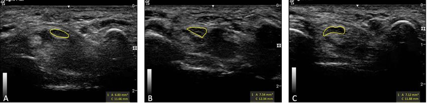
Finally, the examination of differences in average CSA, perimeter, and circularity by nerve
shape is presented in Figure 3. CSA measures were not found to be different among nerves
of different shapes. In the dominant wrist, the average perimeter (14.14mm) was
significantly larger and circularity (0.55) was significantly smaller for median nerves with
irregular shapes as compared to the same measures in nerves with angular shape (12.85mm,
pYao et al. Page 7
the original method lower values indicate the less a less circular object as opposed to higher
Author Manuscript
values over 1.0 when using the reciprocal method. While both techniques provide an
accurate measure of ciruculaty due to the fact that the same data inputs are used, the original
formula may be easier to interpret and therefore should be used across the field to promote
data comparision and eliminate confusion across studies.
Regardless of the method used, there is mixed evidence for the correlation between
measures of median nerve circularity and finger positions. Cowley and colleagues noted a
slight, non-significant decrease in median nerve circularity from 0.57 to 0.54 when the hand
was in a neutral position versus a pinching.15 Using the reciprocal formula, Yoshii and
colleagues found a non-significant change in nerve circularity that increased from 1.69 to
1.75 when measured in full finger extension to full finger flexion.19 This equates to a
decrease in circularity from 0.59 to 0.57, when using normal circularity. Also using the
reciprocal formula, Van Doesburg and colleagues reported a decrease in circularity that
Author Manuscript
equates to 0.03 change (from 0.58 to 0.55) with finger movement.18 In contrast, to these
studies that may suggest that a nerve becomes less round with finger flexion, a fourth study
noted that median nerve circularity improved when moving from finger extension to finger
flexion (i.e., 0.52 to 0.55 using the original formula).
Results in our study showed slightly higher circularity but were similar to the first two
studies noted above,15,19 in that we did not find any significant difference in nerve
circularity measures between resting, gripping, and pinching. However, irrespective of the
position of the fingers, we were able to validate that circularity of the median nerve
significantly decreased when the nerve was identified as ovoid shape versus angular or
irregular. This was especially true for nerves with high circularity (>0.75) being primarily
ovoid and angular and low circularity values (Yao et al. Page 8
compression, to build upon the findings in our data. While a circularity calculation may be
Author Manuscript
most useful when the values are higher or lower, a subjective examination of the nerve may
be more important to identify either intermittent compression during dynamic activity or
sustained compression due to finger positioning that results in change in shape. Employing
these two techniques together further elucidate nuances of nerve compression that could be
useful in research and practice. Future research should consider these measurement
techniques individually and together to identify utility within clinical diagnosis and
intervention, such that individualized care recommendations or splinting may be provided to
assist a patient with CTS in minimizing movements that lead to compression. Alternatively,
longitudinal studies could examine these measures to determine their ability to identify
individuals at high-risk for development of median nerve pathologies, such as CTS, due to
median nerve deformation during functional hand use.
Limitations
Author Manuscript
This article has several limitations as follows. Interpretation and use of our study findings is
primarily limited by having only evaluated the measures in healthy participants. As such, we
do not know how these measures would apply to individuals with median nerve pathologies
who may have enlarged median nerves, edema, or other pathological considerations. Despite
not being able to interpret our findings in clinical care, the results of our study showed
potential usefulness of this subjective classification for median nerve shape. Translation of
this work to determine the value of the classification system in differentiating high-risk
populations from the normal population or as a support to diagnosis or treatment for
individuals with CTS should be explored in future studies.
Conclusion
Author Manuscript
In our study, with a rather clear sonographic image, the shape of the median nerve in the
carpal tunnel could be easily distinguished as ovoid, angular, or irregular by tracing inside
the hypoechoic border of the median nerve. When compared to circularity, irregular shapes
demonstrated the least amount of circularity as compared to ovoid shapes. Moreover,
subjective evaluation of shape was more sensitive than circularity for identifying differences
in nerve shape change during dynamic movement and in static images obtained in various
finger positions. Shape change was highly prevalent in all individuals during movements and
more angular and irregular shaped nerves were identified during functional positioning of
the fingers than when at rest; differences circularity did not identify. These data suggest that
a subjective evaluation may have additional value beyond what circularity may provide in
the assessment of nerve compression for prevention, diagnosis, and intervention as a
component of research and practice.
Author Manuscript
Acknowledgements
The authors wish to thank Jennifer Mitchell for her help in image acquisition and identification, and Ariana Cristino
for her initial contributions in development of a preliminary image analysis protocol that led to the final protocol
deployed in this study.
Funding: This work was supported by the Undergraduate Research Associate Program at the University of
Southern California and the Centers for Disease Control (CDC), National Institute for Occupational Safety and
J Diagn Med Sonogr. Author manuscript; available in PMC 2021 January 01.Yao et al. Page 9
Health (NIOSH) (Grant #: R01-OH010665). Its contents are solely the responsibility of the authors and do not
necessarily represent the official views of the CDC.
Author Manuscript
References
1. Talbott NR. Prevalence of Carpal Tunnel Syndrome in a General Population. Physical Therapy.
1999;79(12):1226.
2. Papanicolaou GD, McCabe SJ, Firrell J. The prevalence and characteristics of nerve compression
symptoms in the general population. Journal of Hand Surgery. 2001;26(3):460–466. [PubMed:
11418908]
3. Atroshi I, Gummesson C, Johnsson R, Ornstein E, Ranstam J, Rosen I. Prevalence of carpal tunnel
syndrome in a general population. JAMA. 1999;282(2):153–158. [PubMed: 10411196]
4. Ibrahim I, Khan WS, Goddard N, Smitham P. Carpal tunnel syndrome: a review of the recent
literature. Open Orthop J. 2012;6:69–76. [PubMed: 22470412]
5. Ashworth NL. Carpal tunnel syndrome. BMJ Clin Evid. 2014;2014:1114.
6. Toosi KK, Impink BG, Baker NA, Boninger ML. Effects of computer keyboarding on
Author Manuscript
ultrasonographic measures of the median nerve. American Journal of Industrial Medicine.
2011;54(11):826–833. [PubMed: 21739468]
7. Jaeschke R, Thoirs K, Bain G, Massy-Westropp N. Systematic review: hand activity and ultrasound
of the median nerve. Occupational Medicine. 2017;67(5):389–393. [PubMed: 28582584]
8. Ashworth NL. Carpal Tunnel Syndrome. Am Fam Physician. 2016;94(10):830–831. [PubMed:
27929273]
9. Ablove RH, Peimer CA, Diao E, Oliverio R, Kuhn JP, Buffalo N. Morphologic changes following
endoscopic and two-portal subcutaneous carpal tunnel release. Journal of Hand Surgery. 1994;19(5):
821–826. [PubMed: 7806811]
10. Wang Y, Zhao C, Passe SM, et al. Transverse ultrasound assessment of median nerve deformation
and displacement in the human carpal tunnel during wrist movements. Ultrasound Med Biol.
2014;40(1):53–61. [PubMed: 24210862]
11. Padua L, Coraci D, Erra C, et al. Carpal tunnel syndrome: clinical features, diagnosis, and
management. The Lancet Neurology. 2016;15(12):1273–1284. [PubMed: 27751557]
Author Manuscript
12. Lu Y, Meng Z, Pan X, Qin L, Wang G. Value of high-frequency ultrasound in diagnosing carpal
tunnel syndrome. Int J Clin Exp Med. 2015;8(12):22418–22424. [PubMed: 26885222]
13. Park D Ultrasonography of the Transverse Movement and Deformation of the Median Nerve and
Its Relationships With Electrophysiological Severity in the Early Stages of Carpal Tunnel
Syndrome. PM R. 2017;9(11):1085–1094. [PubMed: 28433831]
14. Chang Y-W, Hsieh T-C, Tzeng IS, Chiu V, Huang P-J, Horng Y-S. Ratio and difference of the
cross-sectional area of median nerve to ulnar nerve in diagnosing carpal tunnel syndrome: a case
control study. BMC Medical Imaging. 2019;19(1):52. [PubMed: 31272405]
15. Cowley JC, Leonardis J, Lipps DB, Gates DH. The influence of wrist posture, grip type, and grip
force on median nerve shape and cross-sectional area. Clinical Anatomy. 2017;30(4):470–478.
[PubMed: 28281294]
16. Bueno-Gracia E, Malo-Urriés M, Ruiz-de-Escudero-Zapico A, et al. Reliability of measurement of
the carpal tunnel and median nerve in asymptomatic subjects with ultrasound. Musculoskeletal
Science and Practice. 2017;32:17–22. [PubMed: 28800435]
17. Cox EP. A Method of Assigning Numerical and Percentage Values to the Degree of Roundness of
Author Manuscript
Sand Grains. Journal of Paleontology. 1927;1(3):179–183.
18. Van Doesburg MHM, Henderson J, Yoshii Y, et al. Median nerve deformation in differential finger
motions: Ultrasonographic comparison of carpal tunnel syndrome patients and healthy controls.
Journal of Orthopaedic Research. 2012;30(4):643–648. [PubMed: 21953849]
19. Yoshii Y, Villarraga HR, Henderson J, Zhao C, An KN, Amadio PC. Ultrasound assessment of the
displacement and deformation of the median nerve in the human carpal tunnel with active finger
motion. J Bone Joint Surg Am. 2009;91(12):2922–2930. [PubMed: 19952256]
J Diagn Med Sonogr. Author manuscript; available in PMC 2021 January 01.Yao et al. Page 10
20. Marquardt TL, Gabra JN, Li Z-M. Morphological and positional changes of the carpal arch and
median nerve during wrist compression. Clinical Biomechanics. 2015;30(3):248–253. [PubMed:
Author Manuscript
25661267]
21. Roll SC, Evans K. Feasibility of Using a Hand-Carried Sonographic Unit for Investigating Median
Nerve Pathology. Journal of Diagnostic Medical Sonography. 2009;25(5):241–249.
22. Mhoon JT, Juel VC, Hobson‐Webb LD. Median nerve ultrasound as a screening tool in carpal
tunnel syndrome: Correlation of cross-sectional area measures with electrodiagnostic abnormality.
Muscle & Nerve. 2012;46(6):861–870. [PubMed: 22996383]
23. Tai-Tzung K, Ming-Ru L, Yin-Yin L, Jiann-Perng C, Yen-Wei H, Chih-Kuang Y. Assessment of
Median Nerve Mobility by Ultrasound Dynamic Imaging for Diagnosing Carpal Tunnel
Syndrome. PLoS ONE. 2016;11(1):e0147051. [PubMed: 26764488]
24. Marquardt TL, Gabra JN, Li ZM. Morphological and positional changes of the carpal arch and
median nerve during wrist compression. Clin Biomech (Bristol, Avon). 2015;30(3):248–253.
25. Van Doesburg MHM, Yoshii Y, Villarraga HR, et al. Median nerve deformation and displacement
in the carpal tunnel during index finger and thumb motion. Journal of Orthopaedic Research.
2010;28(10):1387–1390. [PubMed: 20225286]
Author Manuscript
Author Manuscript
Author Manuscript
J Diagn Med Sonogr. Author manuscript; available in PMC 2021 January 01.Yao et al. Page 11
Author Manuscript
Figure 1.
Gray-scale transverse images taken of median nerve (yellow circle) at level of pisiform
depicting three different categorizations of median nerve shape: (A) ovoid, (B) angular, and
(C) irregular.
Author Manuscript
Author Manuscript
Author Manuscript
J Diagn Med Sonogr. Author manuscript; available in PMC 2021 January 01.Yao et al. Page 12
Author Manuscript
Author Manuscript
Figure 2.
The distribution of median nerve shape during hand rest, grip, and pinch for dominant
(n=157) and non-dominant (n=156) wrists.
Author Manuscript
Author Manuscript
J Diagn Med Sonogr. Author manuscript; available in PMC 2021 January 01.Yao et al. Page 13
Author Manuscript
Author Manuscript
Figure 3.
Comparison of average cross-sectional area (CSA, A), perimeter (B), and circularity (C)
among ovoid, angular, and irregular median nerves by hand dominance. Results of analysis
of variance testing, with Bonferonni post-hoc testing for pairwise comparisons indicating
significant differences: **P < 0.01; ***P < 0.001.
Author Manuscript
Author Manuscript
J Diagn Med Sonogr. Author manuscript; available in PMC 2021 January 01.Yao et al. Page 14
Author Manuscript
Figure 4.
Gray-scale transverse images taken at the level of pisiform within the carpal tunnel for
measurement of the median nerve (yellow circle) shape and circularity, demonstrating three
median nerves with similar circularity but different shapes due to varying amounts of
Author Manuscript
compression by surrounding structures. (A) Ovoid median nerve with a circularity of 0.63.
(B) Angular median nerve with a circularity of 0.62. (C) Irregular median nerve with a
circularity of 0.63.
Author Manuscript
Author Manuscript
J Diagn Med Sonogr. Author manuscript; available in PMC 2021 January 01.Yao et al. Page 15
Table 1.
Evaluation criteria for categorizing median nerve shape within the carpal tunnel.
Author Manuscript
Evaluation criteria
Ovoid was determined as a nerve with an “oval” shape with curved edges all around and no flat or angled sides. An ovoid median
Ovoid
nerve was not impacted by any of the surrounding structures in a non-uniform manner, making it appear “free-floating.”
Angular was defined as a nerve having at least one “flat” side causing an angle or sharp, distinctive change in direction when
Angular
tracing the border of the nerve. Angular nerves could have two or three angles, to the extent of appearing like a triangle.
Irregular was identified as a nerve compressed by multiple structures, causing a shape that could not be clearly characterized as
Irregular
ovoid or angular.
Author Manuscript
Author Manuscript
Author Manuscript
J Diagn Med Sonogr. Author manuscript; available in PMC 2021 January 01.Yao et al. Page 16
Table 2.
Descriptive statistics of participants enrolled in the study (n=167)
Author Manuscript
Mean/Frequency (SD/%)*
Age, years 24.6 (3.3)
BMI, kg/m2 23.3 (4.0)
Gender, male 25 (15.0%)
Handedness, right 154 (92.2%)
Race
American Indian/Alaska Native 2 (1.2%)
Asian 64 (38.3%)
Native Hawaiian or other Pacific Islander 1 (0.6%)
Black 4 (2.4%)
White 74 (44.3%)
Author Manuscript
Other 22 (13.2%)
Ethnicity, Hispanic 41 (24.6%)
*
Mean (SD) were calculated for continuous data and frequency (%) are reported for categorical data.
Author Manuscript
Author Manuscript
J Diagn Med Sonogr. Author manuscript; available in PMC 2021 January 01.Yao et al. Page 17
Table 3.
Distribution of median nerve shapes across finger positions in static images and shape change during dynamic
Author Manuscript
gripping motion in the dominant and non-dominant sides.
Dominant hand Non-dominant hand p-value*
Rest
Ovoid 76 (48.7%) 60 (38.2%)
Angular 58 (37.2%) 54 (34.4%) 0.01
Irregular 22 (14.1%) 43 (27.4%)
Grip
Ovoid 38 (24.4%) 30 (19.1%)
Angular 84 (53.8%) 82 (52.2%) 0.29
Irregular 34 (21.8%) 45 (28.7%)
Pinch
Author Manuscript
Ovoid 43 (27.6%) 40 (25.5%)
Angular 78 (50.0%) 74 (47.1%) 0.60
Irregular 35 (22.4%) 43 (27.4%)
Shape change
Yes 113 (72.4%) 122 (77.7%)
0.28
No 43 (27.6%) 35 (22.3%)
*
Chi-squared test
Author Manuscript
Author Manuscript
J Diagn Med Sonogr. Author manuscript; available in PMC 2021 January 01.Yao et al. Page 18
Table 4.
Comparison of mean ± SD of cross-sectional Area (CSA), perimeter, and circularity of the median nerve by
Author Manuscript
finger position within the dominant (n=156) and non-dominant sides (n=157).
Rest Grip* Pinch* p-value**
CSA, mm2
Dominant 8.09±1.57 8.53±1.97 8.33±2.05 0.12
Non-dominant 7.88±1.33 8.11±1.51 8.09±1.53 0.31
Perimeter, mm
Dominant 12.75±1.43 13.42±2.19 13.06±2.13 0.01***
Non-dominant 12.62±1.51 12.92±1.77 12.77±1.65 0.27
Circularity
Dominant 0.63±0.08 0.61±0.11 0.62±0.10 0.15
Non-dominant 0.63±0.09 0.62±0.11 0.63±0.11 0.66
Author Manuscript
*
Two-sample independent t-test showed significant difference between dominant and non-dominant for grip CSA, pinch CSA, and pinch perimeter
(pYou can also read